


Abstract
Background/Objectives: Hydroxyapatite (HA) coatings offer an osteoconductive interface and a mineral reservoir for alendronate. Their pharmaceutical value depends on whether binding produces biologically available exposure without impairing healing. This review examines that relationship in HA–alendronate coatings on titanium. Methods: A structured critical narrative review used PubMed/MEDLINE, Scopus and Web of Science Core Collection, with reference-list screening. Screening identified 62 primary studies; contextual literature supported mechanistic and translational interpretation. Results: Adsorption, mineral incorporation, laser deposition and hybrid reservoirs produce different loading and release profiles. Cell and animal findings depend on formulation, dose, comparator and model. Mineral content, bone-to-implant contact and mechanical fixation provide complementary evidence and are not interchangeable outcomes. Human studies have mainly evaluated other bisphosphonates in fibrinogen coatings or alendronate applied as a gel around dental implants. Some reported less marginal bone loss, but improvements in implant stability were inconsistent. These results do not directly establish the efficacy of HA–alendronate coatings. Conclusions: Translation requires reproducible manufacture, measured active-drug availability, preserved physiological remodeling, durable coating adhesion and controlled trials for a defined implant indication. Strong mineral binding alone does not establish effective local therapy.
1. Introduction
Bone is a living mineral–organic composite whose remodeling supports mechanical function [1]. Early studies of bone–implant interfaces and hydroxyapatite (HA(-coated hip prostheses established the rationale for combining a load-bearing substrate with an osteoconductive surface [2,3,4,5].
The bone–implant interface must support fixation while inflammation resolves and new matrix forms. HA resembles skeletal mineral, but natural bone mineral differs from stoichiometric Ca10(PO4)6(OH)2. Von Euw et al. used solid-state nuclear magnetic resonance on excised cortical bone from a two-year-old sheep, examined fresh and after drying, alongside synthetic calcium phosphate reference materials. They identified an amorphous, hydrogen-phosphate-rich surface around the native apatite particles [6]. These were ex vivo bone specimens and synthetic mineral comparisons, rather than measurements in a living animal or on a titanium implant.
HA serves two related functions: it provides a mineral surface for protein adsorption and bone attachment, and it binds alendronate as a drug reservoir. The protein layer encountered by cells can modify both functions [7]. Evidence of apatite deposition or osteoblast attachment therefore cannot, by itself, demonstrate pharmacological drug delivery.
Coupling HA with alendronate adds a pharmacological function to the mineral coating. The phosphonate groups of bisphosphonates bind calcium-containing surfaces, whereas their molecular structure also governs cellular activity. In comparative HA-binding experiments, Nancollas and colleagues found substantial differences among clinically used bisphosphonates; alendronate had greater affinity than risedronate under the tested conditions [8]. This interaction offers a means of retaining a drug near an implant. It also creates a formulation problem: the fraction bound to mineral, the fraction released into tissue fluid and the fraction reaching a cellular target are different quantities.
Alendronate inhibits farnesyl diphosphate synthase (FPPS), interfering with protein prenylation and osteoclast function. Structure–activity studies connect enzyme inhibition with antiresorptive potency, providing a mechanistic basis for local delivery [9]. Early HA–alendronate composites and matrix-assisted pulsed laser evaporation (MAPLE) coatings demonstrated that alendronate-associated mineral could retain biological activity in bone-cell cultures [10,11]. These experiments established feasibility, but their cell-culture outcomes cannot determine an effective human implant dose or long-term fixation.
Recent work has broadened the design space. Chemically bound alendronate can promote calcium phosphate deposition on titanium oxide, while nanotubes, polymer barriers and hybrid mineral reservoirs can alter drug release [12,13,14]. The clinical evidence is less uniform. A multicenter trial of zoledronate-coated dental implants did not show a significant advantage in the principal implant-stability comparison, and a randomized study of locally applied alendronate gel found no significant overall treatment benefit for implant stability or marginal bone levels [15,16]. These studies address related pharmacology, but neither validates the entire class of HA–alendronate coatings.
Previous reviews have surveyed bisphosphonate-functionalized orthopedic implants across drugs and carriers [17], calcium-based scaffolds for regeneration [18], and clinical outcomes of local bisphosphonate application or dental implant coatings [19]. The present review focuses more narrowly on how HA–alendronate binding, coating architecture and biologically available exposure connect to remodeling and fixation on titanium. Recent nanomaterial research in bone metastasis highlights the importance of the bone microenvironment [20], while an amyloid-like protein coating combined with carboxymethyl chitosan/amorphous calcium phosphate illustrates interface-guided enamel remineralization [21]. These adjacent applications inform material design but do not test HA–alendronate implant efficacy. The synthesis follows the sequence from substrate and mineral coating through loading and release to bone remodeling and implant outcomes.
2. Materials and Methods
2.1. Review Design and Question
This article was developed as a structured critical narrative review. The question was whether HA-associated local alendronate delivery can connect a chemically defined implant coating to measurable drug release, appropriate modulation of bone remodeling and improved fixation. The reporting approach follows the emphasis of the Scale for the Assessment of Narrative Review Articles (SANRA) on an explicit purpose, a described literature search, appropriate referencing and interpretation of evidence levels [22]. No systematic-review protocol was registered, and no meta-analysis or formal certainty score was undertaken. The review distinguished between eligible primary studies forming the core evidence base and additional contextual literature used to support mechanistic explanations, historical perspectives, and discussion of clinical and pharmaceutical implications.
Contextual references explain mineral chemistry, drug action, fabrication methods and clinical interpretation; they do not constitute additional independent implant-efficacy studies. Reviews, historical sources and studies of other indications were kept separate from the primary-study corpus.
2.2. Information Sources and Search Strategy
PubMed/MEDLINE, Scopus, and Web of Science Core Collection were searched to identify studies investigating hydroxyapatite-associated alendronate delivery, bisphosphonate-functionalized implant coatings, local drug-release mechanisms, and implant osseointegration.
Three complementary search families were used to identify: (i) physicochemical interactions between alendronate or related bisphosphonates and hydroxyapatite or other calcium phosphate materials; (ii) titanium implant coatings and local drug-delivery systems incorporating alendronate; and (iii) experimental and clinical studies evaluating peri-implant bone remodeling, osseointegration, implant stability, and the translational performance of bisphosphonate-based interventions.
The search concepts combined the material terms “hydroxyapatite” and “calcium phosphate”; drug terms including “alendronate”, “bisphosphonate” and “zoledronate”; and implant, coating, release and bone-response terms. Title/abstract fields were used in PubMed/MEDLINE, TITLE-ABS-KEY in Scopus, and Topic fields in Web of Science Core Collection. The three search families addressed mineral–drug interactions, implant delivery systems and biological or clinical outcomes. Complete database-specific Boolean strings are provided in Supplementary Materials. The strings contain no language, publication-date, species or study-design filters; English-language eligibility was applied during study selection.
The searches were updated on 6 October 2026. The searches retrieved 99 records from PubMed/MEDLINE, 86 records from Scopus, and 82 records from Web of Science Core Collection, yielding 267 records before deduplication. After removal of 49 duplicate records, 218 unique records remained for title and abstract screening.
Reference lists of relevant primary studies and reviews were additionally examined to identify foundational mechanistic investigations and studies addressing the pharmaceutical and clinical translation of hydroxyapatite–alendronate delivery systems. The search was designed to support a structured critical narrative synthesis rather than a quantitative meta-analysis.
2.3. Eligibility Criteria and Study Selection
English-language primary experimental and clinical studies were considered eligible when they investigated a chemically identifiable bisphosphonate associated with hydroxyapatite, calcium phosphate, titanium, or another relevant implant-related delivery platform and reported at least one physicochemical, pharmaceutical, biological, or clinical endpoint.
Eligible experimental investigations included studies evaluating coating fabrication, physicochemical characterization, drug adsorption, drug loading, release kinetics, coating stability, osteoblast and osteoclast responses, bone formation, osseointegration, and mechanical implant fixation. Animal studies were considered when they reported relevant histological, biomechanical, or peri-implant bone outcomes.
Human studies were considered when they investigated local bisphosphonate delivery or bisphosphonate-functionalized implants and reported implant stability, marginal bone changes, osseointegration-related outcomes, implant survival, or treatment-related adverse events. Studies involving other bisphosphonates or non-hydroxyapatite delivery platforms were distinguished from direct HA–alendronate evidence and used to provide mechanistic or translational context.
Studies based exclusively on molecular docking without experimental validation, insufficiently characterized drug-delivery systems, investigations without relevant material or biological endpoints, conference abstracts, case reports, and publications outside the predefined scope were excluded from the selected primary evidence corpus. Reviews and other secondary sources were used for background and contextual interpretation but were not counted among the included primary studies. Two review authors (N.B. and R.M.V.) independently screened the titles and abstracts and subsequently assessed the potentially eligible full-text articles using separate screening records. Screening decisions were compared after both reviewers had completed each stage. Disagreements were initially resolved through discussion and consensus. Any disagreement that could not be resolved between the two reviewers was adjudicated by a third review author (M.S.).
Following deduplication, 218 records underwent title/abstract screening, 44 were excluded and 174 reports underwent full-text assessment. The 112 full-text exclusions comprised irrelevant delivery/implant outcomes (28), reviews/editorials/commentaries/letters (19), conference publications (13), non-English reports (10), case reports/series (8), docking-only studies (8), inadequately characterized delivery systems (8), absent relevant biological/material characterization (7), out-of-scope interventions (5), overlapping data (4) and insufficient information (2), leaving 62 primary studies. Figure S1 summarizes study selection. Follow-up reports from the same clinical cohort were interpreted as longitudinal evidence, not independent replication. Contextual references were not included in the primary-study count.
2.4. Evidence Extraction and Interpretation
The synthesis recorded drug identity, mineral phase, substrate, coating or carrier architecture, loading and release information, model or population, comparator, follow-up and principal endpoints. Direct evidence comprised HA–alendronate coatings on titanium; adjacent evidence differed in mineral, carrier, drug, substrate or administration route. Material studies informed binding or release, cellular studies informed phenotype or activity, animal studies informed integration in a defined model, and clinical studies informed only the tested product and indication. These categories describe applicability, not validated risk-of-bias grades. Drug-free controls and component-removal comparisons were considered when attributing effects.
No pooled effect estimate was calculated because loading units, release media, implantation sites, species, coating compositions and outcome definitions were not sufficiently comparable. Quantitative examples were retained with their original denominator and time point when these could be verified. A change in cell viability, alkaline phosphatase (ALP), bone-to-implant contact (BIC), removal torque or implant stability quotient (ISQ) was not treated as interchangeable with another endpoint or with long-term implant survival.
3. Mineral Affinity and Local Antiresorptive Pharmacology
3.1. Hydroxyapatite as an Active Binding Reservoir
HA behavior depends on accessible surface area, calcium/phosphate surface composition, crystallinity and dissolution. Alendronate bears two phosphonate groups on a P–C–P backbone; their oxygen donors interact with surface calcium, while its hydroxyl group and side-chain chemistry also influence binding [8,23,24]. These drug phosphonates must be distinguished from the phosphate groups already present in HA. Calcium deficiency, carbonate substitution and hydrated non-apatitic surface environments change the sites available for adsorption [6]. Greater specific surface area can increase capacity per gram without increasing affinity per site. Crystallinity and mineral dissolution govern whether buried or strongly bound drug becomes accessible, so loading should be interpreted together with phase composition and accessible area.
Human-bone binding, synthetic-HA adsorption and inhibition of carbonated apatite dissolution interrogate different processes [8,23,24]. Their affinity rankings are therefore assay-specific. Computational estimates, including an early conference abstract, provide hypotheses rather than direct measurements of cellular exposure [25].
Solution pH and competing ions alter the mineral interface and drug partitioning. Commercial biphasic calcium phosphates, for example, adsorbed zoledronate differently, with delayed adsorption on a polymer-covered substitute [26]. This is evidence of carrier-dependent sequestration, not of alendronate elution from titanium or clinical protection against osteonecrosis.
Binding can retain a drug reservoir, but excessive retention may restrict the pharmacologically available fraction. Conversely, substantial measured loading can coexist with rapid release. In a recent comparison of HA and HA–titanium discs, sodium and calcium alendronate showed a pronounced burst and early plateau, whereas the longer release profile belonged to calcium fructoborate [27]. The latter compound has different chemistry and cannot be assigned alendronate’s target mechanism. The clinically relevant question is how much active alendronate becomes available at the tissue interface over time.
3.2. From Mineral Association to Cellular Target Engagement
Bisphosphonate pharmacology separates mineral targeting from intracellular antiresorptive activity [28,29,30,31,32,33,34,35]. For an implant reservoir, both steps must be demonstrated: retention at the interface and subsequent access to a cellular target.
During bone resorption, osteoclast acidification promotes mineral dissolution and can expose mineral-associated alendronate to cellular uptake. Intracellular alendronate inhibits farnesyl diphosphate synthase (FPPS) in the mevalonate pathway, reducing isoprenoid substrates needed to prenylate small Guanosine triphosphate-binding proteins (GTP-binding proteins) [9,35]. Disrupted cytoskeletal organization, vesicular trafficking and resorptive function can then reduce bone turnover. Uptake and target engagement cannot be inferred from coating loading alone; persistence of drug on an inert surface does not establish this intracellular sequence.
Cellular prenylation experiments and structural studies of FPPS support this mechanism, although several foundational experiments used other nitrogen-containing bisphosphonates [36,37]. Reduced resorption may preserve peri-implant bone, but excessive inhibition can also disrupt the remodeling needed to replace damaged tissue.
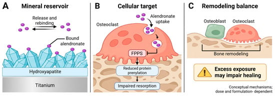
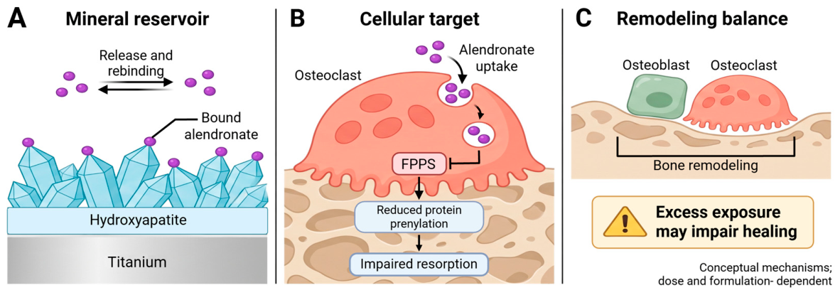
A local coating must preserve drug identity during manufacture and permit sufficient drug access to the relevant cells. A surface-bound alendronate layer that nucleates calcium phosphate may act differently from a reservoir designed to elute alendronate. Experimental and theoretical investigations of titanium oxide–alendronate interfaces support stable surface association, while later work demonstrates calcium phosphate deposition on the modified surface [12,38]. Neither result alone establishes cellular uptake or inhibition of osteoclastic FPPS. Figure 1 separates material binding, cellular action and tissue remodeling to prevent these distinct levels of evidence from being conflated.
Figure 1.
Mineral association and cellular pharmacology of local alendronate. (A) HA provides binding sites and a potential reservoir, with release and rebinding determined by the formulation and environment. (B) Cellularly available alendronate can inhibit FPPS and reduce protein prenylation, impairing osteoclast resorption. (C) Successful integration still requires coordinated remodeling; excessive exposure may compromise healing. Arrows denote conceptual relationships rather than measured transport rates or a validated dose window. The schematic does not establish a direct anabolic effect on osteoblasts. HA, hydroxyapatite; FPPS, farnesyl diphosphate synthase. Created in BioRender. Varut, M. (2026) https://app.biorender.com/illustrations/6ac4dbced5e200e25aee880c [8,9,37].
3.3. Preserving Remodeling While Limiting Excessive Resorption
Exposure responses differ between osteoclasts, mature osteoblasts and progenitors. HA–alendronate composites reduced osteoclast numbers and supported MG63 osteoblast-like differentiation under the tested conditions [10], but their weight-percent drug content does not specify a soluble concentration. In normal human osteoblast cultures, alendronate at ≥10−4 mol/L impaired viability, whereas concentrations ≤ 10−5 mol/L did not increase calcium deposition in that assay [39]. Human marrow cultures showed increased osteoblast-precursor colony formation at 10−8–10−12 mol/L, with differentiation dependent on the culture conditions; higher concentrations inhibited some murine precursor endpoints [40]. These examples, together with the broader in vitro literature [41], support cell- and time-dependent responses rather than a universal osteogenic dose window. Soluble concentrations, release rates and implant areal loadings cannot be directly interconverted without local volume, clearance and binding data.
Octacalcium phosphate (OCP)–alendronate and HA–alendronate produced different cell responses despite similar nominal drug contents, and co-culture changed the response relative to isolated cell populations [42]. Thus, greater mineralization may reflect the carrier, altered cell communication or reduced resorption. Demonstrating an alendronate contribution requires drug-free HA, drug-only and combined-coating controls with comparable topography, together with measured exposure and cytotoxicity.
4. Coating Architecture and Fabrication
4.1. Adsorption and Incorporation into Calcium Phosphates
HA synthesis and deposition routes alter particle size, phases, porosity and surface area. Wet precipitation, hydrothermal, sol–gel, polymeric and emulsion routes provide different mineral precursors [43,44,45,46,47,48,49,50]. Biomimetic and electrodeposition methods additionally depend on solution composition and electrochemical conditions [51,52,53,54]. Drug loading after high-temperature ceramic processing avoids exposing alendronate to that step, whereas incorporation during precipitation requires verification of drug integrity and altered crystal growth.
A reproducible coating therefore requires phase identification, thickness and porosity measurements, surface roughness and wettability, and adhesion testing. Increasing thickness or porosity can increase reservoir volume but also lengthen diffusion paths or weaken cohesion. Roughness affects protein adsorption and osteoblast attachment as well as apparent loading per projected area. These effects make matched surface controls essential.
Adsorption onto a preformed mineral offers a comparatively simple route to drug loading and separates ceramic processing from drug exposure. Its performance depends on the accessible binding sites, solution composition, loading time and subsequent washing. Drug loosely retained in surface liquid or pores can contribute to an early burst.
Coprecipitation and adsorption can yield different drug distributions, as demonstrated in earlier calcium phosphate–antibiotic coatings [55]. Their kinetics cannot be transferred directly to mineral-binding alendronate.
Incorporation during mineral precipitation distributes alendronate differently, but its calcium affinity can also change precipitation, crystal growth and the identity of the resulting phases. The synthesis of HA–alendronate nanocrystals and the calcium alendronate structures obtained with OCP demonstrate why phase analysis is necessary after loading [10,42].
A nominal HA–alendronate formulation should therefore be characterized for both inorganic and organic components. X-ray diffraction (XRD) can identify crystalline phases, while Fourier-transform infrared spectroscopy (FTIR), surface spectroscopy and complementary chemical analysis can support drug association. An unchanged diffraction pattern does not establish uniform drug distribution. Conversely, phosphorus detection alone is not selective for a bisphosphonate in a phosphate-rich matrix. Total loading should be measured with an assay capable of distinguishing alendronate from the carrier, and recovery should account for drug remaining after washing and processing.
4.2. MAPLE and Preservation of Composite Properties
MAPLE is directly relevant to HA–alendronate transfer onto titanium. In the study by Bigi et al., aqueous suspensions of composite nanocrystals containing 0, 3.9 or 7.1 wt% alendronate were frozen and irradiated to deposit thin films. The deposited materials retained a porous morphology and supported distinct osteoblast-like and osteoclast responses over 14 days [11]. This establishes a connection between preparation, coating formation and biological activity under the experimental conditions, rather than demonstrating an implant dose suitable for humans.
The method was subsequently applied to calcium alendronate monohydrate and OCP composites. Structural analyses supported preservation of the powder phases after deposition, and co-cultures derived from osteoporotic subjects showed reduced osteoclast differentiation with favorable osteoblast-associated markers [56]. This is relevant evidence for mild transfer of a bioactive composite. However, calcium alendronate–OCP and HA–alendronate are chemically distinct systems. Their results should be reported separately, and a shared deposition method should not imply equivalent dissolution or tissue exposure.
MAPLE offers mild transfer of composite material, but manufacturing suitability also depends on coverage of threaded or porous implant geometries, deposition reproducibility and insertion resistance. Retained chemical signatures do not establish coating adhesion, fatigue endurance or released-drug potency.
4.3. Nanotubes and Polymer Barriers
Titanium dioxide nanotubes provide a spatial reservoir that can be combined with HA and release-modifying layers. In an osteoporotic rabbit study, HA–TiO2 nanotubes loaded with alendronate showed increased release as the experimental environment became more acidic and improved local integration after three months [57]. This architecture couples mineral and reservoir properties, but the behavior depends on the complete surface treatment. Acidity-responsive release observed in a defined assay does not guarantee the same exposure profile in heterogeneous peri-implant tissue.
Polycaprolactone (PCL) barriers reduced the 24 h alendronate burst from approximately 75% without polymer to 34% and 26% with 2 and 4 wt% PCL, respectively [58]. Release was measured in 5 mL phosphate buffer at pH 7.4 and 37 °C, with 150 rpm shaking and fresh medium at scheduled sampling times through 168 h. These within-study differences support barrier-mediated modulation; they do not establish an optimal tissue exposure or a diffusion-only mechanism.
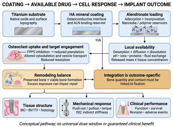
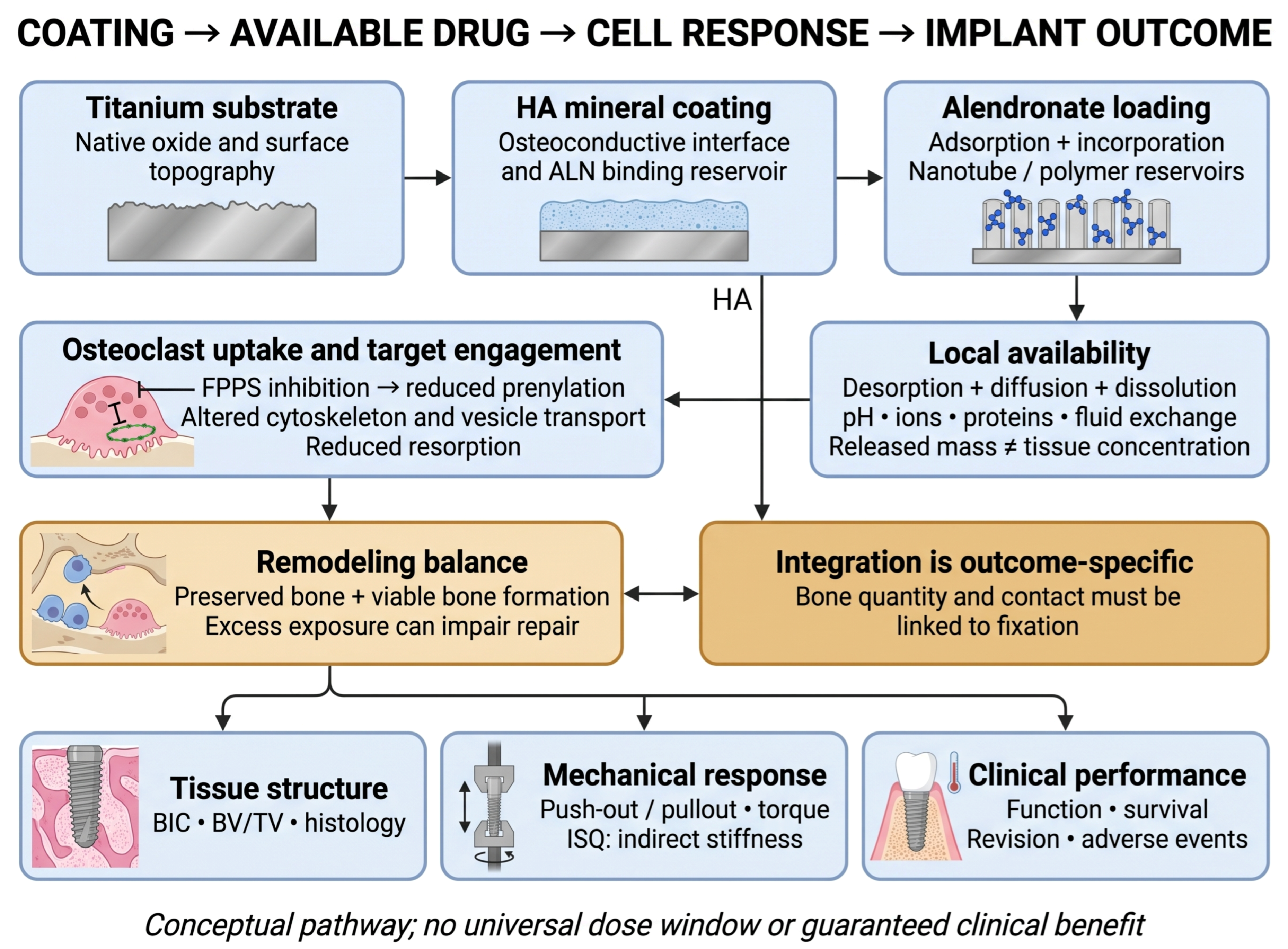
Newer nanotube systems add carboxymethyl chitosan, strontium or antibiotic delivery, increasing both functionality and attribution problems [13,14]. Antibacterial effects in gentamicin-containing groups do not establish antibacterial activity of alendronate. Figure 2 connects the principal coating architectures to exposure, osteoclast action and distinct osseointegration endpoints.
Figure 2.
From coating architecture to osseointegration endpoints. Adsorption, mineral incorporation and nanotube/polymer reservoirs determine where alendronate is retained and how it becomes available. Desorption, diffusion and mineral or matrix dissolution may coexist. Cellular uptake precedes FPPS inhibition, impaired protein prenylation and reduced osteoclast resorption. HA also supports bone attachment independently of the drug. Bone-to-implant contact and bone volume describe tissue structure; push-out, pullout and removal torque test mechanical fixation; implant stability quotient is an indirect stiffness-related measure. Clinical survival and revision require longer follow-up. Arrows show a conceptual pathway, not a validated dose–response or guaranteed benefit. Excess exposure may impair viable bone formation and remodeling. HA, hydroxyapatite; ALN, alendronate; FPPS, farnesyl diphosphate synthase; BIC, bone-to-implant contact; BV/TV, bone volume/tissue volume; ISQ, implant stability quotient [9,11,57,58,59,60]. Created in BioRender. Varut, R. (2026) https://BioRender.com/7oejimv.
4.4. Hybrid and Sequential Systems
Calcium phosphate–alendronate in PLGA microparticles and mineral-containing hydrogels extend reservoir design beyond metal coatings [61,62]. In one microparticle study, encapsulation efficiency increased from 8% to 14% at a 10% feed and from 11.5% to 20.5% at a 20% feed when calcium phosphate was included. The approximately 70% relative improvement reported by the authors is not an absolute encapsulation efficiency of 70% [61]. Such free-standing carriers require separate adhesion and insertion testing before use as implant coatings.
Sequential HA–collagen/BMP-2/alendronate scaffolds and IL-4/alendronate/nano-HA coatings on PEEK aim to coordinate early repair with later antiresorption [63,64]. Their combined effects cannot be attributed to alendronate alone. Appropriate comparisons include the carrier alone, each active component separately and the combination, with matched total composition and release characterization; otherwise, changes in timing, carrier or co-treatment remain alternative explanations.
Other systems include PCL short fibers containing alendronate-modified HA, a casein-phosphopeptide/tannic-acid/alendronate/magnesium coating for osteoporotic fracture repair, HA–alendronate/chitosan microspheres on polyetheretherketone (PEEK) and alendronate-loaded titanium nanotubes with a diclofenac/chitosan overcoat [65,66,67,68]. These are compositionally distinct; the last architecture lacks a reported HA component. Table 1 compares the manufacturing and release implications without treating all platforms as direct HA–alendronate/titanium evidence.
Table 1.
Fabrication, release and manufacturing implications of local alendronate architectures. Direct evidence concerns HA–ALN on titanium; adjacent platforms are identified explicitly.
5. Drug Release and Pharmaceutical Characterization
5.1. Loading Does Not Define the Effective Local Dose
Drug loading (mass per device, area or composite mass), encapsulation efficiency (fraction of input drug retained after processing) and released fraction (fraction of measured loading recovered in medium) describe different quantities. None directly specifies the concentration encountered by cells. Residual drug requires extraction of the used coating and verification of chemical identity; subtracting cumulative release from 100% gives an unreleased balance, not a measured active reservoir. Supplementary Table S1 retains study-specific units and assay conditions.
Pura et al. compared 0.02, 0.06 and 0.18 mg/cm2 alendronate on HA-associated porous titanium implants. The intermediate dose gave the most consistent bone apposition and peri-implant-bone benefits at 12 weeks, with effects depending on whether controls were bare titanium or HA-coated titanium [59]. Ancillary elution tests recovered 40 ± 10% within one hour in distilled deionized water, followed by slower release. Thus, strong HA association can coexist with a burst from other parts of a porous structure. These results define neither a human dose window nor mechanical fixation strength.
For a release assay with complete medium replacement, cumulative recovery must include drug removed at earlier sampling times. If only an aliquot is replaced, the calculation must additionally account for the changing concentration in the remaining medium. These basic mass-balance considerations are particularly important when comparing a burst phase with low later concentrations. Loss through adsorption to vessels, precipitation or analytical interference can otherwise resemble sustained retention. Unrecovered drug should not simply be labeled as still active within the coating.
5.2. Medium Composition and Analytical Selectivity
Release should be interpreted with pH, temperature, ionic strength, calcium/phosphate content, proteins, agitation, bath volume and fluid exchange. Acidification can increase calcium phosphate dissolution; ions and proteins can alter adsorption, rebinding and access to the surface. Full medium replacement maintains a concentration gradient differently from a static or partially exchanged bath. A protein-free buffer cannot reproduce osteoclast acidification, cellular uptake or clearance in peri-implant tissue. Shen et al. measured alendronate/calcium release and calcium in osteoclast medium, providing support for a dissolution-related contribution [57]. These coupled measurements are stronger evidence than an acidic release curve alone.
Alendronate quantification requires particular care because it lacks a strong conventional ultraviolet chromophore. The recent HA/HA–titanium comparison used high-performance liquid chromatography after 9-fluorenylmethyl chloroformate derivatization [27]. Such an approach illustrates a route to selective measurement, but derivatization yield, calibration in the relevant matrix, detection limits and recovery require validation. Gravimetric uptake is useful as a complementary measure and can be affected by retained water, salts or changes in mineral mass. Orthogonal assays should be used when a loading or release claim depends on small mass differences.
Mechanism cannot be assigned from the shape of a cumulative curve alone. Diffusion through a polymer barrier should be tested against thickness and permeability; desorption against binding and wash/rebinding measurements; dissolution-controlled release against calcium/phosphate loss, mineral mass and phase changes. Polymer molecular-mass loss or erosion can support a matrix-degradation contribution. The PCL comparison [58] supports release modulation, while the calcium phosphate-poly(lactic-co-glycolic acid) (CaP–PLGA) study [61] used distilled water to avoid phosphate interference with its copper-complex assay; its proposed osteoclast feedback mechanism was not established by that acellular release experiment. Mixed mechanisms remain plausible. An isolated 92% alendronate (ALN) release value [13] lacks interpretability without its time denominator and validated loading recovery.
5.3. Connecting Release to Biological Response
Direct-contact cell cultures combine surface topography, mineral exchange and drug exposure; eluate cultures emphasize soluble drug. Drug-free HA versus bare titanium tests the carrier contribution, and HA–ALN versus drug-free HA tests the incremental drug-associated effect. A soluble-ALN control or an ALN surface without HA helps distinguish mineral retention from drug action, but is not exposure-matched unless available concentrations are measured. Resorption assays, viability and differentiation/mineralization should be assessed separately; increased ALP or preserved cell viability alone does not demonstrate stronger fixation.
Carrier persistence may also limit repair. PCL/HA-based scaffolds released alendronate for more than 22 days, but persistent scaffold restricted bone formation within rat defects despite increased peripheral bone [69]. Table 2 retains the endpoint and applicability of selected studies; Supplementary Table S2 contrasts animal site, bone condition, follow-up and outcome.
Table 2.
Selected material and preclinical studies connecting formulation, measured response and limits of interpretation.
6. Evidence for Peri-Implant Integration
6.1. Animal Models and the Choice of Comparator
Animal models address different indications. HA–zoledronate intramedullary nails in rats exposed to polyethylene wear particles improved peri-implant bone measures and pullout resistance relative to HA-only controls [70]. This is a wear-osteolysis model using another drug. By contrast, HA–TiO2 nanotube/alendronate implants in osteoporotic rabbits were evaluated after three months by micro-computed tomography, histology and push-out testing [57], linking tissue structure to a mechanical endpoint within that model.
In ten skeletally mature dogs, local alendronate applied before bone compaction increased mechanical fixation around HA-coated Ti-alloy implants in proximal tibial cancellous bone at 12 weeks, without a significant BIC difference [60]. The model represented a cancellous interface relevant to cementless joint replacement but did not directly load the test implant. It also used topical alendronate rather than a preloaded HA–ALN coating. Pura et al. instead studied preloaded porous titanium in canine proximal femoral canals at 12 weeks, using bare-metal and HA-only comparators [59]. These distinctions prevent species, site, delivery mode and surgical technique from being treated as interchangeable.
A rabbit study involving wear-particle-induced high bone turnover compared HA with alendronate–HA and risedronate–HA coatings. Alendronate-associated effects were more pronounced around the implant, whereas risedronate produced greater changes at distant skeletal sites [71]. The observation is consistent with formulation-dependent distribution, but it also cautions against assuming that local delivery eliminates systemic effects. A later rabbit study of PLGA–alendronate-coated titanium reported increased anchorage stability and fewer osteoclasts and macrophages after four weeks [72]. That carrier did not reproduce the HA component of the central review question.
In an eight-sheep pilot study, zoledronate-coated dental implants had an early removal-torque advantage over certain groups, but not at 28 days; ISQ, BIC and bone-area measures did not differ significantly [73]. BIC describes bone contact, bone volume describes tissue abundance, and push-out, pullout or removal torque test resistance under different mechanical geometries. Greater bone volume does not itself distinguish new formation from reduced removal. ISQ is an indirect stiffness-related measure. None alone establishes long-term survival. Multiple implants within one animal also require analysis as clustered observations.
6.2. Human Evidence Is Heterogeneous and Largely Indirect
The most informative clinical studies test related local-bisphosphonate concepts rather than a single HA–alendronate product. In the 2012 split-mouth trial by Abtahi et al., 16 patients received paired titanium dental implants, one carrying a fibrinogen coating with pamidronate and ibandronate. The increase in ISQ at six months favored the coated implant by 6.9 units, and marginal resorption was lower [74]. This was an encouraging proof of concept for local delivery, but the carrier and active drugs differed from HA–alendronate.
The five-year radiographic assessment retained 14 of those original patients. Median marginal bone loss was 0.20 mm for coated implants and 0.70 mm for controls, with all evaluated implants functioning [75]. This follow-up extends the same randomized cohort; it is not an independent trial. Its size and radiographic endpoint cannot establish a reduction in rare adverse events or a survival advantage.
Subsequent studies have produced less consistent stability findings. A 2019 paired trial in 16 patients found no significant early ISQ advantage for zoledronate-coated implants, although marginal-bone findings favored coating [76]. A later multicenter trial randomized 62 patients, of whom 49 completed follow-up. It found no significant improvement in ISQ or one-year marginal bone levels with a fibrinogen/zoledronate coating [15]. These results support clinical feasibility while leaving superiority unresolved. Small samples and restricted eligibility also limit conclusions about patients with severe skeletal compromise.
Local alendronate itself has been evaluated as a gel applied immediately before dental implant insertion. In a four-group study of 27 patients and 71 implants, alendronate, recombinant BMP-2, their combination and untreated controls showed no significant overall between-group advantage in stability; the study also reported no significant treatment effect on marginal bone changes versus control [16]. This result should neither be omitted nor interpreted as proof that all alendronate coatings are ineffective. It addresses a peri-insertion gel with its own exposure profile, not mineral-bound drug elution.
Outside the jaw, a randomized study of 20 osteotomy patients tested zoledronate/fibrinogen-coated external fixation pins against site-specific controls, including separate HA-coated metaphyseal pins [77]. This concerns temporary fracture/osteotomy fixation rather than dental implants or joint-replacement stems. No superiority was demonstrated; the study was not designed to establish noninferiority. Table 3 keeps these clinical settings and products separate.
Table 3.
Human evidence for related local bisphosphonate interventions. None directly validates a standardized HA–alendronate coating.
6.3. Mineral Sequestration After Systemic Administration
Earlier systemic-treatment studies provide a separate context. Von Knoch et al. administered subcutaneous alendronate or intravenous zoledronate in a rabbit implant model and observed increased peri-implant bone stock; these were not drug-eluting coatings [78]. In a small randomized hip-arthroplasty study, Tapaninen et al. followed 16 patients after six months of postoperative alendronate plus calcium or calcium alone. Although early bone preservation favored treatment, the between-group differences in bone mineral density changes were not statistically significant at five years [79]. The study therefore does not establish a durable five-year treatment advantage despite the wording of its title.
A distinct strategy is to implant a calcium phosphate material and subsequently administer a bisphosphonate systemically. A 2024 study used calcium sulfate/HA around fixation devices and showed that the timing of systemic zoledronic acid influenced mineral uptake and peri-implant bone formation in animal experiments. A small human feasibility component extended the concept to trochanteric fracture care [80]. This is relevant to the role of mineral as a local sink, but it retains systemic dosing and does not demonstrate controlled elution from an alendronate-preloaded coating. It should therefore remain a separate translational strategy rather than be combined with coating trials as though the interventions were equivalent.
7. Safety and Requirements for Translation
7.1. A Local Route Requires a Local Safety Assessment
Alendronate distribution and renal phosphate physiology provide background for assessing escape from a local reservoir [81,82,83]. They do not define a safe implant loading. Safety must relate measured local and systemic exposure to tissue effects.
Local delivery may reduce the administered amount needed to expose an implant interface, but reduced systemic exposure must be measured rather than presumed. Mineral-bound drug may be released at a later time or redistributed after dissolution and remodeling. The distant skeletal changes observed in the rabbit coating comparison are a concrete reason to include systemic observations in local-device studies [71]. Safety evaluation should therefore consider both the peri-implant tissue and the drug fraction escaping the implantation site.
Medication-related osteonecrosis of the jaw (MRONJ) is an established concern with antiresorptive therapy and requires attention in the dental setting. The American Association of Oral and Maxillofacial Surgeons position paper emphasizes the clinical complexity of its diagnosis and management [84].
Reports after systemic, particularly intravenous, bisphosphonate treatment characterize a different exposure setting [85,86,87]. Their MRONJ risk estimates cannot be assigned to local coatings, and small coating trials cannot exclude rare events. Local assessment should include mucosal healing, inflammation, infection and remodeling quality alongside mineralized tissue quantity.
Likewise, systemic efficacy in oncology cannot substitute for local-device efficacy because the drug, route, population and endpoints differ [88].
Excess antiresorptive activity could preserve existing mineral while disrupting the turnover needed for repair. The distinction between retained bone and newly formed, mechanically competent bone therefore matters. The dose–response findings around porous titanium and the persistence of PCL in scaffold-treated defects illustrate separate ways in which more drug or longer residence may fail to improve the complete repair process [59,69]. These observations favor a measured exposure–response approach rather than selection of the formulation with the largest loading capacity.
7.2. Product Identity Must Survive Manufacture and Use
An implant coating becomes a pharmaceutical delivery system only if its composition and performance are reproducible. Proposed release specifications should cover drug identity, loading, early release, later release and residual drug. Material specifications should include calcium phosphate phases, coating thickness and uniformity, adhesion and the extent of particulate shedding. The final sterilized and packaged device is the relevant test article; successful characterization of a laboratory precursor does not establish the quality of that product.
The substrate remains part of the product. Anodization changes the titanium oxide/topography; thermal deposition and subsequent processing can alter interface stresses; thicker or poorly adherent layers may crack or shed during insertion and cyclic loading. Adhesion before and after soaking, insertion damage, corrosion/electrochemical behavior, particulate shedding and fatigue must therefore be tested on the final sterilized device. An ALN–hydrolyzed-collagen coating improved electrochemical protection in artificial saliva [89], but that adjacent formulation does not establish corrosion or fatigue performance for HA–ALN coatings. Comparable evidence on stainless steel, cobalt–chromium or degradable magnesium cannot be presumed from titanium because their oxide chemistry, corrosion and mechanics differ. PEEK examples are polymer-substrate evidence, not evidence for other metals.
Manufacturing complexity should be justified by a specific unmet requirement. A polymer layer may moderate a burst but introduce degradation and adhesion variables. A second bioactive agent may improve one model while expanding the need for dose optimization and safety testing. A practical development program should first identify the function that the simpler drug–mineral system cannot provide, then demonstrate that the additional component resolves that limitation under relevant conditions.
7.3. A Staged Evidence Pathway
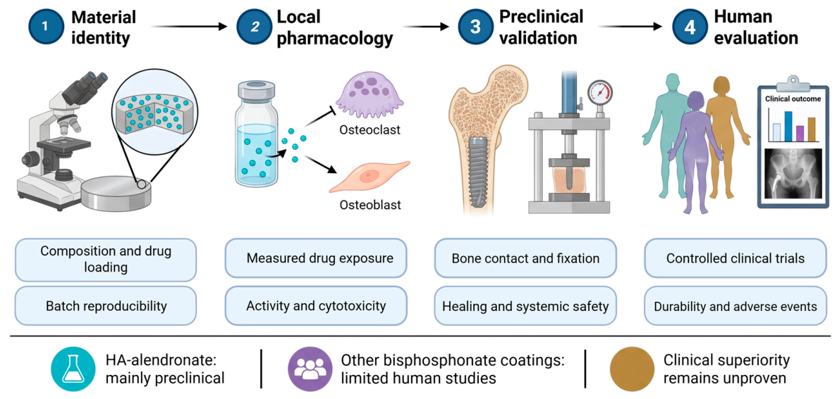
Figure 3 presents a proposed pathway connecting material identity to human evaluation. The first stage is a reproducible coating with a selective loading assay and documented structural integrity. The second connects release under defined conditions to measured cellular effects, including both efficacy-related endpoints and cytotoxicity. The third combines histology with mechanical testing and local and systemic safety in an indication-relevant animal model. Reporting should address allocation, blinding, experimental units, exclusions and sample-size rationale, consistent with Animal Research: Reporting of In Vivo Experiments (ARRIVE) 2.0 principles [90].
Figure 3.
Proposed evidence pathway for a drug-eluting implant. Material identity and batch consistency should precede interpretation of local pharmacology, animal fixation and clinical performance. The stages describe evidence required, not milestones already completed by every HA–alendronate system. Clinical charts and instrument icons are illustrative and contain no numerical results. Human evidence from other bisphosphonate coatings is informative but cannot substitute for validation of a specified HA–alendronate product. Created in BioRender. Varut, M. (2026) https://app.biorender.com/illustrations/6ac553924d7b0b8a66284216 [15,16,59,74,76,90].
Clinical evaluation should compare a defined coating with surface-matched controls and the appropriate standard implant. HA-only controls isolate the incremental effect of drug addition. Dental implants in jawbone, temporary fixation devices and joint-replacement components face different anatomy, loading and failure modes [4,74,77,79]. Statistically significant marginal-bone or ISQ differences need assessment against effect size, precision and a prespecified clinically meaningful threshold. Conversely, a nonsignificant comparison does not establish equivalence. Durable benefit requires function, failure/revision and adverse-event follow-up.
Clinical protocols should also account for multiple implants in one patient, pre-existing antiresorptive exposure and concurrent therapies. The mixed results from the clinical literature argue for a clearly specified primary endpoint and realistic effect assumptions rather than extrapolation from a favorable cell assay. A successful product would demonstrate that its release profile improves an outcome that matters for that implantation context without compromising healing.
8. Limitations of the Review
This is a structured critical narrative review, without protocol registration, meta-analysis or formal risk-of-bias/certainty grading. Differences in coating composition, assay conditions, species, implant sites and endpoints preclude a pooled efficacy claim. Incomplete analytical recovery, residual-drug measurements, durability testing and long-term follow-up further limit comparison. The 62-study selection and contextual literature have different purposes: the latter supports background and interpretation and is not additional independent implant-efficacy evidence.
Coatings, gels and defect-filling scaffolds produce different exposure geometries; healthy, osteoporotic and wear-particle models address different questions. Clinical evidence for a specified HA–alendronate coating remains insufficient. The validation pathway proposed here identifies evidence gaps rather than constituting a clinically validated procedure or a dosing recommendation.
9. Conclusions
HA combines an osteoconductive surface with a reservoir for alendronate, but strong mineral affinity alone is not evidence of effective local therapy. Experimental benefits depend on carrier chemistry, biologically available exposure, cell response, implant site and comparator.
Translation requires reproducible formulation and loading, active-drug availability over a relevant period, preserved physiological remodeling and acceptable coating durability under use. Human findings from other bisphosphonate coatings or local alendronate gels remain indirect. Controlled, indication-specific trials must demonstrate a clinically meaningful improvement before HA–alendronate coatings can be considered validated drug-eluting implants.
Supplementary Materials
The following supporting information can be downloaded at: https://www.mdpi.com/article/10.3390/pharmaceutics18111447/s1, Appendix A Database search strings; Figure S1: Selection flow for the structured critical narrative review; Table S1: Loading and release under defined conditions; Table S2: Animal models and endpoint applicability.
Author Contributions
Conceptualization P.D.F.V., N.B., A.M.K., R.M.V. and M.S.; methodology, P.D.F.V., D.-M.T., A.M.K., R.M.V., P.I.D., D.R., C.C.A. and M.S.; validation, D.-M.T., N.B., A.M.K., R.M.V., P.I.D. and M.S.; formal analysis, C.C.A., P.D.F.V., D.-M.T., D.R. and M.S.; investigation, P.D.F.V., D.-M.T., A.M.K., R.M.V., P.I.D., D.R. and M.S.; resources, A.M.K., R.M.V., N.B. and P.I.D.; data curation, P.D.F.V., D.-M.T., R.M.V., D.R. and M.S.; writing—original draft preparation, C.C.A., P.D.F.V., N.B., R.M.V. and M.S.; writing—review and editing, C.C.A., P.D.F.V., D.-M.T., N.B., A.M.K., R.M.V., P.I.D., D.R. and M.S.; visualization, D.-M.T. and M.S.; supervision, A.M.K., R.M.V. and M.S.; project administration, A.M.K., R.M.V., P.I.D. and M.S. All authors have read and agreed to the published version of the manuscript.
Funding
The Article Processing Charges were funded by the University of Medicine and Pharmacy of Craiova, Romania.
Institutional Review Board Statement
Not applicable.
Informed Consent Statement
Not applicable.
Data Availability Statement
No new data were created or analyzed in this study. Data sharing is not applicable to this article.
Conflicts of Interest
The authors declare no conflicts of interest.
Abbreviations
ALN, alendronate; ALP, alkaline phosphatase; BIC, bone-to-implant contact; BMP-2, bone morphogenetic protein 2; CaP, calcium phosphate; FPPS, farnesyl diphosphate synthase; FTIR, Fourier-transform infrared spectroscopy; HA, hydroxyapatite; GTP, Guanosine triphosphate-binding proteins; HPLC, high-performance liquid chromatography; ISQ, implant stability quotient; MAPLE, matrix-assisted pulsed laser evaporation; MRONJ, medication-related osteonecrosis of the jaw; OCP, octacalcium phosphate; PCL, polycaprolactone; PEEK, polyetheretherketone; PLGA, poly(lactic-co-glycolic acid); XRD, X-ray diffraction.
References
- Marieb, E.N.; Hoehn, K. Human Anatomy & Physiology, 7th ed.; Pearson Benjamin Cummings: San Francisco, CA, USA, 2007. [Google Scholar]
- Puleo, D.A. Bone-Implant Interface. In Encyclopedia of Biomaterials and Biomedical Engineering; Marcel Dekker: New York, NY, USA, 2004; Volume 1, pp. 190–198. [Google Scholar]
- Jarcho, M.; Kay, J.F.; Gumaer, K.I.; Doremus, R.H.; Drobeck, H.P. Tissue, cellular and subcellular events at a bone-ceramic hydroxylapatite interface. J. Bioeng. 1977, 1, 79–92. [Google Scholar] [PubMed]
- Jaffe, W.L.; Scott, D.F. Total hip arthroplasty with hydroxyapatite-coated prostheses. J. Bone Jt. Surg. Am. 1996, 78, 1918–1934. [Google Scholar] [CrossRef] [Scilit] [PubMed]
- Wang, M. Developing bioactive composite materials for tissue replacement. Biomaterials 2003, 24, 2133–2151. [Google Scholar] [CrossRef] [Scilit] [PubMed]
- Von Euw, S.; Wang, Y.; Laurent, G.; Drouet, C.; Babonneau, F.; Nassif, N.; Azaïs, T. Bone mineral: New insights into its chemical composition. Sci. Rep. 2019, 9, 8456. [Google Scholar] [CrossRef] [Scilit] [PubMed]
- Wilson, C.J.; Clegg, R.E.; Leavesley, D.I.; Pearcy, M.J. Mediation of biomaterial-cell interactions by adsorbed proteins: A review. Tissue Eng. 2005, 11, 1–18. [Google Scholar] [CrossRef] [Scilit] [PubMed]
- Nancollas, G.H.; Tang, R.; Phipps, R.J.; Henneman, Z.; Gulde, S.; Wu, W.; Mangood, A.; Russell, R.G.; Ebetino, F.H. Novel insights into actions of bisphosphonates on bone: Differences in interactions with hydroxyapatite. Bone 2006, 38, 617–627. [Google Scholar] [CrossRef] [Scilit] [PubMed]
- Dunford, J.E.; Thompson, K.; Coxon, F.P.; Luckman, S.P.; Hahn, F.M.; Poulter, C.D.; Ebetino, F.H.; Rogers, M.J. Structure-activity relationships for inhibition of farnesyl diphosphate synthase in vitro and inhibition of bone resorption in vivo by nitrogen-containing bisphosphonates. J. Pharmacol. Exp. Ther. 2001, 296, 235–242. [Google Scholar] [CrossRef] [Scilit]
- Boanini, E.; Torricelli, P.; Gazzano, M.; Giardino, R.; Bigi, A. Alendronate-hydroxyapatite nanocomposites and their interaction with osteoclasts and osteoblast-like cells. Biomaterials 2008, 29, 790–796. [Google Scholar] [CrossRef] [Scilit] [PubMed]
- Bigi, A.; Boanini, E.; Capuccini, C.; Fini, M.; Mihailescu, I.N.; Ristoscu, C.; Sima, F.; Torricelli, P. Biofunctional alendronate-Hydroxyapatite thin films deposited by Matrix Assisted Pulsed Laser Evaporation. Biomaterials 2009, 30, 6168–6177. [Google Scholar] [CrossRef] [Scilit] [PubMed]
- Despotović, I.; Petrović, Ž.; Katić, J.; Mikić, D. Alendronate as Bioactive Coating on Titanium Surfaces: An Investigation of CaP-Alendronate Interactions. Materials 2024, 17, 2703. [Google Scholar] [CrossRef] [Scilit] [PubMed]
- Dhinasekaran, D.; Sivakumar, J.P.; Rajendran, A.R. Enhancement of implant performance through synergistic drug delivery and bioactivity by nanotubular titanium with hydroxyapatite coating. Int. J. Pharm. 2025, 681, 125854. [Google Scholar] [CrossRef] [Scilit] [PubMed]
- Jia, K.; Zuo, C.; Xu, Y.; Ma, W.; Wang, L.; Ji, Y.; Chen, J.; Zhang, Q.; Pan, C.; Liu, T. Carboxymethyl chitosan/alendronate sodium/Sr2+ modified TiO2 nanotube arrays enhancing osteogenic activity and antibacterial property. Biomater. Adv. 2025, 167, 214107. [Google Scholar] [CrossRef] [Scilit] [PubMed]
- Mokhtari, R.A.; Olsson, M.; Östman, P.O.; Dahlin, C. A randomized, multicenter, double-blinded parallel study to evaluate the safety and performance of zoledronate-coated versus uncoated dental implants in partially edentulous patients. Clin. Implant Dent. Relat. Res. 2024, 26, 78–87. [Google Scholar] [CrossRef] [Scilit] [PubMed]
- Al-Assaf, D.A.; Bede, S.Y. The Effects of Local Alendronate with or Without Recombinant Human Bone Morphogenetic Protein 2 on Dental Implant Stability and Marginal Bone Level: A Randomized Controlled Study. J. Craniofac. Surg. 2022, 33, 1003–1007. [Google Scholar] [CrossRef] [Scilit] [PubMed]
- Zhang, J.; Bai, H.; Bai, M.; Wang, X.; Li, Z.; Xue, H.; Wang, J.; Cui, Y.; Wang, H.; Wang, Y.; et al. Bisphosphonate-incorporated coatings for orthopedic implants functionalization. Mater. Today Bio 2023, 22, 100737. [Google Scholar] [CrossRef] [Scilit] [PubMed]
- Min, K.H.; Kim, D.H.; Kim, K.H.; Seo, J.-H.; Pack, S.P. Biomimetic Scaffolds of Calcium-Based Materials for Bone Regeneration. Biomimetics 2024, 9, 511. [Google Scholar] [CrossRef] [Scilit] [PubMed]
- Pérez-González, F.; Sánchez-Labrador, L.; Sáez-Alcaide, L.M.; López Carriches, C.; Méniz-García, C. Does local bisphosphonate application or surface coating improve implant success? A systematic review. J. Stomatol. Oral Maxillofac. Surg. 2026, 127, 102780. [Google Scholar] [CrossRef] [Scilit] [PubMed]
- Hao, X.; Jiang, B.; Wu, J.; Xiang, D.; Xiong, Z.; Li, C.; Li, Z.; He, S.; Tu, C.; Li, Z. Nanomaterials for bone metastasis. J. Control. Release 2024, 373, 640–651. [Google Scholar] [CrossRef] [Scilit] [PubMed]
- Zhou, L.; Hou, Y.-H.; Luo, S.-Y.; Wang, H.-J.; Wang, H.-R.; Guo, X.-W.; Liu, J.; Zhang, X.-Y.; Zhang, X. An amyloid-like protein coating synergizes with a carboxymethyl chitosan/amorphous calcium phosphate nanocomposite for biomimetic dental enamel remineralization. J. Dent. 2026, 167, 106573. [Google Scholar] [CrossRef] [Scilit] [PubMed]
- Baethge, C.; Goldbeck-Wood, S.; Mertens, S. SANRA-a scale for the quality assessment of narrative review articles. Res. Integr. Peer Rev. 2019, 4, 5. [Google Scholar] [CrossRef] [Scilit] [PubMed]
- Leu, C.T.; Luegmayr, E.; Freedman, L.P.; Rodan, G.A.; Reszka, A.A. Relative binding affinities of bisphosphonates for human bone and relationship to antiresorptive efficacy. Bone 2006, 38, 628–636. [Google Scholar] [CrossRef] [Scilit] [PubMed]
- Henneman, Z.J.; Nancollas, G.H.; Ebetino, F.H.; Russell, R.G.; Phipps, R.J. Bisphosphonate binding affinity as assessed by inhibition of carbonated apatite dissolution in vitro. J. BioMed. Mater. Res. A 2008, 85, 993–1000. [Google Scholar] [CrossRef] [Scilit] [PubMed]
- Ebetino, F.H.; Barnett, B.L.; Russell, R.G.G. A computational model delineates differences in hydroxyapatite binding affinities of bisphosphonates in clinical use [conference abstract]. J. Bone Miner. Res. 2005, 20, S259. [Google Scholar]
- Paulo, S.; Laranjo, M.; Abrantes, A.M.; Teixo, R.J.; Fonseca, D.A.; Mendes-Abreu, J.; Figueiredo, J.P.; Botelho, M.F.; Ferreira, M.M.; Serra, A. Differential Zoledronate Adsorption by Commercially Available Biphasic Calcium Phosphate Bone Substitutes. Pharmaceuticals 2026, 19, 1281. [Google Scholar] [CrossRef] [Scilit] [PubMed]
- Trasca, D.M.; Pluta, I.D.; Sirbulet, C.; Varut, R.M.; Singer, C.E.; Preoteasa, D.; Stoica, G.A. Comparative Molecular Docking, Molecular Dynamics and Adsorption-Release Analysis of Calcium Fructoborate and Alendronate Salts on Hydroxyapatite and Hydroxyapatite-Titanium Implants. Biomedicines 2026, 14, 44. [Google Scholar] [CrossRef] [Scilit] [PubMed]
- Rodan, G.A.; Fleisch, H.A. Bisphosphonates: Mechanisms of action. J. Clin. Investig. 1996, 97, 2692–2696. [Google Scholar] [CrossRef] [Scilit] [PubMed]
- Ebetino, F.H.; Bayless, A.V.; Amburgey, J.; Ibbotson, K.J.; Dansereau, S.; Ebrahimpour, A. Elucidation of a Pharmacophore for the Bisphosphonate Mechanism of Bone Antiresorptive Activity. Phosphorus Sulfur Silicon Relat. Elem. 1996, 109, 217–220. [Google Scholar] [CrossRef] [Scilit]
- Francis, R.M. Bisphosphonates in the treatment of osteoporosis in 1997: A review. Curr. Ther. Res. 1997, 58, 656–678. [Google Scholar] [CrossRef] [Scilit]
- Fleisch, H. Bisphosphonates in Bone Disease: From the Laboratory to the Patient, 3rd ed.; Parthenon Publishing Group: New York, NY, USA, 1997. [Google Scholar]
- Ebetino, F.H.; Francis, M.D.; Rogers, M.J.; Russell, R.G.G. Mechanisms of action of etidronate and other bisphosphonates. Rev. Contemp. Pharmacother. 1998, 9, 233–243. [Google Scholar] [CrossRef] [Scilit] [PubMed]
- Russell, R.G.; Rogers, M.J. Bisphosphonates: From the laboratory to the clinic and back again. Bone 1999, 25, 97–106. [Google Scholar] [CrossRef] [Scilit] [PubMed]
- Papapoulos, S.E. Bisphosphonates: How do they work? Best Pract. Res. Clin. Endocrinol. Metab. 2008, 22, 831–847. [Google Scholar] [CrossRef] [Scilit] [PubMed]
- Russell, R.G.; Watts, N.B.; Ebetino, F.H.; Rogers, M.J. Mechanisms of action of bisphosphonates: Similarities and differences and their potential influence on clinical efficacy. Osteoporos. Int. 2008, 19, 733–759. [Google Scholar] [CrossRef] [Scilit] [PubMed]
- Luckman, S.P.; Coxon, F.P.; Ebetino, F.H.; Russell, R.G.; Rogers, M.J. Heterocycle-containing bisphosphonates cause apoptosis and inhibit bone resorption by preventing protein prenylation: Evidence from structure-activity relationships in J774 macrophages. J. Bone Min. Res. 1998, 13, 1668–1678. [Google Scholar] [CrossRef] [Scilit] [PubMed]
- Kavanagh, K.L.; Guo, K.; Dunford, J.E.; Wu, X.; Knapp, S.; Ebetino, F.H.; Rogers, M.J.; Russell, R.G.; Oppermann, U. The molecular mechanism of nitrogen-containing bisphosphonates as antiosteoporosis drugs. Proc. Natl. Acad. Sci. USA 2006, 103, 7829–7834. [Google Scholar] [CrossRef] [Scilit] [PubMed]
- Petrović, Ž.; Šarić, A.; Despotović, I.; Katić, J.; Peter, R.; Petravić, M.; Petković, M. A New Insight into Coating’s Formation Mechanism Between TiO2 and Alendronate on Titanium Dental Implant. Materials 2020, 13, 3220. [Google Scholar] [CrossRef] [Scilit] [PubMed]
- García-Moreno, C.; Serrano, S.; Nacher, M.; Farré, M.; Díez, A.; Mariñoso, M.L.; Carbonell, J.; Mellibovsky, L.; Nogués, X.; Ballester, J.; et al. Effect of alendronate on cultured normal human osteoblasts. Bone 1998, 22, 233–239. [Google Scholar] [CrossRef] [Scilit] [PubMed]
- Giuliani, N.; Pedrazzoni, M.; Negri, G.; Passeri, G.; Impicciatore, M.; Girasole, G. Bisphosphonates stimulate formation of osteoblast precursors and mineralized nodules in murine and human bone marrow cultures in vitro and promote early osteoblastogenesis in young and aged mice in vivo. Bone 1998, 22, 455–461. [Google Scholar] [CrossRef] [Scilit] [PubMed]
- Hadad, H.; de Jesus, L.K.; da Silva, M.P.; Oliveira, M.E.F.S.; Guastaldi, F.P.S.; Nilsson, O.; Okamoto, R.; Souza, F.Á. A Systematic Review of the Effects of Bisphosphonates on Osteoblasts In Vitro. Calcif. Tissue Int. 2025, 116, 86. [Google Scholar] [CrossRef] [Scilit] [PubMed]
- Boanini, E.; Torricelli, P.; Gazzano, M.; Fini, M.; Bigi, A. The effect of alendronate doped calcium phosphates on bone cells activity. Bone 2012, 51, 944–952. [Google Scholar] [CrossRef] [Scilit] [PubMed]
- Santos, M.H.; Oliveira, M.D.; Souza, L.P.D.F.; Mansur, H.S.; Vasconcelos, W.L. Synthesis control and characterization of hydroxyapatite prepared by wet precipitation process. Mater. Res. 2004, 7, 625–630. [Google Scholar] [CrossRef] [Scilit]
- Manuel, C.M.; Ferraz, M.P.; Monteiro, F.J. Synthesis of Hydroxyapatite and Tricalcium Phosphate Nanoparticles—Preliminary Studies. Key Eng. Mater. 2003, 240–242, 555–558. [Google Scholar] [CrossRef] [Scilit]
- Ferraz, M.P.; Monteiro, F.J.; Manuel, C.M. Hydroxyapatite nanoparticles: A review of preparation methodologies. J. Appl. Biomater. Biomech. 2004, 2, 74–80. [Google Scholar]
- Nayak, A.K. Hydroxyapatite Synthesis Methodologies: An Overview. Int. J. ChemTech Res. 2010, 2, 903–907. [Google Scholar]
- Suchanek, W.L.; Riman, R.E. Hydrothermal Synthesis of Advanced Ceramic Powders. Adv. Sci. Technol. 2006, 45, 184–193. [Google Scholar] [CrossRef] [Scilit]
- Balamurugan, A.; Michel, J.; Fauré, J.; Benhayoune, H.; Wortham, L.; Sockalingum, G.; Banchet, V.; Bouthors, S.; Laurent-Maquin, D.; Balossier, G. Synthesis and structural analysis of sol gel derived stoichiometric monophasic hydroxyapatite. Ceram. Silik. 2006, 50, 27–31. [Google Scholar]
- Brendel, T.; Engel, A.; Rüssel, C. Hydroxyapatite coatings by a polymeric route. J. Mater. Sci. Mater. Med. 1992, 3, 175–179. [Google Scholar] [CrossRef] [Scilit]
- Kimura, I. Synthesis of Hydroxyapatite by Interfacial Reaction in a Multiple Emulsion. Res. Lett. Mater. Sci. 2007, 2007, 71284. [Google Scholar] [CrossRef] [Scilit]
- Kokubo, T.; Kushitani, H.; Sakka, S.; Kitsugi, T.; Yamamuro, T. Solutions able to reproduce in vivo surface-structure changes in bioactive glass-ceramic A-W. J. BioMed. Mater. Res. 1990, 24, 721–734. [Google Scholar] [CrossRef] [Scilit] [PubMed]
- Tas, A.C. Synthesis of biomimetic Ca-hydroxyapatite powders at 37 degrees C in synthetic body fluids. Biomaterials 2000, 21, 1429–1438. [Google Scholar] [CrossRef] [Scilit] [PubMed]
- Shirkhanzadeh, M. Direct formation of nanophase hydroxyapatite on cathodically polarized electrodes. J. Mater. Sci. Mater. Med. 1998, 9, 67–72. [Google Scholar] [CrossRef] [Scilit] [PubMed]
- Manso, M.; Jiménez, C.; Morant, C.; Herrero, P.; Martínez-Duart, J.M. Electrodeposition of hydroxyapatite coatings in basic conditions. Biomaterials 2000, 21, 1755–1761. [Google Scholar] [CrossRef] [Scilit] [PubMed]
- Stigter, M.; de Groot, K.; Layrolle, P. Incorporation of tobramycin into biomimetic hydroxyapatite coating on titanium. Biomaterials 2002, 23, 4143–4153. [Google Scholar] [CrossRef] [Scilit] [PubMed]
- Boanini, E.; Torricelli, P.; Forte, L.; Pagani, S.; Mihailescu, N.; Ristoscu, C.; Mihailescu, I.N.; Bigi, A. Antiresorption implant coatings based on calcium alendronate and octacalcium phosphate deposited by matrix assisted pulsed laser evaporation. Colloids Surf. B Biointerfaces 2015, 136, 449–456. [Google Scholar] [CrossRef] [Scilit] [PubMed]
- Shen, X.; Ma, P.; Hu, Y.; Xu, G.; Xu, K.; Chen, W.; Ran, Q.; Dai, L.; Yu, Y.; Mu, C.; et al. Alendronate-loaded hydroxyapatite-TiO2 nanotubes for improved bone formation in osteoporotic rabbits. J. Mater. Chem. B 2016, 4, 1423–1436. [Google Scholar] [CrossRef] [Scilit] [PubMed]
- Bose, S.; Vu, A.A.; Emshadi, K.; Bandyopadhyay, A. Effects of polycaprolactone on alendronate drug release from Mg-doped hydroxyapatite coating on titanium. Mater. Sci. Eng. C Mater. Biol. Appl. 2018, 88, 166–171. [Google Scholar] [CrossRef] [Scilit] [PubMed]
- Pura, J.A.; Bobyn, J.D.; Tanzer, M. Implant-Delivered Alendronate Causes a Dose-Dependent Response on Net Bone Formation Around Porous Titanium Implants in Canines. Clin. Orthop. Relat. Res. 2016, 474, 1224–1233. [Google Scholar] [CrossRef] [Scilit] [PubMed]
- Jakobsen, T.; Baas, J.; Kold, S.; Bechtold, J.E.; Elmengaard, B.; Søballe, K. Local bisphosphonate treatment increases fixation of hydroxyapatite-coated implants inserted with bone compaction. J. Orthop. Res. 2009, 27, 189–194. [Google Scholar] [CrossRef] [Scilit] [PubMed]
- Matrali, S.S.H.; Ghag, A.K. Feedback-Controlled Release of Alendronate from Composite Microparticles. J. Funct. Biomater. 2020, 11, 46. [Google Scholar] [CrossRef] [Scilit] [PubMed]
- Klara, J.; Onak, S.; Kowalczyk, A.; Horak, W.; Wójcik, K.; Lewandowska-Łańcucka, J. Towards Controlling the Local Bone Tissue Remodeling-Multifunctional Injectable Composites for Osteoporosis Treatment. Int. J. Mol. Sci. 2023, 24, 4959. [Google Scholar] [CrossRef] [Scilit] [PubMed]
- Lee, D.; Wufuer, M.; Kim, I.; Choi, T.H.; Kim, B.J.; Jung, H.G.; Jeon, B.; Lee, G.; Jeon, O.H.; Chang, H.; et al. Sequential dual-drug delivery of BMP-2 and alendronate from hydroxyapatite-collagen scaffolds for enhanced bone regeneration. Sci. Rep. 2021, 11, 746. [Google Scholar] [CrossRef] [Scilit] [PubMed]
- Zheng, Y.; Gao, A.; Bai, J.; Liao, Q.; Wu, Y.; Zhang, W.; Guan, M.; Tong, L.; Geng, D.; Zhao, X.; et al. A programmed surface on polyetheretherketone for sequentially dictating osteoimmunomodulation and bone regeneration to achieve ameliorative osseointegration under osteoporotic conditions. Bioact. Mater. 2022, 14, 364–376. [Google Scholar] [CrossRef] [Scilit] [PubMed]
- Yang, Y.; Sun, M.; Jia, W.; Jiao, K.; Wang, S.; Liu, Y.; Liu, L.; Dai, Z.; Jiang, X.; Yang, T.; et al. An osteoporosis bone defect regeneration strategy via three-dimension short fibers loaded with alendronate modified hydroxyapatite. Colloids Surf. B Biointerfaces 2024, 233, 113659. [Google Scholar] [CrossRef] [Scilit] [PubMed]
- Yang, S.; Huang, Y.; Lv, Y. Construction of Mg2+ loaded multifunctional casein phosphopeptide/alendronate sodium antioxidative coating for repairing osteoporotic fracture. Int. J. Biol. Macromol. 2025, 305, 141333. [Google Scholar] [CrossRef] [Scilit] [PubMed]
- Li, R.; Lu, R.; Yang, L.; Li, J.; Guo, Z.; Yan, Q.; Li, M.; Ni, Y.; Chen, K.; Li, Y.; et al. Immobilization of chitosan nano-hydroxyapatite alendronate composite microspheres on polyetheretherketone surface to enhance osseointegration by inhibiting osteoclastogenesis and promoting osteogenesis. Chin. Chem. Lett. 2025, 36, 110242. [Google Scholar] [CrossRef] [Scilit]
- Zhang, T.; Liu, N.; Xie, C.; Xiao, X. Electrophoretic loading of alendronate on TiO2 nanotube arrays. Mater. Lett. 2024, 372, 137101. [Google Scholar] [CrossRef] [Scilit]
- Hedvičáková, V.; Žižková, R.; Buzgo, M.; Vištejnová, L.; Klein, P.; Hovořáková, M.; Bartoš, M.; Steklíková, K.; Luňáčková, J.; Šebová, E.; et al. The Gradual Release of Alendronate for the Treatment of Critical Bone Defects in Osteoporotic and Control Rats. Int. J. Nanomed. 2023, 18, 541–560. [Google Scholar] [CrossRef] [Scilit] [PubMed]
- Suratwala, S.J.; Cho, S.K.; van Raalte, J.J.; Park, S.H.; Seo, S.W.; Chang, S.S.; Gardner, T.R.; Lee, F.Y. Enhancement of periprosthetic bone quality with topical hydroxyapatite-bisphosphonate composite. J. Bone Jt. Surg. Am. 2008, 90, 2189–2196. [Google Scholar] [CrossRef] [Scilit] [PubMed]
- Niu, S.; Cao, X.; Zhang, Y.; Zhu, Q.; Zhu, J.; Zhen, P. Peri-implant and systemic effects of high-/low-affinity bisphosphonate-hydroxyapatite composite coatings in a rabbit model with peri-implant high bone turnover. BMC Musculoskelet. Disord. 2012, 13, 97. [Google Scholar] [CrossRef] [Scilit] [PubMed]
- Naemi, V.; Haghighat, A.; Esfahanian, M. Comparing the stability of implants coated with nano PLGA-alendronate sodium: An in-vivo study. Biomater. Investig. Dent. 2025, 12, 43372. [Google Scholar] [CrossRef] [Scilit] [PubMed]
- Mokhtari, R.A.; Kunrath, M.; Darnaud, C.; Aludden, H.; Tollstoy, L.; Palmquist, A.; Dahlin, C. Osseointegration of Nitrogen-Containing Bisphosphonate Coatings on Dental Implants: An Experimental Pilot Study. J. BioMed. Mater. Res. B Appl. Biomater. 2025, 113, e35653. [Google Scholar] [CrossRef] [Scilit] [PubMed]
- Abtahi, J.; Tengvall, P.; Aspenberg, P. A bisphosphonate-coating improves the fixation of metal implants in human bone. A randomized trial of dental implants. Bone 2012, 50, 1148–1151. [Google Scholar] [CrossRef] [Scilit] [PubMed]
- Abtahi, J.; Henefalk, G.; Aspenberg, P. Randomised trial of bisphosphonate-coated dental implants: Radiographic follow-up after five years of loading. Int. J. Oral Maxillofac. Surg. 2016, 45, 1564–1569. [Google Scholar] [CrossRef] [Scilit] [PubMed]
- Abtahi, J.; Henefalk, G.; Aspenberg, P. Impact of a zoledronate coating on early post-surgical implant stability and marginal bone resorption in the maxilla-A split-mouth randomized clinical trial. Clin. Oral Implants Res. 2019, 30, 49–58. [Google Scholar] [CrossRef] [Scilit] [PubMed]
- Toksvig-Larsen, S.; Aspenberg, P. Bisphosphonate-coated external fixation pins appear similar to hydroxyapatite-coated pins in the tibial metaphysis and to uncoated pins in the shaft: A randomized trial. Acta Orthop. 2013, 84, 314–318. [Google Scholar] [CrossRef] [Scilit] [PubMed]
- von Knoch, F.; Eckhardt, C.; Alabre, C.I.; Schneider, E.; Rubash, H.E.; Shanbhag, A.S. Anabolic effects of bisphosphonates on peri-implant bone stock. Biomaterials 2007, 28, 3549–3559. [Google Scholar] [CrossRef] [Scilit] [PubMed]
- Tapaninen, T.S.; Venesmaa, P.K.; Jurvelin, J.S.; Miettinen, H.J.; Kröger, H.P. Alendronate reduces periprosthetic bone loss after uncemented primary total hip arthroplasty—A 5-year follow-up of 16 patients. Scand. J. Surg. 2010, 99, 32–37. [Google Scholar] [CrossRef] [Scilit] [PubMed]
- Markeviciute, V.; Puthia, M.; Arvidsson, L.; Liu, Y.; Törnquist, E.; Tengattini, A.; Huang, J.; Bai, Y.; Vater, C.; Petrolis, R.; et al. Systemically administered zoledronic acid activates locally implanted synthetic hydroxyapatite particles enhancing peri-implant bone formation: A regenerative medicine approach to improve fracture fixation. Acta Biomater. 2024, 179, 354–370. [Google Scholar] [CrossRef] [Scilit] [PubMed]
- Lin, J.H.; Duggan, D.E.; Chen, I.W.; Ellsworth, R.L. Physiological disposition of alendronate, a potent anti-osteolytic bisphosphonate, in laboratory animals. Drug Metab. Dispos. 1991, 19, 926–932. [Google Scholar] [CrossRef] [Scilit]
- Steele, T.H. Interactions of starvation and selective phosphorus depletion on renal phosphate reabsorption. Ren. Physiol. 1982, 5, 44–52. [Google Scholar] [CrossRef] [Scilit] [PubMed]
- Berndt, T.J.; Schiavi, S.; Kumar, R. “Phosphatonins” and the regulation of phosphorus homeostasis. Am. J. Physiol. Ren. Physiol. 2005, 289, F1170–F1182. [Google Scholar] [CrossRef] [Scilit] [PubMed]
- Ruggiero, S.L.; Dodson, T.B.; Aghaloo, T.; Carlson, E.R.; Ward, B.B.; Kademani, D. American Association of Oral and Maxillofacial Surgeons’ Position Paper on Medication-Related Osteonecrosis of the Jaws-2022 Update. J. Oral Maxillofac. Surg. 2022, 80, 920–943. [Google Scholar] [CrossRef] [Scilit] [PubMed]
- Pogrel, M.A. Bisphosphonates and bone necrosis. J. Oral Maxillofac. Surg. 2004, 62, 391–392. [Google Scholar] [CrossRef] [Scilit] [PubMed]
- Schwartz, H.C. Osteonecrosis and bisphosphonates: Correlation versus causation. J. Oral Maxillofac. Surg. 2004, 62, 763–764. [Google Scholar] [CrossRef] [Scilit] [PubMed]
- García-Ferrer, L.; Bagán, J.V.; Martínez-Sanjuan, V.; Hernandez-Bazan, S.; García, R.; Jiménez-Soriano, Y.; Hervas, V. MRI of mandibular osteonecrosis secondary to bisphosphonates. Am. J. Roentgenol. 2008, 190, 949–955. [Google Scholar] [CrossRef] [Scilit] [PubMed]
- Berenson, J.R.; Lichtenstein, A.; Porter, L.; Dimopoulos, M.A.; Bordoni, R.; George, S.; Lipton, A.; Keller, A.; Ballester, O.; Kovacs, M.J.; et al. Efficacy of Pamidronate in Reducing Skeletal Events in Patients with Advanced Multiple Myeloma. N. Engl. J. Med. 1996, 334, 488–493. [Google Scholar] [CrossRef] [Scilit] [PubMed]
- Petrović, Ž.; Šarić, A.; Despotović, I.; Katić, J.; Peter, R.; Petravić, M.; Ivanda, M.; Petković, M. Surface Functionalisation of Dental Implants with a Composite Coating of Alendronate and Hydrolysed Collagen: DFT and EIS Studies. Materials 2022, 15, 5127. [Google Scholar] [CrossRef] [Scilit] [PubMed]
- Percie du Sert, N.; Hurst, V.; Ahluwalia, A.; Alam, S.; Avey, M.T.; Baker, M.; Browne, W.J.; Clark, A.; Cuthill, I.C.; Dirnagl, U.; et al. The ARRIVE guidelines 2.0: Updated guidelines for reporting animal research. PLoS Biol. 2020, 18, e3000410. [Google Scholar] [CrossRef] [Scilit] [PubMed]

Disclaimer/Publisher’s Note: The statements, opinions and data contained in all publications are solely those of the individual author(s) and contributor(s) and not of MDPI and/or the editor(s). MDPI and/or the editor(s) disclaim responsibility for any injury to people or property resulting from any ideas, methods, instructions or products referred to in the content. |
© 2026 by the authors. Licensee MDPI, Basel, Switzerland. This article is an open access article distributed under the terms and conditions of the Creative Commons Attribution (CC BY) license.