


Abstract
Background/Objectives: Osteomyelitis and implant-associated bone infections remain difficult to treat because of persistent bacterial colonization, biofilm formation, limited antibiotic exposure in infected bone, and antimicrobial resistance. Bisphosphonate conjugation has been explored for bone-targeted antibiotic delivery, but targeting a β-lactamase inhibitor itself to our knowledge has not been established. This study aimed to synthesize and evaluate an ester-linked bisphosphonate–tazobactam (BP-Tazo-ester) conjugate as a putative bone-targeted β-lactamase inhibitor prodrug. Methods: BP-Tazo-ester (compound 5) was synthesized and characterized by nuclear magnetic resonance spectroscopy and mass spectrometry. β-lactamase inhibition was assessed using a nitrocefin-based in vitro assay. Hydroxyapatite binding was evaluated using a sample derived from compound 5. Piperacillin combinations were evaluated against β-lactamase-producing Escherichia coli HS and planktonic Staphylococcus aureus CCUG 25925. The same S. aureus strain was used in an ex vivo bone-slice early-colonization model. Results: Compound 5 was obtained in 61% yield and retained measurable β-lactamase inhibitory activity, although its observed activity was lower than that of free tazobactam. TMSBr-treated material derived from compound 5 exhibited high apparent hydroxyapatite binding. Compound 5 supported piperacillin-mediated growth inhibition against E. coli HS. Against S. aureus CCUG 25925, piperacillin–compound 5 and piperacillin–tazobactam showed the same observed MIC, both lower than that of piperacillin alone, while compound 5 alone exhibited limited antibacterial activity. In the bone-slice model, piperacillin–compound 5 reduced recoverable S. aureus counts below the assay limit of detection during 24 h of treatment co-initiated with inoculation. Conclusions: These findings provide preliminary proof-of-concept for a bisphosphonate-based strategy targeting a β-lactamase inhibitor rather than the antibiotic partner. Further research on linker-cleavage kinetics, bone biodistribution, pharmacokinetics, and in vivo efficacy is warranted.
1. Introduction
Bone infections, particularly osteomyelitis and orthopaedic implant-associated infections, remain major clinical challenges because of their complex pathophysiology, persistent bacterial colonization, and increasing antimicrobial resistance [1,2]. Osteomyelitis may develop following trauma, orthopaedic surgery, spread from adjacent tissue in patients with vascular insufficiency, or prosthetic joint implantation and is characterized by local inflammation, bone destruction, and necrosis [3]. Staphylococcus aureus and Staphylococcus epidermidis are among the most common causative organisms of osteomyelitis, with biofilm formation contributing to bacterial persistence and the potential for recurrent infection [4]. Gram-negative organisms are also frequently implicated, and bone infections may be polymicrobial [2]. The ability of S. aureus to persist within biofilms and the osteocyte-lacuno-canalicular network (OLCN), together with the emergence of methicillin-resistant S. aureus (MRSA), contributes to treatment failure, limits therapeutic options, and increases morbidity, mortality, and healthcare costs [5,6,7].
Infections involving β-lactamase-producing organisms present an additional therapeutic challenge because enzymatic degradation can compromise the activity of β-lactam antibiotics [2,8,9,10]. Although current management typically combines prolonged antimicrobial therapy with surgical intervention and infection-control measures, treatment failure and recurrence remain significant concerns [11]. Systemic therapy may also encounter difficulties in maintaining effective drug exposure in infected bone, toxicity associated with prolonged treatment durations, and the selection of resistant organisms [12,13,14]. Together, inadequate local antibiotic exposure, biofilm-associated bacterial persistence, and β-lactamase-mediated antibiotic inactivation represent major barriers to effective treatment of bone infections. These challenges have motivated the development of bone-targeted drug-delivery strategies.
Bisphosphonates (BPs) have high affinity for hydroxyapatite (HA) and are widely used in the treatment of osteoporosis and metastatic bone disease [15,16]. Their mineral-binding properties have also been exploited for bone-specific drug delivery [17,18,19]. Conjugation of antimicrobial agents to BP moieties has been reported to enhance bone binding, prolong local drug exposure, and inhibit biofilm formation. Examples include BP–ciprofloxacin, BP–moxifloxacin, and BP–sitafloxacin conjugates, which demonstrated depot-like pharmacokinetics and improved antimicrobial efficacy in osteomyelitis models [20,21,22,23,24].
To the best of our knowledge, bone-targeted conjugation of β-lactam antibiotics or β-lactamase inhibitors (BLIs) to bisphosphonates has not been previously reported for bone-targeted therapy. Tazobactam, a clinically approved BLI, has the potential to neutralize β-lactamase activity in infected bone tissues and restore the efficacy of co-administered β-lactam antibiotics such as piperacillin [10,25,26]. We therefore hypothesized that conjugation of tazobactam to a BP scaffold will combine β-lactamase inhibition with bone-targeting capability. An ester-linked BP–tazobactam conjugate was designed as a putative prodrug to provide a bone-targeting functionality while retaining the potential for subsequent release of tazobactam. Similar linker-based strategies have been investigated for other BP–antibiotic conjugates to combine bone targeting with release of the active antimicrobial agent [22,27].
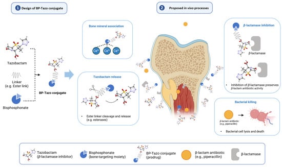
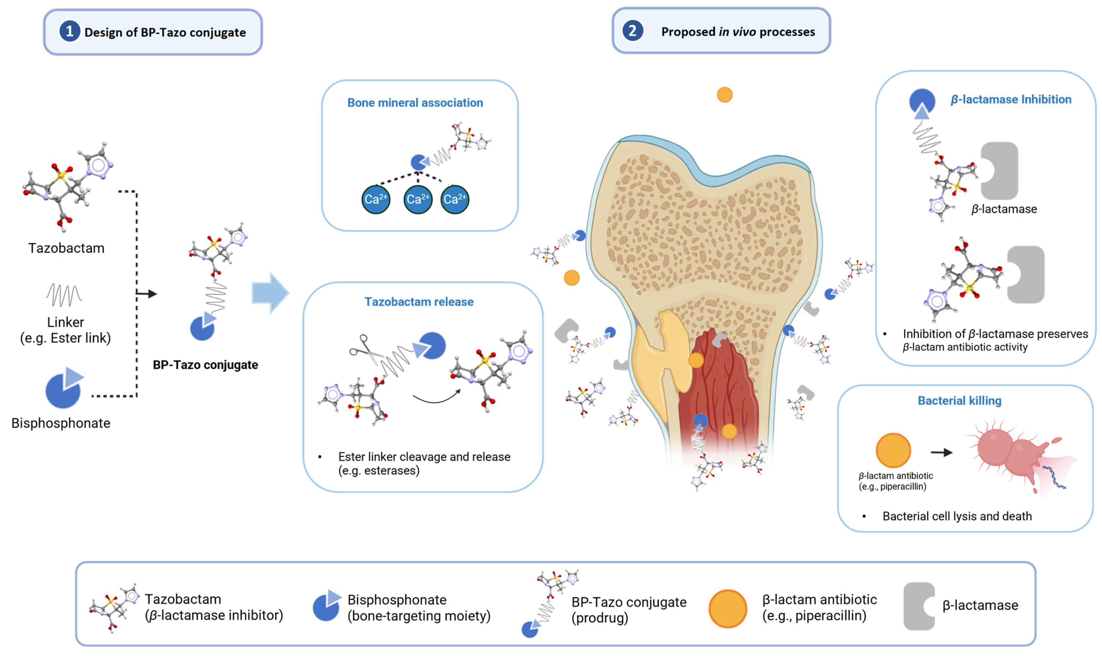
Here, we report the synthesis and characterization of BP-Tazo-ester (compound 5), followed by biochemical, mineral-binding, and antibacterial evaluations. Compound 5 was assessed in combination with piperacillin against β-lactamase-producing Escherichia coli HS and planktonic Staphylococcus aureus CCUG 25925. The same S. aureus strain was evaluated in an ex vivo bone-slice early-colonization model. Together, these studies assess the feasibility of a bisphosphonate-linked β-lactamase-inhibitor platform for potential application in bone infection. The conceptual design and proposed in vivo processes are illustrated in Figure 1.
Figure 1.
Conceptual design of BP–Tazo-ester and proposed in vivo processes. Tazobactam is linked to a bisphosphonate-containing bone-targeting moiety through an ester linker. Phosphonate deprotection may expose mineral-binding groups and promote association with hydroxyapatite-rich bone surfaces. The intact conjugate may contribute directly to β-lactamase inhibition, while ester cleavage may release free tazobactam and provide an additional inhibitory pathway. These processes may occur concurrently and are not intended to imply a fixed temporal sequence. β-lactamase inhibition is proposed to preserve the activity of co-administered β-lactam antibiotics, such as piperacillin, thereby promoting bacterial killing. The illustrated in vivo processes are proposed and require direct validation.
2. Materials and Methods
2.1. Chemical Synthesis
2.1.1. General Information
All solvents and reagents were used from commercial sources without further purification. Anhydrous tetrahydrofuran, citric acid, anhydrous dichloromethane, Amberlyst-15, bromotrimethylsilane, anhydrous ethanol, ethyl acetate, tetraisopropyl methylenediphosphonate (1), 2-(3-bromopropoxy)tetrahydro-2H-pyran (2), tazobactam, piperacillin, ciprofloxacin, dicyclohexylcarbodiimide, 4-dimethylaminopyridine (DMAP), hydroxyapatite powder, Sodium hydride (57–63% oil dispersion), and alendronate were purchased from Sigma-Aldrich (St. Louis, MO, USA).
The progress of all reactions was monitored by LC–MS (Shimadzu LC–MS system, Kyoto, Japan). Mass spectral analyses were performed on a Shimadzu mass spectrometer under electrospray ionization (ESI). NMR spectra were recorded on a Bruker 600 MHz spectrometer (Bruker BioSpin, Ettlingen, Germany); chemical shifts (δ) are reported in ppm relative to TMS as an internal standard. Multiplicities are denoted as s (singlet), d (doublet), t (triplet), q (quartet), m (multiplet), dd (doublet of doublets), tt (triplet of triplets), and br s (broad singlet). Flash column chromatography was performed using silica gel columns and C18 reverse-phase columns from Teledyne. The 31P NMR spectra of compounds 3 and 4 were acquired without 1H decoupling.
2.1.2. Synthesis of Tetraisopropyl 4-(2-Tetrahydro-2H-pyranyloxy)butylene-1,1-bisphosphonate (3)
Compounds 3 and 4 were prepared following the procedure reported by Tanaka et al. [20] and characterized by 1H, 13C, and 31P NMR and ESI-MS. Their 1H NMR data were compared with the previously published values (Table S4).
To a suspension of NaH (60% suspension in mineral oil, 13.9 mg, 0.349 mmol, 1.2 eq.) in dry THF (500 μL) was added dropwise tetraisopropyl methylenediphosphonate (1) (91.8 μL, 0.29 mmol, 1.0 eq.). The resulting clear solution was stirred at room temperature for 45 min, after which 2-(3-bromopropoxy)tetrahydro-2H-pyran (2) (49.2 μL, 0.29 mmol, 1.0 eq.) was added dropwise. The mixture was heated to 55 °C for 24 h. LC–MS analysis confirmed complete consumption of the starting materials. The reaction mixture was poured onto a 5% aqueous citric acid (10 mL) solution and extracted with ethyl acetate (2 × 10 mL). The combined organic layers were washed with brine (10 mL), dried, and concentrated in vacuo. Compound 3 (110.0 mg) was obtained as a pale-yellow oil and used without further purification. Yield: 78%.
1H NMR (600 MHz, CDCl3) δ: 1.30–1.36 (m, 24H, CH3 × 8), 1.50–1.90 (m, 10H, CH2), 2.20 (tt, J = 24.3, 5.9 Hz, 1H, CH–P), 3.45 (m, 2H, CH2–O), 3.80 (m, 2H, CH2–O), 4.59 (m, 1H, CH–O), 4.79 (m, 4H, CH–CH3).
13C NMR (151 MHz, CDCl3): δ 98.6, 71.2, 71.1, 71.0, 67.0, 62.1, 38.2 (t, J = 135.2 Hz), 30.7, 29.2 (t, J = 6.6 Hz), 25.5, 24.2, 24.1, 24.0, 23.9, 23.0 (t, J = 5.0 Hz), 19.4.
31P NMR (243 MHz, CDCl3): δ 21.9.
MS (ESI): m/z 487.30 [M + H]+ (calcd for C21H45O8P2+, 487.26).
2.1.3. Deprotection to Yield Tetraisopropyl 4-Hydroxybutylene-1,1-bisphosphonate (4)
Compound 3 (275 mg, 0.57 mmol, 1.0 eq.) was dissolved in methanol, and Amberlyst-15 (54 mg, catalytic) was added. The mixture was heated at 55 °C for 20 h, until LC–MS analysis showed complete consumption of starting material. The resin was removed by filtration, and the filtrate was concentrated in vacuo. The residue was purified by silica gel flash chromatography using a RediSep Rf silica gel column (0–10% methanol/ethyl acetate) to afford compound 4 (100.0 mg) as a pale-yellow oil. Yield: 44%.
1H NMR (600 MHz, CDCl3) δ: 1.34–1.36 (m, 24H, CH3 × 8), 1.80–1.82 (m, 2H, CH2), 2.00–2.07 (m, 2H, CH2), 2.21 (tt, J = 24.6, 5.4 Hz, 1H, CH–P), 2.79 (m, 1H, OH), 3.66 (m, 2H, CH2–O), 4.75–4.80 (m, 4H, CH–CH3).
13C NMR (151 MHz, CDCl3): δ 71.3, 71.2, 61.2, 37.2 (t, J = 135.5 Hz), 32.0 (t, J = 5.7 Hz), 24.2, 24.1, 24.0, 23.9, 21.6 (t, J = 4.9 Hz).
31P NMR (243 MHz, CDCl3): δ 22.4.
MS (ESI): m/z 403.25 [M + H]+ (calcd for C16H37O7P2+, 403.20).
2.1.4. Synthesis of 4,4-Bis(dipropan-2-ylphosphono)butyl (2S,3S,5R)-3-methyl-4,4,7-trioxo-3-(1H-1,2,3-triazol-1-ylmethyl)-4λ6-thia-1-azabicyclo[3.2.0]heptane-2-carboxylate (5)
Tazobactam (16.4 mg, 0.055 mmol, 1.0 eq.), compound 4 (22.0 mg, 0.055 mmol, 1.00 eq.), and DCC (13.8 mg, 0.067 mmol, 1.22 eq.) were dissolved in dry DCM (4.0 mL). To this solution was added DMAP (about 0.7 mg, 0.005 mmol, 0.10 eq.), and the reaction mixture was stirred overnight at room temperature. The reaction progress was monitored by LC–MS. After completion, the mixture was filtered, and the filtrate was concentrated in vacuo. The crude product was purified by reversed-phase flash chromatography (HP C18 Aq column, 5–60% ACN/H2O). Lyophilization afforded compound 5 (23.0 mg) as a colorless glass. Yield: 61%.
1H NMR (600 MHz, CD3OD) δ: 1.34–1.37 (m, 24H, CH3 × 8), 1.41 (s, 3H, CH3), 2.01–2.03 (m, 4H, CH2), 2.44 (tt, J = 24.6, 5.4 Hz, 1H, CH–P), 3.39 (dd, J = 16.2, 1.8 Hz, 1H, CH2), 3.64 (dd, J = 16.2, 4.2 Hz, 1H, CH2), 4.28 (m, 2H, CH2–O), 4.74–4.77 (m, 4H, CH–CH3), 4.87 (s, 1H, CH), 4.97 (dd, J = 4.8, 1.8 Hz, 1H, CH), 5.06 (d, J = 15 Hz, 1H, CH2), 5.26 (d, J = 15 Hz, 1H, CH2), 7.77 (d, J = 1.2 Hz, 1H, CH–N), 8.06 (d, J = 1.2 Hz, 1H, CH–N).
13C NMR (150 MHz, CD3OD): δ 170.9, 166.6, 133.2, 126.8, 71.8, 67.3, 64.7, 62.4, 60.3, 50.4, 37.8, 37.1 (J = 120 Hz), 27.5, 27.0, 22.4, 15.4.
31P NMR (243 MHz, CD3OD): δ 21.5.
MS (ESI): m/z 685.20 [M + H]+ (calcd for C26H47N4O11P2S+, 685.24), 707.20 [M+Na]+ (calcd for C26H46N4NaO11P2S+, 707.23).
2.2. In Silico Physicochemical Property Analysis
Selected physicochemical descriptors for tazobactam and compound 5 were obtained from database records and structure-based prediction platforms. PubChem (National Center for Biotechnology Information, Bethesda, MD, USA) was used to obtain reference information for tazobactam, including molecular formula and molecular weight. SwissADME (Swiss Institute of Bioinformatics, Lausanne, Switzerland) was used to obtain hydrogen-bond donor and acceptor counts, topological polar surface area (TPSA), fraction of sp3 carbons (Fsp3), molar refractivity, and rotatable-bond count for tazobactam and compound 5. ADMET Predictor version 12 (Simulations Plus, Inc., Lancaster, CA, USA) was used to predict pKa, logP, logD at pH 7.4, and predominant ionization state at pH 7.4. For compound 5, the molecular formula and molecular weight were calculated from its chemical structure.
2.3. Biological Evaluation
Complementary biological evaluations were used. A cell-free nitrocefin assay assessed β-lactamase inhibition. A β-lactamase-producing Escherichia coli HS strain was used to assess whether compound 5 could support piperacillin activity in a bacterial growth assay. Broth microdilution testing of Staphylococcus aureus CCUG 25925 provided planktonic susceptibility data for the same strain used in the bone-slice early-colonization model. These assays address complementary questions; the planktonic MIC and bone-associated colonization endpoints are not directly interchangeable.
2.3.1. Assessment of β-Lactamase Inhibitory Activity of Compound 5
The inhibitory activity of compound 5 against β-lactamase was evaluated using the Amplite® Colorimetric Beta-Lactamase Activity Assay Kit (AAT Bioquest, Pleasanton, CA, USA; Cat. No. 12551). The β-lactamase standard supplied with the kit as Component C was reconstituted according to the manufacturer’s instructions and diluted to a working activity concentration of 250 µU/mL. Nitrocefin supplied with the kit was used as the chromogenic substrate.
Nitrocefin working solution was freshly prepared by mixing 50 µL of Component A with 5 mL of assay buffer. Stock solutions of compound 5 and tazobactam (10 mM) were initially prepared using 5% (v/v) DMSO. Subsequent serial dilutions were prepared in PBS, yielding final assay concentrations of 0.25–250 µM and 0.05–62.5 µM, respectively. For each assay well, 50 µL of inhibitor solution was incubated with 50 µL of β-lactamase working solution (250 µU/mL). Nitrocefin solution (100 µL) was subsequently added to initiate the reaction, yielding a final volume of 200 µL per well and a final β-lactamase activity concentration of 62.5 µU/mL.
Vehicle-control wells contained β-lactamase and nitrocefin without inhibitor. Blank wells contained nitrocefin without enzyme. Compound-only control wells containing the corresponding concentrations of compound 5 without enzyme were included to correct for potential absorbance interference.
The plates were incubated at 25 °C for 30 min in the dark. Absorbance was measured at 490 nm and 380 nm using a SpectraMax i3x microplate reader (Molecular Devices, San Jose, CA, USA). Enzymatic activity was expressed as the A490/A380 ratio after blank correction. Percentage inhibition was calculated as follows:
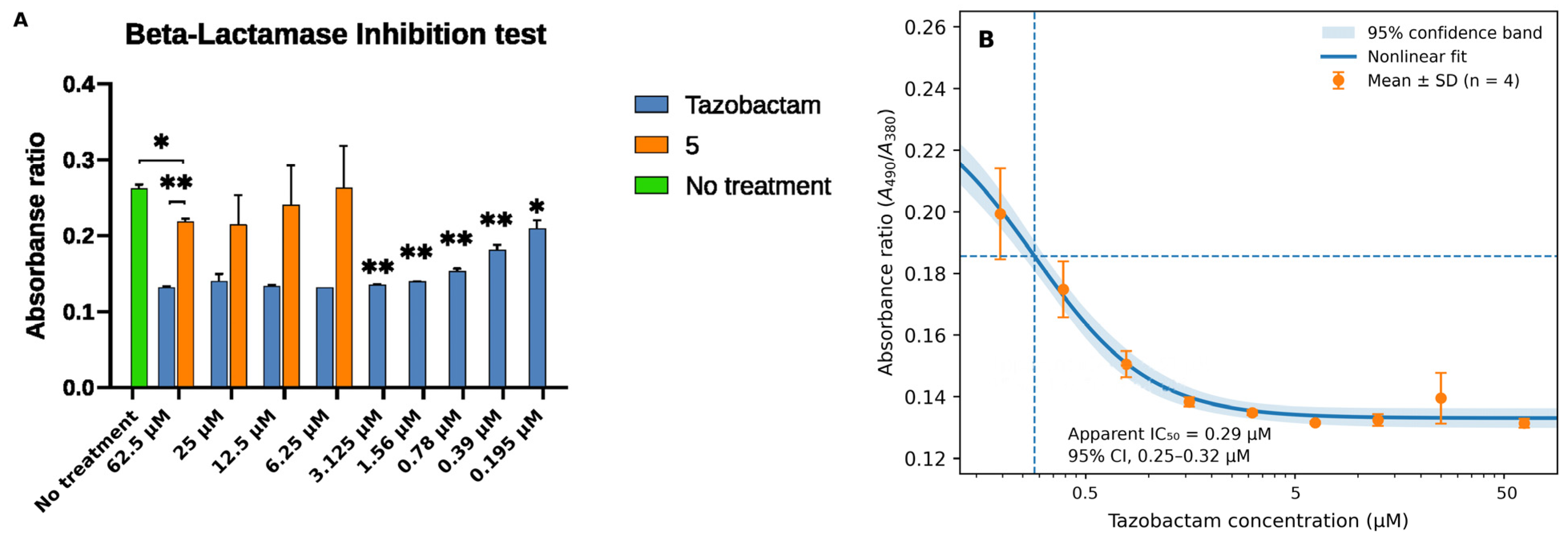
Measurements were performed using four replicate wells per concentration, and the data are presented as the mean ± standard deviation. For estimation of the tazobactam IC50, all four replicate-well measurements at each of the nine tazobactam concentrations shown in Figure 2B were fitted by nonlinear least-squares regression to a three-parameter inhibitory Hill model, with the mean no-inhibitor control fixed as the upper asymptote and the lower asymptote, IC50, and Hill coefficient fitted. An approximate 95% confidence interval for IC50 was calculated from the nonlinear-regression parameter covariance matrix on the log-concentration scale. Pointwise 95% confidence bands for the fitted mean response were calculated from the parameter covariance matrix using the delta method. Here, the apparent IC50 is the fitted concentration at the midpoint between the fixed upper and fitted lower asymptotes; it is not necessarily the concentration producing 50% inhibition relative to the no-inhibitor control.
Figure 2.
(A) Concentration-dependent inhibition of β-lactamase activity by compound 5 and tazobactam, measured using a nitrocefin-based colorimetric assay. Absorbance ratios (A490/A380) reflect enzyme activity, with lower values indicating increased inhibition. Statistical comparisons were performed using one-way ANOVA followed by Dunnett’s multiple-comparisons test. * p < 0.05; ** p < 0.01. (B) Nonlinear concentration–response analysis of tazobactam. Points represent mean ± SD (n = 4), the solid line represents the fitted three-parameter inhibitory Hill curve, and the shaded region indicates the pointwise 95% confidence band of the fitted mean response. The dashed horizontal and vertical lines indicate the midpoint between the fixed upper and fitted lower response asymptotes and the corresponding apparent IC50, respectively. The apparent IC50 was 0.29 µM (95% CI, 0.25–0.32 µM).
2.3.2. In Vitro Hydroxyapatite (HA) Binding Assay
The high mineral affinity of bisphosphonates is associated with accessible, ionizable phosphonate groups that interact with calcium-rich hydroxyapatite surfaces. In intact compound 5, these groups are protected as isopropyl esters, which may limit their mineral-binding interactions. Therefore, a phosphonate-ester deprotection procedure was applied to an aliquot of compound 5 before the HA-binding assay to evaluate the apparent mineral-binding potential of the treated material [20]. The treated material was not isolated or structurally characterized; accordingly, the HA-binding result is reported for “TMSBr-treated material derived from compound 5” rather than for intact compound 5 or a defined deprotected structure. Because the assay measures phosphorus remaining in the supernatant, it cannot distinguish mineral association of tazobactam-bearing material from that of other phosphorus-containing products.
An aliquot of compound 5 was dissolved in dichloromethane and treated with bromotrimethylsilane (16 equivalents) at room temperature for 4 h. The solvent and volatile reagents were removed under reduced pressure, and the residue was reconstituted in water or saline and adjusted to pH 7.0 for the HA-binding assay. Alendronate (ALN) was used as a positive control.
Briefly, HA powder (5 mg) was incubated with samples at concentrations of 0.5 mM and 0.1 mM in 0.3 mL water or saline. Binding reactions were carried out at 37 °C for 2.5 h under gentle agitation. Following incubation, samples were centrifuged for 5 min, and the supernatants were collected for phosphate quantification. Control samples without hydroxyapatite were prepared under otherwise identical conditions to assess nonspecific loss.
Phosphate concentrations were determined using a colorimetric phosphate assay. Calibration curves were prepared with serial dilutions of ALN (0–1.0 mM). Each sample aliquot (50 µL) was mixed with 30 µL of magnesium nitrate solution in 95% ethanol and heated until a white residue was obtained. The residue was dissolved in 0.3 mL of 0.5 M hydrochloric acid and heated in boiling water until a clear solution was obtained. After cooling, 0.6 mL of ammonium molybdate solution (0.42% in 1 M sulfuric acid) and 0.1 mL of 10% ascorbic acid were added. Samples were incubated at 37 °C for 1 h, and absorbance was measured at 820 nm.
The apparent HA-binding percentage was calculated as:
Measurements were performed in duplicate and are reported as the mean ± standard deviation.
2.3.3. Antimicrobial Growth-Inhibition and Concentration–Response Analysis
The ability of compound 5 to enhance the antibacterial activity of piperacillin was evaluated against a β-lactamase-producing Escherichia coli HS strain kindly donated by Dr. Elhenawy, University of Alberta.
For E. coli HS, bacterial cultures were grown in Mueller–Hinton broth to the mid-logarithmic phase and diluted to approximately 5 × 10⁵ CFU/mL. Aliquots were added to 96-well plates containing two-fold serial dilution series spanning 1.95–250 µg/mL nominal concentration for piperacillin alone (PIP), piperacillin–tazobactam (PIP–Tazo), or piperacillin combined with compound 5 (PIP–BP–Tazo). Both combination formulations were prepared at a fixed 4:0.5 (equivalent to 8:1, w/w) mass ratio of piperacillin to inhibitor (tazobactam or compound 5, respectively). At the highest nominal combination concentration (250 µg/mL), this corresponded to 222.2 µg/mL piperacillin and 27.8 µg/mL inhibitor. The premixed combinations were subsequently serially diluted. Accordingly, the reported PIP–Tazo and PIP–BP–Tazo concentrations refer to the total nominal concentrations of the fixed-ratio combinations rather than the concentration of the inhibitor alone.
The plates were incubated at 37 °C for 18–20 h. Bacterial growth was quantified by measuring optical density at 660 nm. Wells containing the corresponding drug concentrations without bacteria were used for background correction, and growth inhibition was calculated relative to the untreated bacterial control.
Percentage growth inhibition was calculated as:
The minimum inhibitory concentration (MIC) was defined as the lowest concentration resulting in no visible growth. Experiments were performed in triplicate.
To further characterize concentration-dependent antimicrobial activity, mean E. coli growth-inhibition values at all eight tested concentrations were fitted by nonlinear least-squares regression to a two-parameter Hill model with the lower and upper asymptotes constrained to 0% and 100% inhibition, respectively. The fitted parameters were IC50 and the Hill coefficient (n), with positive parameters estimated on the log scale. For PIP–Tazo and PIP–BP–Tazo, concentration (C) therefore represents the total nominal concentration of the fixed 8:1 (w/w) piperacillin:inhibitor formulation.
E = 100 × Cn/(IC50n + Cn)
The concentration predicted to produce 90% inhibition (IC90) was derived from the fitted Hill parameters. Approximate 95% confidence intervals (CIs) for IC50 and the Hill coefficient were calculated from the nonlinear-regression parameter covariance matrix on the log-transformed scale using six residual degrees of freedom (eight concentration means minus two fitted parameters). The IC90 CI was propagated on the log scale. Pointwise 95% confidence bands for the fitted mean concentration–response curves were calculated from the parameter covariance matrix using the delta method; these bands represent uncertainty in the fitted mean response rather than prediction intervals for individual wells. Residuals were examined graphically.
IC90 = IC50 × 9 (1/n)
To evaluate model form, two-parameter Hill, three-parameter Hill, and four-parameter logistic models were compared using the corrected Akaike information criterion (AICc), calculated from the eight concentration-level means to avoid treating technical triplicates as additional dose levels. Exploratory extra-sum-of-squares F tests were used to compare treatment-specific two-parameter Hill curves with reduced models sharing a common curve, a common Hill coefficient, or a common IC50. Overall activity across the tested range was additionally summarized by trapezoidal area under the observed growth-inhibition curve plotted against log10 concentration from 1.95 to 250 µg/mL; AUC was normalized by the corresponding log-concentration interval and reported as average percentage growth inhibition across the tested log-dose range.
Additional broth microdilution testing was performed using S. aureus CCUG 25925, the strain used in the bone-slice assay. Four treatment groups (piperacillin alone, PIP–Tazo, PIP–BP–Tazo, and compound 5 alone) were evaluated as part of the same continuous S. aureus MIC study. The three piperacillin-containing groups were evaluated over 11 twofold dilution levels spanning approximately 0.24–250 µg/mL. Both combinations were prepared at a fixed piperacillin:inhibitor mass ratio of 4:0.5 (equivalent to 8:1, w/w); combination concentrations represent total nominal formulation concentrations. The compound 5-alone group was tested in a consecutive concentration series comprising ten twofold dilutions (0.49–250 µg/mL). Three replicate wells per concentration and bacterial-growth and medium-only controls were used for the S. aureus MIC measurements. MIC was defined as the lowest tested concentration showing no visible growth. MIC results for all four treatment groups are summarized together in Table S5; raw OD660 measurements for the compound 5-alone series are provided in Table S6. Free tazobactam alone was not tested.
2.3.4. Ex Vivo Bone Slice-Associated Early Colonization/Biofilm-Formation Assay
Fresh 2.0 mm thick cortical bone slices were cut and collected in a solution of phosphate-buffered saline pH 7.4 (using a Buehler Isomet wafer saw blade) from the femur of Sprague–Dawley (SD) rats being euthanized elsewhere, and bone slices were sterilized by autoclaving at 121 °C for 20 min. Staphylococcus aureus (CCUG 25925, Culture collection University of Gothenburg, Göteborg, Sweden) was cultured overnight in Brain Heart Infusion (BHI) broth. The bacterial suspension was adjusted to approximately 2 × 10⁴ CFU/mL based on OD660 and confirmed by viable counting.
Sterile bone slices were individually placed in a 96-well plate. Each well received 100 µL of bacterial suspension and the designated treatment. The treatment groups consisted of piperacillin (250 µg/mL), ciprofloxacin (0.5 µg/mL), piperacillin–tazobactam (PIP–Tazo; 62.5 µg/mL total nominal concentration), and piperacillin–compound 5 (PIP–BP–Tazo; 62.5 and 125 µg/mL total nominal concentrations). Both combination formulations were prepared at a fixed 8:1 (w/w) mass ratio of piperacillin to inhibitor (tazobactam or compound 5, respectively). A sterile non-infected bone-slice control and an S. aureus-infected untreated bone-slice control were included. Each condition was evaluated using three bone slices. The plates were incubated at 37 °C for 24 h to permit bacterial attachment and biofilm development. Because treatment was co-initiated with bacterial inoculation, this assay evaluates inhibition of bacterial attachment and early biofilm development rather than treatment of an established infection.
After incubation, the planktonic suspension was removed, and each bone slice was washed twice with sterile phosphate-buffered saline (PBS) to remove non-adherent bacteria. The slices were transferred to tubes containing 1 mL of sterile PBS and sonicated for 20 min to detach bone-associated bacteria. This was based on our established protocols, which showed that this sonication time did not negatively influence bacterial viability. The resulting suspensions were serially diluted, and 100 µL aliquots were plated on BHI agar. Colonies were enumerated after incubation at 37 °C for 24 h. Bacterial burden was expressed as log10 CFU/mL.
The assay limit of detection (LOD) was 1 × 10⁴ CFU/mL (4.00 log10 CFU/mL). Samples with no recoverable colonies were considered below the LOD and were assigned one-half of the LOD for graphical presentation and statistical analysis. Statistical comparisons between infected treatment groups and the infected untreated control were performed using one-way ANOVA followed by Dunnett’s multiple-comparisons test (GraphPad Prism, version 8.0, GraphPad Software, San Diego, CA, USA). The sterile non-infected control was excluded from inferential statistical analysis.
3. Results
3.1. Chemistry
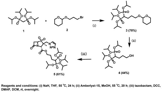
The synthetic route used to prepare BP-Tazo-ester (compound 5) is outlined in Scheme 1. Acid-mediated removal of the tetrahydropyranyl protecting group from compound 3 afforded the hydroxy-functionalized bisphosphonate intermediate 4 as a pale-yellow oil in 44% yield. Esterification of intermediate 4 with tazobactam under carbodiimide-mediated coupling conditions produced compound 5 in 61% yield after C18 reverse-phase purification.
Scheme 1.
Overall synthetic scheme of the tazobactam–bisphosphonate conjugate (compound 5).
The identity of compound 5 was supported by NMR and mass-spectrometric analyses. Compound 5 exhibited the expected molecular ion peaks in ESI–MS (m/z 685 [M+H]+, 707 [M+Na]+). The 1H and 13C NMR spectra were consistent with the proposed structure, and 31P NMR showed a single resonance at δ 21.5 ppm, consistent with the bisphosphonate-containing structure.
3.2. Physicochemical and In Silico Property Comparison
Selected physicochemical descriptors, analytical data, and in silico predictions for tazobactam and compound 5 are summarized in Table 1. Relative to tazobactam, compound 5 showed substantial increases in molecular weight (684.68 vs. 300.29 g/mol), topological polar surface area (210.52 vs. 130.84 Å2), hydrogen-bond acceptor count (13 vs. 7), and number of rotatable bonds (18 vs. 3). ADMET Predictor further indicated a marked shift in lipophilicity and ionization behavior: the intact isopropyl-protected conjugate was predicted to be more lipophilic and predominantly neutral at pH 7.4, whereas tazobactam was predicted to be strongly hydrophilic and predominantly anionic.
Table 1.
Selected physicochemical descriptors, analytical data, and in silico predictions for tazobactam and compound 5.
These computational descriptors were used for comparative physicochemical interpretation and should not be interpreted as experimentally measured pharmacokinetic or tissue-distribution properties. Additional SwissADME outputs, including model-specific lipophilicity estimates, water-solubility predictions, pharmacokinetic classifications, drug-likeness filters, and medicinal-chemistry descriptors, are provided in Table S3.
3.3. Biological Activity
3.3.1. Inhibitory Activity of Compound 5 Against β-Lactamase
The inhibitory activity of compound 5 toward β-lactamase was evaluated using a nitrocefin-based chromogenic assay, with free tazobactam included as the reference inhibitor. Nitrocefin hydrolysis was quantified using the A490/A380 absorbance ratio.
As shown in Figure 2, both compound 5 and tazobactam exhibited concentration-dependent suppression of β-lactamase activity. Increasing concentrations of compound 5 resulted in a progressive decrease in the A490/A380 ratio, confirming its inhibitory potential. However, compound 5 exhibited lower inhibitory activity than free tazobactam across the tested concentration range, with a lower maximum percentage inhibition. As shown in Figure 2B, nonlinear concentration–response analysis of tazobactam yielded an apparent IC50 of 0.29 µM (95% CI, 0.25–0.32 µM) under the present assay conditions. An IC50 was not assigned to compound 5 because the observed responses did not sufficiently define the 50% inhibitory midpoint for a reliable estimate.
These results demonstrate that compound 5 retains measurable β-lactamase inhibitory activity following conjugation to the bisphosphonate moiety, although its observed inhibitory effect under these assay conditions is lower than that of free tazobactam.
3.3.2. In Vitro Hydroxyapatite Binding
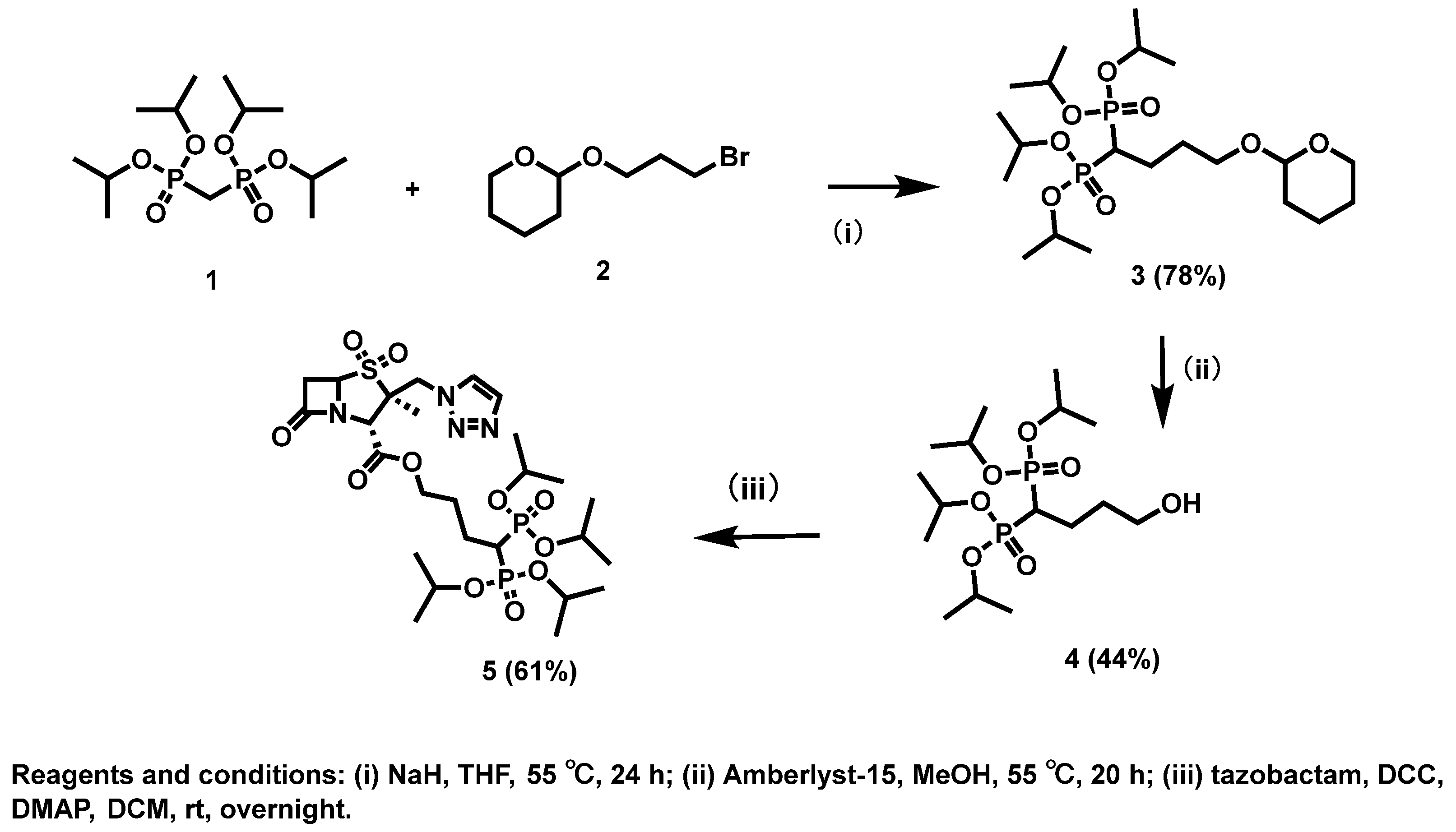
The apparent HA-binding behavior of the TMSBr-treated material derived from compound 5 and ALN (positive control) was assessed in vitro (Figure 3). At a nominal concentration of 0.5 mM, alendronate and the compound 5-derived sample showed apparent HA-binding values of 96.7% and 93.9%, respectively. At 0.1 mM, the corresponding apparent binding values were 101.6% and 92.0%.
Figure 3.
Apparent in vitro hydroxyapatite (HA) binding of TMSBr-treated material derived from compound 5 and alendronate (ALN). Binding assays were conducted at 0.5 mM and 0.1 mM in either saline or water. Samples were incubated with HA at 37 °C for 2.5 h, and phosphate content in the supernatant was quantified using a colorimetric phosphate assay to calculate apparent binding percentage. Data are presented as mean ± SD.
The apparent binding behavior was influenced by the incubation medium. The compound 5-derived material showed consistently high apparent HA binding in saline. Alendronate showed higher apparent binding than the compound 5-derived sample in water. Control samples prepared without HA showed minimal analyte loss under the corresponding conditions. Overall, the TMSBr-treated material derived from compound 5 exhibited high apparent HA binding under the conditions tested.
3.3.3. Antimicrobial Growth-Inhibition and Concentration–Response Analysis
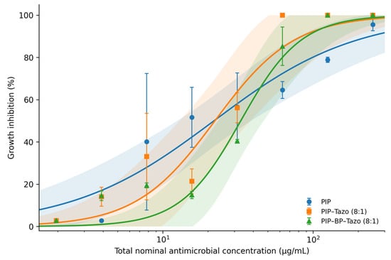
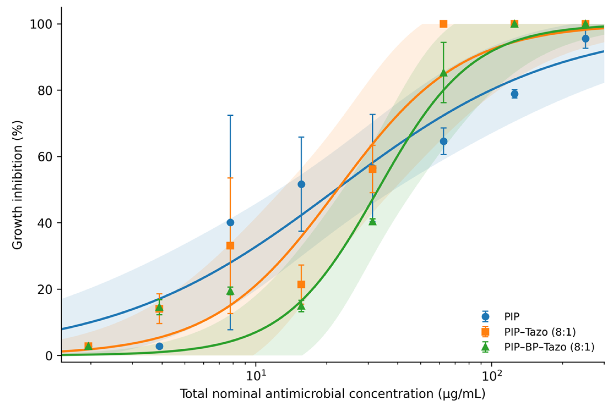
The antimicrobial activity of compound 5 in combination with piperacillin was evaluated against a β-lactamase–producing E. coli strain (Figure 4). Piperacillin alone did not achieve complete growth inhibition, even at concentrations as high as 250 µg/mL. In contrast, PIP–Tazo produced complete growth inhibition at total nominal concentrations of 62.5 µg/mL and above. PIP–BP–Tazo produced 85.4% growth inhibition at 62.5 µg/mL total nominal concentration and complete growth inhibition at 125 µg/mL. The experimentally determined MICs were therefore >250 µg/mL for PIP, 62.5 µg/mL total nominal concentration for PIP–Tazo, and 125 µg/mL total nominal concentration for PIP–BP–Tazo. These observations are consistent with retained β-lactamase-inhibitory function; however, because compound 5 was not tested alone in this assay, the present data do not exclude additional or alternative mechanisms.
Figure 4.
Concentration–response relationships for piperacillin (PIP), piperacillin–tazobactam (PIP–Tazo), and piperacillin combined with compound 5 (PIP–BP–Tazo) against β-lactamase-producing E. coli (HS strain). Points show mean growth inhibition ± SD from triplicate OD660 measurements. Solid curves represent that the selected two-parameter Hill fits with lower and upper asymptotes constrained to 0% and 100% inhibition, and shaded regions show pointwise 95% confidence bands for the fitted mean responses. All eight tested concentrations were included in the analysis. The x-axis represents total nominal antimicrobial concentration on a logarithmic scale. PIP–Tazo and PIP–BP–Tazo were both prepared as fixed 8:1 (w/w) piperacillin:inhibitor formulations and serially diluted as premixed combinations; combination concentrations therefore refer to total nominal formulation concentrations rather than inhibitor concentrations alone.
Because both combinations were prepared at an 8:1 piperacillin:inhibitor mass ratio, the inhibitor component constituted one ninth of the total formulation concentration. At a matched total concentration of 62.5 µg/mL, each formulation therefore contained 6.94 µg/mL inhibitor. This corresponded to 23.1 µM tazobactam but only 10.1 µM compound 5 because of their different molecular weights (300.29 and 684.68 g/mol, respectively). Thus, at any matched total formulation concentration, the molar concentration of compound 5 was 43.9% of that of tazobactam despite an identical inhibitor mass fraction.
The selected two-parameter Hill model yielded an estimated IC50 of 21.90 µg/mL for PIP (95% CI, 12.94–37.08), 22.22 µg/mL total nominal concentration for PIP–Tazo (95% CI, 13.68–36.10), and 33.02 µg/mL total nominal concentration for PIP–BP–Tazo (95% CI, 24.04–45.35) (Table 2). The corresponding Hill coefficients were 0.92 (95% CI, 0.57–1.48), 1.65 (95% CI, 0.81–3.35), and 2.17 (95% CI, 1.18–3.99), respectively. PIP–BP–Tazo therefore showed a numerically steeper fitted concentration–response relationship, although the confidence intervals for the combination-treatment slopes overlapped.
Table 2.
Concentration–response parameters from the selected two-parameter Hill model.
Estimated IC90 values were 240.19 µg/mL for PIP (95% CI, 68.49–842.39), 84.17 µg/mL total nominal concentration for PIP–Tazo (95% CI, 29.14–243.16), and 90.93 µg/mL total nominal concentration for PIP–BP–Tazo (95% CI, 45.39–182.13). Relative to PIP alone, the total nominal IC90 was reduced by 65.0% with PIP–Tazo and by 62.1% with PIP–BP–Tazo. Expressed as the piperacillin component present at the fitted IC90, the corresponding piperacillin concentrations were 74.82 and 80.82 µg/mL, representing 68.9% and 66.4% reductions, respectively, relative to the PIP-alone IC90. At their respective fitted combination IC90 values, PIP–Tazo contained 9.35 µg/mL tazobactam (31.1 µM), whereas PIP–BP–Tazo contained 10.10 µg/mL compound 5 (14.8 µM). These inhibitor concentrations describe formulation content at the combination IC90 and do not represent IC90 values for the inhibitors alone.
Normalized log-concentration AUC values were 49.1% for PIP, 53.7% for PIP–Tazo, and 46.5% for PIP–BP–Tazo. The constrained two-parameter Hill model was preferred by AICc for all three datasets (AICc = 40.87, 44.49, and 39.90, respectively), with all more complex candidate models having ΔAICc ≥ 4.40. In exploratory extra-sum-of-squares comparisons, PIP–Tazo and PIP–BP–Tazo were not significantly different when tested as separate versus common curves (p = 0.321), nor was a difference detected for their Hill coefficients (p = 0.578) or IC50 parameters (p = 0.140). Residual inspection showed deviations around the fitted responses at several concentrations, and the IC90 confidence intervals were broad; therefore, the close IC90 point estimates should be interpreted as comparable rather than formally equivalent.
Planktonic Susceptibility of S. aureus CCUG 25925
For S. aureus CCUG 25925, the MIC was 7.81 µg/mL for piperacillin alone and 0.98 µg/mL total nominal concentration for both PIP–Tazo and PIP–BP–Tazo (Table S5). Both combination MICs were approximately eightfold lower than the PIP-alone MIC on the reported nominal concentration scale. At a total combination concentration of 0.98 µg/mL and a 4:0.5 (8:1, w/w) ratio, each combination contained approximately 0.87 µg/mL piperacillin and 0.109 µg/mL inhibitor. The two combinations therefore had the same observed MIC within the resolution of the twofold dilution series; this does not establish equivalent intrinsic inhibitor potency or pharmacological synergy. In the compound 5-alone arm of the same S. aureus MIC study, bacterial growth was observed at all tested concentrations, including 250 µg/mL, giving an MIC > 250 µg/mL (Table S5). The mean background-corrected growth inhibition at 250 µg/mL was approximately 24.8%, without a clear concentration-dependent response across the tested range (Table S6).
3.3.4. Ex Vivo Bone Slice–Associated Staphylococcus aureus Early Colonization/Biofilm Formation and Quantification Assay
The effect of compound 5 in combination with piperacillin on bone slice-associated S. aureus early colonization was evaluated by quantifying viable bacteria recovered from bone slices after 24 h of incubation. The PIP–BP–Tazo combination was tested at total nominal antimicrobial concentrations of 62.5 and 125 µg/mL.
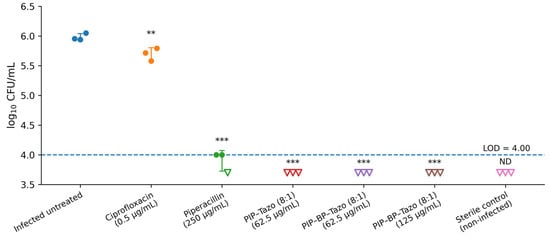
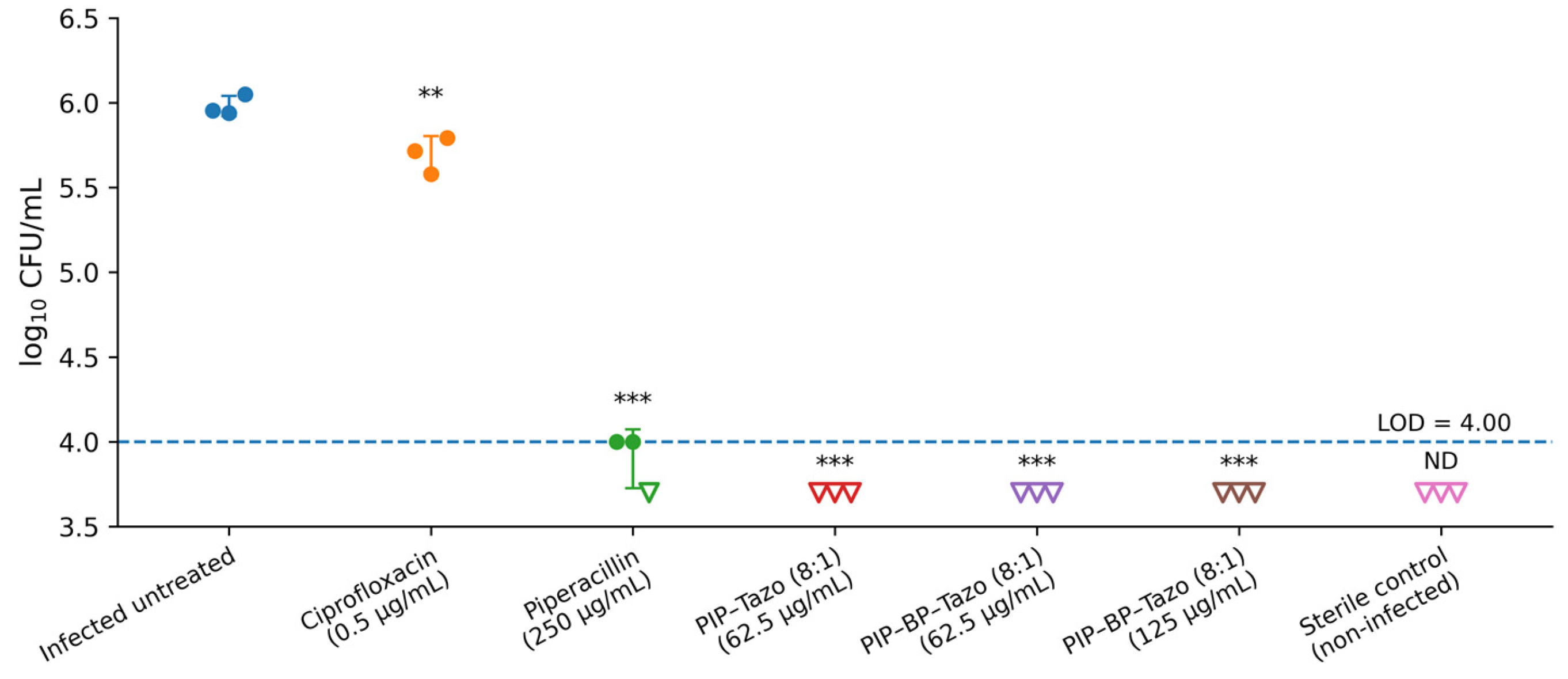
As shown in Figure 5, the infected, untreated bone-slice control exhibited a mean bacterial burden of 5.98 log10 CFU/mL after 24 h. Ciprofloxacin treatment yielded 5.70 log10 CFU/mL, corresponding to a 0.28-log10 reduction relative to the untreated control (p < 0.01). For piperacillin alone, one colony was recovered from each of two bone-slice replicates (1 × 10⁴ CFU/mL per replicate), while the third replicate was below the LOD (Table S2). Substitution of one-half of the LOD for the non-detect yielded a descriptive mean of approximately 3.90 log10 CFU/mL, corresponding to a 2.08-log10 reduction relative to the untreated control (p < 0.001). This mean includes a censored-value substitution and is not a directly measured bacterial burden below the LOD.
Figure 5.
Viable Staphylococcus aureus recovered from bone slices following 24 h of incubation under the indicated treatment conditions. Individual points represent independent bone-slice replicates, and data are presented as mean ± SD (n = 3). For combination treatments, concentrations are expressed as total nominal antimicrobial concentrations of the premixed 8:1 (w/w) formulations. The assay limit of detection (LOD) was 1 × 10⁴ CFU/mL (4.00 log10 CFU/mL). Samples with no recoverable colonies were considered below the LOD and were assigned one-half of the LOD for graphical presentation and statistical analysis; these values are shown as open downward triangles. Relative to the infected untreated mean (5.98 log10 CFU/mL), a result below the LOD corresponds to a reduction of >1.98 log10 CFU/mL. Statistical comparisons were performed using one-way ANOVA followed by Dunnett’s multiple-comparisons test, with each infected treatment group compared with the infected untreated control. The sterile non-infected control was excluded from inferential statistical analysis. ** p < 0.01; *** p < 0.001. Treatment was co-initiated with inoculation; the assay therefore reflects early colonization rather than established infection.
No bacterial colonies were recovered following treatment with PIP–Tazo or with PIP–BP–Tazo at either 62.5 or 125 µg/mL total nominal concentration. Because these samples were below the assay LOD of 4.00 log10 CFU/mL, each combination produced a reduction of >1.98 log10 CFU/mL relative to the infected untreated mean. The half-LOD values used for plotting and statistical analysis represent censored-value substitutions. Each of these treatment groups showed a statistically significant reduction relative to the infected untreated control (p < 0.001). No bacterial colonies were recovered from the uninfected bone-slice control.
Thus, both tested PIP–BP–Tazo concentrations reduced recoverable S. aureus below the assay LOD during early bone-slice colonization.
4. Discussion
The present study provides proof-of-concept that a bisphosphonate-containing tazobactam conjugate can retain β-lactamase inhibitory activity while enhancing the antibacterial effect of piperacillin. The molecular design of compound 5 was intended to integrate two complementary functions: potential affinity for bone mineral through the bisphosphonate moiety and potential release of active tazobactam through cleavage of the ester linkage. Removal or hydrolysis of the isopropyl phosphonate-protecting groups would be required to expose the strongly mineral-binding phosphonate groups, whereas hydrolysis of the linker ester would release free tazobactam [28]. This design integrates bone targeting with the possibility of restoring full β-lactamase inhibitory activity after local activation.
From a synthetic perspective, the high polarity of both tazobactam and the bisphosphonate moiety, together with their weak UV absorbance, posed a significant challenge in reaction monitoring, purification, and structural characterization. These limitations were addressed using an isopropyl-protected bisphosphonate intermediate, which significantly improved synthetic handling and enabled efficient purification of the final conjugate.
The in silico physicochemical comparison provides additional context for the effect of conjugation. The increased size and polarity of compound 5, together with the shift toward a predominantly neutral and more lipophilic protected species, may alter solubility, membrane partitioning, molecular interactions, and steric access to β-lactamase. These physicochemical changes therefore provide a plausible explanation for the reduced intrinsic inhibitory activity observed for intact compound 5 relative to tazobactam. However, these relationships remain mechanistic hypotheses based on structure-derived predictions rather than experimentally measured disposition or enzyme-binding data.
Compound 5 retained measurable, concentration-dependent β-lactamase inhibitory activity, although its effect was weaker than that of free tazobactam and the observed responses did not sufficiently define the 50% inhibitory midpoint for a reliable IC50 estimate. This cell-free result provides direct biochemical evidence that conjugation did not abolish β-lactamase-inhibitor function, but it does not by itself establish that β-lactamase inhibition is the sole mechanism underlying bacterial potentiation. The ester linkage in compound 5 was incorporated as a potential release mechanism for tazobactam. Although linker-cleavage kinetics were not directly evaluated in the present study, hydrolytic or enzymatic cleavage under physiological conditions could generate free tazobactam and enhance β-lactamase inhibition relative to the intact conjugate. The retained enzyme-inhibitory activity therefore supports further evaluation of compound 5 as a putative β-lactamase-inhibitor prodrug while keeping the bacterial mechanism interpretation appropriately cautious.
The TMSBr-treated material derived from compound 5 exhibited high apparent binding to HA, supporting the mineral-binding potential of compound 5-derived material after chemical treatment. The high apparent binding observed after treatment is consistent with the established affinity of ionizable bisphosphonate groups for calcium-rich mineral surfaces [29,30]. Because the treated material was not isolated and structurally characterized as a discrete product, these data do not establish the HA affinity of intact compound 5 or of a specific product of the TMSBr treatment. If phosphonate-ester deprotection occurs under physiological conditions, exposure of ionizable phosphonate groups could promote mineral association and potentially increase local retention at bone surfaces; whether this occurs with compound 5 in vivo remains to be determined.
Compound 5 also supported piperacillin-mediated growth inhibition against β-lactamase-producing E. coli. The concentration–response analysis highlights both the retained function and the trade-off introduced by conjugation. PIP–BP–Tazo had a higher estimated IC50 than PIP–Tazo (33.02 versus 22.22 µg/mL total nominal concentration) and a lower normalized log-concentration AUC (46.5% versus 53.7%), indicating reduced potency over the intermediate concentration range. This is consistent with the lower intrinsic β-lactamase inhibitory activity observed for intact compound 5. However, the fitted PIP–BP–Tazo curve was numerically steeper (Hill coefficient 2.17 versus 1.65), and the estimated IC90 values were similar in magnitude (90.93 versus 84.17 µg/mL), while PIP alone required a much higher estimated concentration (240.19 µg/mL). Thus, conjugation produced a measurable mid-range potency cost but retained substantial inhibitor-supported activity at the high-response end of the curve. The independent nitrocefin assay demonstrates retained enzyme-inhibitory activity, whereas the E. coli combination assay demonstrates enhanced piperacillin activity. Considered together, these findings are consistent with β-lactamase-mediated potentiation; however, because compound 5 was not tested alone against the E. coli strain, they do not establish β-lactamase inhibition as the exclusive mechanism, and additional or alternative mechanisms cannot be excluded. These findings are therefore presented as preliminary proof-of-concept rather than as definitive mechanistic evidence.
The fixed 8:1 formulation ratio is a mass ratio, not a molar ratio. Compound 5 has a molecular weight of 684.68 g/mol, more than twice that of tazobactam (300.29 g/mol); therefore, at the same inhibitor mass concentration, compound 5 is present at only 43.9% of the molar concentration of tazobactam. Each mole of compound 5 contains one mole of the tazobactam-derived β-lactamase-inhibitory moiety, so the nominal molar abundance of inhibitor-bearing moieties in PIP–BP–Tazo is likewise less than one-half that in the mass-matched PIP–Tazo formulation. At the respective fitted IC90 values, the formulations contained approximately 14.8 µM compound 5 versus 31.1 µM free tazobactam. The ability of PIP–BP–Tazo to approach the high-level growth inhibition achieved by PIP–Tazo is therefore notable, particularly because, at matched total formulation concentrations, both combinations contained the same piperacillin concentration while compound 5 was present at less than half the molar concentration of free tazobactam. However, this comparison should not be interpreted as evidence that compound 5 has greater intrinsic molar potency, because the observed response reflects the activity of the complete piperacillin–inhibitor formulation, and compound 5 may undergo time-dependent hydrolysis or release of tazobactam during the assay.
The fitted high-response concentrations of PIP–Tazo and PIP–BP–Tazo can therefore be described as comparable, but this interpretation should remain cautious. Confidence intervals for the combination-treatment Hill coefficients overlapped, and exploratory curve comparisons did not detect significant differences between PIP–Tazo and PIP–BP–Tazo in the overall fitted curves, Hill coefficients, or IC50 parameters. In addition, the IC90 confidence intervals were broad. Accordingly, the present data support describing the two formulations as having comparable estimated high-response concentrations, rather than claiming formal equivalence between them.
The S. aureus CCUG 25925 MIC study provides a strain-matched planktonic comparison for the bone-slice model. Both PIP–Tazo and PIP–BP–Tazo showed an MIC of 0.98 µg/mL total nominal concentration, compared with 7.81 µg/mL for piperacillin alone, whereas compound 5 alone did not fully inhibit growth at concentrations up to 250 µg/mL (MIC > 250 µg/mL). The matched combination MICs indicate similar observed susceptibility at the resolution of the tested dilution series but do not establish equivalent inhibitor potency or pharmacological synergy. The bone-slice combination concentrations of 62.5 and 125 µg/mL were approximately 64- and 128-fold higher than the planktonic MIC; differences in growth state, substrate, and exposure conditions mean that the two assays are not directly interchangeable.
In the ex vivo bone-slice model, both piperacillin–compound 5 combinations reduced recoverable S. aureus counts to below the assay limit of detection, demonstrating a pronounced antibacterial effect in a mineralized, bone-relevant environment. Ciprofloxacin produced only a modest, although statistically significant, reduction in bacterial burden, whereas piperacillin alone resulted in a substantially greater but incomplete reduction, with viable bacteria remaining recoverable. In contrast, no recoverable colonies were detected following treatment with either the piperacillin–compound 5 combination or piperacillin–tazobactam. These findings are consistent with previous studies highlighting reduced antimicrobial susceptibility during biofilm development and the challenges of controlling bacterial colonization in bone and implant-associated infections [31,32]. Because treatment was initiated at the time of inoculation, this model primarily reflects inhibition of bacterial attachment, colonization, and early biofilm development rather than eradication of an established biofilm. Nevertheless, suppression of recoverable bacteria to below detectable levels is encouraging and highlights the potential of the piperacillin–compound 5 combination to prevent or limit establishment of infection on bone surfaces. These findings support further evaluation of this approach as a prophylactic or early-intervention strategy for bone- and implant-associated infections.
Together, the nitrocefin, E. coli HS, and S. aureus CCUG 25925 assays provide complementary biochemical and antibacterial evidence. Differences in observed MICs between the species are specific to the tested strains and experimental conditions. Because compound 5 alone was not tested against E. coli HS, and the S. aureus single-agent result does not identify the cause of potentiation, the available assays do not establish an exclusive β-lactamase-dependent mechanism.
Table 3 summarizes representative bisphosphonate-conjugated antimicrobial strategies and highlights both their demonstrated strengths and their principal design dependencies. Direct antibiotic targeting has produced substantial benefits, including rapid bone association, prolonged local antibacterial activity, reduced dosing burden in some models, and, in the most advanced studies, eradication of established MRSA infection with osseointegration [5,20,21,22,23,24,27]. However, because the bactericidal payload itself is chemically modified, antibacterial performance must be preserved or regenerated for each antibiotic–linker combination. This dependence is evident in the earlier fluoroquinolone prodrug series, in which only a subset of conjugates efficiently regenerated the parent antibiotic, and in the sitafloxacin–tedizolid comparison, in which efficacy differed markedly with linker/payload chemistry [20,21,23]. Thus, payload-specific optimization of release and retained antibacterial activity remains an important design requirement for antibiotic-cargo conjugates.
Table 3.
Representative bisphosphonate–antimicrobial strategies for bone-targeted delivery, highlighting demonstrated advantages, design dependencies, and comparison with the present BP–Tazo-ester adjuvant-targeting strategy.
The present BP–tazobactam strategy addresses a different pharmacological component of therapy: the antimicrobial adjuvant rather than the bactericidal antibiotic. This provides three potential design advantages. First, the partner β-lactam remains chemically unmodified, avoiding direct derivatization of its bactericidal pharmacophore. Second, the antibiotic and the targeting component can in principle be optimized independently. Third, because β-lactamase inhibition is a resistance-modifying function shared across susceptible β-lactam partners, the same targeted-inhibitor concept could potentially be paired with more than one β-lactam, providing greater platform modularity than a payload-specific antibiotic conjugate. Importantly, compound 5 retained measurable β-lactamase-inhibitory activity after conjugation and supported piperacillin-mediated antibacterial activity, indicating that conjugation did not abolish adjuvant function. These potential advantages remain proof-of-concept and must be balanced against the lower intrinsic inhibitor activity relative to free tazobactam, the need for a two-component regimen, and the current absence of in vivo pharmacokinetic and efficacy data.
Taken together, the findings support a testable bone-targeting and release model in which phosphonate deprotection may expose mineral-binding groups and promote association with bone mineral, while the intact conjugate retains measurable β-lactamase inhibitory activity. In parallel or subsequently, ester cleavage may release free tazobactam and further contribute to β-lactamase inhibition, thereby helping preserve the activity of co-administered piperacillin. These processes are not necessarily sequential and may occur concurrently or to different extents depending on the local physiological environment. The present study provides indirect support for individual components of this model, but direct studies of deprotection, mineral association, ester-cleavage kinetics, inhibitor activity, and tazobactam release are required to define their temporal and mechanistic relationships under physiological conditions.
What Is New, Limitations, and Future Directions
What is new? To the best of our knowledge, this study provides the first proof-of-concept for a bisphosphonate–tazobactam conjugate and, more broadly, for targeting a β-lactamase inhibitor rather than the antibiotic partner. The contribution can be considered at three levels: chemically, compound 5 represents a new bisphosphonate–tazobactam conjugate architecture; pharmacologically, the targeting strategy is directed toward an antimicrobial adjuvant rather than a directly bactericidal cargo; and functionally, the conjugate retained measurable β-lactamase inhibitory activity and remained capable of supporting piperacillin-mediated growth inhibition despite the structural modification and lower molar abundance of inhibitor-bearing molecules in the 8:1 mass-matched formulation.
Limitations. Several limitations should be considered. The TMSBr-treated material used for HA binding was not isolated or fully characterized; therefore the HA data demonstrate the mineral-binding potential of treated compound 5-derived material rather than the bone affinity of intact compound 5. Ester-linker cleavage, compound stability, and tazobactam release were not directly characterized, and a formal β-lactamase-inhibition IC50 for intact compound 5 was not obtained. Biological testing involved one E. coli strain and one S. aureus strain with limited replicate numbers. The fixed-ratio concentration–response results reflect formulation-level activity and do not establish pharmacological synergy. Although compound 5 alone was tested against S. aureus (MIC > 250 µg/mL), it was not tested alone against E. coli HS. The contribution of β-lactamase inhibition to the S. aureus combination response remains unresolved. Finally, treatment in the bone-slice assay began at inoculation; the model does not reproduce mature biofilm infection, host immunity, vascular drug delivery, systemic pharmacokinetics, or active bone remodeling in vivo.
Future directions. Future studies should directly define phosphonate deprotection, HA adsorption and desorption, compound stability, ester cleavage, and tazobactam release under physiologically relevant conditions, and should evaluate additional β-lactamase-producing isolates and established biofilms. Appropriately powered in vivo bone-infection studies are also needed to determine pharmacokinetics, biodistribution, bone retention, antimicrobial efficacy, and systemic safety. Because bisphosphonate moieties can influence osteoclast-mediated bone resorption, dose, treatment duration, mineral retention, and potential effects on local and systemic bone turnover should be assessed alongside antimicrobial efficacy.
5. Conclusions
Collectively, this study provides proof-of-concept for a bisphosphonate-containing β-lactamase inhibitor as a potential bone-targeting antimicrobial adjuvant strategy. BP-Tazo-ester (compound 5) retained β-lactamase inhibitory activity and enhanced piperacillin-mediated growth inhibition against β-lactamase-producing E. coli, while TMSBr-treated compound 5-derived material showed high apparent HA binding. In the S. aureus CCUG 25925 broth assay, PIP–BP–Tazo and PIP–Tazo showed the same observed MIC, lower than that of piperacillin alone, while compound 5 alone showed limited antibacterial activity. In the ex vivo S. aureus bone-slice model, piperacillin–compound 5 combinations reduced recoverable bacteria to below the assay limit of detection during early colonization, supporting a prophylactic or early-intervention application. Together, these findings support further development of compound 5 and establish a foundation for subsequent studies of mineral targeting, prodrug activation, pharmacokinetics, and therapeutic efficacy in bone infection.
Supplementary Materials
The following supporting information can be downloaded at: https://www.mdpi.com/article/10.3390/pharmaceutics18111432/s1, Figure S1: 1H NMR spectrum of compound 5; Figure S2: 13C NMR spectrum of compound 5; Figure S3: 31P NMR spectrum of compound 5; Figure S4: ESI–MS spectrum of compound 5; Figure S5: Phosphate-assay calibration curve; Figure S6: 1H NMR spectrum of compound 3; Figure S7: 1H NMR spectrum of compound 4; Table S1: Complete antimicrobial concentration matrix and raw OD660 values; Table S2: Individual bone-slice CFU measurements; Table S3: Additional structural and drug-likeness descriptors for tazobactam and compound 5; Table S4: Comparison of the 1H NMR data for compounds 3 and 4 with the corresponding literature values reported by Tanaka et al. [20]; Table S5: Minimum inhibitory concentration (MIC) results against S. aureus CCUG 25925; Table S6: Raw OD660 readings and blank-corrected growth-inhibition estimates for BP–Tazo-ester (compound 5) alone against S. aureus CCUG 25925.
Author Contributions
Conceptualization, R.T. and M.R.D.; methodology, R.T., C.A.V.-M., P.N., N.M.D. and M.R.D.; validation, R.T., C.A.V.-M., N.P.B. and M.R.D.; formal analysis, R.T. and R.N.; investigation, R.T., R.N. and C.A.V.-M.; resources, C.A.V.-M., P.N., N.M.D. and M.R.D.; data curation, R.T., C.A.V.-M. and R.N.; writing—original draft preparation, R.T. and M.R.D.; writing—review and editing, R.T., R.N., C.A.V.-M., N.P.B., J.B., P.N., N.M.D. and M.R.D.; visualization, R.T. and N.M.D.; supervision, C.A.V.-M., P.N., N.M.D. and M.R.D.; project administration, M.R.D.; funding acquisition, P.N. and M.R.D. All authors have read and agreed to the published version of the manuscript.
Funding
This research was supported by funding from the Mitacs Globalink Research Award IT45587, and funding from the Molecular Imaging Biomarker network (MIB-net) initiative supported by the Taiho Endowment Fund from the Faculty of Pharmacy and Pharmaceutical Sciences, University of Alberta.
Institutional Review Board Statement
Not applicable.
Data Availability Statement
The data supporting the findings of this study are available within the article and its Supplementary Materials.
Acknowledgments
The authors would like to thank Shadab Alam, Preetham Baddam and Jason Li for help with assay development and preliminary literature review respectively. The authors also thank Rong Ling and Chuanjun Gao for their helpful discussions and valuable suggestions related to methods used during compound synthesis. Prasanna Neelakantan is supported by an endowed chairship from the Alberta Dental Association & College.
Conflicts of Interest
The authors declare no conflicts of interest.
References
- Arciola, C.R.; Campoccia, D.; Montanaro, L. Implant infections: Adhesion, biofilm formation and immune evasion. Nat. Rev. Microbiol. 2018, 16, 397–409. [Google Scholar] [CrossRef] [Scilit] [PubMed]
- Masters, E.A.; Ricciardi, B.F.; Bentley, K.L.M.; Moriarty, T.F.; Schwarz, E.M.; Muthukrishnan, G. Skeletal infections: Microbial pathogenesis, immunity and clinical management. Nat. Rev. Microbiol. 2022, 20, 385–400. [Google Scholar] [CrossRef] [Scilit] [PubMed]
- Lew, D.P.; Waldvogel, F.A. Osteomyelitis. Lancet 2004, 364, 369–379. [Google Scholar] [CrossRef] [Scilit] [PubMed]
- Kavanagh, N.; Ryan, E.J.; Widaa, A.; Sexton, G.; Fennell, J.; O’Rourke, S.; Cahill, K.C.; Kearney, C.J.; O’Brien, F.J.; Kerrigan, S.W. Staphylococcal Osteomyelitis: Disease Progression, Treatment Challenges, and Future Directions. Clin. Microbiol. Rev. 2018, 31, e00084-17. [Google Scholar] [CrossRef] [Scilit] [PubMed]
- Ren, Y.; Weeks, J.; Xue, T.; Rainbolt, J.; de Mesy Bentley, K.L.; Shu, Y.; Liu, Y.; Masters, E.; Cherian, P.; McKenna, C.E.; et al. Evidence of bisphosphonate-conjugated sitafloxacin eradication of established methicillin-resistant S. aureus infection with osseointegration in murine models of implant-associated osteomyelitis. Bone Res. 2023, 11, 51. [Google Scholar] [CrossRef] [Scilit] [PubMed]
- Jensen, L.K.; Birch, J.M.; Jensen, H.E.; Kirketerp-Moller, K.; Gottlieb, H. Bacterial invasion of the submicron osteocyte lacuna-canaliculi network (OLCN): A part of osteomyelitis disease biology. APMIS 2023, 131, 325–332. [Google Scholar] [CrossRef] [Scilit]
- Li, B.; Webster, T.J. Bacteria antibiotic resistance: New challenges and opportunities for implant-associated orthopedic infections. J. Orthop. Res. 2018, 36, 22–32. [Google Scholar] [CrossRef] [Scilit] [PubMed]
- Masters, E.A.; Trombetta, R.P.; de Mesy Bentley, K.L.; Boyce, B.F.; Gill, A.L.; Gill, S.R.; Nishitani, K.; Ishikawa, M.; Morita, Y.; Ito, H.; et al. Evolving concepts in bone infection: Redefining “biofilm”, “acute vs. chronic osteomyelitis”, “the immune proteome” and “local antibiotic therapy”. Bone Res. 2019, 7, 20. [Google Scholar] [CrossRef] [Scilit] [PubMed]
- Muthukrishnan, G.; Masters, E.A.; Daiss, J.L.; Schwarz, E.M. Mechanisms of Immune Evasion and Bone Tissue Colonization That Make Staphylococcus aureus the Primary Pathogen in Osteomyelitis. Curr. Osteoporos. Rep. 2019, 17, 395–404. [Google Scholar] [CrossRef] [Scilit] [PubMed]
- Higashitani, F.; Hyodo, A.; Ishida, N.; Inoue, M.; Mitsuhashi, S. Inhibition of β -lactamases by tazobactam and in-vitro antibacterial activity of tazobactam combined with piperacillin. J. Antimicrob. Chemother. 1990, 25, 567–574. [Google Scholar] [CrossRef] [Scilit] [PubMed]
- Osmon, D.R.; Berbari, E.F.; Berendt, A.R.; Lew, D.; Zimmerli, W.; Steckelberg, J.M.; Rao, N.; Hanssen, A.; Wilson, W.R.; Infectious Diseases Society of America. Diagnosis and management of prosthetic joint infection: Clinical practice guidelines by the Infectious Diseases Society of America. Clin. Infect. Dis. 2013, 56, e1–e25. [Google Scholar] [CrossRef] [Scilit] [PubMed]
- Thabit, A.K.; Fatani, D.F.; Bamakhrama, M.S.; Barnawi, O.A.; Basudan, L.O.; Alhejaili, S.F. Antibiotic penetration into bone and joints: An updated review. Int. J. Infect. Dis. 2019, 81, 128–136. [Google Scholar] [CrossRef] [Scilit] [PubMed]
- Bue, M.; Hanberg, P.; Koch, J.; Jensen, L.K.; Lundorff, M.; Aalbaek, B.; Jensen, H.E.; Soballe, K.; Tottrup, M. Single-dose bone pharmacokinetics of vancomycin in a porcine implant-associated osteomyelitis model. J. Orthop. Res. 2018, 36, 1093–1098. [Google Scholar] [CrossRef] [Scilit] [PubMed]
- Incavo, S.J.; Ronchetti, P.J.; Choi, J.H.; Wu, H.; Kinzig, M.; Sörgel, F. Penetration of piperacillin-tazobactam into cancellous and cortical bone tissues. Antimicrob. Agents Chemother. 1994, 38, 905–907. [Google Scholar] [CrossRef] [Scilit] [PubMed]
- Farrell, K.B.; Karpeisky, A.; Thamm, D.H.; Zinnen, S. Bisphosphonate conjugation for bone specific drug targeting. Bone Rep. 2018, 9, 47–60. [Google Scholar] [CrossRef] [Scilit] [PubMed]
- Sun, S.; Tao, J.; Sedghizadeh, P.P.; Cherian, P.; Junka, A.F.; Sodagar, E.; Xing, L.; Boeckman, R.K., Jr.; Srinivasan, V.; Yao, Z.; et al. Bisphosphonates for delivering drugs to bone. Br. J. Pharmacol. 2021, 178, 2008–2025. [Google Scholar] [CrossRef] [Scilit] [PubMed]
- Cole, L.E.; Vargo-Gogola, T.; Roeder, R.K. Targeted delivery to bone and mineral deposits using bisphosphonate ligands. Adv. Drug Deliv. Rev. 2016, 99, 12–27. [Google Scholar] [CrossRef] [Scilit] [PubMed]
- Hirabayashi, H.; Fujisaki, J. Bone-specific drug delivery systems: Approaches via chemical modification of bone-seeking agents. Clin. Pharmacokinet. 2003, 42, 1319–1330. [Google Scholar] [CrossRef] [Scilit] [PubMed]
- Landesberg, R.; Eisig, S.; Fennoy, I.; Siris, E. Alternative indications for bisphosphonate therapy. J. Oral Maxillofac. Surg. 2009, 67, 27–34. [Google Scholar] [CrossRef] [Scilit] [PubMed]
- Tanaka, K.S.; Houghton, T.J.; Kang, T.; Dietrich, E.; Delorme, D.; Ferreira, S.S.; Caron, L.; Viens, F.; Arhin, F.F.; Sarmiento, I.; et al. Bisphosphonated fluoroquinolone esters as osteotropic prodrugs for the prevention of osteomyelitis. Bioorg. Med. Chem. 2008, 16, 9217–9229. [Google Scholar] [CrossRef] [Scilit] [PubMed]
- Houghton, T.J.; Tanaka, K.S.; Kang, T.; Dietrich, E.; Lafontaine, Y.; Delorme, D.; Ferreira, S.S.; Viens, F.; Arhin, F.F.; Sarmiento, I.; et al. Linking bisphosphonates to the free amino groups in fluoroquinolones: Preparation of osteotropic prodrugs for the prevention of osteomyelitis. J. Med. Chem. 2008, 51, 6955–6969. [Google Scholar] [CrossRef] [Scilit] [PubMed]
- Sedghizadeh, P.P.; Sun, S.; Junka, A.F.; Richard, E.; Sadrerafi, K.; Mahabady, S.; Bakhshalian, N.; Tjokro, N.; Bartoszewicz, M.; Oleksy, M.; et al. Design, Synthesis, and Antimicrobial Evaluation of a Novel Bone-Targeting Bisphosphonate-Ciprofloxacin Conjugate for the Treatment of Osteomyelitis Biofilms. J. Med. Chem. 2017, 60, 2326–2343. [Google Scholar] [CrossRef] [Scilit] [PubMed]
- Adjei-Sowah, E.; Peng, Y.; Weeks, J.; Jonason, J.H.; de Mesy Bentley, K.L.; Masters, E.; Morita, Y.; Muthukrishnan, G.; Cherian, P.; Hu, X.E.; et al. Development of Bisphosphonate-Conjugated Antibiotics to Overcome Pharmacodynamic Limitations of Local Therapy: Initial Results with Carbamate Linked Sitafloxacin and Tedizolid. Antibiotics 2021, 10, 732. [Google Scholar] [CrossRef] [Scilit] [PubMed]
- Ren, Y.; Xue, T.; Rainbolt, J.; Bentley, K.L.M.; Galloway, C.A.; Liu, Y.; Cherian, P.; Neighbors, J.; Hofstee, M.I.; Ebetino, F.H.; et al. Efficacy of Bisphosphonate-Conjugated Sitafloxacin in a Murine Model of S. aureus Osteomyelitis: Evidence of “Target & Release” Kinetics and Killing of Bacteria Within Canaliculi. Front. Cell. Infect. Microbiol. 2022, 12, 910970. [Google Scholar] [CrossRef] [Scilit] [PubMed]
- Bush, K.; Bradford, P.A. beta-Lactams and beta-Lactamase Inhibitors: An Overview. Cold Spring Harb. Perspect. Med. 2016, 6, a025247. [Google Scholar] [CrossRef] [Scilit] [PubMed]
- Gin, A.; Dilay, L.; Karlowsky, J.A.; Walkty, A.; Rubinstein, E.; Zhanel, G.G. Piperacillin-tazobactam: A beta-lactam/beta-lactamase inhibitor combination. Expert Rev. Anti-Infect. Ther. 2007, 5, 365–383. [Google Scholar] [CrossRef] [Scilit] [PubMed]
- Herczegh, P.; Buxton, T.B.; McPherson, J.C., 3rd; Kovacs-Kulyassa, A.; Brewer, P.D.; Sztaricskai, F.; Stroebel, G.G.; Plowman, K.M.; Farcasiu, D.; Hartmann, J.F. Osteoadsorptive bisphosphonate derivatives of fluoroquinolone antibacterials. J. Med. Chem. 2002, 45, 2338–2341. [Google Scholar] [CrossRef] [Scilit] [PubMed]
- Erez, R.; Ebner, S.; Attali, B.; Shabat, D. Chemotherapeutic bone-targeted bisphosphonate prodrugs with hydrolytic mode of activation. Bioorg. Med. Chem. Lett. 2008, 18, 816–820. [Google Scholar] [CrossRef] [Scilit] [PubMed]
- Lawson, M.A.; Xia, Z.; Barnett, B.L.; Triffitt, J.T.; Phipps, R.J.; Dunford, J.E.; Locklin, R.M.; Ebetino, F.H.; Russell, R.G. Differences between bisphosphonates in binding affinities for hydroxyapatite. J. Biomed. Mater. Res. Part B Appl. Biomater. 2010, 92, 149–155. [Google Scholar] [CrossRef] [Scilit] [PubMed]
- Drake, M.T.; Clarke, B.L.; Khosla, S. Bisphosphonates: Mechanism of action and role in clinical practice. Mayo Clin. Proc. 2008, 83, 1032–1045. [Google Scholar] [CrossRef] [Scilit] [PubMed]
- Paleczny, J.; Brozyna, M.; Dudek-Wicher, R.; Dydak, K.; Oleksy-Wawrzyniak, M.; Madziala, M.; Bartoszewicz, M.; Junka, A. The Medium Composition Impacts Staphylococcus aureus Biofilm Formation and Susceptibility to Antibiotics Applied in the Treatment of Bone Infections. Int. J. Mol. Sci. 2022, 23, 11564. [Google Scholar] [CrossRef] [Scilit] [PubMed]
- Monzon, M.; Garcia-Alvarez, F.; Lacleriga, A.; Gracia, E.; Leiva, J.; Oteiza, C.; Amorena, B. A simple infection model using pre-colonized implants to reproduce rat chronic Staphylococcus aureus osteomyelitis and study antibiotic treatment. J. Orthop. Res. 2001, 19, 820–826. [Google Scholar] [CrossRef] [Scilit] [PubMed]

Disclaimer/Publisher’s Note: The statements, opinions and data contained in all publications are solely those of the individual author(s) and contributor(s) and not of MDPI and/or the editor(s). MDPI and/or the editor(s) disclaim responsibility for any injury to people or property resulting from any ideas, methods, instructions or products referred to in the content. |
© 2026 by the authors. Licensee MDPI, Basel, Switzerland. This article is an open access article distributed under the terms and conditions of the Creative Commons Attribution (CC BY) license.