


Abstract
Background/Objectives: The c-Myc oncoprotein is a master regulator of cell cycle progression, cellular metabolism, and ribosome biogenesis, and its dysregulation occurs in more than 70% of human malignancies. Its intrinsically disordered structure and absence of deep hydrophobic binding pockets have historically limited direct pharmacological targeting, leading to its classification as an “undruggable” protein. Targeted protein degradation offers strategies to reduce c-Myc abundance by recruiting endogenous degradation machinery or introducing an exogenous protease. Methods/Results: This review evaluates recent strategies for reducing c-Myc abundance—classified into direct c-Myc degraders, indirect destabilization pathways, and proposed platform adaptations—encompassing established small-molecule modalities, including heterobifunctional proteolysis-targeting chimeras (PROTACs) and molecular glues, oligonucleotide-based PROTACs, proteomimetic polymers, MIDN-dependent ubiquitin-independent proteasomal degraders, and orthogonal bacterial proteases. Adaptable platforms, including KRAS-directed TKD constructs and anti-c-Myc modular nanotransporters (MNTs), are discussed separately because c-Myc degradation has not yet been demonstrated for these adaptations. Conclusions: Collectively, these approaches broaden the potential of c-Myc-targeted degradation to address therapeutic resistance, influence the tumor immune microenvironment, and facilitate the clinical translation of intranuclear degraders.
1. Introduction: c-Myc Oncogenicity and the “Undruggability” Paradigm
C-Myc is among the most important yet challenging targets in modern molecular oncology [1,2]. Encoded by the proto-oncogene MYC, this transcription factor serves as a global regulator of gene expression, influencing approximately 15% of the human genome [1,2]. It coordinates core cellular processes, including cell cycle progression, metabolic reprogramming, ribosome biogenesis, differentiation, and apoptosis [1,2].
Dysregulated or elevated c-Myc expression occurs in more than 70% of human malignancies [3,4]. These include aggressive solid tumors, such as breast, prostate, ovarian, pancreatic, and colorectal cancers, as well as hematologic malignancies, including Burkitt lymphoma, acute myeloid leukemia, and T-cell acute lymphoblastic leukemia [3,4]. High c-Myc expression is associated with poor clinical outcomes, increased metastatic potential, and resistance to conventional chemotherapy and targeted therapies [3,5].
Felsher and Bishop convincingly demonstrated the oncogenic role of c-Myc and the potential to reverse malignant disease by suppressing its expression [6]. Using transgenic mice with conditionally regulated c-Myc expression in hematopoietic cells, they showed that sustained oncoprotein expression induced T-cell lymphomas and acute myeloid leukemia [6]. Conversely, c-Myc suppression triggered rapid tumor regression, accompanied by proliferative arrest, differentiation, apoptosis, and restoration of normal hematopoiesis [6]. These findings established the concept of oncogene addiction and stimulated the development of targeted approaches that inhibit c-Myc activity or promote its physical elimination [7].
Embryonic knockout and antisense studies demonstrate an essential role for MYC during early development [8,9]. The consequences of c-Myc suppression in adult tissues depend on their proliferative state and on the depth, duration, and mode of intervention [7,10]. Evidence supporting this therapeutic window came from studies of systemic expression of Omomyc, a dominant-negative c-Myc peptide [10]. In the mouse model studied, reversible Omomyc expression induced tumor regression and affected proliferating normal tissues; these normal-tissue effects were reversible after Omomyc withdrawal under the experimental conditions tested [10]. As of 19 October 2026, ClinicalTrials.gov listed the Phase II pilot study of OMO-103 in advanced high-grade osteosarcoma (NCT06650514) as “Recruiting”; the record was last updated on 10 January 2025 [11]. These findings support further investigation of a therapeutic window for c-Myc targeting, while safety and recovery must be evaluated for each modality, tissue, and dosing regimen [7,10,12]. Genetic deletion, functional inhibition, and targeted degradation should not be assumed to have equivalent effects on normal tissues. Although numerous reviews have addressed approaches to targeting c-Myc [3,7,12,13,14,15,16], targeted degradation of the oncoprotein has emerged as a particularly promising strategy.
Despite the strong biological rationale for targeting c-Myc, the protein was long considered “undruggable” [3,15]. This designation reflects its distinctive biophysical properties. Unlike conventional enzymes or receptors, c-Myc is an intrinsically disordered protein [3,17]. As a monomer, it lacks a stable tertiary structure and well-defined hydrophobic pockets that enable high-affinity binding by conventional small molecules [3]. Its biological activity depends on protein–protein interactions, notably heterodimerization with the obligate partner MAX through the bHLH-LZ domain, followed by binding to E-box regulatory DNA sequences (5′−CACGTG−3′) [17,18]. These structural features complicate the development of small molecules that selectively disrupt c-Myc interactions [19].
The emergence of targeted protein degradation (TPD) has fundamentally shifted this paradigm [20,21,22,23]. Rather than sterically blocking active sites, TPD seeks to remove the target protein from the cell. By artificially recruiting a target protein to intracellular proteolytic machinery, including the ubiquitin–proteasome system (UPS) or lysosomal pathways, small molecules such as PROTACs and molecular glues, as well as biomacromolecules, can induce its destruction [20,21,22,23]. Event-driven degradation can permit repeated target turnover without sustained target occupancy [20,23].
2. Biological Basis of c-Myc Stability, Domain Organization, and Endogenous Turnover
2.1. Structural Architecture and Regulatory Domains
In normal cells, c-Myc has an extremely short half-life of approximately 20–30 min, enabling a dynamic transcriptional response to mitogenic signals [1,24]. The c-Myc protein comprises three main functional domains (Table 1) [2,4].
Table 1.
Domain architecture of the c-Myc protein and regulatory functions of its domains.
The key molecular switch governing c-Myc stability is the MBI region within the TAD [1]. It contains threonine 58 (Thr58) and serine 62 (Ser62) residues [1]. Initial phosphorylation of Ser62 by kinases of the MAPK or CDK families stabilizes the protein [1]. Subsequent phosphorylation of Thr58 by glycogen synthase kinase 3 (GSK-3β) creates a consensus phosphodegron that triggers Ser62 dephosphorylation and subsequent proteasomal degradation [1].
2.2. Natural Degradation Pathways via the UPS
The principal mechanism of c-Myc clearance is ubiquitin-dependent proteolysis by the 26S proteasome, which is regulated by several E3 ubiquitin ligases [1]:
- Fbw7 ligase (SCFFbw7): A classical tumor suppressor that specifically recognizes the Thr58-phosphorylated MBI motif [1]. Fbw7 ubiquitinates c-Myc during the G1 phase [1,25]. Mutations in the FBXW7 gene or in the c-Myc phosphodegron, which are characteristic of lymphomas, block proteolysis and cause pathological hyperstabilization of the oncoprotein [1].
- Skp2 ligase (SCFSkp2): Acts during the S phase of the cell cycle [1]. Skp2 promotes c-Myc degradation while simultaneously acting as a transcriptional coactivator of its oncogenic activity [1].
- CHIP ligase (U-box type): Functions in the chaperone-mediated protein quality-control system in conjunction with Hsp70/90 [1,18,26]. CHIP can directly interact with c-Myc and target it for degradation, providing a basis for the design of chimeric degraders [1,18,26].
- UBE3B promotes c-Myc ubiquitination and proteasomal turnover through a pathway that can operate independently of Fbw7 but remains dependent on c-Myc Thr58 phosphorylation. c-Myc-T58A substitution reduced UBE3B binding and ubiquitination, while GSK3 inhibition attenuated UBE3B-mediated c-Myc ubiquitination. UBE3B depletion increased c-Myc stability, and Ube3b deletion shortened survival in c-Myc-driven lymphoma mouse models [27].
2.3. The Role of the TRIB3–Myc Axis in Protection from Degradation
An important regulator of c-Myc stability is Tribbles Pseudokinase 3 (TRIB3) [28]. TRIB3 binds to c-Myc and interferes with its interaction with UBE3B, thereby reducing UBE3B-mediated ubiquitination and degradation [27]. This interaction increases c-Myc stability and enhances its transcriptional activity in complex with MAX [28]. Genetic depletion or pharmacological inhibition of TRIB3 reduces intracellular c-Myc levels, suppresses target-gene expression, and inhibits the proliferation of B-cell lymphomas and solid carcinomas [28]. The development of helical peptides and small molecules that bind to the Glu344 pocket of TRIB3, as well as PROTAC chimeras designed to eliminate TRIB3, represents a promising strategy that shows pronounced synergy with BET inhibitors, CDK7 inhibitors, and DNA-damaging agents [28].
3. TPD Technology Platforms: PROTACs, Molecular Glues and Structural Barriers
3.1. Mechanisms of Action: PROTACs vs. Molecular Glues
Two major small-molecule approaches to ubiquitin-dependent targeted degradation are heterobifunctional PROTACs and molecular glue degraders (MGDs) [20,22]. Both platforms employ the principle of proximity-induced pharmacology but differ in their structural architecture and pharmacodynamics (Table 2) [20,22].
Table 2.
Comparative characteristics of PROTAC platforms and molecular glues.
- PROTAC chimeras: These consist of three structural modules: a ligand for the target protein (warhead), a flexible or conformationally rigid chemical linker, and a ligand that binds to an E3 ubiquitin ligase or its substrate receptor, such as cereblon (CRBN), von Hippel–Lindau tumor suppressor (VHL), or an inhibitor of apoptosis protein [20,23]. A PROTAC recruits the protein of interest (POI) to an endogenous E3 ubiquitin ligase, forming a ternary complex [POI–PROTAC–E3] by orienting the target protein relative to the ligase to enable polyubiquitin transfer to lysine residues [23,29,30]. Complex dissociation can release the PROTAC for subsequent degradation cycles [23,31].
- Molecular glues (MGDs): These small molecules induce or stabilize interactions between a target protein and an E3 ligase or its associated machinery, promoting target ubiquitination and degradation without the modular target-ligand–linker–E3-ligand architecture of classical PROTACs [22].
3.2. Incomplete c-Myc Depletion and N-Terminal Truncation: The MTP3 Example
MTP3 illustrates that treatment with an MYC-targeting PROTAC can produce outcomes beyond complete target elimination [19]. MTP3 was synthesized from a modified KJ-Pyr-9 ligand and a ligand that recruits CRBN, the substrate receptor of the CRL4 E3 ubiquitin ligase complex, and selectively bound to c-Myc [19]. However, rather than undergoing complete degradation, c-Myc accumulated as an N-terminally truncated variant, tMyc [19].
The tMyc fragment lacks approximately 10 kDa of the N-terminal transactivation domain but fully retains the C-terminal bHLH-LZ domain [19]. Consequently, tMyc continues to bind to MAX and DNA and retains its ability to promote cancer cell proliferation [19]. The accumulation of functionally active tMyc indicates that treatment can result in incomplete c-Myc degradation [19]. Proteasomal degradation requires substrate engagement, unfolding, and translocation into the proteolytic core. Limitations at one or more of these steps may contribute to the formation of truncated products; however, the specific contributions of substrate geometry, MAX binding, and proteasomal engagement remain to be established. These findings underscore the importance of assessing both full-length c-Myc depletion and the persistence of biologically active fragments when evaluating c-Myc-directed degraders.
4. Direct Approaches to c-Myc Degradation
4.1. WBC100: A Small-Molecule c-Myc Degrader with a Proposed Molecular Glue Mechanism
An important step in the clinical exploration of c-Myc TPD was the synthesis and early-stage evaluation of a water-soluble derivative of the natural product triptolide, 14-D-valyl-triptolide (WBC100) [32,33].
WBC100 binds to the NLS1–Basic–NLS2 region of c-Myc and promotes its proteasomal degradation [32]. Treatment increased the association of c-Myc with CHIP and Hsp70, while pull-down experiments with purified CHIP supported direct compound–ligase binding. CHIP knockdown attenuated c-Myc depletion, supporting a role for this E3 ligase [32]. A post-translational degradation mechanism is supported by early MYC mRNA measurements, increased c-Myc ubiquitination, and rescue by the proteasome inhibitor MG132 [32]. In cycloheximide (CHX)-chase assays in MOLM-13 and H9 cells, WBC100 reduced the estimated c-Myc protein half-life from approximately 55 to 31 min and from 50 to 32 min, respectively [32]. These findings support the proposed molecular glue mechanism; however, the study did not establish the structure or binding cooperativity of a c-Myc–WBC100–CHIP ternary complex [32]. In preclinical models, oral administration of WBC100 caused regression of acute myeloid leukemia (AML) patient-derived tumor xenografts in NSG mice, as well as cell line-derived xenografts of MOLM-13 (AML), MIA PaCa-2 (pancreatic cancer), and MGC-803 (gastric cancer) [32].
Clinical evidence is currently limited to an early, uncontrolled Phase I clinical trial (NCT05100251) reported in an ASCO 2025 abstract, which involved 28 patients with advanced solid tumors refractory to standard therapies [33]. The drug was administered orally every other day at doses of 0.5–3.5 mg, and the maximum tolerated dose was not reached [33]. Among the 19 evaluable patients, one patient with pancreatic ductal adenocarcinoma had a partial response, while six (31.6%) evaluable patients, including those with liver and pancreatic cancer, achieved stable disease [33]. These early observations from a small, uncontrolled cohort do not establish clinical efficacy or a broadly favorable safety profile [33]. As of 19 October 2026, ClinicalTrials.gov listed NCT05100251 as “Terminated” following the sponsor’s decision to change its development strategy; the every-other-day dosing cohort had been completed. The record was last updated on 17 February 2026. This registry status alone does not establish toxicity or lack of activity [34].
4.2. Dual Degraders Targeting the c-Myc/GSPT1 Oncogenic Axis
An innovative TPD strategy involves the development of dual degraders that simultaneously target c-Myc and the translation termination factor GSPT1 (eRF3a) [35,36,37]. GT19715 was described in earlier reports as the salt form of GT19630 [35,36]. Mechanistic studies focused on GT19630, which was developed by structurally optimizing the c-Myc:MAX inhibitor GT19077 and the CRBN ligand GT19406 [37]. In a homogeneous time-resolved fluorescence (HTRF) assay assessing CRBN binding, GT19630 showed an IC50 of 26.9 nM [37]. Biochemical binding assays and cellular target-engagement experiments supported direct interaction between GT19630 and c-Myc. Cryo-electron microscopy resolved a DDB1–CRBN–GSPT1–GT19630 complex, providing structural evidence for GSPT1 recruitment. However, a corresponding c-Myc-containing complex was not structurally resolved, leaving the geometry of c-Myc recruitment unestablished [37].
The rationale for dual targeting is supported by a positive regulatory relationship between c-Myc and GSPT1: c-Myc promotes GSPT1 transcription, whereas GSPT1 contributes to proper termination of MYC mRNA translation [37].
GT19630 reduced c-Myc and GSPT1 protein levels in responsive hematologic models, with concentration- and time-dependent effects demonstrated in the experimental systems examined. Responses were not uniform; for example, resistant Raji cells exhibited GSPT1 depletion without a corresponding reduction in c-Myc. A separate viability screen evaluated GT19630 across more than 90 cell lines [37]. In GT19630-treated cells, c-Myc stop-codon readthrough, activation of the integrated stress response, and metabolic alterations affecting oxidative phosphorylation and the tricarboxylic acid cycle were observed [37]. These findings indicate effects on both protein homeostasis and cellular metabolism, although the individual contributions of c-Myc depletion, GSPT1 depletion, and downstream stress responses remain to be fully resolved. Proteasome inhibitors rescued c-Myc protein levels in GT19630-treated HL-60 cells, whereas CRBN knockout abrogated treatment-induced cell death. Comparison with the GSPT1-directed molecular glue CC-90009 supported an effect on c-Myc beyond that attributable to GSPT1 depletion alone [37]. Nevertheless, concurrent effects on c-Myc translation and protein turnover complicate quantitative attribution of the reduction in c-Myc abundance to degradation versus altered synthesis.
Early reports showed that GT19715 induced cell death in AML models irrespective of TP53 status, reduced circulating tumor cells in a systemic Daudi lymphoma model, and decreased leukemia burden in a chemotherapy-resistant AML patient-derived xenograft model [35,36]. Subsequent studies evaluated GT19715 in T-cell acute lymphoblastic leukemia and relapsed/refractory T-cell prolymphocytic leukemia models [38,39]. GT19715 was also investigated in FLT3-amplified, midostaurin-resistant AML cells [40].
Separately, GT19630 demonstrated antitumor activity in systemic Daudi lymphoma, subcutaneous HL-60 and MM.1S xenografts, and a chemotherapy-refractory AML patient-derived xenograft model [37]. GT19630 pretreatment restored venetoclax sensitivity in resistant MV4;11 cells in vitro. In mice bearing venetoclax-sensitive or -resistant MV4;11 leukemia, GT19630 reduced leukemia burden and prolonged survival [37].
In the GT19630 study, treatment at 3 mg/kg reduced white blood cell counts in CRBNI391V mice with humanized CRBN, whereas hemoglobin and platelet levels were not reduced under the tested conditions [37]. Potential resistance determinants included reduced CRBN expression in resistant HL-60 cells and the c-Myc-T58I mutation identified in Raji cells, although their contributions require further characterization. GT19630 also showed greater cytotoxic activity against primary CD34+ AML cells than against normal hematopoietic stem/progenitor cells in experiments involving AML samples from 10 patients and normal bone-marrow samples from five donors [37]. Earlier GT19715 reports likewise described differential in vitro sensitivity between malignant cells and normal hematopoietic progenitors [35,36]. These findings support further investigation of a therapeutic window, but do not establish a clinical therapeutic index.
4.3. Oligonucleotide PROTACs
The natural DNA-binding activity of c-Myc has enabled the design of hybrid nucleic acid–chemical degraders [41,42,43].
4.3.1. TNA–E-Box–Pomalidomide (TEP)
A bivalent chimera synthesized by Li et al. combines a threose nucleic acid (TNA)–DNA recognition element with pomalidomide, a CRBN recruiter [41]. In Hs578T and 4T1 triple-negative breast cancer cells, TEP induces c-Myc reduction and growth inhibition, sensitizing cells to palbociclib [41]. In mouse allograft models using the 4T1 breast cancer cell line, intratumoral injection of liposomes containing TEP caused tumor regression when evaluated as a combination treatment with palbociclib; however, this combination efficacy cannot be assigned exclusively to TEP-mediated c-Myc degradation [41].
4.3.2. ProMyc and Bispecific circPA1-ProMyc
The ProMyc construct developed by Wang et al. links the c-Myc-binding DNA aptamer MA9C1 to pomalidomide [42]. To promote receptor-directed delivery, circPA1-ProMyc was developed as a circular chimera carrying an additional PA1 aptamer targeting Programmed Death-Ligand 1 (PD-L1) [42]. In transfected HCT116 cells, ProMyc yielded a DC50 of 5.02 nM and approximately 95% loss at 50 nM after 24 h, whereas the A549 DC50 was approximately 98.52 nM (HeLa and MDA-MB-231 cells were also evaluated) [42]. c-Myc/CRBN association, ligand competition, ubiquitination assays, MG132/MLN4924 rescue experiments, and CRBN genetic manipulation support involvement of the UPS [42]. However, unchanged MYC mRNA levels do not exclude a translational contribution, and accompanying MAX loss limits exclusive-target claims [42]. This design enabled selective endocytosis in PD-L1-positive tumor cells, and peritumoral administration of circPA1-ProMyc in BALB/c nude mice bearing HCT116 xenografts (5 mg/kg every four days) suppressed growth of HCT116 xenografts under the tested local dosing regimen [42].
4.3.3. Deoxyoligonucleotide PROTACs (MP-16 and MP-17)
MP-16 and MP-17 were developed as approximately 10-kDa DNA–VH032 PROTACs containing a shortened E-box duplex conjugated to a ligand for the VHL E3 ligase [43]. Electrophoretic mobility shift assays support c-Myc/MAX recognition, while biochemical pull-down assays support VHL-complex recruitment, although a proposed c-Myc homodimer explanation for c-Myc-only pull-down has not been directly demonstrated [43]. In transfected SK-Hep-1 and SNU-398 cells treated at 0.4 μM, c-Myc loss begins by 4 h, is strongest near 8 h, and weakens by 24 h [43]. VH032 competition, inactive E-box mutants, accelerated CHX-chase assays, MG132 rescue, unchanged mRNA levels and increased c-Myc ubiquitination provide substantial mechanistic support [43]. In female BALB/c nude mice bearing subcutaneous SNU-398 tumors, PEI-formulated MP-16 or MP-17 was administered intravenously at 5 or 10 mg/kg every other day. Approximately 50% tumor growth inhibition and reduced tumor c-Myc were reported [43]. Limited organ histology and biochemical measurements support tolerability under these conditions [43].
4.4. Heterobifunctional Proteomimetic Copolymers (HYDRACs)
Wang et al. developed macromolecular degraders termed Hybrid Degrading Copolymers (HYDRACs) built on a ring-opening metathesis polymerization (ROMP)-derived norbornene polymer backbone [17]. HYDRACs are assembled modularly from a target-binding peptide ligand (e.g., the c-Myc-binding H1 peptide), cationic elements, and a C-terminal RRRG peptide degron designed to promote proteasomal recruitment, enabling multivalent binding to conformationally labile oncoproteins [17]. In PC3 cells, endogenous c-Myc reduction was detectable at 0.3 μM after 24 h. Separately, c-Myc-T58A decreased in engineered PC3 cells treated with 2.5 or 5 μM for 6 h. Sustained c-Myc suppression after washout was assessed following 10 μM treatment for 24 h, with monitoring for a further 72 h [17]. The sustained reduction in c-Myc after washout indicates a prolonged cellular effect but does not, by itself, establish repeated degradation cycles by the same HYDRAC molecule [17]. MG132/MLN4924 rescue supports proteasome- and neddylation-dependent activity; the E3 machinery underlying RRRG-mediated degradation remains unresolved. Separate c-Myc-directed variants incorporated VHL, Keap1, or CRBN recruiters. KRAS degradation was achieved using distinct KRAS-targeting HYDRACs [17]. In immunocompetent FVB mice bearing subcutaneous MycCaP prostate allografts, intraperitoneal (i.p.) HYDRAC administration (25 mg/kg three times weekly for 25 days) achieved 52% tumor growth inhibition (TGI), accompanied by reduced Ki67 expression and increased cleaved caspase-3 in tumor tissue, without detectable body-weight loss. In healthy mice, single intraperitoneal doses of MYC-targeting HYDRACs up to 40 mg/kg were associated with minimal histopathological changes [17]. In Luc-MV4-11 models, IVIS imaging following administration of Cy5.5-labeled HYDRAC showed tumor-associated fluorescence for up to 72 h, with minor hepatic and renal signals [17].
4.5. Ubiquitin-Independent Degradation via Midnolin (MbTAC and the c-Myc-Directed Peptide MbMD)
A major limitation of classical PROTACs is their dependence on ubiquitination: mutations in ligase receptors (CRBN, VHL) can confer primary resistance to therapy [20,22]. Zhao et al. proposed a strategy to circumvent this resistance by developing MbTACs (midnolin-based targeting chimeras), which function independently of ubiquitin [44]. This mechanism is based on the nuclear protein midnolin (MIDN), which binds to short-lived transcription factors through a conserved Catch domain and directs them for proteasomal degradation [44]. The MbTAC construct is a multifunctional peptide comprising the following components [44]:
- (a)
- A c-Myc-binding module;
- (b)
- A midnolin-binding module (KLVFF peptide);
- (c)
- A proteolytically cleavable linker specifically cleaved by the tumor-associated protease legumain;
- (d)
- A cell-penetrating peptide.
Intracellular c-Myc/MIDN engagement by MbMD is supported by nuclear colocalization, subcellular fractionation, co-immunoprecipitation (co-IP), NanoBRET, and binding-defective mutant controls [44]. Furthermore, the proteasome inhibitor MG132 attenuates c-Myc loss, the E1 ubiquitin-activating enzyme inhibitor TAK-243 fails to block degradation under the tested conditions, and MIDN genetic knockout abolishes the effect, collectively supporting MIDN-dependent, ubiquitin-independent proteasomal degradation [44]. In HCT116, PC-3, and SK-OV-3 cells, MbMD induced proteasomal degradation of c-Myc [44], yielding reported 24-h DC50 values of 5.483 μM, 4.451 μM, and 12.35 μM, respectively [44]. Importantly, these quantitative DC50 values derive from plasmid-expressed HiBiT–c-Myc rather than an endogenous knock-in reporter, whereas endogenous c-Myc depletion was confirmed separately by immunoblotting across 12, 24, and 48 h time courses at 10 μM [44]. Additional antiproliferative activity was observed across SNU119, 769-P, A2780, and MDA-MB-231 cell lines, which served as viability models rather than quantitative degradation models [44]. In vivo, intravenous administration of MbMD to male nude mice bearing HCT116 xenografts at 10 mg/kg every other day (n = 5; endpoint at day 16) suppressed tumor growth and reduced intratumoral c-Myc abundance, demonstrating the feasibility of ubiquitin-independent degradation of nuclear oncoproteins [44].
4.6. MYC-LYSO and Se-MYC-LYSO
The use of generative structural AI has enabled the design of peptide agents that promote lysosomal degradation via chaperone-mediated autophagy (CMA) [45]. Based on the c-Myc/MAX dimer structure, Liu et al. designed a bifunctional peptide degrader, MYC-LYSO, which incorporates a peptide dimerization inhibitor and a CMA-targeting signal, whereas the Se-MYC-LYSO formulation incorporates selenium nanoparticles as a delivery platform [45]. Across cellular models including DU145, C4-2, CWR22Rv1, and HeLa cells, the degrader directs c-Myc to the lysosomal compartment and induces apoptosis, with lysosomal-pathway and LAMP2A controls supporting CMA involvement [45].
4.7. Max-CHIP
Hatakeyama et al. developed the artificial E3 ubiquitin ligase Max–CHIP (Max-U) by genetically fusing the c-Myc-binding MAX element to the catalytic U-box-containing region of CHIP [18]. Experiments in HEK293T, HeLa, NIH3T3, and Namalwa cells, including a catalytically impaired P270A control, support c-Myc polyubiquitination and proteasomal degradation. Expression of the fusion reduced c-Myc stability and suppressed colony formation in agar. Because Max–CHIP contains its own E3 catalytic module, its activity does not require recruitment of endogenous CHIP. In vivo, implantation of engineered NIH3T3-Myc/Bcl2 cells expressing Max–CHIP into BALB/c nude mice resulted in reduced tumor development. This experiment provides proof of principle for suppression of tumorigenicity, but does not establish therapeutic efficacy of a systemically administered Max–CHIP agent [18].
4.8. Direct Orthogonal Proteolysis by the Bacterial Lon Protease (rLon)
An alternative approach to c-Myc targeting uses recombinant bacterial Lon (rLon), an ATP-dependent serine protease from Escherichia coli, as an orthogonal proteolytic tool [46,47]. Unlike agents that recruit endogenous degradation machinery, rLon can directly cleave c-Myc without requiring a host E3 ubiquitin ligase, substrate ubiquitination, or the 26S proteasome. Purified-protein assays and experiments in A498 cells support this mechanism [46,47]. However, the reported local efficacy in animal models does not establish systemic delivery to distant tumors or the absence of host immunogenicity against this bacterial enzyme.
The effects of rLon involve both direct proteolysis and inhibition of c-Myc function. Lon recognizes and cleaves intrinsically disordered, serine-rich regions of c-Myc, compromising structural elements required for DNA binding and interaction with MAX. Separately, the N-terminal domain of Lon binds to the C-terminal regions of c-Myc and MAX with reported submicromolar affinity (Kd < 1 μM), interfering with c-Myc/MAX complex formation and E-box binding. These inhibitory effects, and associated changes in transcription, should be distinguished from destruction of the target protein. Because direct Lon-mediated proteolysis does not require the canonical Thr58–Fbw7 pathway, it may circumvent resistance arising from defects in this pathway [46,47]. Preclinical studies in MB49 bladder tumor models using intravesical administration and in ApcMin intestinal tumor models using oral administration provide evidence of antitumor activity, including reduced c-Myc abundance, suppression of tumor progression, and improved survival in the investigated settings [46].
5. Indirect Pathways That Destabilize or Reduce c-Myc
Natural compounds can modulate protein stability through several mechanisms, including enhanced E3-dependent ubiquitination of target proteins and degradation of factors that normally protect other proteins from turnover. Studies of targets other than c-Myc illustrate this mechanistic diversity. Berberine has been reported to act as a molecular glue degrader that promotes TRIM28-mediated ubiquitination and degradation of HIF-1α, contributing to ferroptosis in resistant chronic myeloid leukemia models [48]. Echinacoside binds to FBXO11 and promotes its ubiquitin–proteasome-dependent degradation in colorectal cancer models. Loss of FBXO11 reduces protective K63-linked ubiquitination at Lys15 of TMBIM6, facilitating its subsequent K48-linked ubiquitination and proteasomal degradation [49]. Okanin binds to PRDX5 and promotes its WSB1-mediated ubiquitination and degradation, accompanied by reduced GPX4 expression [50]. Cathayanon E binds to MCL1 and enhances its β-TRCP-mediated ubiquitination and proteasomal degradation, increasing sensitivity to oxaliplatin in colorectal cancer models [51]. These findings illustrate how natural compounds can alter proteostasis through distinct molecular interactions. Their relevance to c-Myc destabilization, however, requires target-specific evidence, as discussed below.
With respect to c-Myc, natural small molecules can promote its degradation through several mechanisms. One such mechanism involves the N-terminal MBI phosphodegron, where priming phosphorylation at Ser62 (mediated by the Ras/ERK axis) enables subsequent Thr58 phosphorylation by GSK-3β, recognition by the Fbw7 E3 ligase, and proteolysis by the 26S proteasome [1,25]. The compounds discussed below differ in their mechanisms and in the strength of evidence for accelerated c-Myc turnover.
Oridonin, an ent-kaurane diterpenoid isolated from Rabdosia rubescens, promotes GSK3/Fbw7-associated c-Myc turnover. Experiments involving K562, KU812, HL-60, and Daudi cells include CHX-chase analysis, MG132 rescue, ubiquitination assays, MYC-mRNA measurements, and phosphodegron and genetic controls; K562 xenografts in NOD/SCID mice provide additional in vivo support [52]. These findings support indirect proteasomal destabilization rather than established direct binding to c-Myc. Oridonin also affects other oncogenic pathways, including miR-17/20a regulation and HSF1/Hsp70-associated BCR–ABL degradation, emphasizing its pleiotropic activity [53].
Antimycin A, a microbial inhibitor of mitochondrial respiratory complex III, accelerates c-Myc turnover through ROS/GSK3-associated regulation [54]. Evidence from an E-H1 reporter system and experiments involving HCT116, HeLa, MIA PaCa-2, A549, PANC-1, and HL-60 cells includes accelerated CHX-chase kinetics, increased c-Myc ubiquitination, unchanged MYC mRNA, pharmacological GSK3 controls, and relative resistance of the T58A variant. These results support indirect destabilization, but MG132-assisted accumulation of ubiquitinated c-Myc should not be equated with an independently quantified rescue of protein loss [54].
Dihydroartemisinin (DHA), an artemisinin derivative, reduces c-Myc expression in Jurkat and HuT78 cells at 15–30 μM over 48 h, alongside changes in AKT/GSK3 signaling [55]. However, MYC mRNA levels also decrease, making it difficult to unequivocally distinguish between accelerated protein degradation and reduced protein synthesis. DHA is therefore considered an indirect c-Myc-lowering agent with an unproven contribution of accelerated degradation.
Platycodin D, a triterpenoid saponin from Platycodon grandiflorum, promotes c-Myc ubiquitination and MG132-sensitive turnover in gastric cancer models, including AZ521 and NUGC3 cells, with GES1 cells used as a nonmalignant comparator. CHX-chase experiments and unchanged MYC mRNA support a post-translational contribution to protein loss [56]. Nevertheless, the required E3 ligase and direct molecular target remain unidentified.
Lanatoside C, a cardiac glycoside from Digitalis lanata, affects Wnt/β-catenin signaling and USP28-associated stabilization of c-Myc [57]. The principal mechanistic study uses MKN45 and SGC7901 cells, with additional experiments involving HN4, MCF7, HepG2, and HEK293-related systems. MG132 rescue, ubiquitination assays, and USP28 manipulation support a proteasomal contribution to c-Myc loss. The mechanistic exposure of 5 μM for 6 h should be distinguished from nanomolar concentrations affecting cell viability. A subsequent prostate-cancer study implicating TNF/IL-17 signaling broadens the reported biological effects of lanatoside C [58].
The translational potential of natural compounds requires compound-specific assessment, as bioavailability, selectivity, and tolerability vary across agents. Where such limitations are identified, structural optimization or targeted delivery strategies may help improve their therapeutic potential [53,57,58]. Rational modification of the core chemical structures of natural products through medicinal chemistry—including the introduction of thiazole and triazole heterocycles into the A-ring of oridonin (CYD0617, CYD0618), D-ring aziridination (YD0514), and the design of water-soluble prodrugs (HAO472 [53], WBC100 [32,33])—may in the future help overcome pharmacokinetic barriers and facilitate the use of natural products as starting points for developing selective degraders of oncoproteins.
6. Adaptable and Proposed Platforms for Which c-Myc Degradation Has Not Yet Been Experimentally Demonstrated
6.1. Nanobody-Mediated Lysosomal Degradation of Intracellular Regulatory Oncoproteins (TKD Platform)
Yang et al. [59] developed a new modular strategy for the destruction of intracellular oncogenic proteins and created a chimeric biodegrader, TKD (tumor-targeting KRAS degrader), that directs target proteins to the lysosomal compartment.
The construct is a recombinant polypeptide that combines three functional domains:
- (a)
- Cell-penetrating peptide BR2: supporting preferential uptake of TKD in the examined tumor-cell models; labeled TKD uptake was detected in HT29 and HCT116 cells but not in the FHC normal colon epithelial comparator under the tested conditions [59];
- (b)
- Single-domain antibody (nanobody) to KRAS: a high-affinity binding unit (Kd = 6.57 nM) with high thermal stability (Tm ≈ 78.9 °C);
- (c)
- Lysosomal peptide degron CTM (KFERQKILDQRFFE): a signal sequence recognized by lysosomal receptors that triggers uptake of the resulting protein complex into lysosomes for subsequent proteolysis.
The platform operates through ubiquitin-independent lysosomal degradation: degradation is completely blocked by lysosomal inhibitors (Lys05 and bafilomycin A1) but persists in the presence of the 26S proteasome inhibitor MG132 [59].
Although the platform has been validated across multiple KRAS variants, including secondary G12C inhibitor-resistance mutations (R68S, Y96C, and H95D/Q/R), TKD treatment reduced RAF/ERK signaling and c-Myc abundance in the examined models [59]. Consequently, it must be emphasized that direct experimental evidence for TKD so far concerns intracellular KRAS degradation, whereas anti-c-Myc TKD constructs represent a proposed, prospective platform extension. The authors note that this three-component paradigm could theoretically be extended to other targets: replacing the KRAS-binding nanobody with a c-Myc-binding module could provide a starting point for a c-Myc-directed construct, subject to independent validation of intracellular delivery, target engagement, and lysosome-dependent c-Myc degradation.
In addition, TKD-mediated degradation restores the transcription factor IRF2, suppresses expression of the immune checkpoint PD-L1 on the tumor cell surface, and reduces myeloid-derived suppressor cell (MDSC) infiltration, thereby synergizing with anti-PD-1 immunotherapy and the anti-EGFR monoclonal antibody cetuximab in vivo [59].
6.2. Modular Nanotransporter Technology and “Diving Antibodies”
The development of highly specific protein degraders has long been hindered by the inability of macromolecules to cross biological membranes [60]. To address this fundamental challenge, Sobolev’s group developed modular nanotransporters (MNTs)—artificial recombinant polypeptides that enable the precise intracellular delivery of bioactive molecules [61,62]. MNTs have provided the basis for a new class of targeted agents: “diving antibodies” [63,64,65,66,67,68,69,70].
Each MNT molecule comprises the following complementary modules [61,62]:
- (a)
- The ligand module: provides high-affinity binding to internalizable receptors on cancer cells (e.g., EGFR), thereby triggering receptor-mediated endocytosis [61,62];
- (b)
- The endosomolytic module: a pH-dependent peptide (e.g., a peptide derived from the diphtheria toxin translocation domain) that forms pores in the endosomal membrane during acidification, enabling MNT release into the cytosol [61,62];
- (c)
- The intracellular transport module: contains a canonical nuclear localization signal (NLS) from the SV40 large T antigen, enabling importin-dependent transport into the nucleoplasm [61,62];
- (d)
- The carrier module: stabilizes the overall three-dimensional architecture of the molecule [61,62];
- (e)
- Antibody mimetic: a monobody or nanobody that selectively binds to an intracellular target [65,70].
Lupanova et al. developed an MNT-based “diving antibody” containing the anti-c-Myc nanobody CMY-CVH-12-321. This “diving antibody” bound to recombinant EGFP–c-Myc (Kd = 46 ± 14 nM) and interacted with EGFR on A431 epidermoid carcinoma cells (Kd = 20.1 ± 2.1 nM) [70]. While the inclusion of an NLS motif and pH-dependent liposomal leakage assays support the design of the transport modules, they do not by themselves establish functional nuclear delivery in cells [70]. In A431 cells, 500 nM “diving antibody” reduced abundance of nucleolin protein (its gene is a c-Myc target gene) after 5 h, as assessed by Western blotting. In MCF7 cells, treatment at 500 nM for six days reduced the MTT-based measure of proliferative activity to approximately 81 ± 5% of the control MNT condition [70]. Thus, this study demonstrated binding to the model target and receptor interaction, together with cellular effects consistent with inhibition of c-Myc-associated signaling; these findings do not by themselves establish direct intracellular target engagement or MNT-mediated c-Myc protein degradation [70].
A strategic extension of this platform involves incorporating peptide motifs that recruit endogenous E3 ubiquitin ligases (e.g., Keap1, VHL, or CHIP) or other degradation factors into “diving antibodies” [63,68]. The authors previously demonstrated the effectiveness of this approach for the targeted proteasomal degradation of the intracellular SARS-CoV-2 nucleocapsid (N) protein using an MNT carrying an anti-N monobody and a Keap1-binding peptide degron [63,64,68]. The incorporation of similar degrons into anti-c-Myc “diving antibodies” could, in the future, combine antibody epitope selectivity with catalytic proteasomal degradation of c-Myc within the nucleus, potentially offering an alternative to conventional PROTACs. Figure 1 depicts proposed degradation routes for MNT-based anti-c-Myc “diving antibodies”. Our laboratory is already working on obtaining experimental data on the feasibility of implementing these mechanisms.
Figure 1.
Conventional PROTAC recruitment and proposed degradation-enabled MNT. Left: a heterobifunctional PROTAC recruits a protein of interest (POI) to an E3 ligase, enabling ubiquitination and proteasomal degradation when a productive complex forms. Right: candidate MNT designs combine a delivery ligand or cell-penetrating peptide (CPP), an endosomolytic module (EM), an optional nuclear localization signal (NLS), an antibody mimetic (AbM), and a proposed proteolytic recruitment motif. R/TM denotes a recruitment/targeting motif; CTM, CMA-targeting motif; E3BP, an E3-binding peptide; MRM, a midnolin-recruiting motif; α/β-imp, importins. Dashed arrows indicate proposed steps in degradation-enabled anti-c-Myc MNT designs; c-Myc degradation by these constructs has not yet been demonstrated.
7. Comparative Evidence and Translational Limitations
Table 3 evaluates evidence for c-Myc degradation using concentration- and time-dependent protein loss; rescue by proteasomal or lysosomal inhibition, as appropriate; genetic or pharmacological validation of the recruited E3 ligase or alternative pathway; target ubiquitination for ubiquitin-dependent UPS mechanisms; and controls distinguishing accelerated turnover from reduced transcription or translation. Criteria are applied according to mechanism.
Table 3.
Comparative evidence and requirements for confirming degradation.
8. Resistance Mechanisms, Pharmacokinetic Barriers, On-Target Toxicity, and Therapeutic Window of TPD Therapy
8.1. Modality-Specific Mechanisms of Drug Resistance and Pharmacokinetic Exposure Barriers
Resistance to c-Myc-directed degradation may arise through modality-specific changes in target engagement, degradation-pathway competence, or exposure. Altered c-Myc binding sites or epitope accessibility are candidate mechanisms that require direct testing for each construct. Resistance mechanisms identified in other CRBN-dependent therapies and degrader systems provide additional hypotheses. In multiple myeloma, hypermethylation of an intronic CRBN enhancer has been associated with reduced CRBN expression and resistance to immunomodulatory drugs; the examined promoter remained unmethylated [71]. Experimental loss of CUL4B conferred lenalidomide resistance, whereas the tested CUL4B missense variants did not reproduce this effect [72]. Genetic perturbation studies of other targeted degraders have implicated UBE2M-dependent neddylation and CRL regulatory factors, including the COP9 signalosome and CAND1, in drug sensitivity [73]. These findings do not establish the same resistance mechanisms for c-Myc-directed degraders. For GT19630, reduced CRBN expression was reported in resistant HL-60 cells, and CRBN knockout protected cells from treatment-induced death [37]. Strategies that warrant evaluation include recruitment of alternative E3 ligase machinery, as exemplified by VHL- and CHIP-based c-Myc-directed approaches [18,43]; Keap1-recruiting designs have also been evaluated within the c-Myc-directed HYDRAC platform [17], while Keap1 recruitment in MNTs has been demonstrated for a different target [68]. Ubiquitin-independent MIDN-recruiting chimeras [44] and the orthogonal bacterial protease rLon [46,47] provide mechanistically distinct approaches, but their ability to overcome resistance to a particular c-Myc degrader requires direct testing.
Classical PROTACs often have relatively high molecular weight, substantial polar surface area, and conformational flexibility [20,22,23], which may limit membrane permeability and contribute to metabolic liabilities, including oxidation of susceptible polyethylene glycol linkers [23]. PROTAC optimization can include linker rigidification and reduction in molecular size or polarity [23]. Molecular glue development offers a distinct design strategy [22]. Degrader–antibody conjugates (DACs) use antibodies against internalizing cell-surface antigens, such as HER2, to promote selective cellular uptake. Following intracellular processing and payload release, the active degrader must reach the cytosol and, where required, the nucleus to engage its intracellular target; the release mechanism depends on the conjugate and linker design [26,74]. In contrast, LYTACs bridge extracellular or membrane-associated target proteins to cell-surface lysosome-shuttling receptors, such as CI-M6PR, promoting target internalization and lysosomal degradation without requiring delivery of an active small-molecule degrader to the cytosol [26,75,76,77]. Anti-drug antibodies (ADAs) are a potential source of altered exposure or reduced activity for protein-based constructs; their incidence and functional effects require construct-specific assessment, as discussed in Section 8.4. Compensatory activation of signaling pathways, including AKT and ERK1/2, may contribute to resistance in some settings [5,40]. Such findings provide a rationale for testing mechanism-informed combinations with pathway inhibitors or immunotherapy [28,40,75,78]. The durability of c-Myc depletion depends on the balance between induced turnover and ongoing protein synthesis. Recovery after washout may reflect target resynthesis, loss of intracellular active agent, or both, and should not by itself be interpreted as acquired resistance. Time-resolved measurements of target abundance, nascent synthesis, and active-agent exposure are therefore needed to distinguish these possibilities; polymer retention is a particular consideration for HYDRACs [17].
Additional candidate resistance mechanisms include changes in c-Myc/MAX association or epitope accessibility, adaptation of proteasomal processing, and impaired endosomal escape or nuclear delivery. For MIDN- and CMA-dependent approaches, the availability and function of MIDN, HSPA8, LAMP2A, and the relevant proteolytic machinery warrant investigation. The pathway dependencies described in the reviewed studies provide a rationale for testing these possibilities, rather than evidence of acquired resistance to a specific c-Myc-directed construct [44,45]. Intratumoral heterogeneity in receptor expression, delivery, and pathway competence may also produce uneven target depletion and should be examined alongside population-average responses.
8.2. Physiological Barriers and On-Target Toxicity of Systemic c-Myc Elimination
c-Myc regulates ribosome biogenesis, metabolism, cell-cycle progression, and nucleotide synthesis [15,79,80], and contributes to the maintenance and regeneration of several healthy tissues [81,82,83]. These physiological functions raise concerns about on-target toxicity, particularly in proliferating intestinal, hematopoietic, and immune-cell populations. Genetic MYC deletion, pharmacological inhibition, and targeted degradation differ in their effects on target abundance, function, and recovery. Their safety therefore requires modality-specific assessment of tissue and developmental context, exposure, depth and duration of c-Myc suppression, and dosing schedule. In the intestinal epithelium, c-Myc participates in Wnt/β-catenin/TCF-4-dependent regulation of crypt progenitor cells [82,83]. Context-dependent effects on crypt proliferation and maintenance in genetic studies identify intestinal integrity and recovery as priorities for degrader safety assessment [82,83].
In hematopoietic tissues, c-Myc contributes to the balance between stem-cell self-renewal and differentiation [81,84]. Embryonic and conditional deletion studies establish these physiological roles [8,84,85,86]; their relevance to degrader safety follows the modality-specific assessment outlined above. Altered adhesion and retention of hematopoietic stem cells within their niches have been described in genetic models [84]. The extent to which a particular c-Myc degrader affects mature blood-cell production therefore requires direct assessment of its exposure, target depletion, dosing schedule, and effects on normal progenitors. MYC-dependent metabolic reprogramming contributes to lymphocyte activation, as demonstrated by conditional c-Myc deletion in primary T cells [87]. Effects on lymphocyte proliferation, viability, and function should be assessed at the exposures achieved by each degrader.
8.3. Therapeutic Window and the Concept of “Myc Addiction”: Molecular Basis of Selectivity
Functional dependence on c-Myc may provide a basis for differential responses between tumors and normal tissues [15,79]. Increased MYC expression and altered transcriptional programs can contribute to tumor maintenance, but high c-Myc abundance alone does not establish oncogene addiction or predict sensitivity to a degrader. The consequences of c-Myc suppression also depend on the cellular context, including checkpoint function, compensatory pathways, and the extent and duration of target modulation [15,79]. In specific mouse models using reversible Omomyc expression, tumor regression occurred alongside effects on proliferating normal tissues that were reversible after Omomyc withdrawal [10]. Together with early Phase I clinical experience with the c-Myc inhibitor OMO-103 [88], these findings support further investigation of a therapeutic window. Applying the modality-specific principle outlined in Section 8.2, degrader studies should quantify normal-tissue effects and recovery in relation to exposure and target depletion.
8.4. Engineering Strategies to Expand the Therapeutic Window: Targeted Delivery, E3 Ligase Profiling, and Deimmunization
Targeted delivery strategies aim to improve the distribution of degraders and reduce unwanted exposure in normal tissues [26,78]. DACs directed against internalizing tumor-associated antigens, such as HER2, can facilitate selective uptake and intracellular payload release [26,74]. The effects of antigen, linker, payload, and dosing regimen on systemic and tissue exposure should be related to bone-marrow and intestinal tolerability. LYTACs instead direct accessible extracellular or membrane-associated targets to lysosomes through receptors such as CI-M6PR/IGF2R [26,75,76,77]. Their relevance to c-Myc-associated signaling depends on the selected surface or extracellular target. Nanobody-based formats may offer useful delivery properties, but tumor penetration, normal-tissue accumulation, and clearance require construct-specific assessment [89,90]. Differences in the abundance and activity of the recruited E3 ligase machinery may contribute to cellular selectivity [91]. For CRBN-, VHL-, and Keap1-recruiting constructs, tumor and normal hematopoietic cells should be compared for ligase activity, MYC dependence, intracellular exposure, degradation capacity, and viability to establish whether recruitment confers a therapeutic advantage.
For protein-based degraders, repeated-dose studies should assess binding ADAs and, where detected, characterize their neutralizing activity and potential effects on pharmacokinetics, pharmacodynamics, and tolerability. Cytokine changes and infusion reactions should be evaluated separately, as they do not by themselves demonstrate ADA-mediated neutralization. Relevant clinical precedents include separate ADA and neutralizing-antibody measurements for ozoralizumab [92] and ADA monitoring in the Phase I OMO-103 study [88]; neither study establishes the immunogenicity profile of a c-Myc degrader. Studies of rLon have evaluated intravesical delivery in mouse bladder cancer models and oral delivery in mouse colon cancer models [46]. Evaluation of systemic rLon requires route- and construct-specific pharmacokinetic, immunogenicity, and toxicology studies.
For VHH-based constructs, humanization and framework engineering may reduce immunogenicity risk [89], but do not guarantee its elimination, as illustrated by ADAs detected against humanized ozoralizumab [92]. Optimization should include experimental assessment of immunogenicity and preservation of stability, c-Myc-binding affinity, and biological activity, with affinity values reported for the specific engineered construct and assay.
8.5. Candidate Biomarkers and Validation Requirements for Prospective Patient Stratification
Prospective studies should distinguish candidate predictors of treatment response from pharmacodynamic measures of MYC inhibition and assessments of exposure, safety, and immunogenicity. Each candidate requires validation for the specific therapeutic platform and disease context. MYC amplification, translocations, and c-Myc protein abundance should be evaluated alongside functional MYC dependence rather than treated as interchangeable measures. The WBC100 study provides preclinical evidence relating c-Myc abundance to compound sensitivity [32], whereas neither MYC amplification nor overexpression was an inclusion criterion in the Phase I OMO-103 trial [88]. In that trial, changes in MYC transcriptional signatures in paired biopsies supported pharmacodynamic assessment; these findings concern a c-Myc inhibitor and do not validate biomarkers for degraders [88]. The status of p53, p21, and p16INK4a may be considered as exploratory biological covariates, but is not established as a predictive panel for c-Myc degrader therapy. Experimental analyses of MYC promoter occupancy at different c-Myc concentrations provide a mechanistic basis for investigating lower-affinity regulatory sites [93]; their use in patient selection would require assay standardization and prospective clinical validation.
For ubiquitin-dependent platforms, candidate biomarkers of degradation-pathway competence should be matched to the recruited machinery. For GT19630, CRBN knockout and studies of resistant HL-60 cells support investigation of CRBN expression and function [37]. For WBC100, CHIP knockdown supports investigation of CHIP-dependent activity [32]. VHL recruitment in the deoxyoligonucleotide PROTAC study provides a rationale for assessing VHL-pathway competence, but does not establish a predictive expression threshold [43]. CRBN enhancer methylation, CUL4B loss, and perturbation of neddylation or CRL regulation are supported by studies of other therapies and remain exploratory candidates for c-Myc-directed platforms [71,72,73]. FBXW7-mediated c-Myc turnover [25] and the TRIB3–UBE3B pathway [27] provide mechanistic grounds for candidate assessments only where the intervention depends on those endogenous pathways. These factors are not interchangeable predictors, and their clinical predictive value requires platform-specific validation.
For antibody-mediated delivery platforms, candidate delivery determinants include surface-antigen abundance, internalization, and productive intracellular payload release. The HER2-directed DACs study provides a primary example of receptor-associated selective delivery, but its payload targeted RIPK2 rather than c-Myc [74]. For LYTACs, accessible extracellular or membrane-associated targets and the recruited lysosome-shuttling receptor, such as CI-M6PR/IGF2R, are mechanistically relevant [77]; these features do not establish response biomarkers for intracellular c-Myc degradation. Serial measurements of AKT and ERK1/2 phosphorylation could be explored as pharmacodynamic readouts of adaptive signaling. Their ability to predict response or guide combination treatment remains to be demonstrated for each c-Myc-directed platform.
For protein- and nanobody-based constructs, binding ADAs, neutralizing activity, and associated changes in drug exposure should be evaluated as immunogenicity and pharmacokinetic endpoints, rather than as markers of tumor c-Myc dependence. Construct-specific immunogenicity assessment is discussed in Section 8.4. Circulating cytokines and selected immune-cell populations may support exploratory pharmacodynamic or safety analyses. The soluble-factor signatures reported for OMO-103 require further validation and cannot be transferred directly to degrader-based treatment [88]. Immune measurements should distinguish tumor-associated immune changes from unwanted suppression of activated immune cells and from inflammatory responses to the administered construct. Together, these assessments define a research framework; predictive value, assay performance, sampling schedules, and treatment-decision thresholds require prospective validation.
9. Promising Directions, Research Priorities, and Combination Strategies
9.1. Synergy with Immuno-Oncology and Microenvironment Remodeling
MYC suppression can alter tumor–immune interactions. In mouse tumors and human tumor cells, c-Myc bound to the promoters of CD47 and CD274 (PD-L1), and MYC suppression reduced their expression; enforced expression of these immune regulators limited immune-mediated tumor regression in the investigated models [94]. These findings provide a rationale for testing combinations of c-Myc-directed degraders with immune checkpoint inhibitors, but do not establish that each degrader reproduces these effects or converts an immunologically “cold” tumor into an immunotherapy-responsive one. Therapeutic synergy requires direct testing in immunocompetent models. Such studies should distinguish tumor-intrinsic checkpoint modulation from effects on normal immune cells, including c-Myc-dependent metabolic responses in activated T cells [87], and from nonspecific inflammation induced by delivery carriers or exogenous proteins.
9.2. Nanotechnology-Based Delivery and Independent Biological Degradation Platforms
To overcome the pharmacokinetic barriers of PROTACs and macromolecular degraders, targeted delivery platforms are being actively developed; however, delivery vehicles must be evaluated as an integral part of the active drug product rather than as a generic solution to all degrader limitations [31]:
- Nanoparticles, polymeric carriers, and nucleic acid formulations: Encapsulation of degraders into lipid nanoparticles, complexation with polyethyleneimine (PEI), circularization of aptamers, or conjugation with selenium nanoparticles may improve specific delivery steps—such as enzymatic stability or cellular uptake—but their benefits, liabilities, and levels of evidence depend strictly on the specific payload and route of administration [31,42,43,45,95]. Specifically, local peritumoral administration of circular circPA1-ProMyc [42], intravenous administration of PEI-formulated MP-16/MP-17 [43], and strictly cellular (in vitro) evaluations of the nano-selenium conjugate Se-MYC-LYSO [45] represent fundamentally different levels of delivery validation.
- Degrader–Antibody Conjugates (DACs): These conjugates combine the high receptor selectivity of monoclonal antibodies with the degradative activity of PROTAC [21]. Nevertheless, effective c-Myc targeting via DACs requires validated receptor-dependent internalization and productive cytosolic release of an active degrader [21,31].
- Amorphous calcium phosphate nanoplatform for antibody/TRIM21 co-delivery: This system enables the cytosolic co-delivery of antibodies and the TRIM21 E3 ubiquitin ligase, supporting intracellular delivery and UPS-mediated c-Myc degradation, cell cycle arrest, and apoptosis in the mammalian cell and mouse models examined [96]. This system is discussed here as an example of antibody/E3 ligase co-delivery, outside the selected mechanistic comparison in Table 3.
9.3. Computational Design, Artificial Intelligence, and Experimental Validation
Structure-based and AI-assisted design can support the development of peptide-based c-Myc degraders, as illustrated by the approaches discussed above [44,45]. Computational predictions cannot by themselves establish target degradation, nuclear delivery, proteome selectivity, or safety. A prospective workflow could use computational models to prioritize candidates for synthesis and initial biochemical and cellular testing, followed by evaluation in patient-derived organoids to assess activity in more complex tumor models. Meaningful cross-platform comparisons would require matched intracellular exposure and quantitative target-loss endpoints.
10. Conclusions
Targeted c-Myc degradation is supported by biochemical and preclinical evidence across small-molecule, oligonucleotide, polymeric, and protein-based approaches, although mechanistic validation varies among platforms [17,18,19,32,37,41,42,43,44,45,46,47]. Indirect agents can accelerate endogenous turnover or reduce MYC expression, whereas c-Myc-directed adaptations of TKD and degradation-enabled MNTs remain proposed strategies [52,53,54,55,56,57,58,59,70]. Preliminary Phase I observations for WBC100 provide an early clinical signal, but clinical efficacy of c-Myc degradation remains unestablished [33].
Comparison and translation are limited by differences in assay systems, exposure, delivery methods, and the completeness of degradation evidence. Selectivity of delivery of an agent targeting c-Myc into cancer cells, its target binding, reduction in protein abundance, and antitumor activity each require distinct experimental support. Active fragments, concurrent effects on translation, and reporter-dependent measurements complicate interpretation [19,37,44]. Further barriers include productive intracellular delivery, active-agent pharmacokinetics, proteome selectivity, resistance, manufacturing reproducibility, and tolerability in normal tissues; local animal efficacy provides limited information about systemic treatment [17,42,43,46,47].
Priority studies should apply mechanism-appropriate validation of target loss and pathway dependence while separating degradation from reduced synthesis. Comparisons should report exposure, time-dependent degradation, and recovery alongside functional outcomes. Development should then link active-agent PK/PD to tumor and normal-tissue responses, investigate resistance and immunogenicity where relevant, and establish reproducible manufacturing. Proposed platforms require direct c-Myc-degradation evidence, while combination strategies require testing in models that capture both tumor and host immune effects. These steps will help identify approaches with a measurable therapeutic window and a credible basis for clinical evaluation [5,31,44,45,75,87,94]. Future studies should also prioritize tumor-cell-selective delivery of c-Myc degraders to limit exposure of normal cells and reduce potential adverse effects.
Author Contributions
Conceptualization, T.N.L. and A.S.S.; writing—original draft preparation, T.N.L. and A.S.S.; writing—review and editing, G.P.G. and A.S.S. All authors have read and agreed to the published version of the manuscript.
Funding
The research was supported by grant 26-14-00039 from the Russian Science Foundation.
Institutional Review Board Statement
Not applicable.
Informed Consent Statement
Not applicable.
Data Availability Statement
No new data were created or analyzed in this study. Data sharing is not applicable to this article.
Acknowledgments
The authors used ChatGPT (GPT-4o) for Microsoft Word 1.3.1.0 for grammar editing and Google Gemini 3.6 Flash for figure and graphical abstract design.
Conflicts of Interest
The authors declare no conflicts of interest.
Abbreviations
The following abbreviations are used in this manuscript:
| AbM | Antibody mimetic (monobody/nanobody) |
| ADA | Anti-drug antibody |
| AKT | Protein kinase B |
| AML | Acute myeloid leukemia |
| ATP | Adenosine triphosphate |
| bHLH-LZ | Basic helix–loop–helix and leucine zipper |
| CHIP | C-terminus of Hsc70-interacting protein (STUB1) |
| CHX | Cycloheximide |
| CI-M6PR | Cation-independent mannose-6-phosphate receptor |
| CMA | Chaperone-mediated autophagy |
| c-Myc | Cellular myelocytomatosis transcription factor |
| CPP | Cell-penetrating peptide |
| CRBN | Cereblon (E3 ligase substrate receptor) |
| CRL | Cullin–RING ubiquitin ligase |
| CTM | Chaperone-mediated autophagy targeting motif |
| DAC | Degrader-antibody conjugate |
| E3BP | E3 ligase-binding peptide |
| EGFR | Epidermal growth factor receptor |
| EM | Endosomolytic module |
| Fbw7 | F-box and WD repeat domain-containing 7 |
| GSK-3β | Glycogen synthase kinase 3 beta |
| GSPT1/eRF3a | G1 to S phase transition protein 1/eukaryotic translation termination factor 3a |
| Hsp70/Hsp90 | Heat shock proteins 70/90 |
| HYDRAC | Hybrid degrading copolymer |
| IL-17 | Interleukin 17 |
| IMiD | Immunomodulatory drug |
| ISR | Integrated stress response |
| Kd | Dissociation constant |
| LYTAC | Lysosome-targeting chimera |
| MAX | Myc-associated factor X |
| MBI, MBII, MBIII, MBIV | Myc boxes I–IV |
| MbTAC | Midnolin-based targeting chimera |
| MDSC | Myeloid-derived suppressor cells |
| MGD | Molecular glue degrader |
| MIDN | Midnolin |
| MNT | Modular nanotransporter |
| MRM | Midnolin-Recruiting Motif |
| NLS | Nuclear localization signal |
| PD-1 | Programmed cell death protein 1 |
| PD-L1 | Programmed death-ligand 1 (CD274) |
| POI | Protein of interest |
| PR | Partial response |
| PROTAC | Proteolysis-targeting chimera |
| rLon | Recombinant Lon protease |
| ROS | Reactive oxygen species |
| SD | Stable disease |
| Skp2 | S-phase kinase-associated protein 2 |
| TAD | Transactivation domain |
| TKD | Tumor-targeting KRAS degrader/TKD platform |
| TME | Tumor microenvironment |
| tMyc | N-terminally truncated c-Myc |
| TNA | Threose nucleic acid |
| TNF | Tumor necrosis factor |
| TP53 | Cellular tumor antigen p53 |
| TPD | Targeted protein degradation |
| TRIB3 | Tribbles pseudokinase 3 |
| TRIM21 | Tripartite motif-containing protein 21 |
| UBE3B | Ubiquitin Protein Ligase E3B |
| UPS | Ubiquitin–proteasome system |
| VHL | von Hippel–Lindau tumor suppressor |
References
- Farrell, A.S.; Sears, R.C. MYC degradation. Cold Spring Harb. Perspect. Med. 2014, 4, a014365. [Google Scholar] [CrossRef] [Scilit] [PubMed]
- Dhimitriu, R.; Tsimpili, H.; Zoidis, G. Key breakthroughs in small molecule MYC inhibitors. Future Med. Chem. 2025, 17, 1097–1100. [Google Scholar] [CrossRef] [Scilit] [PubMed]
- Yu, J.; Liu, D.; Yuan, Y.; Sun, C.; Su, Z. Rethinking MYC inhibition: A multi-dimensional approach to overcome cancer’s master regulator. Front. Cell Dev. Biol. 2025, 13, 1601975. [Google Scholar] [CrossRef] [Scilit] [PubMed]
- Chen, H.; Song, J.; Wang, Y.; Peng, Z.; Zeng, H.; Zhou, H.; Shang, W.; Chen, Y. Alternative Strategies of Reducing MYC Expression for the Treatment of Cancers. J. Med. Chem. 2026, 69, 744–764. [Google Scholar] [CrossRef] [Scilit] [PubMed]
- Donati, G.; Amati, B. MYC and therapy resistance in cancer: Risks and opportunities. Mol. Oncol. 2022, 16, 3828–3854. [Google Scholar] [CrossRef] [Scilit] [PubMed]
- Felsher, D.W.; Bishop, J.M. Reversible Tumorigenesis by MYC in Hematopoietic Lineages. Mol. Cell 1999, 4, 199–207. [Google Scholar] [CrossRef] [Scilit] [PubMed]
- Huang, H.; Weng, H.; Zhou, H.; Qu, L. Attacking C-Myc: Targeted and Combined Therapies for Cancer. Curr. Pharm. Des. 2014, 20, 6543–6554. [Google Scholar] [CrossRef] [Scilit] [PubMed]
- Davis, A.C.; Wims, M.; Spotts, G.D.; Hann, S.R.; Bradley, A. A Null C-Myc Mutation Causes Lethality before 10.5 Days of Gestation in Homozygotes and Reduced Fertility in Heterozygous Female Mice. Genes Dev. 1993, 7, 671–682. [Google Scholar] [CrossRef] [Scilit] [PubMed]
- Paria, B.C.; Dey, S.K.; Andrews, G.K. Antisense C-Myc Effects on Preimplantation Mouse Embryo Development. Proc. Natl. Acad. Sci. USA 1992, 89, 10051–10055. [Google Scholar] [CrossRef] [Scilit] [PubMed]
- Soucek, L.; Whitfield, J.; Martins, C.P.; Finch, A.J.; Murphy, D.J.; Sodir, N.M.; Karnezis, A.N.; Swigart, L.B.; Nasi, S.; Evan, G.I. Modelling Myc Inhibition as a Cancer Therapy. Nature 2008, 455, 679–683. [Google Scholar] [CrossRef] [Scilit]
- ClinicalTrials.gov. A Phase 2 Pilot Study to Evaluate the Safety and the Anti-Tumour Activity of the Myc Inhibitor OMO-103 Administered Intravenously in Patients with Advanced High-Grade Osteosarcoma (Osteomyc). NCT06650514. Status: Recruiting; Record Updated 10 January 2025. Available online: https://clinicaltrials.gov/study/NCT06650514 (accessed on 19 October 2026).
- Weber, L.I.; Hartl, M. Strategies to Target the Cancer Driver MYC in Tumor Cells. Front. Oncol. 2023, 13, 1142111. [Google Scholar] [CrossRef] [Scilit] [PubMed]
- Madden, S.K.; de Araujo, A.D.; Gerhardt, M.; Fairlie, D.P.; Mason, J.M. Taking the Myc out of Cancer: Toward Therapeutic Strategies to Directly Inhibit c-Myc. Mol. Cancer 2021, 20, 3. [Google Scholar] [CrossRef] [Scilit] [PubMed]
- Zhou, Y.; Gao, X.; Yuan, M.; Yang, B.; He, Q.; Cao, J. Corrigendum: Targeting Myc Interacting Proteins as a Winding Path in Cancer Therapy. Front. Pharmacol. 2021, 12, 785732. [Google Scholar] [CrossRef] [Scilit] [PubMed]
- Llombart, V.; Mansour, M.R. Therapeutic Targeting of “Undruggable” MYC. EBioMedicine 2022, 75, 103756. [Google Scholar] [CrossRef] [Scilit] [PubMed]
- Whitfield, J.R.; Soucek, L. MYC in Cancer: From Undruggable Target to Clinical Trials. Nat. Rev. Drug Discov. 2025, 24, 445–457. [Google Scholar] [CrossRef] [Scilit] [PubMed]
- Wang, M.M.; Truica, M.I.; Gattis, B.S.; Oktawiec, J.; Sagar, V.; Basu, A.A.; Bertin, P.A.; Zhang, X.; Abdulkadir, S.A.; Gianneschi, N.C. Heterobifunctional proteomimetic polymers for targeted degradation of MYC and KRAS. Nat. Commun. 2026, 17, 1706. [Google Scholar] [CrossRef] [Scilit] [PubMed]
- Hatakeyama, S.; Watanabe, M.; Fujii, Y.; Nakayama, K.I. Targeted Destruction of C-Myc by an Engineered Ubiquitin Ligase Suppresses Cell Transformation and Tumor Formation. Cancer Res. 2005, 65, 7874–7879. [Google Scholar] [CrossRef] [Scilit] [PubMed]
- Boyd, S.R.; Chamakuri, S.; Trostle, A.J.; Chen, H.; Liu, Z.; Jian, A.; Wang, J.; Malovannaya, A.; Young, D.W. MYC-Targeting PROTACs Lead to Bimodal Degradation and N-Terminal Truncation. ACS Chem. Biol. 2025, 20, 896–906. [Google Scholar] [CrossRef] [Scilit] [PubMed]
- Faryal, B.; Ul Abideen, Z.; Irfan, M.; Ahmed, H.; Jalilov, F.; Abduraximova, L.; Ashraf, G.A. Targeted Protein Degradation in Cancer: PROTACs, New Targets, and Clinical Mechanisms. Biomolecules 2026, 16, 325. [Google Scholar] [CrossRef] [Scilit] [PubMed]
- Kacin, E.; Sewduth, R.N. Molecular Design of Novel Protein-Degrading Therapeutics Agents Currently in Clinical Trial. Pharmaceutics 2025, 17, 744. [Google Scholar] [CrossRef] [Scilit]
- Zhang, Y.; Li, L.; Xu, J.; Che, C.; Jia, J.; Lu, H.; You, Q.; Xu, X. A Novel Paradigm for Targeting Challenging Targets: Advancing Technologies and Future Directions of Molecular Glue Degraders. Molecules 2026, 31, 459. [Google Scholar] [CrossRef] [Scilit]
- Peng, W.; Pan, X.; Wang, S.; Huang, G.; Qian, Y. New PROTAC Designs for Targeted Protein Degradation in 2021–2025: Novel E3 Ligases and Pre-PROTACs. J. Med. Chem. 2026, 69, 8688–8718. [Google Scholar] [CrossRef] [Scilit] [PubMed]
- Gregory, M.A.; Hann, S.R. c-Myc proteolysis by the ubiquitin-proteasome pathway: Stabilization of c-Myc in Burkitt’s lymphoma cells. Mol. Cell. Biol. 2000, 20, 2423–2435. [Google Scholar] [CrossRef] [Scilit] [PubMed]
- Yada, M.; Hatakeyama, S.; Kamura, T.; Nishiyama, M.; Tsunematsu, R.; Imaki, H.; Ishida, N.; Okumura, F.; Nakayama, K.; Nakayama, K.I. Phosphorylation-dependent degradation of c-Myc promoted by the F-box protein Fbw7. EMBO J. 2004, 23, 2116–2125. [Google Scholar] [CrossRef] [Scilit] [PubMed]
- Wang, H.; Zhou, R.; Xu, F.; Yang, K.; Zheng, L.; Zhao, P.; Shi, G.; Dai, L.; Xu, C.; Yu, L.; et al. Beyond canonical PROTAC: Biological targeted protein degradation (bioTPD). Biomater. Res. 2023, 27, 72. [Google Scholar] [CrossRef] [Scilit] [PubMed]
- Li, K.; Wang, F.; Yang, Z.-N.; Zhang, T.-T.; Yuan, Y.-F.; Zhao, C.-X.; Yeerjiang, Z.; Cui, B.; Hua, F.; Lv, X.-X.; et al. TRIB3 promotes MYC-associated lymphoma development through suppression of UBE3B-mediated MYC degradation. Nat. Commun. 2020, 11, 6316. [Google Scholar] [CrossRef] [Scilit] [PubMed]
- Kamel, E.M.; Alsalama, S.A.; Allam, A.A.; Ahmed, N.A.; Alhail, F.F.A.; Lamsabhi, A.M. Targeting the TRIB3–MYC axis in cancer: Mechanistic insights and therapeutic disruption strategies. Investig. New Drugs 2025, 43, 1109–1124. [Google Scholar] [CrossRef] [Scilit] [PubMed]
- Anaya, Y.A.; Barragan, M.; Bracho, R.P.; Shaham, S.H.; Bandyopadhyay, D.; George, E.; Nguyen, D.; Tripathi, M.K. Proteolysis-targeting chimeras in cancer therapy: Targeted protein degradation for next-generation treatment. Cancer 2025, 131, e70132. [Google Scholar] [CrossRef] [Scilit] [PubMed]
- Wu, K.Y.; Hung, T.I.; Chang, C.A. PROTAC-induced protein structural dynamics in targeted protein degradation. eLife 2025, 13, RP101127. [Google Scholar] [CrossRef] [Scilit]
- Yang, X.; Wang, D.; Li, C.; Zhang, Y.; Yang, J.; Liu, M.; Xing, D.; Jiang, W.; Xu, S.; Wang, C. Nano-PROTACs for precision medicine: Engineering strategies for enhanced targeting and potency. J. Nanobiotechnol. 2026, 24, 120. [Google Scholar] [CrossRef] [Scilit] [PubMed]
- Xu, Y.; Yu, Q.; Wang, P.; Wu, Z.; Zhang, L.; Wu, S.; Li, M.; Wu, B.; Li, H.; Zhuang, H.; et al. A Selective Small-Molecule c-Myc Degrader Potently Regresses Lethal c-Myc Overexpressing Tumors. Adv. Sci. 2022, 9, e2104344. [Google Scholar] [CrossRef] [Scilit] [PubMed]
- Zhang, Q.; Fu, Q.; Wang, H.; Shan, J.; Liu, Z.; Wu, S.; Zhang, B.; Xu, R.; Liang, T. Safety and Efficacy of a Small-Molecule c-Myc Degrader WBC100 in Solid Tumors: A First-in-Human, Phase I Trial. J. Clin. Oncol. 2025, 43, 3120. [Google Scholar] [CrossRef] [Scilit]
- ClinicalTrials.gov. Study of WBC100 in Patients with Advanced Solid Tumors. NCT05100251. Status: Terminated; Record Updated 17 February 2026. Available online: https://clinicaltrials.gov/study/NCT05100251 (accessed on 19 October 2026).
- Nishida, Y.; Scruggs, D.A.; Ayoub, E.; Patsilevas, T.; Ruvolo, V.R.; Mak, P.Y.; Carter, B.Z.; Boettcher, S.; Maiti, A.; Zhou, Q.; et al. AML-147 C-MYC targeting by degradation: Novel dual c-Myc/GSPT1 degrader GT19715 exerts profound cell kill in vitro and in vivo in acute myeloid leukemia and lymphomas. Clin. Lymphoma Myeloma Leuk. 2022, 22, S218. [Google Scholar] [CrossRef] [Scilit]
- Nishida, Y.; Scruggs, D.A.; Ayoub, E.; Patsilevas, T.; Ruvolo, V.; Mak, P.Y.; Carter, B.Z.; Boettcher, S.; Maiti, A.; Zhou, Q.; et al. C-MYC targeting by degradation: Novel dual c-Myc/GSPT1 degrader GT19715 induces TP53-independent cell death in acute myeloid leukemia and lymphomas. Blood 2022, 140, 2933–2934. [Google Scholar] [CrossRef] [Scilit]
- Nishida, Y.; Impedovo, V.; Ayoub, E.; Baran, N.; Scruggs, D.A.; Mizuno, H.; Khazaei, S.; Ostermann, L.B.; Kamachi, K.; Zhang, L.; et al. Dual MYC and GSPT1 Protein Degrader for MYC-Driven Hematologic Malignancies. Blood 2026, blood.2025030170, Epub ahead of print. [Google Scholar] [CrossRef] [Scilit] [PubMed]
- Garza, M.; Khazaei, S.; Munir, F.; Calkins, P.; Threat, D.; Mizuno, H.; Ostermann, L.; Cuglievan, B.; Garcia, M.; Ma, L.; et al. Protein degradation of MYC/GSPT1 is a novel treatment modality for aggressive T-cell acute lymphoblastic leukemias. Blood 2025, 146, 1539. [Google Scholar] [CrossRef] [Scilit]
- Nishida, Y.; Kettlun, C.; Ostermann, L.; Kadia, T.; Andreeff, M.; Vega, F. MYC/GSPT1 protein degradation in refractory/relapsed T-cell prolymphocytic leukemias. Blood 2025, 146, 3314. [Google Scholar] [CrossRef] [Scilit]
- Zhang, X.; Katayama, K.; Nomura, N.; Sugihara, E.; Saito, K.; Sato, F.; Mizuno, H.; Fuakase, M.; Gao, P.; Bai, J.; et al. Overcoming midostaurin resistance via dual MYC and GSPT1 degradation by the protac GT19715 in FLT3-amplified AML cells. Blood 2025, 146, 3271. [Google Scholar] [CrossRef] [Scilit]
- Li, X.; Zhang, Z.; Gao, F.; Ma, Y.; Wei, D.; Lu, Z.; Chen, S.; Wang, M.; Wang, Y.; Xu, K.; et al. c-Myc-targeting PROTAC based on a TNA-DNA bivalent binder for combination therapy of triple-negative breast cancer. J. Am. Chem. Soc. 2023, 145, 9334–9342. [Google Scholar] [CrossRef] [Scilit] [PubMed]
- Wang, Y.; Yang, G.; Zhang, X.; Bai, R.; Yuan, D.; Gao, D.; He, Q.; Yuan, Y.; Zhang, X.; Kou, J.; et al. Antitumor Effect of Anti-c-Myc Aptamer-Based PROTAC for Degradation of the c-Myc Protein. Adv. Sci. 2024, 11, 2309639. [Google Scholar] [CrossRef] [Scilit] [PubMed]
- Ai, M.; Ma, H.; He, J.; Xu, F.; Ming, Y.; Ye, Z.; Zheng, Q.; Luo, D.; Yang, K.; Li, J.; et al. Targeting oncogenic transcriptional factor c-myc by oligonucleotide PROTAC for the treatment of hepatocellular carcinoma. Eur. J. Med. Chem. 2024, 280, 116978. [Google Scholar] [CrossRef] [Scilit] [PubMed]
- Zhao, J.; Wu, H.; Yu, H.; Li, C.; Mao, Y.; Yuan, H.; Li, S. Targeted degradation of c-Myc through the midnolin–proteasome pathway. Proc. Natl. Acad. Sci. USA 2026, 123, e2520128123. [Google Scholar] [CrossRef] [Scilit]
- Liu, D.; Jiang, Y.; Ma, B.; Li, L. Structure-based artificial intelligence-aided design of MYC-targeting degradation drugs for cancer therapy. Biochem. Biophys. Res. Commun. 2025, 766, 151870. [Google Scholar] [CrossRef] [Scilit] [PubMed]
- Butler, D.S.C.; Cafaro, C.; Putze, J.; Wan, M.L.Y.; Tran, T.H.; Ambite, I.; Ahmadi, S.; Kjellström, S.; Welinder, C.; Chao, S.M.; et al. A bacterial protease depletes c-Myc and increases survival in mouse models of bladder and colon cancer. Nat. Biotechnol. 2021, 39, 754–764. [Google Scholar] [CrossRef] [Scilit] [PubMed]
- Ambite, I.; Wan, M.; Tran, H.T.; Nazari, A.; Chaudhuri, A.; Krintel, C.; Gomes, I.; Sabari, S.; Ahmadi, S.; Carneiro, A.N.B.M.; et al. Multitarget mechanism of MYC inhibition by the bacterial lon protease in disease. Sci. Rep. 2025, 15, 6778. [Google Scholar] [CrossRef] [Scilit] [PubMed]
- Su, R.; Xu, R.; Shi, W.; Song, W.; Zhan, X.; Long, Y.; Wang, X.; Nie, H.; Fei, J. Berberine, a natural molecular glue degrader, overcomes CML resistance via TRIM28-mediated HIF1α ubiquitination and subsequent ferroptosis. Cell Rep. 2026, 45, 117889. [Google Scholar] [CrossRef] [Scilit] [PubMed]
- He, C.; Xu, J.; Bai, Y.; Yu, M.; Chen, K.; Zhao, Y.; Gao, X.; Luo, H.; Li, T.; Weng, C.; et al. FBXO11 enhances colorectal cancer progression through TMBIM6 stabilization: Echinacoside as a natural inhibitor. Phytomedicine 2026, 158, 158271. [Google Scholar] [CrossRef] [Scilit]
- Zhao, J.Z.; Li, Y.F.; Yuan, F.K.; Zhao, M.L.; Han, Y.W.; Wang, J.X.; Yang, Q.; Ye, H.Y.; Lu, Y.C.; Lee, S.C. Okanin Suppresses the Growth of Colorectal Cancer Cells by Targeting at Peroxiredoxin 5. Adv. Sci. 2025, 12, e17148. [Google Scholar] [CrossRef] [Scilit] [PubMed]
- Cen, L.; Hu, X.; An, G.; Wang, L.; Hu, Y.; He, J.; Qin, H.; Li, Y.; Cui, H. Cathayanon E induces apoptosis and enhances oxaliplatin sensitivity in colorectal cancer through suppression of MCL1. Apoptosis 2025, 30, 2830–2847. [Google Scholar] [CrossRef] [Scilit] [PubMed]
- Huang, H.L.; Weng, H.Y.; Wang, L.Q.; Yu, C.H.; Huang, Q.J.; Zhao, P.P.; Wen, J.Z.; Zhou, H.; Qu, L.H. Triggering Fbw7-Mediated Proteasomal Degradation of c-Myc by Oridonin Induces Cell Growth Inhibition and Apoptosis. Mol. Cancer Ther. 2012, 11, 1155–1165. [Google Scholar] [CrossRef] [Scilit] [PubMed]
- Liu, X.; Xu, J.; Zhou, J.; Shen, Q. Oridonin and Its Derivatives for Cancer Treatment and Overcoming Therapeutic Resistance. Genes Dis. 2021, 8, 448–462. [Google Scholar] [CrossRef] [Scilit] [PubMed]
- Liu, Z.; Ishikawa, K.; Sanada, E.; Semba, K.; Li, J.; Li, X.; Osada, H.; Watanabe, N. Identification of Antimycin A as a C-Myc Degradation Accelerator via High-Throughput Screening. J. Biol. Chem. 2023, 299, 105083. [Google Scholar] [CrossRef] [Scilit] [PubMed]
- Wei, W.; Zhao, X.; Wu, S.; Zhao, C.; Zhao, H.; Sun, L.; Cui, Y. Dihydroartemisinin Triggers C-Myc Proteolysis and Inhibits Protein Kinase B/Glycogen Synthase Kinase 3β Pathway in T-Cell Lymphoma Cells. Oncol. Lett. 2018, 16, 6838–6846. [Google Scholar] [CrossRef] [Scilit] [PubMed]
- Xu, Q.; Pan, G.; Wang, Z.; Wang, L.; Tang, Y.; Dong, J.; Qin, J.J. Platycodin-D Exerts Its Anti-Cancer Effect by Promoting c-Myc Protein Ubiquitination and Degradation in Gastric Cancer. Front. Pharmacol. 2023, 14, 1138658. [Google Scholar] [CrossRef] [Scilit]
- Hu, Y.; Yu, K.; Wang, G.; Zhang, D.; Shi, C.; Ding, Y.; Hong, D.; Zhang, D.; He, H.; Sun, L.; et al. Lanatoside C Inhibits Cell Proliferation and Induces Apoptosis through Attenuating Wnt/β-Catenin/c-Myc Signaling Pathway in Human Gastric Cancer Cell. Biochem. Pharmacol. 2018, 150, 280–292. [Google Scholar] [CrossRef] [Scilit] [PubMed]
- Huang, S.; Huang, D.; Jin, Y.; Shao, C.; Su, X.; Yang, R.; Jiang, J.; Wu, J. Lanatoside C Inhibits Proliferation and Induces Apoptosis in Human Prostate Cancer Cells Through the TNF/IL-17 Signaling Pathway. Int. J. Mol. Sci. 2025, 26, 2558. [Google Scholar] [CrossRef] [Scilit]
- Yang, J.; Wang, Q.L.; Wang, G.N.; Ye, J.C.; Li, Z.Q.; Wang, J.Y.; Liang, Z.H.; Li, S.X.; Sun, C.; Liao, W.T.; et al. A pan-KRAS degrader for the treatment of KRAS-mutant cancers. Cell Discov. 2024, 10, 70. [Google Scholar] [CrossRef] [Scilit]
- Nandave, M.; Jain, P. (Eds.) PROTAC-Mediated Protein Degradation: A Paradigm Shift in Cancer Therapeutics; Springer Nature: Singapore, 2024. [Google Scholar] [CrossRef] [Scilit]
- Sobolev, A.S. Modular Nanotransporters for Nuclear-Targeted Delivery of Auger Electron Emitters. Front. Pharmacol. 2018, 9, 952. [Google Scholar] [CrossRef] [Scilit] [PubMed]
- Sobolev, A.S. The Delivery of Biologically Active Agents into the Nuclei of Target Cells for the Purposes of Translational Medicine. Acta Naturae 2020, 12, 47–56. [Google Scholar] [CrossRef] [Scilit]
- Khramtsov, Y.V.; Ulasov, A.V.; Lupanova, T.N.; Georgiev, G.P.; Sobolev, A.S. Modular Nanotransporters Capable of Causing Intracellular Degradation of the N-Protein of the SARS-CoV-2 Virus in A549 Cells with Temporary Expression of This Protein Fused with a Fluorescent Protein MRuby3. Dokl. Biochem. Biophys. 2024, 513, S60–S62. [Google Scholar] [CrossRef] [Scilit]
- Khramtsov, Y.V.; Ulasov, A.V.; Lupanova, T.N.; Georgiev, G.P.; Sobolev, A.S. Quantitative Description of the N-Protein of the SARS-CoV-2 Virus Degradation in Cells Stably Expressing It under the Influence of New Modular Nanotransporters. Dokl. Biochem. Biophys. 2024, 513, S63–S66. [Google Scholar] [CrossRef] [Scilit] [PubMed]
- Ulasov, A.V.; Khramtsov, Y.V.; Lupanova, T.N.; Tsvetkova, A.D.; Rosenkranz, A.A.; Slastnikova, T.A.; Georgiev, G.P.; Sobolev, A.S. MNT Optimization for Intracellular Delivery of Antibody Fragments. Dokl. Biochem. Biophys. 2018, 479, 62–65. [Google Scholar] [CrossRef] [Scilit] [PubMed]
- Khramtsov, Y.V.; Ulasov, A.V.; Rosenkranz, A.A.; Slastnikova, T.A.; Lupanova, T.N.; Georgiev, G.P.; Sobolev, A.S. Modular Nanotransporters Deliver Anti-Keap1 Monobody into Mouse Hepatocytes, Thereby Inhibiting Production of Reactive Oxygen Species. Pharmaceutics 2024, 16, 1345. [Google Scholar] [CrossRef] [Scilit] [PubMed]
- Khramtsov, Y.V.; Ulasov, A.V.; Slastnikova, T.A.; Rosenkranz, A.A.; Lupanova, T.N.; Georgiev, G.P.; Sobolev, A.S. Modular Nanotransporters Delivering Biologically Active Molecules to the Surface of Mitochondria. Pharmaceutics 2023, 15, 2687. [Google Scholar] [CrossRef] [Scilit]
- Khramtsov, Y.V.; Ulasov, A.V.; Lupanova, T.N.; Slastnikova, T.A.; Rosenkranz, A.A.; Bunin, E.S.; Georgiev, G.P.; Sobolev, A.S. Intracellular Degradation of SARS-CoV-2 N-Protein Caused by Modular Nanotransporters Containing Anti-N-Protein Monobody and a Sequence That Recruits the Keap1 E3 Ligase. Pharmaceutics 2024, 16, 4. [Google Scholar] [CrossRef] [Scilit] [PubMed]
- Khramtsov, Y.V.; Rosenkranz, A.A.; Ulasov, A.V.; Slastnikova, T.A.; Lupanova, T.N.; Alieva, R.T.; Georgiev, G.P.; Sobolev, A.S. Modular Nanotransporters Containing Keap1 Monobodies Are Capable of Reducing the Toxic Effect of Acetaminophen on the Liver of Mice. Dokl. Biochem. Biophys. 2025, 521, 174–177. [Google Scholar] [CrossRef] [Scilit] [PubMed]
- Lupanova, T.N.; Ulasov, A.V.; Khramtsov, Y.V.; Rozenkranz, A.A.; Georgiev, G.P.; Sobolev, A.S. Intracellular Delivery of an Antibody-Like Molecule Capable of Inhibiting c-Myc. Dokl. Biochem. Biophys. 2023, 509, 70–72. [Google Scholar] [CrossRef] [Scilit] [PubMed]
- Haertle, L.; Barrio, S.; Munawar, U.; Han, S.; Zhou, X.; Vogt, C.; Alonso, R.; Bittrich, M.; Ruiz-Heredia, Y.; Da-Via, M.; et al. Cereblon enhancer methylation and IMiD resistance in multiple myeloma. Blood 2021, 138, 1721–1726. [Google Scholar] [CrossRef] [Scilit] [PubMed]
- Barrio, S.; Munawar, U.; Zhu, Y.X.; Giesen, N.; Shi, C.-X.; Da Viá, M.; Sanchez, R.; Bruins, L.; Demler, T.; Müller, N.; et al. IKZF1/3 and CRL4CRBN E3 ubiquitin ligase mutations and resistance to immunomodulatory drugs in multiple myeloma. Haematologica 2020, 105, e237–e241. [Google Scholar] [CrossRef] [Scilit] [PubMed]
- Mayor-Ruiz, C.; Jaeger, M.G.; Bauer, S.; Brand, M.; Sin, C.; Hanzl, A.; Mueller, A.C.; Menche, J.; Winter, G.E. Plasticity of the Cullin-RING Ligase Repertoire Shapes Sensitivity to Ligand-Induced Protein Degradation. Mol. Cell 2019, 75, 849–858.e8. [Google Scholar] [CrossRef] [Scilit] [PubMed]
- Chan, K.; Sathyamurthi, P.S.; Queisser, M.A.; Mullin, M.; Shrives, H.; Coe, D.M.; Burley, G.A. Antibody-Proteolysis Targeting Chimera Conjugate Enables Selective Degradation of Receptor-Interacting Serine/Threonine-Protein Kinase 2 in HER2+ Cell Lines. Bioconjug. Chem. 2023, 34, 2049–2054. [Google Scholar] [CrossRef] [Scilit] [PubMed]
- Li, S.; Jin, Y.; Wu, H.; Yuan, H.; Zhao, J. Recent advances in targeting protein degradation for tumor immunotherapy. J. Hematol. Oncol. 2026, 19, 5. [Google Scholar] [CrossRef] [Scilit] [PubMed]
- Gauthier, C.; El Cheikh, K.; Basile, I.; Daurat, M.; Morère, E.; Garcia, M.; Maynadier, M.; Morère, A.; Gary-Bobo, M. Cation-independent mannose 6-phosphate receptor: From roles and functions to targeted therapies. J. Control. Release 2024, 365, 759–772. [Google Scholar] [CrossRef] [Scilit] [PubMed]
- Banik, S.M.; Pedram, K.; Wisnovsky, S.; Ahn, G.; Riley, N.M.; Bertozzi, C.R. Lysosome-targeting chimaeras for degradation of extracellular proteins. Nature 2020, 584, 291–297. [Google Scholar] [CrossRef] [Scilit] [PubMed]
- Zhang, F.; Wang, Q.; Li, M.; Wang, T.; Lei, P.; Huang, T.; Wu, Z. Targeted protein degradation dismantles undruggable targets to reverse immune evasion and therapy resistance in cancer. Front. Immunol. 2026, 17, 1935909. [Google Scholar] [CrossRef] [Scilit] [PubMed]
- Kuehl, W.M.; Bergsagel, P.L. MYC addiction: A potential therapeutic target in MM. Blood 2012, 120, 2351–2352. [Google Scholar] [CrossRef] [Scilit] [PubMed]
- Zhong, W.-J.; Ma, L.; Yang, F.; Cao, J.; Tan, J.; Li, B. Matrine, a potential c-Myc inhibitor, suppresses ribosome biogenesis and nucleotide metabolism in myeloid leukemia. Front. Pharmacol. 2022, 13, 1027441. [Google Scholar] [CrossRef] [Scilit] [PubMed]
- Delgado, M.D.; León, J. Myc Roles in Hematopoiesis and Leukemia. Genes Cancer 2010, 1, 605–616. [Google Scholar] [CrossRef] [Scilit] [PubMed]
- Bettess, M.D.; Dubois, N.; Murphy, M.J.; Dubey, C.; Roger, C.; Robine, S.; Trumpp, A. c-Myc Is Required for the Formation of Intestinal Crypts but Dispensable for Homeostasis of the Adult Intestinal Epithelium. Mol. Cell. Biol. 2005, 25, 7868–7878. [Google Scholar] [CrossRef] [Scilit] [PubMed]
- Muncan, V.; Sansom, O.J.; Tertoolen, L.; Phesse, T.J.; Begthel, H.; Sancho, E.; Cole, A.M.; Gregorieff, A.; Moreno de Alboran, I.; Clevers, H.; et al. Rapid Loss of Intestinal Crypts upon Conditional Deletion of the Wnt/Tcf-4 Target Gene c-Myc. Mol. Cell. Biol. 2006, 26, 8418–8426, Correction in Mol. Cell. Biol. 2018, 38, e00482-18. https://doi.org/10.1128/MCB.00482-18. [Google Scholar] [CrossRef] [Scilit] [PubMed]
- Wilson, A.; Murphy, M.J.; Oser, G.M.; Oskarsson, T.; Kaloulis, K.; Bettess, M.D.; Pasche, A.C.; Knabenhans, C.; MacDonald, H.R.; Trumpp, A. c-Myc controls the balance between hematopoietic stem cell self-renewal and differentiation. Genes Dev. 2004, 18, 2747–2763. [Google Scholar] [CrossRef] [Scilit] [PubMed]
- Sheng, Y.; Ma, R.; Yu, C.; Wu, Q.; Zhang, S.; Paulsen, K.; Zhang, J.; Ni, H.; Huang, Y.; Zheng, Y.; et al. Role of c-Myc haploinsufficiency in the maintenance of HSCs in mice. Blood 2021, 137, 610–623. [Google Scholar] [CrossRef] [Scilit] [PubMed]
- He, C.; Hu, H.; Braren, R.; Fong, S.-Y.; Trumpp, A.; Carlson, T.R.; Wang, R.A. c-myc in the hematopoietic lineage is crucial for its angiogenic function in the mouse embryo. Development 2008, 135, 2467–2477. [Google Scholar] [CrossRef] [Scilit] [PubMed]
- Wang, R.; Dillon, C.P.; Shi, L.Z.; Milasta, S.; Carter, R.; Finkelstein, D.; McCormick, L.L.; Fitzgerald, P.; Chi, H.; Munger, J.; et al. The transcription factor Myc controls metabolic reprogramming upon T lymphocyte activation. Immunity 2011, 35, 871–882. [Google Scholar] [CrossRef] [Scilit] [PubMed]
- Garralda, E.; Beaulieu, M.-E.; Moreno, V.; Casacuberta-Serra, S.; Martínez-Martín, S.; Foradada, L.; Alonso, G.; Massó-Vallés, D.; López-Estévez, S.; Jauset, T.; et al. MYC targeting by OMO-103 in solid tumors: A phase 1 trial. Nat. Med. 2024, 30, 762–771. [Google Scholar] [CrossRef] [Scilit] [PubMed]
- Blueggel, M.; Gül, D.; Stauber, R.H.; Knauer, S.K. Small but mighty: The versatility of nanobodies in modern medicine. Nanoscale Horiz. 2025, 10, 2158–2171. [Google Scholar] [CrossRef] [Scilit] [PubMed]
- Chan, A.; Tsourkas, A. Intracellular Protein Delivery: Approaches, Challenges, and Clinical Applications. BME Front. 2024, 5, 0035. [Google Scholar] [CrossRef] [Scilit] [PubMed]
- Liu, Z.; Zhang, Y.; Xiang, Y.; Kang, X. Small-Molecule PROTACs for Cancer Immunotherapy. Molecules 2022, 27, 5439. [Google Scholar] [CrossRef] [Scilit] [PubMed]
- Tanaka, Y.; Kawanishi, M.; Nakanishi, M.; Yamasaki, H.; Takeuchi, T. Efficacy and safety of anti-TNF multivalent NANOBODY® compound ‘ozoralizumab’ without methotrexate co-administration in patients with active rheumatoid arthritis: A 52-week result of phase III, randomised, open-label trial (NATSUZORA trial). Mod. Rheumatol. 2023, 33, 875–882. [Google Scholar] [CrossRef] [Scilit] [PubMed]
- Lorenzin, F.; Benary, U.; Baluapuri, A.; Walz, S.; Jung, L.A.; von Eyss, B.; Kisker, C.; Wolf, J.; Eilers, M.; Wolf, E. Different promoter affinities account for specificity in MYC-dependent gene regulation. eLife 2016, 5, e15161. [Google Scholar] [CrossRef] [Scilit] [PubMed]
- Casey, S.C.; Tong, L.; Li, Y.; Do, R.; Walz, S.; Fitzgerald, K.N.; Gouw, A.M.; Baylot, V.; Gütgemann, I.; Eilers, M.; et al. MYC regulates the antitumor immune response through CD47 and PD-L1. Science 2016, 352, 227–231, Erratum in Science 2016, 352, aaf7984. https://doi.org/10.1126/science.aaf7984. [Google Scholar] [CrossRef] [Scilit] [PubMed]
- Ma, Y.M.; Xia, H.-F.; Xiao, Y.-L.; Yu, Z.-L. Unlocking the “undruggable”: Current landscape and emerging frontiers in lysosomal receptor-mediated protein degradation. Drug Deliv. 2026, 33, 2636323. [Google Scholar] [CrossRef] [Scilit] [PubMed]
- Peng, L.; Chen, J.; Liu, X.; Yan, J.; Gan, Y.; Fang, C.; Ge, J.; Yao, S.Q.; Yuan, P. Intracellular Delivery of Antibody-Based Therapeutics for Targeted Oncogenic c-Myc Degradation in Mammalian Cells and Mice. Angew. Chem. Int. Ed. 2026, 65, e6002660. [Google Scholar] [CrossRef] [Scilit] [PubMed]
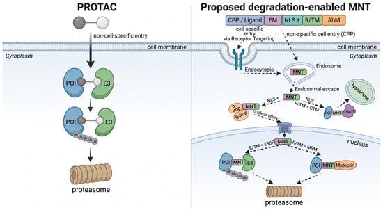
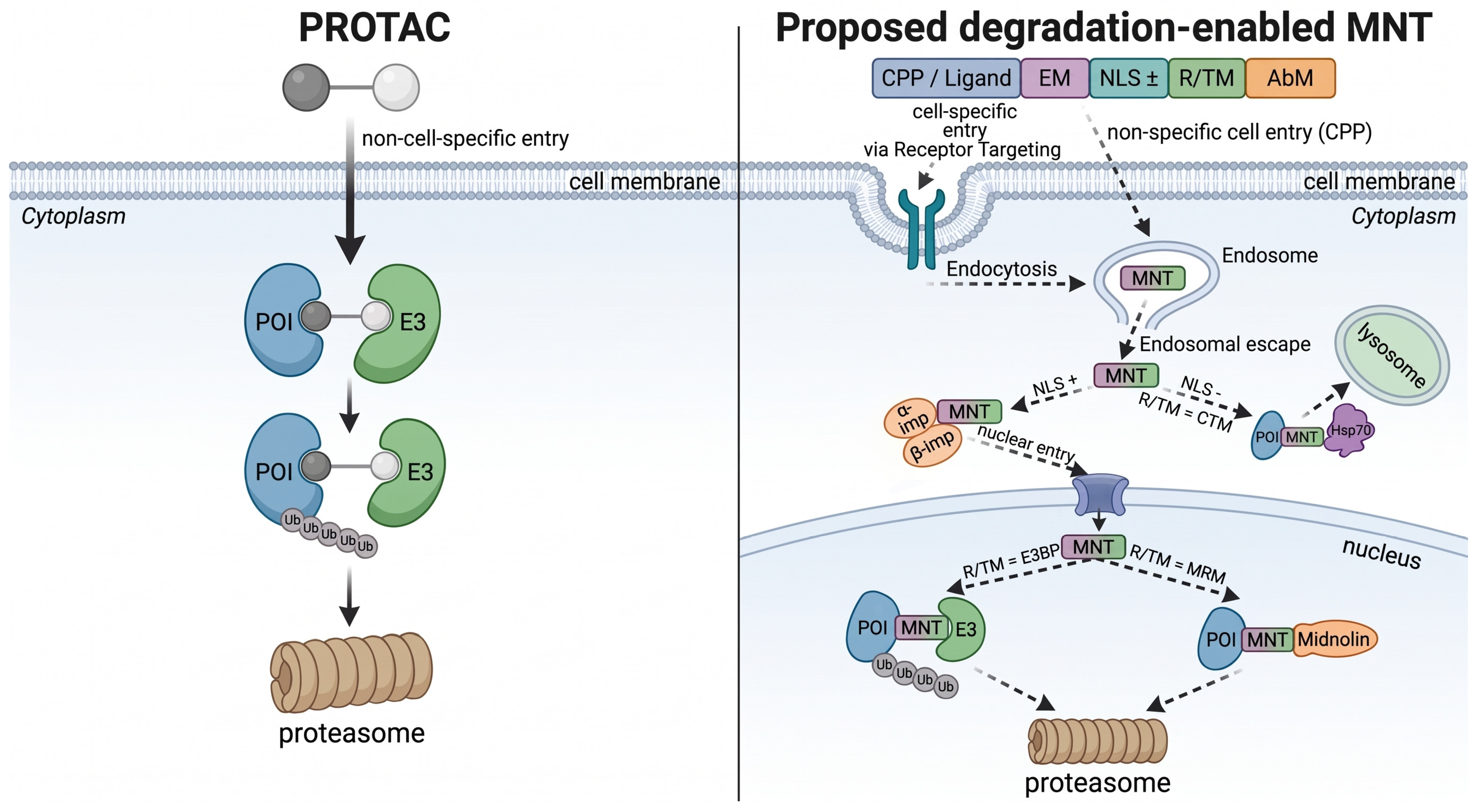

Disclaimer/Publisher’s Note: The statements, opinions and data contained in all publications are solely those of the individual author(s) and contributor(s) and not of MDPI and/or the editor(s). MDPI and/or the editor(s) disclaim responsibility for any injury to people or property resulting from any ideas, methods, instructions or products referred to in the content. |
© 2026 by the authors. Licensee MDPI, Basel, Switzerland. This article is an open access article distributed under the terms and conditions of the Creative Commons Attribution (CC BY) license.