


Abstract
Background/Objectives: Boron neutron capture therapy requires the efficient delivery of sufficient amounts of 10B to tumor cells. In this study, we evaluated a liposomal boron-delivery system based on the B5-TAT-lipopeptide, which contains five boronophenylalanine (BPA) residues within its peptide portion and is anchored to the liposomal membrane through a hydrophobic lipid moiety. Methods: B5-TAT-lipopeptide 5% liposomes with a particle size of approximately 100 nm were prepared, and boron delivery to CT26 murine colon carcinoma cells was evaluated. Results: At an exposure concentration of 25 ppm boron, B5-TAT-lipopeptide 5% liposomes achieved a cell-associated boron level of approximately 4.5 µg B per 1 × 10⁶ cells. Separately, sodium mercaptoundecahydro-closo-dodecaborate (BSH)-encapsulated B5-TAT-lipopeptide 5% liposomes achieved approximately 11 µg B per 1 × 10⁶ cells when evaluated at an exposure concentration of 50 ppm. Following thermal neutron irradiation, B5-TAT-lipopeptide 5% liposome-treated cells showed marked loss of clonogenic survival, with surviving fractions of approximately 0.12 and 0.01 after 15 and 45 min of irradiation, respectively. In the preliminary in vivo tolerability study, rapid intravenous administration of B5-TAT-lipopeptide 5% liposomes without encapsulated BSH at 10 mg B/kg resulted in the death of all treated mice within 5–10 min, whereas no mortality occurred following the administration of phosphate-buffered saline (PBS), free BSH, or BSH-encapsulated PEGylated liposomes. Conclusions: These findings demonstrate substantial cell-associated boron delivery and neutron-induced clonogenic cell killing by B5-TAT-lipopeptide-modified liposomes while revealing a critical limitation in acute in vivo tolerability under the examined administration conditions.
1. Introduction
Boron neutron capture therapy (BNCT) is a binary radiotherapeutic modality that relies on the selective accumulation of 10B in tumor cells followed by neutron irradiation, resulting in the 10B(n,α)7Li reaction and the generation of high-linear-energy-transfer particles with short path lengths. The clinical feasibility of accelerator-based BNCT has been demonstrated using 10B-enriched boronophenylalanine (BPA; borofalan) in patients with recurrent or locally advanced head and neck cancer, and accelerator-based clinical evaluation has recently been extended to newly diagnosed glioblastoma [1,2]. Because the cytotoxic effect of BNCT is produced only in the vicinity of 10B atoms undergoing neutron capture, therapeutic efficacy critically depends on achieving a sufficiently high and spatially favorable boron distribution within tumors while limiting the exposure of normal tissues. Although BPA and sodium mercaptoundecahydro-closo-dodecaborate (BSH) have been the principal boron carriers investigated clinically, limitations such as the insufficient tumor retention of BPA and limited tumor selectivity of BSH continue to motivate the development of more effective boron delivery strategies [3].
Liposomes are a versatile platform for boron delivery because their distinct structural compartments allow the incorporation of boron-containing molecules with different physicochemical properties into a single carrier. Hydrophilic boron compounds can be encapsulated in an aqueous core, whereas boron-conjugated lipids or other amphiphilic boron-containing molecules can be incorporated into the lipid bilayer, providing complementary approaches for increasing the boron payload of the carrier [4,5]. Additionally, liposomal surfaces can be functionalized with peptides or other ligands to modulate their interactions with target cells and promote cellular association or internalization. Previous studies have demonstrated that liposomal formulations containing BSH or boron-containing lipids can enhance boron delivery compared to free boron compounds and support effective neutron capture reactions in experimental models [4,5,6]. These characteristics make liposomes particularly suitable for the development of multifunctional boron delivery systems in which boron loading and cellular delivery can be independently optimized.
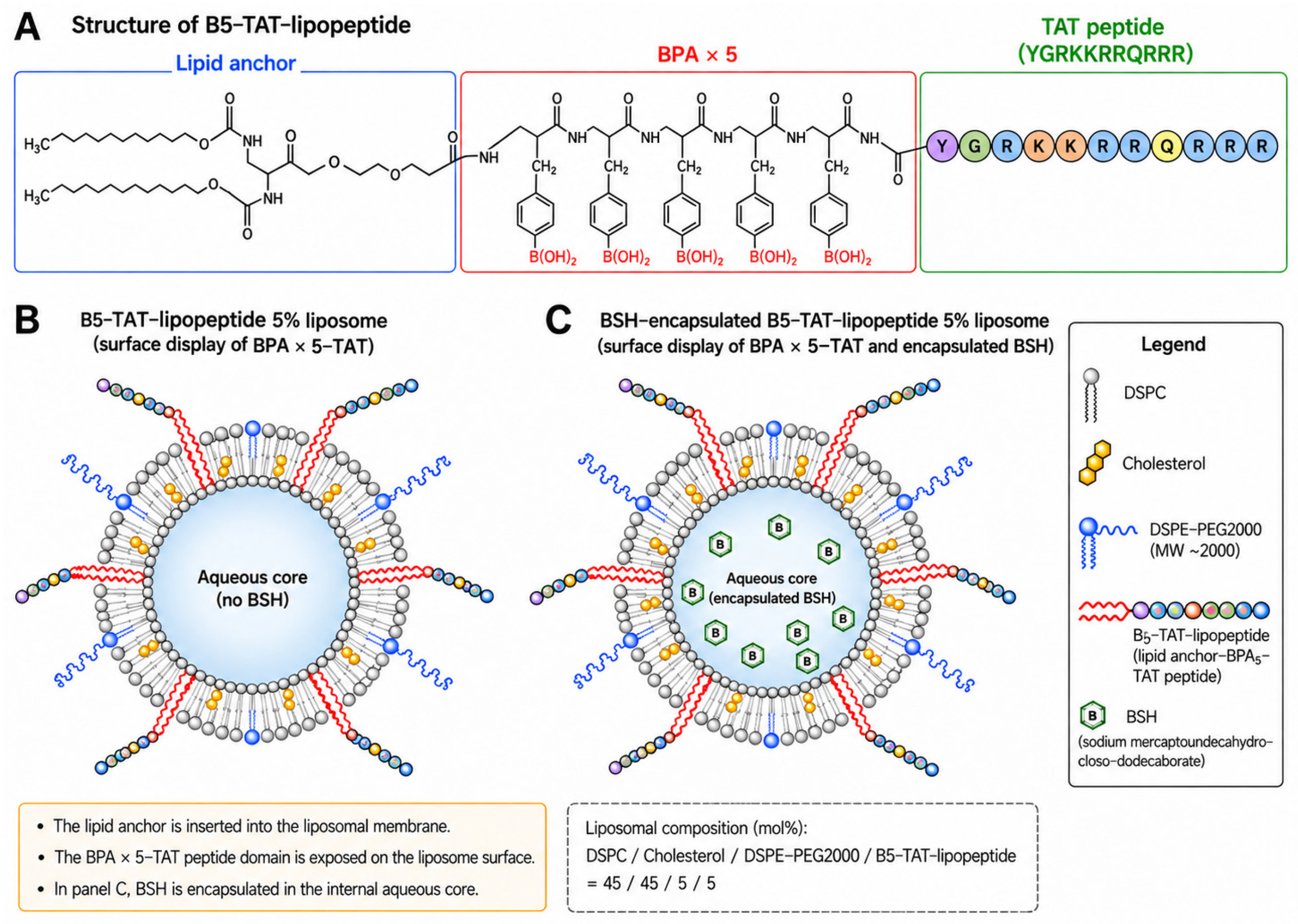
To exploit the advantages of liposomal boron delivery, we previously developed a series of BPA-bearing TAT-lipopeptides, in which multiple BPA residues were conjugated to a peptide containing the HIV-1 transactivator of transcription (TAT) domain and a lipid moiety [7]. Among these constructs, the B3-TAT- and B5-TAT-lipopeptides contained three and five BPA residues, respectively, and were successfully incorporated into liposomal membranes [7]. In the present study, B5-TAT-lipopeptide was selected for further evaluation because of its higher boron payload. TAT is a well-established cell-penetrating peptide rich in basic amino acid residues, which has been widely investigated as a means of enhancing the cellular delivery of molecular and nanoscale cargo through mechanisms involving endocytic pathways, including macropinocytosis [8,9]. B5-TAT-lipopeptide contains five BPA residues covalently incorporated into its peptide chain, together with a TAT-derived cell-penetrating domain and a hydrophobic lipid moiety that anchors the molecule to the liposomal membrane. In this configuration, the BPA residues are located within the peptide portion extending from the membrane surface, whereas the internal aqueous compartment of the liposome remains available for the encapsulation of an additional hydrophilic boron compound, such as BSH.
In this study, we evaluated B5-TAT-lipopeptide-modified liposomes as a multifunctional boron-delivery platform for BNCT. The physicochemical characteristics of the B5-TAT-lipopeptide 5% liposomes and their association with CT26 murine colon carcinoma cells were examined. The ability of the liposomal system to accommodate additional BSH in the internal aqueous phase was evaluated separately by measuring cell-associated boron following exposure to BSH-encapsulated B5-TAT-lipopeptide 5% liposomes. The functional effect of B5-TAT-lipopeptide-mediated boron delivery was further assessed by clonogenic survival following thermal neutron irradiation and compared to that of conventional boron compounds and control liposomal formulations. Finally, the preliminary in vivo tolerability of B5-TAT-lipopeptide 5% liposomes without encapsulated BSH was evaluated following rapid intravenous administration and compared to that of phosphate-buffered saline (PBS), free BSH, and BSH-encapsulated PEGylated liposomes. These experiments were designed to characterize the potential of the B5-TAT-lipopeptide platform for boron delivery and neutron-induced cell killing, as well as its limitations for systemic administration.
2. Materials and Methods
2.1. Materials
1,2-Distearoyl-sn-glycero-3-phosphocholine (DSPC; Coatsome MC-8080) and polyethylene glycol-conjugated phosphatidylethanolamine (DSPE-PEG2000; SUNBRIGHT DSPE-020CN) were purchased from NOF Corporation (Tokyo, Japan). Cholesterol was obtained from Wako Pure Chemical Industries, Ltd. (Osaka, Japan). BSH (Na2B12H11SH; 10B enrichment > 98%) was purchased from Katchem Ltd. (Prague, Czech Republic). L-Boronophenylalanine (10B-BPA; 10B enrichment > 98%) was purchased from Interpharma Praha Ind., Ltd. (Prague, Czech Republic). All other reagents were of reagent grade and used without further purification.
2.2. Synthesis of B5-TAT-Lipopeptide
The B5-TAT-lipopeptide was synthesized by Fmoc solid-phase peptide synthesis on TGS-RAM resin using an automated peptide synthesizer (PSSM-8; Shimadzu Corporation, Kyoto, Japan), as previously described [7]. A tryptophan residue was introduced immediately N-terminal to the HIV-1 TAT sequence as a fluorescent probe, and five BPA residues were incorporated to generate B5-TAT. Subsequently, Fmoc-AEEA (9-fluorenylmethoxycarbonyl-8-amino-3,6-dioxaoctanoic acid) as the linker domain, Fmoc-Asp-OtBu as the hydrophilic domain, and Fmoc-Dap(Fmoc)-OH as the glycerol-mimetic domain were sequentially coupled. Peptide coupling reactions were performed using benzotriazol-1-yloxytripyrrolidinophosphonium hexafluorophosphate (PyBOP), N-hydroxybenzotriazole (HOBt), and N-methylmorpholine (NMM) at molar equivalents of 1.0, 1.0, and 1.5, respectively, relative to the amino acid. Fmoc-protected amino acids and alkyl chains were used in seven- and six-fold molar excesses, respectively, relative to the resin loading, and each coupling reaction was performed for 30 min. Palmitic acid was introduced during the final condensation step, which was performed manually and monitored using the ninhydrin test. The synthesized lipopeptide was cleaved from the resin and deprotected for 16 h at room temperature using a cleavage cocktail consisting of trifluoroacetic acid/H2O/thioanisole/1,2-ethanedithiol/ethyl methyl sulfide/phenol (82:5:5:3:2:3, v/v) containing 10 mg/mL 2-methylindole. The crude product was precipitated with excess diethyl ether and subsequently purified by reversed-phase HPLC using a CAPCELL PAK C18 column (5 µm, 10 × 250 mm). The column temperature was 35 °C, and UV detection was performed at 215 nm. Mobile phase A was Milli-Q water containing 0.1% TFA, and mobile phase B was acetonitrile containing 0.1% TFA. A linear gradient from 0% to 100% B over 60 min was applied at a flow rate of 1.0 mL/min. The fraction corresponding to the main peak was collected and lyophilized, and the resulting material was used for liposome preparation. The identity of the B5-TAT-lipopeptide was confirmed using high-performance liquid chromatography, electrospray ionization time-of-flight mass spectrometry, and 1H nuclear magnetic resonance spectroscopy as previously reported [7].
2.3. Preparation of B5-TAT-Lipopeptide-Modified Liposomes
B5-TAT-lipopeptide-modified liposomes were prepared using the conventional lipid film method. The lipid concentration used for preparation was 10 mg/mL. DSPC, cholesterol, DSPE-PEG2000, and B5-TAT-lipopeptide were mixed in a molar ratio of 45:45:5:5 and dissolved in an organic solvent mixture of chloroform and methanol. After formation of the lipid film, the film was hydrated with PBS, and the resulting liposomal suspension was extruded through a polycarbonate membrane with a pore size of 100 nm to obtain B5-TAT-lipopeptide 5% liposomes. The designation “5%” refers to the nominal molar proportion of B5-TAT-lipopeptide added during preparation, rather than its measured proportion in the final liposomal formulation. The compositions of the liposomal formulations used in this study are summarized in Table 1.
Table 1.
Composition of liposomal formulations used in this study.
2.4. Preparation of BSH-Encapsulated Liposomes
BSH-encapsulated liposomes were prepared using reverse-phase evaporation. BSH-encapsulated bare liposomes, BSH-encapsulated PEGylated liposomes, and BSH-encapsulated B5-TAT-lipopeptide 5% liposomes were prepared using the lipid compositions shown in Table 1. The lipids were dissolved in a chloroform/diethyl ether mixture (1:1, v/v), and an aqueous solution of BSH (125 mM) was added to the organic phase to form a water-in-oil emulsion. The volume ratio of the aqueous phase to the organic phase was maintained at 1:2. The emulsion was sonicated and subsequently concentrated to a gel by rotary evaporation under reduced pressure at 30 °C for 1 h. The resulting preparation was extruded 21 times through a polycarbonate membrane with a pore size of 100 nm at 60 °C using a Lipex extruder (Lipex Biomembranes, Vancouver, BC, Canada). Following extrusion, the preparation was subjected to size-exclusion chromatography to separate the liposomal fraction from unencapsulated BSH and other non-liposomal components. The liposomal fraction was collected, and its boron content was determined by inductively coupled plasma atomic emission spectroscopy (ICP-AES; Shimadzu Corporation, Kyoto, Japan). Liposomes were subsequently recovered from the collected fraction by ultracentrifugation at 100,000× g for 1.5 h at 4 °C using a CS120 ultracentrifuge equipped with an S100AT5 rotor (Hitachi Koki Co., Ltd., Tokyo, Japan). The resulting pellet was resuspended in PBS to obtain the desired boron concentration, and the suspension was filter-sterilized before use in subsequent experiments. All liposomal formulations were used promptly after preparation for the experiments, without storage.
2.5. Physicochemical Characterization of Liposomes
The particle-size and zeta-potential characterization of B5-TAT-lipopeptide 5% liposomes was previously reported [7]. The physicochemical values presented here are based on the original measurement records associated with that characterization, rather than newly acquired measurements. The original particle-size measurements were performed by dynamic light scattering, whereas zeta potential was calculated from electrophoretic mobility measured by laser Doppler electrophoresis. Samples were diluted with water for particle-size and zeta-potential measurements. Both measurements were performed using a Zetasizer Nano ZS (Malvern Instruments Ltd., Worcestershire, UK) at 25 °C. The particle-size measurement duration was 90 s. The zeta-potential measurement comprised 13 runs on the same sample within a single measurement. The physicochemical properties of BSH-encapsulated liposomes and their dependence on preparation conditions were investigated in our previous study [10]. The morphology of the B5-TAT-lipopeptide liposomes was further examined by transmission electron microscopy (TEM). Liposomal suspensions were applied to carbon-film-supported 400-mesh copper grids (EM Fine Grid; Nisshin EM Co., Ltd., Tokyo, Japan) and negatively stained with phosphotungstic acid. The samples were observed under a JEM-2000EX transmission electron microscope (JEOL Ltd., Tokyo, Japan) at an accelerating voltage of 100 kV. Liposome formation and particle morphology were evaluated using electron microscopy.
2.6. Determination of Lipopeptide Incorporation and Boron Content
Size-exclusion chromatography was used to separate liposomal, micellar, and monomeric fractions, and the collected liposomal fraction was used to prepare the formulations for biological experiments. Boron was measured in this fraction before ultracentrifugation and subsequent resuspension in PBS. The assessment of B5-TAT-lipopeptide incorporation into the liposomal fraction using tryptophan fluorescence was previously reported [7]. The boron content of the liposomal preparations was determined using ICP-AES. For BSH-encapsulated B5-TAT-lipopeptide liposomes, the measured total boron content includes boron from both the lipopeptide and BSH. We previously reported a BSH encapsulation efficiency of 3.13% for PEGylated liposomes with a DSPC/cholesterol/DSPE-PEG2000 molar ratio of 47.5:47.5:5 prepared by reverse-phase evaporation using a BSH solution containing 1000 µg B/mL [10].
2.7. Cell Culture
The murine colon carcinoma cell line CT26.WT was obtained from the American Type Culture Collection (ATCC; CRL-2638, Lot No. 70009864; Manassas, VA, USA) and cultured in Eagle’s minimum essential medium (MEM; Sigma-Aldrich Japan, Tokyo, Japan) supplemented with 10% fetal bovine serum and 1% (v/v) penicillin–streptomycin solution (Sigma-Aldrich, Inc., St. Louis, MO, USA). The cells were maintained at 37 °C in a humidified atmosphere containing 5% CO2. For subsequent in vitro experiments, CT26 cells were washed twice with PBS, resuspended in complete culture medium, and incubated for 24 h before treatment with boron-containing formulations.
2.8. Measurement of Cell-Associated Boron
Cell-associated boron was quantified in CT26 cells following exposure to the boron-containing formulations. For each condition, four separate cell samples were individually treated with the formulation and washed, and the boron content of each sample was measured separately (n = 4 independent biological replicates). Unless otherwise specified, the boron concentration during cell exposure was adjusted to 25 ppm. The BSH-encapsulated B5-TAT-lipopeptide 5% liposome formulation was evaluated at a boron concentration of 50 ppm. CT26 cells were incubated with 1 mL of each formulation for 2 h at 37 °C. After incubation, the cells were washed twice with PBS to reduce the amount of unassociated boron compounds and liposomes, and subsequently detached using 0.25% trypsin. For boron analysis, 0.8 mL of concentrated nitric acid was added to 1 mL of the resulting cell suspension, and the samples were digested at 115 °C for 2 h. After digestion, the samples were diluted with water to a final volume of 5 mL and passed through a membrane filter. Total elemental boron concentrations were determined by inductively coupled plasma atomic emission spectroscopy (ICP-AES; Shimadzu Corporation) and expressed as µg B per 1 × 10⁶ cells. Although the boron-containing reagents were enriched in 10B (>98%), the ICP-AES results are reported as total boron rather than isotope-specific 10B amounts.
2.9. Neutron Irradiation and Colony Formation Assay
Clonogenic survival following thermal neutron irradiation was evaluated in CT26 cells treated with the boron-containing formulations. CT26 cells were seeded in 75 cm2 culture flasks at 4 × 10⁵ cells per flask and cultured for 24 h. The cells were then exposed for 2 h at 37 °C in a humidified atmosphere containing 5% CO2 to 1 mL of B5-TAT-lipopeptide 5% liposomes, BPA–fructose, free BSH, or BSH-encapsulated PEGylated liposomes, with the final boron concentration in the culture medium adjusted to 32 ppm. An irradiation-only control group was included without the addition of any formulation or other substance. Cells in this group were washed twice with PBS before neutron irradiation.
Following treatment with B5-TAT-lipopeptide 5% liposomes, BPA–fructose, or BSH-encapsulated PEGylated liposomes, the cells were washed twice with PBS to reduce the amount of extracellular boron compounds or liposomes. In contrast, cells treated with free BSH were not washed before neutron irradiation. This condition was intentionally used because free BSH exhibits limited cellular uptake, and the BSH group was designed to evaluate the cell-killing effect of neutron capture reactions occurring in the presence of extracellular BSH. After treatment and, where applicable, the indicated washing procedure, cells were detached and resuspended in complete Eagle’s minimum essential medium containing 10% fetal bovine serum at 1 × 10⁵ cells in 2 mL. The corresponding non-irradiated controls underwent the same formulation treatment and washing procedures described above and were handled identically except that neutron irradiation was omitted.
Aliquots (500 µL) of the cell suspension were transferred to irradiation tubes and subjected to thermal neutron irradiation at the Kyoto University Research Reactor (KUR), Kyoto University Research Reactor Institute, Osaka, Japan. The samples were irradiated for 15, 30, or 45 min at the KUR heavy-water neutron irradiation facility with a reactor power of 1 MW. Based on the facility dosimetry report, the mean thermal neutron fluences were 1.06 × 10¹², 2.65 × 10¹², and 3.73 × 1012 cm−2, respectively. The corresponding mean non-boron physical dose estimates were 0.57, 1.33, and 1.65 Gy, respectively. These values were calculated as arithmetic means across four measurement positions. The non-boron physical doses included thermal, epithermal, and fast neutron components and the gamma-ray component; the neutron dose calculations assumed a nitrogen concentration of 2%, as specified in the dosimetry report. Boron-capture doses to the cells were not determined. Following irradiation, cells were seeded at 100, 200, and 1000 cells per dish for the 15, 30, and 45 min irradiation conditions, respectively, and cultured for 10 days to allow colony formation. Colonies were stained with methylene blue, and visually distinguishable, discrete colonies were counted. For each treatment and irradiation time, one treated and irradiated cell suspension was divided among five dishes for the colony-formation assay. Treatment and irradiation were performed once per condition. Colonies were counted separately in each dish; thus, n = 5 denotes five dishes derived from one experimental sample, rather than five independently treated and irradiated samples.
2.10. Preliminary In Vivo Tolerability Assessment
The animal experiment was originally planned as a pharmacokinetic study with measurements extending to 72 h after administration. Six-week-old female BALB/cA mice were purchased from CLEA Japan, Inc. (Tokyo, Japan). Mice weighing approximately 20 g were assigned to one of four treatment groups (n = 6 per group): PBS, free BSH, BSH-encapsulated PEGylated liposomes, and B5-TAT-lipopeptide 5% liposomes without encapsulated BSH. Randomization and blinding were not performed. Free BSH, BSH-encapsulated PEGylated liposomes, and B5-TAT-lipopeptide 5% liposomes were adjusted to a boron concentration of 1 mg B/mL and administered at a boron dose of 10 mg B/kg. The injection volume was adjusted to each mouse’s body weight at 10 mL/kg, corresponding to 200 µL for a 20 g mouse. Each formulation was administered manually as a single tail-vein intravenous bolus injection under isoflurane inhalation anesthesia. The injection duration was not measured. Mice were monitored continuously immediately after administration, and mortality and time to death were recorded. Surviving mice were observed for 72 h, corresponding to the originally planned pharmacokinetic observation period. Humane endpoints included marked inactivity, respiratory distress, inability to eat or drink, and weight loss. All animal experiments were approved by the Animal Experiment Safety and Ethics Committee of Fukuyama University on April 28, 2016 (Approval No. H28-A-7).
2.11. Statistical Analysis
Data are presented as the mean ± standard deviation (SD), unless otherwise indicated. For the cell-associated boron study shown in Figure 3A, data were log10-transformed and analyzed using ordinary one-way analysis of variance (ANOVA), followed by Dunnett’s multiple comparisons test to compare the B5-TAT-lipopeptide 5% liposome group with the free BSH and BSH-encapsulated bare liposome groups. No statistical comparison was performed for Figure 3B because the B5-TAT-lipopeptide 5% liposomes and BSH-encapsulated B5-TAT-lipopeptide 5% liposomes were evaluated at different nominal boron concentrations (25 and 50 ppm, respectively). For the clonogenic survival assay, surviving fractions were normalized to the corresponding non-irradiated control for each formulation. Separate irradiation tubes were used for the 15, 30, and 45 min irradiation conditions, and clonogenic survival was evaluated using the respective samples. The same sample was not measured repeatedly across irradiation times; therefore, a repeated-measures analysis was not performed. Statistical analyses of clonogenic survival were performed on log10-transformed surviving fractions at 15, 30, and 45 min of neutron irradiation. The effects of formulation and irradiation time were evaluated using ordinary two-way ANOVA, followed by Dunnett’s multiple comparisons test comparing the B5-TAT-lipopeptide 5% liposome group with each comparator at each irradiation time. In addition, surviving fractions at 45 min irradiation were analyzed separately using one-way ANOVA followed by Tukey’s multiple comparisons test on log10-transformed data. For each condition in the clonogenic survival assay, the five dishes originated from one treated and irradiated cell suspension, with treatment and irradiation performed once. Accordingly, the statistical comparisons reflect dish-level variation within this experiment and do not evaluate variation between independently repeated treatment and irradiation experiments. All statistical tests were two-sided, and statistical significance was set at p < 0.05. Statistical analyses were performed using GraphPad Prism version 6 (GraphPad Software, San Diego, CA, USA).
2.12. Use of Generative Artificial Intelligence
ChatGPT (OpenAI, San Francisco, CA, USA) was used to assist in generating and refining the schematic illustration presented in Figure 1, based on molecular structures and design specifications provided by the authors. The scientific content, molecular structures, labels, and final composition of the figure were reviewed and verified by the authors. Generative artificial intelligence was not used to generate, modify, or analyze any experimental data, quantitative graphs, microscopy images, or statistical results.
Figure 1.
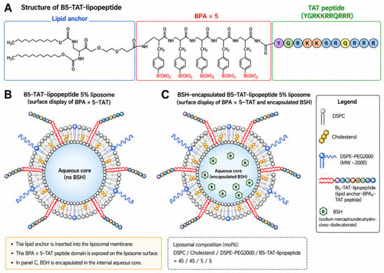
Schematic illustration of the B5-TAT-lipopeptide and the liposomal boron-delivery systems used in this study. (A) Simplified schematic representation of the B5-TAT-lipopeptide, highlighting its lipid-anchor region, five boronophenylalanine (BPA) residues, and TAT-containing peptide domain. (B) Schematic representation of the B5-TAT-lipopeptide 5% liposome, in which the lipopeptide is anchored to the liposomal membrane and the BPA-bearing peptide portion is displayed on the membrane surface. (C) Schematic representation of the BSH-encapsulated B5-TAT-lipopeptide 5% liposome, in which sodium mercaptoundecahydro-closo-dodecaborate (BSH) is encapsulated within the internal aqueous phase in addition to the membrane-anchored B5-TAT-lipopeptide.
3. Results
3.1. Physicochemical Characterization of B5-TAT-Lipopeptide 5% Liposomes
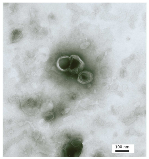
B5-TAT-lipopeptide 5% liposomes were successfully prepared using DSPC, cholesterol, DSPE-PEG2000, and B5-TAT-lipopeptide at a molar ratio of 45:45:5:5. The particle diameter of 105.9 nm and zeta potential of +21 mV were reported previously [7]; the original measurement records are described here in greater detail, including the polydispersity index, intensity-weighted size distribution, and distinction between the overall mean zeta potential and the predominant population. Dynamic light scattering analysis of B5-TAT-lipopeptide 5% liposomes revealed a Z-average hydrodynamic diameter of 105.9 nm with a polydispersity index of 0.066. The intensity-weighted size distribution revealed a single major population centered at 115.2 nm. Zeta-potential analysis yielded a value of +18.6 mV, with the predominant population centered at +21.0 mV and accounting for 85.1% of the distribution, indicating that the liposomes possessed a positively charged surface. TEM following negative staining revealed vesicular particles with diameters of approximately 100 nm, consistent with the particle size determined by dynamic light scattering (Figure 2). At a nominal lipopeptide addition of 5 mol%, an incorporation efficiency of 66.1% into the liposomal fraction was previously reported [7].
Figure 2.
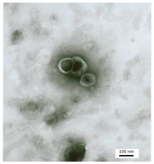
Transmission electron micrograph of B5-TAT-lipopeptide 5% liposomes. Liposomes were negatively stained with phosphotungstic acid and observed by transmission electron microscopy after extrusion through a 100 nm polycarbonate membrane. Scale bar, 100 nm.
3.2. Cell-Associated Boron Levels
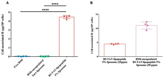
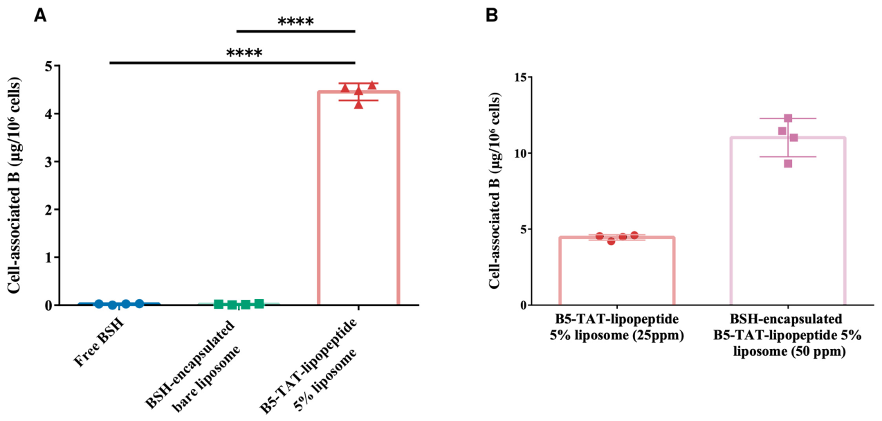
Cell-associated boron levels in CT26 cells were evaluated after 2 h of exposure to different boron-containing formulations, followed by two washes with PBS. At a nominal boron concentration of 25 ppm, free BSH and BSH-encapsulated bare liposomes showed low levels of cell-associated boron. In contrast, treatment with B5-TAT-lipopeptide 5% liposomes at the same nominal boron concentration resulted in a cell-associated boron level of approximately 4.5 µg B per 1 × 10⁶ cells. The cell-associated boron level in the B5-TAT-lipopeptide 5% liposome group was significantly higher than those in both the free BSH and BSH-encapsulated bare liposome groups (p < 0.0001; Figure 3A).
Figure 3.
Cell-associated boron levels in CT26 cells following exposure to boron-containing formulations. (A) Cell-associated boron levels after 2 h of exposure at a nominal boron concentration of 25 ppm to free BSH, BSH-encapsulated bare liposomes, or B5-TAT-lipopeptide 5% liposomes, followed by two washes with PBS. Statistical analysis was performed on log10-transformed data using ordinary one-way ANOVA followed by Dunnett’s multiple comparisons test comparing the B5-TAT-lipopeptide 5% liposome group with the free BSH and BSH-encapsulated bare liposome groups. **** p < 0.0001. (B) Cell-associated boron levels following 2 h of exposure to B5-TAT-lipopeptide 5% liposomes at a nominal boron concentration of 25 ppm or BSH-encapsulated B5-TAT-lipopeptide 5% liposomes at a nominal boron concentration of 50 ppm, followed by two washes with PBS. No statistical comparison was performed between the groups in panel (B) because the nominal boron concentrations differed. Individual values and mean ± SD are shown (n = 4 per group). Symbols indicate individual measurements: blue circles, free BSH; green squares, BSH-encapsulated bare liposomes; and red triangles, B5-TAT-lipopeptide 5% liposomes in panel (A); red circles, B5-TAT-lipopeptide 5% liposomes; and pink squares, BSH-encapsulated B5-TAT-lipopeptide 5% liposomes in panel (B).
To further increase the amount of boron carried by the liposomal system, BSH was encapsulated within the aqueous compartment of B5-TAT-lipopeptide 5% liposomes. When this dual-boron-loaded formulation was evaluated at a nominal boron concentration of 50 ppm, the cell-associated boron level reached approximately 11 µg B per 1 × 10⁶ cells (Figure 3B). Because this formulation was evaluated at twice the nominal boron concentration used for the formulations shown in Figure 3A, no statistical comparison was performed between the 25 and 50 ppm conditions. Accordingly, the higher absolute value observed at 50 ppm should not be interpreted as evidence that BSH encapsulation itself increased boron-association efficiency.
3.3. Clonogenic Cell-Killing Effect Following Thermal Neutron Irradiation
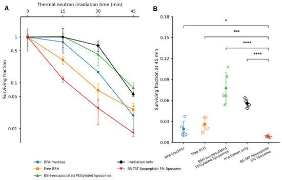
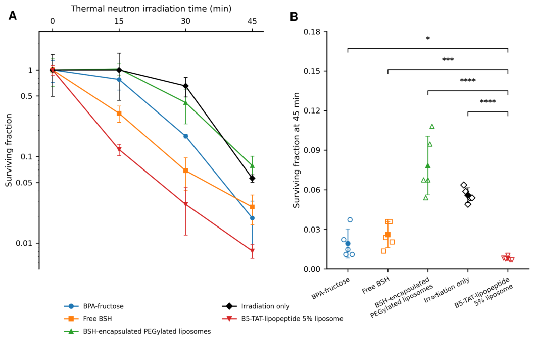
Clonogenic survival of CT26 cells was evaluated following thermal neutron irradiation for 15, 30, and 45 min, corresponding to mean thermal neutron fluences of 1.06 × 10¹², 2.65 × 10¹², and 3.73 × 1012 cm−2, respectively. Surviving fractions were normalized to the corresponding non-irradiated control for each formulation. B5-TAT-lipopeptide 5% liposomes produced a marked irradiation-time-dependent reduction in clonogenic survival, with surviving fractions decreasing to approximately 0.12, 0.03, and 0.01 after 15, 30, and 45 min of irradiation, respectively (Figure 4).
Figure 4.
Clonogenic survival of CT26 cells following thermal neutron irradiation. (A) Irradiation-time-dependent surviving fractions presented as mean ± SD. (B) Clonogenic survival at 45 min irradiation. In panel (B), open symbols represent measurements from individual dishes, and filled symbols with error bars represent mean ± SD across five dishes. For each formulation and irradiation time, the five dishes were derived from a single treated and irradiated cell suspension. CT26 cells were exposed to the indicated boron formulations at a boron concentration of 32 ppm in the culture medium. Irradiation times of 15, 30, and 45 min corresponded to mean thermal neutron fluences of 1.06 × 10¹², 2.65 × 10¹², and 3.73 × 1012 cm−2, respectively. The corresponding mean non-boron physical dose estimates were 0.57, 1.33, and 1.65 Gy, respectively. Surviving fractions were normalized to the corresponding non-irradiated control for each formulation. Cells treated with boron formulations other than free BSH were washed twice with PBS before irradiation, whereas free BSH was not removed before irradiation. Statistical analysis of log10-transformed surviving fractions at 15, 30, and 45 min was performed using ordinary two-way ANOVA followed by Dunnett’s multiple comparisons test. For panel (B), log10-transformed surviving fractions at 45 min were additionally analyzed using one-way ANOVA followed by Tukey’s multiple comparisons test. The statistical comparisons reflect variation among dishes within a single treatment and irradiation experiment per condition, rather than variation between independently repeated experiments. Asterisks indicate significant differences relative to B5-TAT-lipopeptide 5% liposomes (* p < 0.05, *** p < 0.001, **** p < 0.0001; Tukey-adjusted p-values).
Ordinary two-way ANOVA of the log10-transformed surviving fractions showed significant effects of irradiation time [F(2,60) = 449.1, p < 0.0001] and formulation [F(4,60) = 130.9, p < 0.0001], together with a significant irradiation time × formulation interaction [F(8,60) = 7.13, p < 0.0001]. Dunnett’s multiple comparisons test showed that the surviving fraction in the B5-TAT-lipopeptide 5% liposome group was significantly lower than those in the irradiation-only, BPA-fructose, free BSH, and BSH-encapsulated PEGylated liposome groups at each irradiation time examined (all adjusted p ≤ 0.0026). To further evaluate the differences among formulations at the longest irradiation time (45 min), a separate one-way ANOVA was performed on log10-transformed surviving fractions. This analysis revealed significant differences among the experimental groups [F(4,20) = 37.36, p < 0.0001]. Tukey’s multiple comparisons test demonstrated significantly lower surviving fractions for B5-TAT-lipopeptide 5% liposomes than for BPA-fructose (adjusted p = 0.0114), free BSH (adjusted p = 0.000284), BSH-encapsulated PEGylated liposomes (adjusted p < 0.0001), and the irradiation-only control (adjusted p < 0.0001) (Figure 4B).
The experimental conditions for free BSH differed from those for the other boron formulations because free BSH was not removed by washing with PBS before neutron irradiation, whereas the other formulations were washed twice. Thus, the free BSH group represents an irradiation condition in which extracellular BSH remained present.
3.4. Preliminary In Vivo Tolerability
Preliminary in vivo tolerability was evaluated in female BALB/cA mice following rapid intravenous administration. No mortality was observed during the 72 h observation period in mice administered PBS (0/6), free BSH at 10 mg B/kg (0/6), or BSH-encapsulated PEGylated liposomes at 10 mg B/kg (0/6). In contrast, all mice administered B5-TAT-lipopeptide 5% liposomes without encapsulated BSH at 10 mg B/kg died within 5–10 min of administration (6/6) (Table 2). Administration to all six mice had been completed before the first adverse signs were noticed. Mild convulsive movements and mild incontinence were observed, and the mice died before euthanasia was performed. These results demonstrated the markedly poor acute tolerability of the B5-TAT-lipopeptide 5% liposome formulation under the rapid intravenous administration conditions used in this study. The absence of mortality in the BSH-encapsulated PEGylated liposome group indicated that the rapid mortality was not a general consequence of intravenous administration of a BSH-containing liposomal formulation. The mechanism responsible for the rapid mortality associated with the B5-TAT-lipopeptide 5% liposome formulation was not determined.
Table 2.
Preliminary in vivo tolerability following rapid intravenous administration in BALB/cA mice.
4. Discussion
This study evaluated the physicochemical characteristics, cell-associated boron delivery, neutron-induced clonogenic cell killing, and preliminary in vivo tolerability of B5-TAT-lipopeptide-modified liposomes as a boron delivery system for BNCT. B5-TAT-lipopeptide 5% liposomes had a Z-average diameter of 105.9 nm, a polydispersity index of 0.066, and a positive zeta potential of +18.6 mV; their vesicular morphology was confirmed by TEM. In CT26 cells, B5-TAT-lipopeptide 5% liposomes produced substantially higher cell-associated boron levels than free BSH and BSH-encapsulated bare liposomes when evaluated at the same nominal boron concentration of 25 ppm. In a separate experiment, BSH-encapsulated B5-TAT-lipopeptide 5% liposomes achieved a cell-associated boron level of approximately 11 µg B per 106 cells at a nominal boron concentration of 50 ppm; because the exposure concentration differed from that used for the 25 ppm formulations, this result cannot be interpreted as a direct increase in boron-association efficiency. Following thermal neutron irradiation, B5-TAT-lipopeptide 5% liposomes reduced the clonogenic survival of CT26 cells after washing to reduce the amount of unassociated extracellular formulation. In the preliminary in vivo study, rapid intravenous administration of BSH-free B5-TAT-lipopeptide 5% liposomes, in which boron was provided by the BPA residues of the B5-TAT-lipopeptide, at 10 mg B/kg resulted in the death of all treated mice within 5–10 min. In contrast, no mortality was observed after administration of PBS, free BSH, or BSH-encapsulated PEGylated liposomes, with both BSH-containing groups receiving the same boron dose of 10 mg B/kg. These findings demonstrate both the potential of B5-TAT-lipopeptide-modified liposomes for enhancing cell-associated boron delivery and neutron-induced clonogenic cell killing and a critical limitation in the acute in vivo tolerability of the B5-TAT-lipopeptide 5% formulation under the administration conditions used in this study.
The relatively high level of cell-associated boron observed with B5-TAT-lipopeptide 5% liposomes may be related, at least in part, to the presence of the TAT domain on the liposomal surface. TAT is a highly cationic cell-penetrating peptide, and the incorporation of B5-TAT-lipopeptide into the liposomal membrane resulted in positively charged particles, consistent with the surface presentation of the basic peptide domain [7]. Previous studies have shown that TAT modification enhances the binding of liposomes to cell-surface proteoglycans and promotes their subsequent internalization via endocytic pathways [8,9]. Fretz et al. directly demonstrated that TAT-modified liposomes initially bind to the plasma membrane and are subsequently internalized into endosomal and lysosomal compartments, whereas studies of TAT-containing cargoes have also implicated macropinocytosis in cellular entry [8,9]. These observations suggest a plausible mechanism by which the B5-TAT-lipopeptide formulation could increase the amount of boron retained in association with CT26 cells after washing. However, because the intracellular localization of boron was not directly determined in the present study, the measured boron should be interpreted as cell-associated rather than exclusively intracellular. In addition, the increased boron level cannot be attributed solely to TAT-mediated cellular association because the B5-TAT-lipopeptide itself carries five BPA residues and therefore also increases the boron payload of the liposomal carrier.
Another design feature of this liposomal system is its ability to accommodate boron in two spatially distinct regions of the carrier. The B5-TAT-lipopeptide is anchored to the liposomal membrane through its hydrophobic lipid moiety, whereas the five BPA residues are positioned within the peptide portion extending away from the membrane surface. In this design, B5-TAT-lipopeptide functions as a membrane-associated, boron-bearing surface component rather than an encapsulated peptide payload intended for controlled or sustained release. Although incorporation into the liposomal fraction was assessed by size-exclusion chromatography and tryptophan fluorescence, the kinetics of lipopeptide dissociation from the membrane under physiological conditions were not determined. In parallel, the hydrophilic boron compound BSH can be encapsulated within the internal aqueous phase. This spatial arrangement may be advantageous because direct interactions between high concentrations of BSH and phosphatidylcholine membranes have been reported to induce substantial structural alterations, including liposome aggregation, membrane rupture, and leakage of encapsulated solutes [11]. Thus, increasing the boron payload solely by increasing the amount of BSH associated with a liposomal formulation may be constrained by effects on membrane stability. In contrast, the present molecular architecture provides an additional source of boron through the BPA-bearing peptide domain while requiring only the hydrophobic anchor of the lipopeptide to be incorporated into the lipid bilayer. A conceptually similar strategy for positioning boron groups away from the lipid membrane has been shown to minimize interference with liposomal membrane structure [12]. In the present study, BSH-encapsulated B5-TAT-lipopeptide 5% liposomes achieved a cell-associated boron level of approximately 11 µg B per 1 × 10⁶ cells when applied at a nominal boron concentration of 50 ppm. This value was higher than the approximately 4.5 µg B per 1 × 10⁶ cells observed with B5-TAT-lipopeptide 5% liposomes at a nominal boron concentration of 25 ppm; however, the two formulations were evaluated at different nominal boron concentrations. Therefore, the present data do not establish that BSH encapsulation itself increased cellular association of boron. Rather, they demonstrate that a liposomal architecture combining BPA-bearing, membrane-anchored B5-TAT-lipopeptide with encapsulated BSH provides two complementary routes for increasing the boron payload without relying exclusively on high levels of encapsulated BSH.
The marked reduction in clonogenic survival following thermal neutron irradiation indicates that boron retained in association with CT26 cells after exposure to B5-TAT-lipopeptide 5% liposomes was sufficient to enhance neutron-induced clonogenic cell killing. This finding is particularly relevant because the liposome-treated cells were washed before irradiation, thereby reducing the amount of unassociated extracellular formulation. The observed clonogenic cell killing is therefore consistent with boron that remained closely associated with the cells after washing. The spatial distribution of 10B is a critical determinant of BNCT efficacy because the α particles and 7Li nuclei generated by the 10B(n,α)7Li reaction have ranges comparable to cellular dimensions. Recent microdosimetric simulations using a virtual cell model containing intracellular organelles have further demonstrated that the dose delivered to the cell nucleus can vary substantially depending on the intracellular localization of boron, even at an equivalent total boron concentration [13]. These findings indicate that the biological effectiveness of a boron carrier cannot be predicted solely from its bulk boron concentration and that its cellular and subcellular distribution should also be considered. In contrast, free BSH was intentionally retained in the extracellular medium during irradiation in the present study because of its limited cellular uptake. Therefore, the BSH group should be regarded as a reference condition in which neutron capture reactions occurred in the continued presence of extracellular BSH rather than as a direct comparator of cell-associated boron delivery. The substantial clonogenic cell killing observed after B5-TAT-lipopeptide 5% liposome-treated cells were washed is consistent with the concept that maintaining boron in close association with tumor cells is an important determinant of BNCT efficacy.
BPA and BSH are the two principal boron agents with a long history of clinical use in BNCT [3]. Accordingly, BPA-fructose and free BSH were deliberately selected as clinically relevant reference agents for evaluating the efficacy of B5-TAT-lipopeptide 5% liposomes. In addition, BSH-encapsulated PEGylated liposomes were included as a liposomal comparator based on previous investigations of BSH-loaded PEGylated liposomes for BNCT, particularly the study by Maruyama et al. [14]. Thus, the present study incorporated the two clinically established boron agents and a previously investigated liposomal boron-delivery system as scientifically relevant comparators. Notably, B5-TAT-lipopeptide 5% liposomes demonstrated significantly lower clonogenic surviving fractions than all four comparator groups at 15, 30, and 45 min of neutron irradiation, highlighting the substantial enhancement of neutron-induced cell-killing efficacy achieved by the BPA-bearing lipopeptide formulation under the experimental conditions examined.
The preliminary in vivo study revealed a major limitation of the B5-TAT-lipopeptide formulation. Rapid intravenous administration of B5-TAT-lipopeptide 5% liposomes without encapsulated BSH at a boron dose of 10 mg B/kg resulted in the death of all treated mice within 5–10 min. In contrast, no mortality was observed following administration of PBS, free BSH, or BSH-encapsulated PEGylated liposomes, with both BSH-containing groups receiving the same boron dose of 10 mg B/kg. These findings indicate that the rapid mortality was associated with the B5-TAT-lipopeptide 5% liposome formulation under the conditions used in this study. However, the present experimental design does not identify which property of the formulation was responsible for this effect. B5-TAT-lipopeptide 5% liposomes exhibited a positive surface charge, which may influence the biological interactions and tolerability of cationic liposomal formulations [15]. Furthermore, intravenous administration of liposomes can induce acute complement-mediated cardiopulmonary and hemodynamic responses, the magnitude of which depends on formulation characteristics and administration conditions [16,17]. In particular, bolus administration has been reported to produce substantially greater pulmonary vasoactive responses than slow infusion [16]. Therefore, the rapid intravenous administration used in the present study may have amplified a formulation-dependent acute response. However, because complement activation, cardiopulmonary responses, hematological changes, aggregation, and histopathological alterations were not evaluated, the mechanism responsible for the rapid mortality remains undetermined.
The present study has several limitations. First, the boron measurements represent cell-associated boron and do not establish the intracellular or subcellular localization of boron. This is particularly relevant for BNCT because microdosimetric analyses indicate that biological effectiveness depends not only on total cellular boron content but also on its intracellular distribution [13]. Second, BSH-encapsulated B5-TAT-lipopeptide 5% liposomes were evaluated at a nominal boron concentration of 50 ppm, whereas the formulations in Figure 3A were evaluated at 25 ppm. Therefore, the higher absolute cell-associated boron level obtained with the BSH-encapsulated formulation cannot be interpreted as evidence of greater boron-association efficiency. The BSH encapsulation efficiency of the formulations prepared in this study was not measured. Third, although marked acute mortality was observed specifically in mice receiving B5-TAT-lipopeptide 5% liposomes under the administration conditions examined, the mechanism responsible for this response was not investigated. Cytotoxicity toward healthy cells, off-target effects, and organ-specific toxicity were not directly evaluated. Thus, the present findings do not establish biocompatibility or safety for systemic administration. Despite the HPLC purification, a contribution from residual synthesis or purification components, including trifluoroacetate counterions, to the observed acute toxicity cannot be excluded. Furthermore, the B5-TAT-lipopeptide 5% liposome formulation and the BSH-encapsulated PEGylated liposome formulation differed not only in surface modification but also in the chemical form and location of boron within the formulation. In addition, adjustment of the formulations to equal boron concentrations or doses did not ensure equal lipid or lipopeptide concentrations or doses. Consequently, the present in vivo comparison cannot identify the individual factor responsible for the difference in tolerability. The stability of the liposomal formulations under the experimental conditions was not assessed over time. Future studies should therefore examine the effects of B5-TAT-lipopeptide density, surface charge, lipid composition, blood compatibility, and administration rate, together with a direct assessment of intracellular boron localization and in vivo biodistribution.
5. Conclusions
B5-TAT-lipopeptide 5% liposomes provided a multifunctional boron-delivery platform in which BPA-bearing lipopeptides were displayed on the liposomal surface and additional BSH could be encapsulated within the internal aqueous phase. B5-TAT-lipopeptide modification produced substantial cell-associated boron levels in CT26 cells and enhanced neutron-induced clonogenic cell killing following thermal neutron irradiation. However, rapid intravenous administration of B5-TAT-lipopeptide 5% liposomes without encapsulated BSH at a boron dose of 10 mg B/kg resulted in the death of all treated mice, revealing a critical limitation in acute systemic tolerability under the administration conditions examined. Further optimization of the formulation and administration conditions, together with clarification of the mechanism underlying the acute response, will be required before in vivo therapeutic application.
Author Contributions
Conceptualization, M.S. (Makoto Shirakawa); methodology, M.S. (Makoto Shirakawa); validation, A.Z.; formal analysis, M.S. (Makoto Shirakawa); investigation, M.S. (Makoto Shirakawa), K.N., K.B., M.O., A.M. (Aoi Machida), M.S. (Minoru Suzuki); resources, K.N., A.M. (Akira Matsumura), M.S. (Minoru Suzuki); data curation, M.S. (Makoto Shirakawa); writing—original draft preparation, M.S. (Makoto Shirakawa); writing—review and editing, M.S. (Makoto Shirakawa), A.Z., K.B., M.O., A.M. (Aoi Machida), K.N., M.S. (Minoru Suzuki), A.M. (Akira Matsumura); visualization, M.S. (Makoto Shirakawa); supervision, M.S. (Makoto Shirakawa), A.M. (Akira Matsumura); project administration, M.S. (Makoto Shirakawa); funding acquisition, M.S. (Makoto Shirakawa), A.M. (Akira Matsumura). All authors have read and agreed to the published version of the manuscript.
Funding
This research was funded by JSPS KAKENHI, grant numbers 12J01947 and 25893226.
Institutional Review Board Statement
The animal study protocol was approved by the Animal Experiment Safety and Ethics Committee of Fukuyama University on 28 April 2016 (Approval No. H28-A-7).
Informed Consent Statement
Not applicable.
Data Availability Statement
The raw data supporting the conclusions of this article will be made available by the authors on request.
Acknowledgments
The authors thank Yoshinori Sakurai, Hiroki Tanaka, and Takushi Takata of the Institute for Integrated Radiation and Nuclear Science, Kyoto University, for their assistance with neutron irradiation and dosimetry at the Kyoto University Research Reactor (KUR). This work was partially supported by the Joint Usage/Research of the Institute for Integrated Radiation and Nuclear Science, Kyoto University (KURNS). During the preparation of this manuscript, the authors used ChatGPT (OpenAI) to assist in generating and refining the schematic illustration in Figure 1. The authors reviewed and edited the output and take full responsibility for the content of this publication.
Conflicts of Interest
The authors declare no conflicts of interest.
Abbreviations
The following abbreviations are used in this manuscript:
| AEEA | 8-Amino-3,6-dioxaoctanoic acid |
| ANOVA | Analysis of variance |
| BNCT | Boron neutron capture therapy |
| BPA | p-Boronophenylalanine |
| BSH | Sodium mercaptoundecahydro-closo-dodecaborate |
| DSPC | 1,2-Distearoyl-sn-glycero-3-phosphocholine |
| DSPE-PEG2000 | Polyethylene glycol-conjugated phosphatidylethanolamine |
| Fmoc | 9-Fluorenylmethoxycarbonyl |
| HIV-1 | Human immunodeficiency virus type 1 |
| HOBt | N-Hydroxybenzotriazole |
| ICP-AES | Inductively coupled plasma atomic emission spectroscopy |
| KUR | Kyoto University Research Reactor |
| KURNS | Institute for Integrated Radiation and Nuclear Science, Kyoto University |
| MEM | Minimum essential medium |
| NMM | N-Methylmorpholine |
| PBS | Phosphate-buffered saline |
| PyBOP | Benzotriazol-1-yloxytripyrrolidinophosphonium hexafluorophosphate |
| SD | Standard deviation |
| TAT | Transactivator of transcription |
| TEM | Transmission electron microscopy |
References
- Hirose, K.; Konno, A.; Hiratsuka, J.; Yoshimoto, S.; Kato, T.; Ono, K.; Otsuki, N.; Hatazawa, J.; Tanaka, H.; Takayama, K.; et al. Boron neutron capture therapy using cyclotron-based epithermal neutron source and borofalan (10B) for recurrent or locally advanced head and neck cancer (JHN002): An open-label phase II trial. Radiother. Oncol. 2021, 155, 182–187. [Google Scholar] [CrossRef] [Scilit] [PubMed]
- Nakai, K.; Kumada, H.; Matsumoto, Y.; Baba, K.; Murakami, M.; Ishida, T.; Saito, T.; Nakamura, M.; Numajiri, H.; Mizumoto, M.; et al. Boron neutron capture therapy (BNCT) phase I clinical trial for newly diagnosed glioblastoma by newly developed accelerator at University of Tsukuba. Appl. Radiat. Isot. 2025, 226, 112152. [Google Scholar] [CrossRef] [Scilit] [PubMed]
- Barth, R.F.; Mi, P.; Yang, W. Boron delivery agents for neutron capture therapy of cancer. Cancer Commun. 2018, 38, 35. [Google Scholar] [CrossRef] [Scilit] [PubMed]
- Shelly, K.; Feakes, D.A.; Hawthorne, M.F.; Schmidt, P.G.; Krisch, T.A.; Bauer, W.F. Model studies directed toward the boron neutron-capture therapy of cancer: Boron delivery to murine tumors with liposomes. Proc. Natl. Acad. Sci. USA 1992, 89, 9039–9043. [Google Scholar] [CrossRef] [Scilit] [PubMed]
- Feakes, D.A.; Shelly, K.; Hawthorne, M.F. Selective boron delivery to murine tumors by lipophilic species incorporated in the membranes of unilamellar liposomes. Proc. Natl. Acad. Sci. USA 1995, 92, 1367–1370. [Google Scholar] [CrossRef] [Scilit] [PubMed]
- Kueffer, P.J.; Maitz, C.A.; Khan, A.A.; Schuster, S.A.; Shlyakhtina, N.I.; Jalisatgi, S.S.; Brockman, J.D.; Nigg, D.W.; Hawthorne, M.F. Boron neutron capture therapy demonstrated in mice bearing EMT6 tumors following selective delivery of boron by rationally designed liposomes. Proc. Natl. Acad. Sci. USA 2013, 110, 6512–6517. [Google Scholar] [CrossRef] [Scilit] [PubMed]
- Shirakawa, M.; Yamamoto, T.; Nakai, K.; Aburai, K.; Kawatobi, S.; Tsurubuchi, T.; Yamamoto, Y.; Yokoyama, Y.; Okuno, H.; Matsumura, A. Synthesis and evaluation of a novel liposome containing BPA–peptide conjugate for BNCT. Appl. Radiat. Isot. 2009, 67, S88–S90. [Google Scholar] [CrossRef] [Scilit] [PubMed]
- Fretz, M.M.; Koning, G.A.; Mastrobattista, E.; Jiskoot, W.; Storm, G. OVCAR-3 cells internalize TAT-peptide modified liposomes by endocytosis. Biochim. Biophys. Acta 2004, 1665, 48–56. [Google Scholar] [CrossRef] [Scilit] [PubMed]
- Wadia, J.S.; Stan, R.V.; Dowdy, S.F. Transducible TAT-HA fusogenic peptide enhances escape of TAT-fusion proteins after lipid raft macropinocytosis. Nat. Med. 2004, 10, 310–315. [Google Scholar] [CrossRef] [Scilit] [PubMed]
- Shirakawa, M.; Nakai, K.; Sato, Y.; Nakamura, S.; Harada, M.; Ishihara, K.; Yoshida, F.; Matsumura, A.; Tomida, H. Optimization of preparation methods for high loading content and high encapsulation efficiency of BSH into liposomes. Appl. Radiat. Isot. 2021, 169, 109260. [Google Scholar] [CrossRef] [Scilit] [PubMed]
- Awad, D.; Damian, L.; Winterhalter, M.; Karlsson, G.; Edwards, K.; Gabel, D. Interaction of Na2B12H11SH with dimyristoyl phosphatidylcholine liposomes. Chem. Phys. Lipids 2009, 157, 78–85. [Google Scholar] [CrossRef] [Scilit] [PubMed]
- Shirakawa, M.; Zaboronok, A.; Nakai, K.; Sato, Y.; Kayaki, S.; Sakai, T.; Tsurubuchi, T.; Yoshida, F.; Nishiyama, T.; Suzuki, M.; et al. A novel boron lipid to modify liposomal surfaces for boron neutron capture therapy. Cells 2021, 10, 3421. [Google Scholar] [CrossRef] [Scilit] [PubMed]
- Shigehira, T.; Hanafusa, T.; Igawa, K.; Kasai, T.; Furuya, S.; Nishimori, H.; Maeda, Y.; Michiue, H.; Fujimura, A. Particle and Heavy Ion Transport Code System-Based Microdosimetry for the Development of Boron Agents for Boron Neutron Capture Therapy. Adv. Theory Simul. 2023, 6, 2300163. [Google Scholar] [CrossRef] [Scilit]
- Maruyama, K.; Ishida, O.; Kasaoka, S.; Takizawa, T.; Utoguchi, N.; Shinohara, A.; Chiba, M.; Kobayashi, H.; Eriguchi, M.; Yanagie, H. Intracellular targeting of sodium mercaptoundecahydrododecaborate (BSH) to solid tumors by transferrin-PEG liposomes, for boron neutron-capture therapy (BNCT). J. Control. Release 2004, 98, 195–207. [Google Scholar] [CrossRef] [Scilit] [PubMed]
- Andrade, S.; Loureiro, J.A.; Ramirez, S.; Catumbela, C.S.G.; Soto, C.; Morales, R.; Pereira, M.C. Multi-Dose Intravenous Administration of Neutral and Cationic Liposomes in Mice: An Extensive Toxicity Study. Pharmaceuticals 2022, 15, 761. [Google Scholar] [CrossRef] [Scilit] [PubMed]
- Szebeni, J.; Baranyi, L.; Savay, S.; Bodo, M.; Morse, D.S.; Basta, M.; Stahl, G.L.; Bünger, R.; Alving, C.R. Liposome-induced pulmonary hypertension: Properties and mechanism of a complement-mediated pseudoallergic reaction. Am. J. Physiol. Heart Circ. Physiol. 2000, 279, H1319–H1328. [Google Scholar] [CrossRef] [Scilit] [PubMed]
- Fülöp, T.; Kozma, G.T.; Vashegyi, I.; Mészáros, T.; Rosivall, L.; Urbanics, R.; Storm, G.; Metselaar, J.M.; Szebeni, J. Liposome-induced hypersensitivity reactions: Risk reduction by design of safe infusion protocols in pigs. J. Control. Release 2019, 309, 333–338. [Google Scholar] [CrossRef] [Scilit] [PubMed]

Disclaimer/Publisher’s Note: The statements, opinions and data contained in all publications are solely those of the individual author(s) and contributor(s) and not of MDPI and/or the editor(s). MDPI and/or the editor(s) disclaim responsibility for any injury to people or property resulting from any ideas, methods, instructions or products referred to in the content. |
© 2026 by the authors. Licensee MDPI, Basel, Switzerland. This article is an open access article distributed under the terms and conditions of the Creative Commons Attribution (CC BY) license.