


Abstract
Background/Objectives: Nanotherapeutics integrated with drug repurposing represent an emerging approach to overcome ongoing cancer treatment resistance, particularly in breast cancer (BC) and hepatoblastoma (HB). This study comparatively evaluated an optimally designed smart anticancer nanoselenium hybrid repaglinide (RG)-loaded plurosomes (SHPs) against MDA-MB-231 and HepG2 cell lines. Methods: SHPs were fabricated by the thin-film hydration method and optimized using D-optimal design to investigate the impact of formulation parameters, including Pluronic® amount, sonication time, and hydrophilic–lipophilic balance of Pluronic® on entrapment efficacy (EE%), particle size (PS), and zeta potential (ZP). Results: The optimum formulation (OP.SHP) exhibited high EE% (70.50 ± 1.86%), desired nanosize (242.90 ± 13.77 nm), and excellent stability (ZP = −66.40 ± 2.12 mV). The thermal profile of OP.SHP confirmed successful RG entrapment. The in vitro drug release profile demonstrated a pH-dependent behavior with enhanced drug release at pH 5.5 (91.29 ± 5.67% over 10 h) and a slower release rate at pH 7.4 than the RG suspension. The MTT assay showed cell line-dependent cytotoxicity, with OP.SHP showing stronger antiproliferative activity against MDA-MB-231 (IC50 = 107.50 µg/mL) than HepG2 cells (IC50 = 215.40 µg/mL), compared with RG suspension. Additionally, cell-cycle analysis revealed S and G2/M phase accumulation in MDA-MB-231 cells and S phase accumulation in HepG2 cells, indicating cell-cycle disruption. Furthermore, the Annexin-V/PI assay showed apoptotic-necrotic cell death in BC and predominant apoptotic cell death in HB cells. Notably, OP.SHP treatment markedly reduced Bcl-2 levels and significantly increased caspase-3 levels. Molecular docking analysis showed good binding affinity of RG toward AMPK and Bcl-2 (−8.7 and −8.0 kcal/mol, respectively). Conclusions: Lastly, OP.SHP demonstrated significant antitumor activity against both cell lines, highlighting its potential as a nanotherapeutic strategy for cancer treatment.
1. Introduction
Multidrug resistance (MDR) contributes to a major cancer treatment challenge, particularly in breast cancer (BC), which is the second most commonly diagnosed cancer worldwide, and liver cancer, which ranks as the third leading cause of cancer-related mortality [1,2,3,4]. As a result, recent studies are exploring innovative treatment strategies against BC and liver cancer, such as drug repurposing.
Drug repurposing has emerged as a promising approach for cancer therapy due to its time and cost efficiency by utilizing drugs with established safety profiles for novel indications [5]. Among these approaches is the modulation of ATP-sensitive potassium (KATP) channels using selective blockers, which has demonstrated significant antitumor effects, owing to the critical role of KATP channels in regulating tumor development and apoptosis [6,7,8]. Therefore, blocking KATP channels induces apoptosis by modulating the antiapoptotic protein B cell lymphoma (Bcl-2), which is overexpressed in tumors, promoting MDR [7,9].
Besides the role of KATP channels in tumor development, other key regulatory proteins are also associated with tumor progression, including the cellular energy sensor adenosine monophosphate-activated protein kinase (AMPK) [10]. Thus, targeting KATP channels along with Bcl-2 and AMPK represents a promising therapeutic modality for inhibiting tumor growth, particularly triple-negative BC and hepatoblastoma (HB). In this regard, several antidiabetic drugs that target KATP channels have been shown to exhibit strong anticancer effects. For example, repaglinide (RG), an insulin secretagogue and KATP channel inhibitor, has demonstrated significant anticancer activity against various cancer cell lines [11]. Although only a limited number of studies have explored its anticancer potential, Xiao et al. [12] reported that RG inhibits the proliferation and migration of glioblastoma multiforme cells by targeting the Bcl-2 protein. In addition, RG suppresses the transcription factor FOXO3 in neuroblastoma, which is involved in chemoresistance and tumor angiogenesis [13]. Also, another study investigated the cytotoxic profile of RG and highlighted its marked antiproliferative activity in hepatocellular carcinoma, BC, and cervical cancer cell lines [14].
Collectively, these studies demonstrate the potent anticancer potential of RG against diverse cancer types through its ability to modulate key signaling pathways. However, its therapeutic efficacy remains limited by its poor physicochemical properties, including low water solubility, as it belongs to the class II Biopharmaceutical Classification System; limited oral bioavailability; and a short half-life (60 min) [11,15]. Such limitations highlight the need for advanced nanocarrier drug delivery systems to enhance the stability and therapeutic efficacy of the drug. Therefore, encapsulating RG in polymeric nanoparticles (NPs) represents an effective approach to optimize its physicochemical characteristics, facilitate targeted drug delivery, provide sustained drug release at the target site, enhance drug stability, and reduce systemic toxicity [16,17]. Among the polymeric NPs, Pluronic®-based NPs (plurosomes) offer distinctive advantages that enhance the therapeutic performance of nanocarriers, owing to their triblock copolymer structure, which promotes lipophilic drug encapsulation within the hydrophobic core formed by the polypropylene oxide (PPO) segment. In addition, the surrounding polyethylene oxide (PEO) chains provide a steric stabilization layer that protects the encapsulated drug from degradation in the biological milieu, prolongs systemic circulation, and enhances the in vivo half-life of the formulation [18]. Also, Pluronic® tends to facilitate drug release under acidic conditions while retarding drug diffusion at pH 7.4, providing pH-responsive behavior. This smart stimuli-responsive nanosystem enables pH-targeted tumor drug delivery that promotes tumor selectivity while minimizing the side effects associated with premature drug release [19,20,21].
To further potentiate the anticancer efficacy, incorporating additional biologically active components into the nanocarrier presents a promising approach to boost the overall therapeutic performance of the nanosystem. In this regard, selenium NPs (SeNPs) exert a potent anticancer effect through multiple mechanisms, including the generation of reactive oxygen species (ROS), downregulation of antiapoptotic proteins such as Bcl-2, upregulation of proapoptotic genes, and activation of caspase cascades, leading to cell death and cell cycle arrest [22]. It has been reported that SeNPs exert their anticancer activity against several cell lines, including BC and liver cancer, with targeted toxicity against tumor cells and a limited effect on normal cells at an optimum concentration [22].
Overall, we speculate that integrating RG with SeNP hybrid plurosomes may enhance the anticancer activity of the drug against BC and HB cells. Accordingly, the present study introduces an optimally designed nanoselenium hybrid RG-loaded plurosome (SHP) to augment BC and HB therapy by enhancing RG delivery via improving its stability and tumor delivery, thereby potentiating its overall therapeutic performance. To the best of our knowledge, the present work is the first to investigate the antitumor potential of SHP against the human epithelial triple-negative breast adenocarcinoma (MDA-MB-231) and the human HB (HepG2) cell lines, providing comparative mechanistic insights into the therapeutic efficacy of SHP across both cancer cell lines.
To optimize the nanoformulation, a D-optimal experimental design was employed to evaluate the effects of Pluronic® amount (mg) (X1), sonication time (min) (X2), and hydrophilic–lipophilic balance (HLB) of Pluronic® (X3) on the key responses, including entrapment efficacy percent (EE%) (Y1), particle size (PS) (Y2), and zeta potential (ZP) (Y3). The optimum SHP (OP.SHP) was further evaluated through additional characterization studies, involving morphological assessment, in vitro drug release profile, evaluation of the thermal profile using differential scanning calorimetry (DSC), and a short-term storage stability study. In addition, molecular docking analysis was conducted to explore the potential interactions of OP.SHP components against multiple molecular targets. Finally, the anticancer activity of OP.SHP was investigated using MDA-MB-231 and HepG2 cell lines.
2. Materials and Methods
2.1. Materials
Phospholipid from soybean (PC), Span 60, and cellulose membrane (12,000–14,000 molecular weight cutoff) were purchased from Sigma-Aldrich Chemical Co. (St. Louis, MO, USA). Pluronic®P123 (P123) and Pluronic®F127 (F127) were obtained from BASF Co. (Ludwigshafen, Germany). Repaglinide (RG) was generously provided by EPICO Pharmaceuticals (Cairo, Egypt). Selenium nanoparticle (SeNP) suspension (8 ppm) was supplied by NanoTech Egypt for photoelectronics (Giza, Egypt).
2.2. Methods
2.2.1. D-Optimal Design Development for SHP Optimization
Response surface methodology was employed using Design Expert® software (Version 13, State-Ease Inc., Minneapolis, MN,USA). Specifically, the D-optimal design approach was implemented to assess the impact of formulation factors on the measured responses with relevance to the constraints described in Table 1. Pluronic®amount (mg) (X1), sonication time (min) (X2), and HLB of Pluronic®(X3) were identified as independent factors, while the dependent variables included EE% (Y1), PS (Y2), and ZP (Y3). The investigated factors were defined within the specified low and high limits (Table 1). For the HLB factor, an HLB value of 8 was achieved using Pluronic® P123 alone, whereas an HLB value of 15 was obtained using a combination of Pluronic® P123 and F127. Analysis of variance (ANOVA) was employed to analyze each response, where p < 0.05 was regarded as statistically significant. Consequently, the optimum formulation was selected in accordance with the desirability function.
Table 1.
The investigated factors and measured responses for SHP optimization using D-optimal design.
2.2.2. Fabrication of SHP
The thin-film hydration method was adopted to prepare SHP [23]. Firstly, 300 mg of PC were weighed along with 30 mg of Span 60 and RG (20 mg), then dissolved in 10 mL chloroform and transferred to a 250 mL round-bottom flask. Using a rotary evaporator (Rotavapor VV 2000; Heidolph, Schwabach, Germany) under vacuum, the organic solvent was evaporated for 30 min at a temperature of 60 °C and 120 rpm, leaving a uniformly dried lipid film. This film was then hydrated with 10 mL phosphate buffer saline 7.4, into which the supplied SeNPs suspension was directly added to obtain a final concentration of 1 μg/mL and varying amounts of Pluronic® at two HLB values, either in a fixed 1:1 ratio of F127 and P123 mixture or as P123 alone (Table 1). The film was left to completely hydrate for 45 min while being continuously rotated with the aid of small glass beads. Following complete hydration, the resultant NPs were subjected to bath sonication (Ultrasonicator, Model LC 60/H, Elma, Singen, Germany) for varying periods (Table 1) [24]. Lastly, the SHPs were stored at a temperature of 4 °C in a refrigerator to allow complete maturation overnight.
2.3. In Vitro Characterization of SHP
2.3.1. EE% Assessment
The amount of drug entrapped within the NPs was determined by employing the direct method. A 1 mL aliquot of SHP was centrifuged at 20,000 rpm for 1 h under cooling at 4 °C utilizing a cooling centrifuge (Sigma 3K 30, Steinheim, Germany) [25,26]. The clear supernatant was removed post centrifugation, and the pellet was lysed using 1 mL methanol and diluted using a chloroform:methanol mixture (2:1 v/v) for UV-Vis spectrophotometry analysis at λmax 242 nm using a UV-Vis spectrophotometer (Shimadzu UV 1650 Spectrophotometer, Tokyo, Japan) [27]. The linearity of the analytical method was confirmed through the construction of a calibration curve for RG across a suitable concentration range, demonstrating excellent linearity with a regression coefficient of R2 > 0.99. The sample was measured in triplicate ± SD to ensure reproducibility, and EE% was then calculated by applying the following equation [28]:
2.3.2. PS, ZP, Polydispersity Index (PDI) Evaluation
SHP was subjected to PS, ZP, and PDI measurement using Zetasizer (Nano ZS, Malvern Panalytical Ltd., Malvern, UK). A 100-fold dilution was performed prior to measurement using phosphate buffer saline 7.4. In more detail, tracking mobile particles when exposed to an electric field enables the determination of ZP [29]. All measurements were conducted at 25 °C employing 173° noninvasive backscatter optics [26,30]. The sample was measured in triplicate ± SD to maintain reproducibility.
2.3.3. Selection of the Optimum SHP
Employing the desirability function facilitates the selection of the optimum formulation, as it concurrently optimizes multiple responses according to their predefined criteria. The formulation with the highest desirability value (near to one) was selected as the optimum formulation. In order to ensure model accuracy, the selected formulation was then subjected to characterization testing and compared to the predicted values [31]. Based on the constraints provided in Table 1, the optimum SHP (OP.SHP) should exhibit a high EE%, ZP, and minimal PS. Comparing the observed and predicted values is essential to ensure model validity and robustness; thus, the bias percentage (bias %) was calculated using the equation below [32,33]:
2.3.4. Morphological Assessment
OP.SHP was visualized by transmission electron microscope (TEM) (JEM 1230, JEOL, Tokyo, Japan). 1 drop of OP.SHP dispersion was mounted on a carbon-coated copper grid followed by negative staining with 2% w/v aqueous phosphotungstic acid. The sample was then air-dried prior to the TEM examination [33,34].
2.3.5. Differential Scanning Calorimetry (DSC) Based Thermal Profiling
Lyophilized OP.SHP and pure RG thermal properties were analyzed using DSC (DSC-60, Shimadzu Corp., Kyoto, Japan). 5 mg of each sample was weighed and deposited in an aluminum pan, which was heated from 30 °C to 350 °C at a rate of 10 °C/min under a continuous nitrogen flow. Prior to this, the instrument was calibrated using purified indium as a reference standard [29,35].
2.3.6. Fourier-Transform Infrared (FT-IR)
The pure RG and OP.SHP were assessed using an FT-IR spectrophotometer (Shimadzu IR-Affinity-1, Kyoto, Japan) to examine the possibility of any interactions between the RG and formulation components. Each sample (3 mg) was mixed with potassium bromide and pressed into a disk, followed by scanning over the wavenumber range of 500–4000 cm−1 at 25 °C [36,37].
2.3.7. In Vitro Release Profile and Kinetic Modelling
Using a shaking water bath (LSB-O15S, Labtech, Seoul, South Korea), the in vitro drug release was assessed at two different pH values using phosphate buffer solution 5.5 and phosphate buffer saline 7.4 to mimic the tumor microenvironment (TME) and physiological pH, respectively. At a temperature of 37 ± 0.5 °C and 50 rpm, the drug release profiles from the RG suspension and OP.SHP were evaluated by adding 1 mL of each sample into tubes having a diffusion area of 3.14 cm2, each containing an amount equivalent to 2 mg of RG. The tubes were closed tightly using a cellulose membrane at one end, while the other receptor compartment was filled with 50 mL of buffer medium. During the experiment, a 1 mL aliquot was withdrawn at fixed time intervals (1, 2, 3, 4, 5, 6, 7, 8, 9, 10, 11, 12 h) and replaced with an equivalent volume using fresh dissolution medium. Finally, the amount of RG released was determined using a UV-Vis spectrophotometer at λmax 242 nm [38]. The cumulative amount of RG released was calculated using the following equation, which accounts for the dilution effect resulting from the withdrawal and replacement of aliquots during sampling:
The symbols in the equation represent the following: CnRG: RG in the measured sample, CpRG: RG concentration measured at each previous sample, V: Total volume of the dissolution medium, ν: Volume of each aliquot withdrawn, TRG: Total amount of RG in the experiment. The measurements were conducted in triplicate ± SD. By employing OriginPro® software (Version 10.3, OriginLab Corporation, Northampton, MA, USA), one-way ANOVA was applied, followed by Tukey’s multiple comparisons to determine statistical significance.
The mechanism of drug release was identified by fitting the experimental data to mathematical models, including Zero-order, First-order, Higuchi, Hixson–Crowell, and Korsmeyer–Peppas models. The model with the highest regression coefficient (R2) was chosen as the best-fitting model [39].
2.3.8. Assessment of Short-Term Storage
The influence of short-term storage on the physical stability of OP.SHP was evaluated after 0, 45, and 90 days of storage at a temperature of 4 °C. A freshly prepared OP.SHP was assessed for the following parameters: EE%, PS, ZP, PDI, and the amount of drug released after 8 h (Q8h%) and was then stored in sealed amber-glass vials for the specified periods [40]. After 45 and 90 days, the stored OP.SHP was re-evaluated for the aforementioned parameters under the same experimental conditions to monitor any alterations before and after storage in terms of NPs’ integrity and drug encapsulation. The data obtained were then statistically analyzed using a paired student’s t-test through OriginPro® software (Version 10.3, OriginLab Corporation, Northampton, MA, USA). Moreover, the release profiles of the stored and freshly prepared OP.SHP were compared using the similarity factor (ƒ2) according to the equation below [41]:
where n denotes the number of sampling points, Rt is the cumulative percentage of RG released from the freshly prepared formulation at time t, and Tt represents the RG release from the stored formulation. An ƒ2 value in the range of 50–100 indicates similarity between the release profiles, demonstrating that the optimum formulation maintained its colloidal stability, drug EE%, and release characteristics during the storage period [42].
2.4. In Vitro Studies of the Anticancer Effects of OP.SHP
2.4.1. Cell Culture
The MDA-MB-231, human HB (HepG2), and normal human skin fibroblasts (hFB) cell lines were sourced from ATCC® (Manassas, VI, USA) and cultured in Roswell Park Memorial Institute 1640 (RPMI-1640) medium (SPL Life Sciences, Seoul, South Korea). The medium was supplemented with 10% fetal bovine serum, L-glutamine (2 mM), penicillin G sodium (100 units/mL), streptomycin sulphate (100 units/mL), and amphotericin B (250 ng/mL). Cell cultures were maintained under subconfluent conditions at 37 °C in a humidified atmosphere containing 5% CO2 [43].
2.4.2. Cytotoxicity Activity
Cell viability was evaluated using the 3-[4,5-dimethylthiazole-2-yl]-2,5-diphenyltetrazolium bromide (MTT) assay to determine the cytotoxicity profile of the RG suspension and OP.SHP against MDA-MB-231 and HepG2. hFB cells were used as a non-tumorigenic reference to assess the cytotoxicity of the tested formulations toward normal cells. The cells were seeded in a flat-bottom 96-well microplate at a density of 0.5 × 10⁵ cells/well. Moreover, the cells were exposed to 20 µL of the samples to achieve final concentrations of 6.25–200 µg/mL for MDA-MB-231 and 15.625–500 µg/mL for HepG2 cells, followed by 72 h incubation. For hFB cells, the samples were adjusted to final concentrations of 7.80–500 µg/mL. Following incubation, 40 µL of MTT solution (5 mg/mL) was added to each well and then incubated for another 4 h. Subsequently, 180 µL of acidified isopropanol was added to each well to solubilize the formazan crystals. Lastly, using a microplate ELISA reader (FLUOstar OPTIMA, BMG Labtech, Ortenberg, Germany) at 570 nm, the optical density was measured. The relative viability percentage was determined by applying the equation below. Additionally, OriginPro® software (Version 10.3, OriginLab Corporation, Northampton, MA, USA) was used to plot the dose-response curve through nonlinear regression analysis using a 5-parametric logistic curve [43]:
2.4.3. Cell Cycle Analysis
Alterations in the cell cycle distribution following treatment with RG suspension and OP.SHP were evaluated using a CytoFLEX flow cytometer (Beckman Coulter, Inc., Brea, CA, USA) in MDA-MB-231 and HepG2 cells. Initially, the cells (1 × 10⁶ cells/mL) were plated in 25 cm2 culture flasks. Afterwards, the cells were treated with RG suspension or OP.SHP, while untreated cells served as the negative control. The treated groups received their respective IC50 concentrations, followed by a 72 h incubation period. After the incubation period, the cells were harvested, rinsed twice with phosphate buffer saline 7.4, centrifuged at 500× g for 5 min at 4 °C, and then resuspended in phosphate buffer saline 7.4. After that, the cells were fixed overnight in ice-cold ethanol (70%). Subsequently, the fixed cells were treated with the Coulter DNA PREP Reagents Kit (Beckman Coulter, Inc., Brea, CA, USA) containing 50 μg/mL propidium iodide (PI), which was utilized according to the protocol provided by the manufacturer. The CytExpert software (Version 2.6, Beckman Coulter, Inc., Brea, CA, USA) was used to determine the percentage of cells in G0/G1, S, and G2/M phases by analyzing a minimum of 10,000 events per sample [44].
2.4.4. Apoptosis Assay: Annexin-V-FITC Analysis
Apoptosis and necrosis were evaluated in MDA-MB-231 and HepG2 cells using the Annexin V-FITC Apoptosis Detection Kit (Beckman Coulter, Inc., CA, USA). The cells were cultured in 25 cm2 culture flasks at a 1 × 10⁶ cells/mL cell density and treated with either RG suspension or OP.SHP at their IC50 concentrations, with untreated cells serving as the negative control. Following 72 h of incubation, the cells were harvested and washed twice with phosphate buffer saline 7.4, followed by centrifugation at 500× g for 5 min at 4 °C and resuspension of the cells in ice-cold 1× binding buffer. For cell staining, 1 μL Annexin V-FITC and 5 μL PI solution were added to 100 μL of the prepared cell suspension and then incubated for 15 min in the dark. Afterwards, 400 μL of 1X binding buffer was added. Finally, flow cytometric analysis of the stained cells was conducted using a CytoFLEX flow cytometer (Beckman Coulter, Inc., CA, USA) [45]. OriginPro® software (Version 10.3, OriginLab Corporation, Northampton, MA, USA) was used to evaluate statistical significance using one-way ANOVA followed by Tukey’s multiple comparisons test, with p < 0.05 regarded as statistically significant.
2.4.5. Assessment of Biomarkers
The levels of Bcl-2 and caspase-3 were quantified using enzyme-linked immunosorbent assay (ELISA) kits, whereas the phosphorylated AMPK (p-AMPK) levels were evaluated using Chemiluminescent Immunoassay (CLIA) kit, both obtained from Elabscience® (Houston, TX, USA). The tumor necrosis factor-α (TNF-α) levels were determined using Quantikine™ Human TNF-α Immunoassay kit (R&D Systems, Minneapolis, USA). MDA-MB-231 and HepG2 cells were seeded at a density of 1 × 10⁵ cells/well in tissue culture plates. Experimental groups included untreated control cells, RG suspension, and OP.SHP-treated cells were added at their respective IC50 concentrations and incubated for 72 h. Thereafter, the samples were processed according to the manufacturer’s instructions. Statistical analysis was performed using OriginPro® software (Version 10.3, OriginLab Corporation, Northampton, MA, USA). Differences among groups were analyzed with a one-way ANOVA followed by Tukey’s multiple comparisons.
2.5. In Silico Study
2.5.1. Molecular Docking of OP.SHP
In order to assess the binding affinity of RG, PC, F127, P123, and Span 60 with Bcl-2 and AMPK proteins, molecular docking analysis was conducted [46]. Using the RCSB Protein Data Bank, the 3D structures of Bcl-2 and AMPK proteins were retrieved using the PDB IDs: 4LXD and 3AQV, respectively. Moreover, the CASTpFold server was used to identify the binding pocket by predicting the ligand-binding sites based on structure-based geometric analysis methods. Furthermore, water molecules were removed along with nonessential ligands to prepare the protein structure, after which polar hydrogens were added and Kollman charges were assigned using AutoDock Tools (Version 1.5.7) [47,48]. Using Modeller 10.6, the missing residues were modeled. The ligands were also optimized into 3D structure using Open Babel and subsequently energy-minimized with the UFF and MMFF94 force fields in RDKit [49,50]. Molecular docking simulations were carried out using AutoDock Vina (Version 1.5.7), with a grid box centered on the CASTpFold-predicted binding site of Bcl-2 (coordinates: x = 25.549, y = 32.158, z = 9.241; dimensions: 40 × 62 × 42 Å), and AMPK (coordinates: x = − 4.647, y = 44.1241, z = 9.725; dimensions: 40 × 40 × 40 Å), respectively. The exhaustiveness value was set to 32 to facilitate comprehensive exploration of the binding site, while the number of output poses (num_modes) was set to 10 to provide conformational diversity. An energy range of 8.0 kcal/mol was employed to cover a broad range of binding modes within an appropriate energy window. The resulting docking scores were reported by the software as binding free energies (ΔG; kcal/mol). The optimal docking pose was chosen based on the highest docking score, along with the formation of key polar and hydrophobic interactions between the ligand and the target protein [35]. Using Discovery Studio 2025 (BIOVIA, Dassault Systèmes, San Diego, CA, USA), the ligand-receptor interactions were analyzed.
3. Results
3.1. Analysis of D-Optimal Experimental Design
The implementation of the design of experiments approach is crucial for optimizing SHP fabrication, as it allows the systemic evaluation of the effects of selected formulation parameters on the critical responses of the developed nanosystem. This strategy improves formulation performance and product quality while enhancing process robustness and limiting the number of experimental runs, which ultimately reduces the time and costs for formulation development [51].
As shown in Table 2, the applied D-optimal design generated 17 experimental runs, in which the measured responses, EE%, PS, and ZP demonstrated statistically significant model fits, while the PDI model was found to be statistically nonsignificant; consequently, PDI was omitted from design optimization. Briefly, EE% was represented by a two-factor interaction (2FI) model, whereas PS and ZP were best fitted using quadratic and linear models, respectively. The model accuracy was determined by the adequate precision values, which represent the signal-to-noise ratio. All measured responses demonstrated values greater than 4, indicating an acceptable level for model navigation within the design space with high accuracy and reliability (Table 3) [52]. Furthermore, the D-optimal design ANOVA analysis showed good agreement between the predicted and adjusted R2 values for all investigated responses, indicating the robustness of the model, as summarized in Table 3 [53].
Table 2.
SHP-prepared formulations and measured responses in accordance with D-optimal design.
Table 3.
A summary of D-optimal design analysis of SHP formulations showing the observed and predicted values of the selected SHP (F1).
3.1.1. The Impact of Formulation Parameters on the EE% (Y1)
The EE% represents the proportion of drug successfully incorporated within the SHP matrix; therefore, it serves as a critical parameter for evaluating the effectiveness of SHP formulations. As shown in Table 2, the EE% values ranged from 40.00 ± 1.19% to 94.00 ± 2.74%, reflecting a relatively high amount of RG entrapped within SHP. Such large values could be attributed to the high lipophilicity of RG, which exhibits a log p value of 3.97 [54]. In addition, RG incorporation within plurosomes containing PC promotes drug-lipid interaction, which further improves EE% [54,55]. Also, the inclusion of Span 60 provides an additional hydrophobic domain within the PC bilayer, which increases drug entrapment [56]. The equation below represents the EE% (Y1) model:
EE% = +58.13 + 5.61X1 + 5.77X2 − 9.32X3 + 2.66X1X2 − 5.69X1X3 − 10.32 X2X3
As demonstrated in Figure 1A, increasing the Pluronic®amount (mg) (X1) from 40 mg to 60 mg increased the EE% significantly (p = 0.0377). This could be justified by the distinctive triblock copolymer structures of Pluronic® F127 and P123 that are composed of a hydrophilic PEO moiety and a hydrophobic PPO region. Therefore, increasing the total amount of Pluronic® mixture increases the overall hydrophobic PPO content and micellar core volume, providing a larger capacity for RG encapsulation [57,58].
Figure 1.
SHP ANOVA statistical analysis demonstrating the effects of formulation parameters on EE% and PS (nm). (A) Pluronic®amount (mg) on EE%; (B) sonication time (min) on EE%; (C) HLB of Pluronic® on EE%; (D) Pluronic® amount (mg) on PS; (E) sonication time (min) on PS; and (F) HLB of Pluronic® on PS.
Regarding the influence of sonication time (min) (X2) on EE%, it was found that increasing the sonication time from 5 to 10 min resulted in a significant enhancement of EE% (p = 0.0321) (Figure 1B). Such observation could be attributed to the fact that, within an optimal duration range, prolonged sonication (10 min) causes a rearrangement in the PC bilayer and enhances the total hydrophobic bilayer volume available, thereby improving drug encapsulation and EE% [35]. Additionally, the acoustic cavitation induced by ultrasonic waves produced by the bath sonicator might promote stronger intermolecular interactions. The mechanical forces generated during cavitation enhance mass transfer within the nanosystem, thereby facilitating RG incorporation and augmenting the EE% [59,60,61,62]. These findings were consistent with Albash et al. [35], who developed Pluronic®-based elastic cerosomes encapsulating albendazole and demonstrated that optimizing sonication time between 10 and 20 min resulted in higher EE% values. Also, similar results have been reported by Zhang et al. [63], who observed an increase in EE% upon increasing the sonication time of a liposomal formulation encapsulating meloxicam.
With respect to the relationship between the HLB of Pluronic® (X3) and EE%, ANOVA results revealed that formulations prepared using P123 alone (HLB = 8) exhibited a significantly lower EE% than SHP prepared with the F127/P123 mixture (HLB = 15) (p = 0.0010), as shown in Figure 1C. Compared with P123 alone, the Pluronic® mixture forms a more kinetically stable dispersion due to the presence of F127, whereas P123 provides an additional hydrophobic fraction through its longer PPO segment, thereby enhancing drug solubilization [64,65]. Consequently, combining both types of Pluronic®augments the EE% by simultaneously enhancing colloidal stability and increasing drug incorporation.
3.1.2. The Impact of Formulation Parameters on the PS (Y2)
PS is a vital parameter for optimizing SHP preparation, as it directly influences cellular uptake, colloidal stability, and drug targeting. According to the literature, optimal tumor accumulation through the enhanced permeability and retention (EPR) effect requires the NPs to exhibit a PS larger than 50 nm to prevent rapid systemic clearance [66]. Table 2 reveals that the PS of the prepared SHP ranged from 119.60 ± 3.50 nm to 395.30 ± 17.56 nm. The following equation demonstrates the PS model (Y2):
Figure 1D illustrates the influence of increasing the Pluronic® amount (mg) (X1) on PS, showing a quadratic relationship between the formulation parameter and the measured response. In more detail, increasing the Pluronic®amount (mg) from 40 mg to 50 mg resulted in a significant reduction in the mean PS; however, a further increase in Pluronic® amount (mg) to 60 mg led to a larger PS (p = 0.0001). The observed behavior could be attributed to the reduction in interfacial tension caused by increasing the surfactant amount to an optimal level, thereby enhancing the vesicle curvature. In addition, the increased surfactant amount improved the steric stabilization at the NPs’ surface, preventing NP aggregation. However, a further increase in the Pluronic® amount (mg) results in a larger PS due to the depletion-flocculation phenomenon caused by excess surfactant. Furthermore, when the surfactant amount exceeds a certain level, the excess surfactant molecules self-assemble into micelles within the continuous phase rather than adsorbing onto the NPs’ surface. Consequently, the steric stabilization layer surrounding the NPs decreases, leading to PS enlargement [35].
Regarding the effect of sonication time (min) (X2) on PS, ANOVA revealed a significant positive correlation between this parameter and PS (p = 0.0002). As demonstrated in Figure 1E, a gradual increase in PS was found upon increasing the sonication time. This observed PS enlargement coincided with the enhanced EE% of SHP formulations at longer sonication times, which could be attributed to the increased drug incorporation within the hydrophobic regions of the PC. As a result, an increase in the spacing between lipid bilayers could occur, leading to an increased hydrodynamic diameter [35,67]. Therefore, the enlargement in PS might be primarily correlated to the increased drug incorporation rather than particle reaggregation. Such findings have been supported by El-Nabarawi et al. [68], who reported the increased PS associated with higher drug entrapment during the fabrication of dapsone-loaded bilosomes. In addition, our results were in agreement with Pisani et al. [69], who observed an increase in liposomal size upon prolonged sonication.
Upon analyzing the effect of the HLB of Pluronic® (X3) on PS, it was found that SHP formulations with lower HLB values exhibited a significantly larger PS compared to those prepared with higher HLB values (p = 0.0473) (Figure 1F). This finding could be related to the ability of surfactants with higher HLB values to enhance the surface energy of NPs, which promotes the formation of smaller vesicles [25]. Also, the presence of F127 in SHP formulations with a high HLB value creates a hydrophilic corona around the NPs, forming a steric barrier that minimizes PS [70]. Tilley et al. [71] supported our findings and reported that the incorporation of F127 within phytantriol-based cubosomes resulted in a smaller PS.
3.1.3. The Impact of Formulation Parameters on the ZP (Y3)
The determination of ZP is essential for evaluating the nanosystem’s stability, as it reflects the magnitude of surface charge-induced repulsive forces that hinder NP aggregation. Particularly, when the ZP values are lower than −30 mV or higher than +30 mV, the NPs exhibit improved electrostatic colloidal stability [72]. Furthermore, recent studies showed that negatively charged NPs exhibit good cellular uptake and are more effectively distributed in tumors than positively charged NPs as a result of prolonged circulation time [73]. Accordingly, the ZP of the SHP formulations exhibited a highly negative charge, where the absolute values of ZP lie in a range of −28.20 ± 0.75 mV to −69.30 ± 7.91 mV (Table 2). This imparted negative charge could be a result of the incorporation of the negatively charged head group of the PC [74]. The equation of the ZP(Y3) model is presented below:
ZP = +48.77 − 13.81X1 + 2.12 X2 − 2.72X3
As revealed in Figure 2A, altering the Pluronic® amount (mg) (X1) in SHP formulations showed a negative correlation with ZP, in which increasing the Pluronic® amount (mg) markedly reduced the ZP absolute values (p < 0.0001). This could be explained by the increased thickness of the steric stabilization layer upon raising the amount of Pluronic®. The resulting shift of the slipping plane away from the particle surface lowers the charge density at the plane of measurement compared with that at the actual NPs’ surface, causing a decrease in the measured ZP values [75].
Figure 2.
The effect of formulation parameters on ZP (mV). (A) Pluronic® amount (mg); (B) sonication time (min), and (C) HLB of Pluronic®. (D) Illustration of the desirability plot produced from D-optimal design for SHP optimization.
Regarding the sonication time (min) (X2), prolonging the sonication duration resulted in an increase in the ZP magnitude (Figure 2B) (p = 0.0480). The observed positive correlation between ZP and sonication time is in agreement with Sharma et al. [76], who documented that prolonged sonication time increases the magnitude of ZP of liposomes encapsulating salmon frame protein hydrolysate.
Lastly, the influence of the HLB of Pluronic® (X3) on ZP showed that at a high HLB value, the ZP magnitude increased compared to formulations prepared with P123 only (p = 0.0073) (Figure 2C). Similar results have been reported by Seth et al. [77], who described that polylactide-co-glycolide (PLGA) microparticles containing Pluronic® F127 exhibited higher ZP values than Pluronic® with lower HLB values. This occurred as a result of the increased interaction of F127 with water that altered the measured ZP.
3.1.4. The Impact of Formulation Parameters on the PDI
Assessing the PS distribution within the NP population by measuring the PDI is crucial for evaluating the homogeneity of the SHP dispersion. Accordingly, the PDI ranges from 0.0, indicating uniform PS distribution with high homogeneity, to 1.0, which reflects a broad PS distribution [78,79]. As demonstrated in Table 2, the SHP formulations exhibited a PDI range of 0.317 ± 0.19 to 0.660 ± 0.11, which suggests relatively uniform and homogenous SHP formulations, with values lying within the acceptable range [28,80,81]. According to ANOVA, the PDI model was found to be statistically nonsignificant.
3.2. Selection of the Optimum SHP
Design Expert® software was used to determine the SHP formulation with the optimum physicochemical properties based on the desirability function, which identifies the optimal levels of the formulation parameters by simultaneously evaluating the measured responses. Accordingly, the desirability criteria were set to achieve the highest EE% and ZP while reducing the PS (Figure 2D). As previously mentioned, the PDI was excluded from optimization criteria. Consequently, the identified optimum SHP formulation (F1) acquired the highest desirability value (0.795) and consisted of 40 mg of Pluronic® F127/P123 mixture with an HLB value of 15 and was subjected to 10 min of sonication. As summarized in Table 3, the predicted responses demonstrated good agreement with the experimentally observed values for F1, suggesting that the implemented design exhibited a high degree of reliability and robustness. Moreover, the high predictive accuracy of the model was confirmed by the low bias % values, in which all evaluated responses were below 10% (Table 3) [82].
3.3. In Vitro Characterization of the OP.SHP
3.3.1. Morphological Evaluation Using TEM
Visualization of the OP.SHP is vital to ensure the absence of aggregation and evaluate their morphological characteristics. Figure 3A presents the micrograph of OP.SHP, showing its spherical shape with a smooth surface and a relatively consistent morphology of the examined sample. Additionally, no aggregation was observed in the investigated sample, which indicates the structural integrity, physical stability, and good homogeneity of the nanoformulation. Moreover, the mean PS values obtained from TEM analysis were reasonably consistent with the values obtained using the Malvern Zetasizer.
Figure 3.
In vitro characterization of OP.SHP. (A) Transmission electron microscopy (TEM) micrograph of OP.SHP (scale bar = 200 nm). (B) DSC thermograms of pure RG and OP.SHP. (C) Fourier transform infrared spectra of pure RG and OP.SHP. (D) In vitro drug release profiles of the RG suspension and OP.SHP at pH 5.5. (E) In vitro drug release profiles of the RG suspension and OP.SHP at pH 7.4. Data are expressed as the mean ± SD (n = 3).
3.3.2. DSC Thermal Profile Analysis
The DSC was employed to characterize the thermal transitions of pure RG and OP.SHP in order to assess their crystallinity and thermal stability. Figure 3B shows the thermogram of pure RG with a characteristic endothermic peak at 135 °C corresponding to its melting point, which indicates the crystalline nature of the drug [83]. In contrast, the thermal profile of OP.SHP illustrates the absence of the drug’s distinctive peak, confirming successful drug entrapment within the OP.SHP matrix and suggesting the transformation of RG from a crystalline to an amorphous state [38].
3.3.3. FT-IR Analysis
Figure 3C illustrates the FT-IR spectral profiles of pure RG and OP.SHP to evaluate the potential interactions between the drug and the nanoformulation. The FT-IR spectrum of pure RG exhibits characteristic peaks corresponding to the stretching of the primary N–H group at 3308 cm−1 and the C=O stretching band at 1701 cm−1. In contrast, the OP.SHP spectrum showed the masking and broadening of the RG peaks, indicating its successful encapsulation within NPs [38]. Furthermore, the absence of newly emerged peaks indicates that no major chemical incompatibility formed, supporting the physical encapsulation of RG within the OP.SHP system [84].
3.3.4. In Vitro Drug Release Profile and Kinetic Modelling
As illustrated in Figure 3D,E, the release profiles of RG from the RG suspension and OP.SHP were evaluated at two pH values simulating the TME (phosphate buffer solution 5.5) and physiological conditions (phosphate buffer saline 7.4). This evaluation was conducted to predict the behavior of the drug at the target site relative to the blood circulation over a 12 h period. Since RG is a pH-sensitive drug, its solubility may vary between the TME and systemic circulation, which could affect the overall therapeutic performance of the nanoformulation.
By comparing the drug release profiles of both formulations at pH 5.5, it was observed that the RG suspension exhibited a significantly slower drug release, reaching 27.66 ± 3.25% over 8 h (p = 0.0024) and 32.45 ± 1.27% after 10 h (p < 0.0001), while the OP.SHP demonstrated a higher release pattern, achieving 65.59 ± 7.04% and 98.06 ± 6.07% at the corresponding time points (Figure 3D). In contrast, at pH 7.4, the drug release behavior was markedly altered. As shown in Figure 3E, the RG suspension exhibited the highest release rate, reaching 100% cumulative release after 8 h. Conversely, OP.SHP showed a more sustained release pattern, releasing 60.43 ± 5.35% over 8 h (p < 0.05) and 87.89 ± 5.28% after 10 h (p = 0.007). The difference in the drug release profiles of RG suspension under acidic conditions compared to pH 7.4 could be attributed to the amphoteric nature of RG. At pH 5.5, the drug primarily exists in a zwitterionic form, which reduces the aqueous solubility of the drug in acidic media, resulting in a lower drug release rate [85]. However, at pH 7.4, RG solubility increases, leading to enhanced drug dissolution and a more rapid release rate. This phenomenon was supported by El-Nabarawi et al. [38], who compared the solubility of RG at pH 6.8 and 5.5 and reported a relatively lower solubility at pH 5.5 (286.4 μg/mL) compared to pH 6.8 (560.7 μg/mL). In addition, this trend was consistent with Purvis et al.’s [86] findings, who demonstrated that RG solubility increases as a function of increasing pH.
Regarding the release profile of OP.SHP, a biphasic release pattern in both buffered media was observed, involving an early burst release within the first hour, reaching 19.61 ± 0.98% in acidic media and 13.52 ± 7.17% at pH 7.4, followed by a sustained release phase. The early burst release could be justified by the presence of loosely bound RG on the surface of the NPs, while the sustained release phase reflects the gradual diffusion of the entrapped drug within the OP.SHP matrix [25].
In more detail, OP.SHP exhibited a more rapid yet controlled drug release profile at pH 5.5, achieving a uniform preferential drug release under mildly acidic conditions [87]. At physiological pH, the retarded drug release could be attributed to the PEO and PPO segments of Pluronic® which form hydrogen-bonding interactions that slow drug release [88]. Under acidic conditions, Pluronic® tends to behave differently, as the polymer chains may undergo slight protonation, leading to an increased hydrophilicity and enhanced water intercalation. This can be further justified by the ionization of hydroxyl groups in PEO blocks, which may improve interchain mobility and promote the presence of hydronium or hydroxide ions between the polymer chains, which in turn facilitates drug diffusion [19,20]. These findings were in agreement with a previous study that demonstrated drug release from the hyaluronic acid-coated Pluronic® mixed micelles, in which, at pH 5.5, the formulation exhibited a controlled but increased release rate, whereas the drug release remained limited at pH 7.4 [89].
Lastly, the release kinetics analysis indicated that the RG release profile was best fitted to the Higuchi model at both pH 5.5 (R2 = 0.934) and pH 7.4 (R2 = 0.950), reflecting a primarily diffusion-controlled RG release from the OP.SHP matrix [79,84]. Overall, the OP.SHP demonstrated a controlled and sustained drug release behavior under physiological conditions while providing enhanced drug release under mildly acidic conditions, which may be advantageous for localized drug delivery in the acidic TME.
3.3.5. The Impact of Short-Term Storage
Upon examining the optimum nanoformulation after 45 and 90 days of storage, it was found that the stored OP.SHP maintained its physicochemical characteristics. In more detail, statistical t-test analysis revealed no significant differences (p > 0.05) between the fresh and stored formulations regarding the key formulation parameters, including EE%, PS, ZP, PDI, and Q8h (%) (Table 4). In addition, throughout the storage period, no visible signs of aggregation or changes in the physical appearance were observed, indicating that OP.SHP maintained its physical stability. The ƒ2 values at pH 5.5 were 65.51 and 59.41 after 45 and 90 days, respectively, while at pH 7.4, the ƒ2 values reached 63.32 and 70.38 for the corresponding time points. These results confirm the high degree of colloidal stability, which could be attributed to the highly negative ZP and the steric stabilization created by the Pluronic® mixture, particularly the hydrophilic PEO shell that stabilizes the OP.SHP [58]. These favorable physicochemical characteristics highlight the promising potential of OP.SHP as an effective anticancer delivery system, even after prolonged storage [39].
Table 4.
The impact of short-term storage on the physical stability of OP.SHP.
3.4. In Vitro Cytotoxicity Assessment
The antiproliferative activity of the RG suspension and OP.SHP was evaluated in two different cell lines, MDA-MB-231 and HepG2, using the MTT assay. As shown in Figure 4A,B, after treatment of MDA-MB-231 cells with RG suspension and OP.SHP, it was found that RG suspension exhibited a higher IC50 value, reaching 164.77 µg/mL, compared to OP.SHP, which acquired a significantly lower IC50 value of 107.50 µg/mL (p = 0.0025). This 1.53-fold reduction in IC50 indicates the enhanced dose-dependent antiproliferative activity of OP.SHP relative to RG suspension, highlighting the improved anticancer activity achieved by loading the drug into the prepared NPs. Similar results were reported by Basheer et al. [90], who demonstrated the enhanced cytotoxicity of metformin and celecoxib encapsulated in niosomes, which showed reduced IC50 values compared to the free drug against MDA-MB-231 cells.
Figure 4.
Dose–response curves of the MTT cytotoxic assay following treatment with the tested formulations after 72 h incubation. (A) Effect of RG suspension on MDA-MB-231 cells. (B) Effect of OP.SHP on MDA-MB-231 cells. (C) Effect of RG suspension on HepG2 cells. (D) Effect of OP.SHP on HepG2 cells. (E) Bar chart comparing the IC50 values of the tested formulations on both cell lines. Data are expressed as the mean ± SD.
In contrast, HepG2 cells exhibited an opposite trend compared with the MDA-MB-231 cells, where treatment with OP.SHP resulted in a higher IC50 value (215.40 µg/mL) compared with RG suspension, which showed an IC50 of 85.75 µg/mL (p = 0.0096) (Figure 4C,D). This observation might be attributed to the metabolic variations between the two cell lines that may alter the therapeutic response of the drug. According to previous studies, the MDA-MB-231 cells exhibit higher endocytic activity and increased sensitivity to apoptotic signaling, which facilitates greater NPs internalization. However, as reported in previous literature, the HepG2 cells have a relatively lower expression of membrane receptors, reduced uptake transporters, and phase I enzymes than normal hepatocytes, which may influence intracellular drug accumulation within tumors [91,92,93]. Notably, a previous study reported a higher IC50 value for liposomes entrapping ceramide and sorafenib against the HepG2 cell line relative to the free drug [94].
Overall, the difference in the antiproliferative activity of OP.SHP suggests a cell line-dependent cytotoxicity, with a higher sensitivity of the BC cells toward OP.SHP compared to HB cells (Figure 4E). To further assess the cytotoxicity of RG suspension and OP.SHP on normal cells, their effects on hFB cells were investigated. Compared with the cancer cell lines, hFB cells exhibited a relatively higher viability following treatment with the RG suspension and OP.SHP (Figure S5).
3.5. Cell Cycle Analysis
The eukaryotic cell cycle is divided into four major phases: organelle reorganization during the G1 phase, DNA replication in the S phase, protein synthesis during the G2 phase, and chromosomal segregation and cytokinesis during the M phase [95]. By employing nuclear PI staining and flow cytometry analysis, the cell cycle distribution of MDA-MB-231 and HepG2 cells has been assessed post-treatment with RG suspension and OP.SHP (Figure 5A–H).
Figure 5.
Cell cycle analysis of MDA-MB-231 and HepG2 cells treated with RG suspension and OP.SHP at their respective IC50 concentrations and incubated for 72 h. (A–C) Represent the MDA-MB-231 cell cycle distribution of (A) untreated control, (B) RG suspension-treated, and (C) OP.SHP-treated cells. (D) Bar chart representing the percentage of MDA-MB-231 cells in the G0/G1, S, and G2/M phases. (E–G) Represent the HepG2 cell cycle distribution of (E) untreated control, (F) RG suspension-treated, and (G) OP.SHP-treated cells. (H) Bar chart representing the percentage of HepG2 cells in the G0/G1, S, and G2/M phases.
In MDA-MB-231 cells, the control group exhibited 97.33% of cells in the G0/G1 phase, 2.36% in the S phase, and 0.32% in the G2/M phase (Figure 5A). Upon treatment with RG suspension, around 69.62%, 27.62%, and 2.00% of the cells aggregated in G0/G1, S, and G2/M phases, respectively (Figure 5B). In contrast, OP.SHP treatment resulted in a major reduction in the G0/G1 phase population (60.34%) with an increased accumulation of cells in the S phase (28.88%) and G2/M phase (10.25%) (Figure 5C).
Collectively, the RG suspension demonstrated a 1.4-fold reduction in the proportion of cells in the G0/G1 phase, an 11.7-fold increase in the S phase, and a 6.25-fold increase in the G2/M phase. Similarly, the OP.SHP treatment resulted in a 1.6-fold reduction in the number of cells in the G0/G1 phase, an 11.8-fold increase in the S phase, and a 32-fold increase in the G2/M phase, indicating a stronger effect on cell cycle progression relative to the RG suspension (Figure 5D). These findings indicate a more pronounced redistribution of cells toward the G2/M phase post OP.SHP treatment, which could be justified by the presence of SeNPs within the nanosystem, as it has been reported that SeNPs induce G2/M arrest through increased ROS production, leading to tumor growth inhibition [96].
Regarding the HepG2 cells, a different pattern was observed. The control group exhibited 88.93%, 7.46%, and 3.61% of cells accumulated in the G0/G1, S, and G2/M phases, respectively (Figure 5E). Following treatment with RG suspension, a slight increase in the G0/G1 (91.17%) and in the S phase (8.26%) populations was noticed, along with a decrease in the cells distributed in the G2/M phase, reaching 0.56% (Figure 5F). In contrast, treatment with OP.SHP produced a markedly different distribution pattern, with 80.10%, 18.18%, and 1.71% of the cells accumulated in the G0/G1, S, and G2/M phases, respectively (Figure 5G). These findings suggest that OP.SHP exerts a moderate antiproliferative effect on HepG2 cells, as demonstrated by the increased S phase accumulation with a reduction in the G0/G1 phase compared to the RG suspension, suggesting a lower sensitivity of the HB cells toward the proposed OP.SHP (Figure 5H) [97]. This behavior could be attributed to the enhanced antioxidant capacity of HepG2 cells, which could prevent excessive ROS production by SeNPs, thereby limiting progression to the G2/M phase compared with the BC cells [98]. This was further supported by the relatively higher IC50 value of OP.SHP was observed in HepG2 cells compared to BC cells. Consistently, these findings were confirmed by Kung et al. [97], who reported that zingerone NPs exerted a strong disruption of cell cycle progression, particularly by reducing cells accumulated in the G0/G1 phase and increasing the cells aggregated in the S and G2/M phases in human urothelial carcinoma compared to the free drug.
All in all, OP.SHP induced a more pronounced disruption of cell cycle progression than RG suspension, particularly at the S and G2/M phases. This effect may reflect the combined influence of the SeNP-containing nanosystem and the reported proapoptotic activity of RG that is strongly associated with cell cycle disruption [99]. Collectively, these results highlight the augmented therapeutic performance of RG upon encapsulation in OP.SHP.
3.6. Apoptosis Evaluation Using Annexin-V/PI Assay
To determine the mechanism of cell death post-treatment with RG suspension and OP.SHP, Annexin-V-FITC/PI staining was performed. As illustrated in Figure 6A–H, the flow cytometry analysis revealed that in both cell lines, the number of viable cells was significantly reduced upon treatment with the tested formulations. In particular, OP.SHP treatment resulted in a marked decrease in the percentage of viable cells compared to the control (MDA-MB-231: 65.32%; HepG2: 84.90%).
Figure 6.
Flow cytometric analysis using the Annexin V-FITC/PI assay following treatment with RG suspension or OP.SHP in MDA-MB-231 and HepG2 cells at their respective IC50 concentrations and incubated for 72 h. (A–C) Represent the apoptosis and necrosis profiles of (A) untreated control, (B) RG suspension-treated, and (C) OP.SHP-treated MDA-MB-231 cells. (D) Bar chart demonstrating the percentage of cell count analysis on MDA-MB-231. (E–G) Represent the apoptosis and necrosis profiles of (E) untreated control, (F) RG suspension-treated, and (G) OP.SHP-treated HepG2 cells. (H) Bar chart demonstrating the percentage of cell count analysis on HepG2. Data are expressed as the mean ± SD (n = 3). Note: * significance at p < 0.05, **** significance at p < 0.0001, as measured by one-way ANOVA followed by Tukey’s multiple comparisons test.
In more detail, OP.SHP demonstrated a significantly higher total apoptotic rate (4.45%), comprising 1.65% early apoptosis and 2.80% late apoptosis, compared with the RG suspension, which showed a 2.80% total apoptotic population distributed as 2.32% early and 0.48% late apoptosis in MDA-MB-231 cells (p = 0.005) (Figure 6B,C). Notably, OP.SHP induced a marked increase in necrotic cells (30.24%), compared to the RG suspension (20.84%) (p < 0.0001). Relative to the control, OP.SHP significantly increased both apoptotic (p = 0.015) and necrotic populations (p < 0.0001), indicating enhanced cytotoxic activity through stimulating multiple cell death pathways (Figure 6D). Although it has been widely reported that apoptosis is the predominant cell death mechanism of NPs used in cancer therapy with limited induction of necrosis, the observed increase in the necrotic cell population upon OP.SHP treatment might be associated with the oxidative stress reported for SeNPs, which might interfere with cellular processes [100]. Previous studies have demonstrated that RG induces apoptosis through modulation of proapoptotic markers, including the upregulation of Bax and caspase-9, along with the downregulation of Bcl-2 [99]. Similar results have been reported by Niazvand et al. [100], who described an increase in necrotic and apoptotic rates by quercetin-loaded solid lipid NPs after treatment of BC cells, suggesting the NPs’ modulation of multiple cell death pathways by manipulating the Bax/Bcl-2 ratio. Additionally, silver NPs encapsulating paclitaxel have been shown to exhibit higher necrotic and apoptotic rates in BC cells [101]. Another study correlated the enhanced cytotoxicity and high necrotic population to the ability of quercetin to induce oxidative stress [102].
In contrast, in HepG2 cells, treatment with RG suspension resulted in a significant increase in the total apoptotic rate of 12.25%, distributed as 11.30% early apoptosis and 0.95% late apoptosis, compared to control (3.86%) (p < 0.0001), with a slight increase in necrosis, reaching 2.96%. This was accompanied by a marked reduction in viable cells to 84.80%, compared with 94.47% in the control group (p < 0.0001) (Figure 6E,F). Upon treatment with OP.SHP, a slight increase in the total apoptosis was observed (13.86%), with a significant reduction in the necrosis rate (1.24%), compared to the RG suspension (p = 0.002) (Figure 6G,H). These findings suggest that the mechanism of cell death induced by OP.SHP was mainly apoptosis in HepG2 cells. Consistently, our results were in agreement with Abd-Rabou et al. [103], who described an increase in the total apoptotic cells in HepG2 cells following treatment with viramidine NPs.
Taken together, these results indicate that OP.SHP triggers primarily necrotic cell death combined with apoptosis in BC cells, while an apoptosis-dominant response in HB cells was observed. Thus, we speculate that this differential response may reflect cell-line-specific differences in the response to OP.SHP.
3.7. The Impact of OP.SHP on Metabolic, Inflammatory, and Apoptotic Biomarkers
p-AMPK, TNF-α, Bcl-2, and caspase-3 were assessed to investigate the effect of RG suspension and OP.SHP treatment on these biomarkers in MDA-MB-231 and HepG2 cells, which represent cellular metabolic regulation, inflammation, and apoptotic pathways, respectively. Treatment with RG suspension resulted in a marked reduction in p-AMPK (p < 0.05) and Bcl-2 levels by 19.56 ± 0.99% and 21.70 ± 2.05%, respectively, in MDA-MB-231 cells relative to the control. Additionally, TNF-α and caspase-3 levels were significantly increased (p < 0.05) compared with the control by 36.19 ± 1.80% and 73.29 ± 8.13%, respectively (Figure 7A–D). Likewise, compared to the control HepG2 cells, p-AMPK and Bcl-2 levels were significantly reduced (p < 0.05) by 18.28 ± 3.71% and 34.10 ± 2.17%, respectively, along with a pronounced elevation in TNF-α (p < 0.05) and caspase-3 levels by 42.80 ± 14.32% and 13.50 ± 0.49%, respectively (Figure 7E–H).
Figure 7.
Quantitative analysis of p-AMPK, Bcl-2, TNF-α, and caspase-3 levels following treatment with RG suspension or OP.SHP in MDA-MB-231 and HepG2 cells at their respective IC50 values and incubated for 72 h. (A–D) MDA-MB-231 cells. (E–H) HepG2 cells. Data are expressed as the mean ± SD (n = 3). Note: * significance at p < 0.05, ** significance at p < 0.01, *** significance at p < 0.001, as measured by one-way ANOVA followed by Tukey’s multiple comparisons test.
Consistently, OP.SHP treatment of MDA-MB-231 cells markedly reduced p-AMPK and Bcl-2 levels (p < 0.05) by 37.80 ± 0.74% and 48.77 ± 8.94%, respectively, compared with the control group, while TNF-α and caspase-3 levels (p < 0.05) were significantly elevated by 80.79 ± 3.75% and 94.46 ± 11.48%, respectively (Figure 7A–D). Similarly, in HepG2 cells, treatment with OP.SHP significantly decreased p-AMPK (32.05 ± 0.11%) and Bcl-2 (44.43 ± 5.29%) levels (p < 0.05), accompanied by a notable increase in TNF-α and caspase-3 levels (p < 0.05) by 83.37 ± 21.81% and 49.04 ± 3.00%, respectively, in comparison to the control group (Figure 7E–H).
By comparing both formulations as demonstrated in Figure 8, OP.SHP showed a marked reduction in p-AMPK and Bcl-2 levels compared with the free drug, accompanied by significant elevation of TNF-α and caspase-3 levels. The observed downregulation of p-AMPK might be associated with the antitumor activity of OP.SHP. AMPK is known to maintain cellular energy homeostasis and promote cell survival under moderate energy stress conditions by stimulating ATP-generating catabolic pathways while suppressing ATP-consuming anabolic processes. However, under conditions of severe oxidative and metabolic stress accompanied by excessive ATP depletion, this adaptive response may become disrupted, resulting in activation of apoptotic pathways [104]. Therefore, the reduced p-AMPK levels observed following OP.SHP treatment may reflect impaired adaptive responses to severe metabolic stress, potentially contributing to the observed cytotoxic effects.
Figure 8.
Heatmap representing the percentage change in biomarker levels relative to the corresponding control following treatment with RG suspension or OP.SHP against MDA-MB-231 and HepG2 cells.
With respect to the antiapoptotic protein Bcl-2, its suppression plays a critical role in tumor inhibition through the induction of apoptotic cell death, as Bcl-2 maintains the membrane stability of mitochondria [99]. Raafat et al. [105] demonstrated similar findings and described the reduction in Bcl-2 levels after treatment with metformin-loaded nanospanlastics in HEP-2 laryngeal cancer cells.
Furthermore, TNF-α exhibits anticancer activity through the induction of apoptosis via binding to tumor cell surface receptors, as well as its association with ROS generation and oxidative stress-mediated cytotoxicity, which may activate necrotic-apoptotic cell death, since excessive ROS generation impairs cellular homeostasis and induces oxidative damage to lipids, proteins, and DNA [106,107]. Therefore, the marked increase in the necrotic and apoptotic population, particularly in MDA-MB-231 cells, observed in the Annexin V/PI assay might be attributed to the elevated TNF-α levels induced following treatment [108]. These findings were supported by Khaled et al. [109], who described an elevation in TNF-α expression after berberine-loaded SeNPs treatment in HepG2, which activates apoptosis by upregulating inflammatory mediators. Lastly, the executioner caspase-3 enzyme mediates downstream processes of apoptosis, such as the degradation of cellular structures, leading to programmed cell death. Therefore, the increased caspase-3 expression observed following OP.SHP treatment demonstrates its enhanced proapoptotic activity [110]. These results were consistent with Elmahboub et al. [111], who reported an elevation in caspase-3 following treatment with metformin-loaded zein NPs in a BC model.
3.8. In Silico Study
3.8.1. Molecular Docking Evaluation of OP.SHP
To explore the binding interactions of RG and OP.SHP components with AMPK and Bcl-2, molecular docking analysis was conducted to evaluate their potential involvement in cell survival and apoptotic pathways. The docking analysis revealed that RG exhibited a stable binding behavior towards both AMPK and Bcl-2, interacting with key amino acid residues within their respective binding pockets through diverse noncovalent interactions. As demonstrated in Table 5, RG showed high binding affinity toward both AMPK and Bcl-2 proteins with binding energies of −8.7 and −8.0 kcal/mol, respectively. These findings suggest that RG might possess relatively strong binding characteristics and a potential ability to modulate the activity of both targets.
Table 5.
Summary of the docking results.
In AMPK, RG formed a combination of hydrogen bonds, electrostatic interactions, and aromatic contacts, suggesting effective accommodation within the catalytic region of the enzyme. The presence of a conventional hydrogen bond with Asp216, along with multiple carbon–hydrogen bonds and π–π interactions involving aromatic residues, such as Phe102 and Phe169, supports stable ligand anchoring. Additional carbon–hydrogen bonds were observed with Glu100, Asp103, Phe169, and Pro177 (Figure 9A). Also, an electrostatic π–cation interaction was detected involving Pro177, while hydrophobic interactions included π–sigma contacts with Phe102 and Leu170, as well as a π–π stacked interaction with Phe102. A π–sulfur interaction was observed with Cys106. Further stabilization was provided by amide–π stacking involving Phe169 and Leu170, along with multiple alkyl interactions with Leu212, Pro142, and Val202 (Figure 9A). Notably, the observed π–cation and π–sulfur interactions contribute to the enhanced electrostatic and hydrophobic stabilization, indicating a possible balanced interaction profile within the AMPK active site (Table 5). Furthermore, the docking analysis of the formulation ingredients revealed measurable binding affinities toward AMPK, with calculated binding energies of PC (−6.1 kcal/mol), F127 (−4.1 kcal/mol), P123 (−4.8 kcal/mol), and Span 60 (−6.2 kcal/mol). These values could reflect a predicted interaction within the AMPK binding region to varying degrees.
Figure 9.
3D visualization of the binding interaction of RG with target sites. (A) AMPK. (B) Bcl-2.
Regarding Bcl-2, RG demonstrated a distinct binding pattern, characterized by strong hydrogen bonding with Arg143 and Leu134, in addition to an electrostatic π–anion interaction with Asp108 (Figure 9B). As shown in Figure 9B, the hydrophobic interactions included alkyl contacts with Met112 and Ala146, as well as a π–alkyl interaction with Phe101. These interactions provide insight into the binding profile of RG within the Bcl-2 active site. These residues are located within the antiapoptotic groove of Bcl-2, suggesting effective ligand positioning within a functionally relevant region. The accompanying hydrophobic interactions further stabilize the complex and support consistent ligand accommodation within the binding pocket. Similarly, the formulation ingredients demonstrated detectable binding affinities for Bcl-2, including PC (−6.1 kcal/mol), F127 (−3.9 kcal/mol), P123 (−3.6 kcal/mol), and Span 60 (−4.8 kcal/mol), which represents computational predictions of possible molecular interactions with the apoptotic regulatory site.
Collectively, the docking results suggest that RG possesses a multi-target interaction profile, engaging both metabolic signaling (AMPK) and apoptosis-regulating (Bcl-2) proteins. The diversity of hydrogen bonding, electrostatic, and hydrophobic interactions observed across both targets supports the potential of RG to modulate multiple BC and HB–related pathways through stable and structurally complementary binding.
Building on the previous results, statistical analysis was conducted by employing SciPy’s statistical algorithms, which demonstrated that RG exhibits favorable binding scores against both Bcl-2 and AMPK targets [112]. Pandas (Version 2.2.3), Seaborn (Version 0.13.2), and Matplotlib (Version 3.10.1) were employed for data processing and visualization, which confirmed consistent and reproducible docking trends across the two studied proteins [113,114,115]. RG showed a stable affinity toward Bcl-2 (−8.0 ± 0.1 kcal/mol), although this interaction was statistically weaker compared to the reference inhibitor Navitoclax (−9.73 ± 0.3 kcal/mol, p = 0.001). Similarly, RG demonstrated a consistent binding affinity toward AMPK (−8.7 ± 0.1 kcal/mol), while the reference inhibitor Compound C exhibited a stronger interaction (−9.5 ± 0.05 kcal/mol, p < 0.001). Although the reference inhibitors displayed higher absolute binding scores, RG maintained reproducible and statistically distinguishable interactions with both targets.
In summary, the experimental biological findings demonstrated that OP.SHP exerted potent anticancer activity against BC and HB cell lines. This effect was confirmed by the pronounced antiproliferative activity in the MTT assay, disruption of cell cycle progression, and induction of apoptosis by modulating key regulatory proteins, including the downregulation of p-AMPK and Bcl-2 and the upregulation of TNF-α and caspase-3 (Table 6). Additionally, the computational results suggested the potential of RG to interact with both metabolic and apoptosis-related regulatory proteins. The predicted binding interactions of the formulation components with the target sites may further contribute to the overall binding profile. These results highlight the promising anticancer potential of the proposed nanoformulation through simultaneously targeting multiple cancer hallmarks, including evasion of apoptosis and dysregulated cellular metabolism.
Table 6.
A summary of the anticancer effects of OP.SHP against the tested cell lines.
4. Conclusions
This study demonstrated the successful design and development of smart nanoselenium hybrid plurosomes that augmented the overall therapeutic efficacy of RG. The developed system illustrated the efficacy of RG against MDA-MB-231 and HepG2 cells. OP.SHP exhibited a pH-dependent drug release behavior that may facilitate preferential drug delivery under acidic conditions, as evidenced by the in vitro drug release profile, while maintaining a slower and more sustained drug release at physiological pH. The MTT assay of OP.SHP revealed a cell line-dependent cytotoxicity with superior activity towards MDA-MB-231 cells (IC50 = 107.50 µg/mL) compared to HepG2, which exhibited a relatively higher IC50 value (IC50 = 215.40 µg/mL). Additionally, OP.SHP induced cell cycle disruption through S and G2/M phase accumulation in MDA-MB-231 cells, while S phase accumulation predominated in HepG2 cells. Furthermore, the Annexin-V/PI assay revealed that tumor inhibition in BC was mediated through both apoptotic and necrotic pathways, while in HB, apoptosis was the predominant mechanism of cell death. Consequently, OP.SHP resulted in reduced p-AMPK and Bcl-2 protein levels while elevating TNF-α and caspase-3 levels. Molecular docking analysis further supported the experimental findings, demonstrating that RG exhibited strong binding affinity towards AMPK and Bcl-2 (−8.7 and −8.0 kcal/mol, respectively). Finally, these results demonstrate the strong anticancer potential of OP.SHP through its ability to target multiple oncogenic pathways. Nevertheless, further investigations are required to determine the in vivo, pharmacokinetic, and pharmacodynamic profiles of the proposed formulation. Such investigations would provide further mechanistic and translational insight, particularly with regard to dose optimization, biodistribution, and safety assessment.
Supplementary Materials
The following supporting information can be downloaded at: https://www.mdpi.com/article/10.3390/pharmaceutics18111369/s1, Figure S1: Transmission electron microscope micrograph showing SeNPs’ morphology; Figure S2: Fourier transform infrared spectra of OP.SHP, pure RG, SeNPs, blank OP.SHP and physical mixture [116,117]; Figure S3: Structural alignment and interaction analysis of the co-crystallized and redocked ligands within the active site of Bcl-2: (A) 2D interaction map of the co-crystallized ligand. (B) 2D interaction map of the redocked ligand. (C) 3D visualization showing the alignment between the co-crystallized and redocked ligands; Figure S4: Structural alignment and interaction analysis of the co-crystallized and redocked ligands within the active site of AMPK: (A) 2D interaction map of the co-crystallized ligand. (B) 2D interaction map of the redocked ligand. (C) 3D visualization shows the alignment between the co-crystallized and redocked ligands; Figure S5: Dose–response curves of MTT cytotoxic assay on normal human skin fibroblast cells (hFB) following treatment with (A) RG suspension and (B) OP.SHP; Figure S6: Representative TEM micrographs of the selected formulation (F1) obtained from multiple representative fields (A–D), with quantitative measurements of individual particle diameters. (E) Dynamic light scattering (DLS) particle-size distribution of the selected formulation (F1); Figure S7: Diagnostic plots for the fitted response surface models of EE%, PS, and ZP. Predicted-versus-actual plots (upper panel) and normal probability plots of residuals (lower panel) of (A,D) EE%; (B,E) PS; and (C,F) ZP, respectively; Figure S8: DSC thermogram of (A) pure RG, (B) OP.SHP, (C) blank OP.SHP and the physical mixture; Table S1: Validation summary of the molecular docking protocol; Table S2: Release Kinetics models of the OP.SHP.
Author Contributions
Conceptualization, Y.E., S.S., S.A. and R.A.; Methodology, Y.E., S.S., S.A. and R.A.; Software, Y.E., S.A. and R.A.; Validation, Y.E., S.S., S.A. and R.A.; Formal analysis, Y.E., S.S., S.A. and R.A.; Investigation, Y.E., S.A. and R.A.; Resources, Y.E., S.S., S.A., A.A.K.E.-S. and R.A.; Data curation, Y.E. and R.A.; Writing—original draft preparation, Y.E., S.A. and R.A.; Writing—review and editing, Y.E., S.S., S.A., A.A.K.E.-S. and R.A.; Visualization, Y.E. and R.A.; Supervision, S.S., S.A. and R.A.; Project administration, S.S., S.A. and R.A.; Funding acquisition, A.A.K.E.-S. All authors have read and agreed to the published version of the manuscript.
Funding
This work was also supported by Princess Nourah bint Abdulrahman University Researchers Supporting Project number (PNURSP2026R91), Princess Nourah bint Abdulrahman University, Riyadh, Saudi Arabia.
Institutional Review Board Statement
Not applicable.
Informed Consent Statement
Not applicable.
Data Availability Statement
The data presented in this study are available in article. Further inquiries can be directed to the corresponding authors.
Conflicts of Interest
The authors declare no conflicts of interest.
References
- Bray, F.; Laversanne, M.; Sung, H.; Ferlay, J.; Siegel, R.L.; Soerjomataram, I.; Jemal, A. Global cancer statistics 2022: GLOBOCAN estimates of incidence and mortality worldwide for 36 cancers in 185 countries. CA Cancer J. Clin. 2024, 74, 229–263. [Google Scholar] [CrossRef] [Scilit] [PubMed]
- Deng, Y.W.; Hao, W.J.; Li, Y.W.; Li, Y.X.; Zhao, B.C.; Lu, D. Hsa-miRNA-143-3p Reverses Multidrug Resistance of Triple-Negative Breast Cancer by Inhibiting the Expression of Its Target Protein Cytokine-Induced Apoptosis Inhibitor 1 In Vivo. J. Breast Cancer 2018, 21, 251–258. [Google Scholar] [CrossRef] [Scilit] [PubMed]
- Yu, X.; Zhang, Q.; Wang, L.; Zhang, Y.; Zhu, L. Engineered nanoparticles for imaging and targeted drug delivery in hepatocellular carcinoma. Exp. Hematol. Oncol. 2025, 14, 62. [Google Scholar] [CrossRef] [Scilit] [PubMed]
- Wen, L.; Liang, C.; Chen, E.; Chen, W.; Liang, F.; Zhi, X.; Wei, T.; Xue, F.; Li, G.; Yang, Q.; et al. Regulation of Multi-drug Resistance in hepatocellular carcinoma cells is TRPC6/Calcium Dependent. Sci. Rep. 2016, 6, 23269. [Google Scholar] [CrossRef] [Scilit] [PubMed]
- Elmahboub, Y.; Albash, R.; Ahmed, S.; Salah, S. The Road to Precision Nanomedicine: An Insight on Drug Repurposing and Advances in Nanoformulations for Treatment of Cancer. AAPS PharmSciTech 2025, 26, 237. [Google Scholar] [CrossRef] [Scilit] [PubMed]
- Moon, D.-O. Exploring the Role of Surface and Mitochondrial ATP-Sensitive Potassium Channels in Cancer: From Cellular Functions to Therapeutic Potentials. Int. J. Mol. Sci. 2024, 25, 2129. [Google Scholar] [CrossRef] [Scilit] [PubMed]
- Xia, C.; Liu, C.; Ren, S.; Cai, Y.; Zhang, Q.; Xia, C. Potassium channels, tumorigenesis and targeted drugs. Biomed. Pharmacother. 2023, 162, 114673. [Google Scholar] [CrossRef] [Scilit] [PubMed]
- Zúñiga, L.; Cayo, A.; González, W.; Vilos, C.; Zúñiga, R. Potassium Channels as a Target for Cancer Therapy: Current Perspectives. OncoTargets Ther. 2022, 15, 783–797. [Google Scholar] [CrossRef] [Scilit] [PubMed]
- Qian, S.; Wei, Z.; Yang, W.; Huang, J.; Yang, Y.; Wang, J. The role of BCL-2 family proteins in regulating apoptosis and cancer therapy. Front. Oncol. 2022, 12, 985363. [Google Scholar] [CrossRef] [Scilit] [PubMed]
- He, X.; Li, C.; Ke, R.; Luo, L.; Huang, D. Down-regulation of adenosine monophosphate–activated protein kinase activity: A driver of cancer. Tumour Biol. 2017, 39, 1010428317697576. [Google Scholar] [CrossRef] [Scilit] [PubMed]
- Faisal, M.M.; Gomaa, E.; Attia, M.S.; Abdelnaby, R.M.; Ibrahim, A.E.; Al-Harrasi, A.; El Deeb, S.; Al Ashmawy, A.Z.G. Albumin-Based Nanoparticles with Factorial Design as a Promising Approach for Remodeled Repaglinide: Evidence from In Silico, In Vitro, and In Vivo Evaluations. Pharmaceutics 2025, 17, 350. [Google Scholar] [CrossRef] [Scilit] [PubMed]
- Xiao, Z.X.; Chen, R.Q.; Hu, D.X.; Xie, X.Q.; Yu, S.B.; Chen, X.Q. Identification of repaglinide as a therapeutic drug for glioblastoma multiforme. Biochem. Biophys. Res. Commun. 2017, 488, 33–39. [Google Scholar] [CrossRef] [Scilit] [PubMed]
- Salcher, S.; Spoden, G.; Huber, J.M.; Golderer, G.; Lindner, H.; Ausserlechner, M.J.; Kiechl-Kohlendorfer, U.; Geiger, K.; Obexer, P. Repaglinide Silences the FOXO3/Lumican Axis and Represses the Associated Metastatic Potential of Neuronal Cancer Cells. Cells 2019, 9, 1. [Google Scholar] [CrossRef] [Scilit] [PubMed]
- El Sharkawi, F.Z.; El Shemy, H.A.; Khaled, H.M. Possible anticancer activity of rosuvastatine, doxazosin, repaglinide and oxcarbazepin. Asian Pac. J. Cancer Prev. 2014, 15, 199–203. [Google Scholar] [CrossRef] [Scilit] [PubMed]
- Wu, L.; Zhao, L.; Su, X.; Zhang, P.; Ling, G. Repaglinide-loaded nanostructured lipid carriers with different particle sizes for improving oral absorption: Preparation, characterization, pharmacokinetics, and in situ intestinal perfusion. Drug Deliv. 2020, 27, 400–409. [Google Scholar] [CrossRef] [Scilit] [PubMed]
- Elmahboub, Y.S.M.; Elkordy, A.A. Polymeric nanoparticles: A promising strategy for treatment of Alzheimer’s disease. J. Taibah Univ. Med. Sci. 2024, 19, 549–565. [Google Scholar] [CrossRef] [Scilit] [PubMed]
- Samad, M.A.; Ahmad, I.; Zughaibi, T.A.; Suhail, M.; Zaidi, S.K.; Al-Abbasi, F.A.; Tabrez, S. Nanotechnology-based drug delivery for breast cancer treatment: Current applications and future directions. Eur. J. Med. Chem. Rep. 2025, 14, 100268. [Google Scholar] [CrossRef] [Scilit]
- De Castro, K.C.; Coco, J.C.; dos Santos, É.M.; Ataide, J.A.; Martinez, R.M.; do Nascimento, M.H.M.; Prata, J.; da Fonte, P.R.M.L.; Severino, P.; Mazzola, P.G.; et al. Pluronic® triblock copolymer-based nanoformulations for cancer therapy: A 10-year overview. J. Control Release 2023, 353, 802–822. [Google Scholar] [CrossRef] [Scilit] [PubMed]
- Gutiérrez-Saucedo, R.A.; Gómez-López, J.C.; Villanueva-Briseño, A.A.; Topete, A.; Soltero-Martínez, J.F.A.; Mendizábal, E.; Jasso-Gastinel, C.F.; Taboada, P.; Figueroa-Ochoa, E.B. Pluronic F127 and P104 Polymeric Micelles as Efficient Nanocarriers for Loading and Release of Single and Dual Antineoplastic Drugs. Polymers 2023, 15, 2249. [Google Scholar] [CrossRef] [Scilit] [PubMed]
- Derakhshandeh, K.; Fashi, M.; Seifoleslami, S. Thermosensitive Pluronic® hydrogel: Prolonged injectable formulation for drug abuse. Drug Des. Devel Ther. 2010, 4, 255. [Google Scholar] [CrossRef] [Scilit] [PubMed]
- Shinn, J.; Kwon, N.; Lee, S.A.; Lee, Y. Smart pH-responsive nanomedicines for disease therapy. J. Pharm. Investig. 2022, 52, 427. [Google Scholar] [CrossRef] [Scilit] [PubMed]
- Anjum, S.; Hashim, M.; Imran, M.; Babur, S.; Adnan, S.; Hano, C.; Ibrahim, W.N. Selenium Nanoparticles in Cancer Therapy: Unveiling Cytotoxic Mechanisms and Therapeutic Potential. Cancer Rep. 2025, 8, e70210. [Google Scholar] [CrossRef] [Scilit] [PubMed]
- Albash, R.; Abdelbari, M.A.; Elbesh, R.M.; Khaleel, E.F.; Badi, R.M.; Eldehna, W.M.; Elkaeed, E.B.; El Hassab, M.A.; Ahmed, S.M.; Mosallam, S. Sonophoresis mediated diffusion of caffeine loaded Transcutol® enriched cerosomes for topical management of cellulite. Eur. J. Pharm. Sci. 2024, 201, 106875. [Google Scholar] [CrossRef] [Scilit] [PubMed]
- Albash, R.; Hassan, M.; Agiba, A.M.; Abd-Elsalam, W.H.; Aziz, D.; Hassan, Y.R.; Kassem, A.B.; Saleh, A.; Eltabeeb, M.A. PEGylated Terpesome-Loaded 3D-Printed Aripiprazole Ocuserts for the Treatment of Ocular Candidiasis. Pharmaceutics 2025, 17, 1616. [Google Scholar] [CrossRef] [Scilit] [PubMed]
- El-Naggar, M.M.; El-Nabarawi, M.A.; Hassan, M.; Hamed, M.I.A.; Elrashedy, A.A.; Teaima, M.H.; Albash, R. PEG-decorated emulsomal gel loaded levocetirizine dihydrochloride as repurposed cure for effective management of methicillin-resistant Staphylococcus aureus (MRSA) vaginal infection: D-optimal design optimization, ex-vivo, in-silico, and in-vivo studies. J. Drug Deliv. Sci. Technol. 2026, 115, 107680. [Google Scholar] [CrossRef] [Scilit]
- Sayyed, M.E.; Ahmed, S.; Attallah, K.M. Advanced modafinil-loaded transethosomes for brain targeting: Development, ex-vivo permeation and radio-distribution evaluations. Drug Deliv. Transl. Res. 2026; online ahead of print. [CrossRef] [Scilit] [PubMed]
- Desai, N.S.; Nagarsenker, M.S. Design and Evaluation of Self-Nanoemulsifying Pellets of Repaglinide. AAPS PharmSciTech 2013, 14, 994. [Google Scholar] [CrossRef] [Scilit] [PubMed][Green Version]
- Hammoodi, H.A.; Albash, R.; Hamed, M.I.A.; Darwish, K.M.; Kassem, A.B.; Saleh, A.; Alamoudi, J.A.; Badawi, N.M. Harnessing natural terpenes via PEGylated mucoadhesive vesicles for improved urinary bladder delivery: From in vitro evaluation to in vivo assessment. Front. Pharmacol. 2025, 16, 1685423. [Google Scholar] [CrossRef] [Scilit] [PubMed]
- Tawfik, M.A.; Ahmed, S.; El-Dahmy, R.M.; Aziz, D.E. Oleic acid Enriched Leciplexes as Novel Mucoadhesive Cationic Nanocarriers of Agomelatine for Glaucoma Treatment. AAPS PharmSciTech 2025, 27, 4. [Google Scholar] [CrossRef] [Scilit] [PubMed]
- AbouHussein, L.H.; Shalaby, R.A.; Ahmed, S.; Fatouh, A.M. Enhanced oral bioavailability of ticagrelor via novel cremosomes: Development, characterization, and in-vivo pharmacokinetic evaluation. J. Drug Deliv. Sci. Technol. 2026, 123, 108528. [Google Scholar] [CrossRef] [Scilit]
- Elmahboub, Y.; Albash, R.; Salah, S.; Ahmed, S. Engineering a pH-responsive injectable zein hybrid chondrosomal hydrogel for enhanced repurposed amlodipine therapy in breast cancer: Integrated in-vitro, in-vivo and in-silico evaluations. J. Drug Deliv. Sci. Technol. 2026, 126, 108820. [Google Scholar] [CrossRef] [Scilit]
- Hathout, R.M.; Mahmoud, O.A.; Ali, D.S.; Mamdouh, M.; Metwally, A.A. Modeling Drugs-PLGA Nanoparticles Interactions Using Gaussian Processes: Pharmaceutics Informatics Approach. J. Clust. Sci. 2021, 33, 2031–2036. [Google Scholar] [CrossRef] [Scilit]
- Ahmed, S.; Ibrahim, M.M.; Balkhi, B.; Attia, H.; Aziz, D.E. From bench to biology: Unraveling the efficiency of novel brijosomes for trans-tympanic drug delivery. J. Drug Deliv. Sci. Technol. 2025, 114, 107493. [Google Scholar] [CrossRef] [Scilit]
- Albash, R.; Fahmy, A.M.; Ragaie, M.H.; Ahmed, S.S.; El-Gazar, R.A.; Kassem, A.B.; Abdelbari, M.A.; Salem, H.A.; Saleh, A.; Mosallam, S. Innovative Nanocarriers: Elastic Aspasomes Loaded with Metformin Hydrochloride for Effective Management of Melasma: In Vitro Studies and Clinical Appraisal. Pharmaceutics 2026, 18, 364. [Google Scholar] [CrossRef] [Scilit] [PubMed]
- Albash, R.; Azzazy, H.M.E.; Mosallam, S.; Hamed, M.I.A.; Darwish, K.M.; Abdel-Fattah, M.M.; Alshabani, L.A.; Yousef, E.M.; Selim, H.M.R.M.; El-Fadaly, G.; et al. Stearyl amine-modified elastic cerosomes for boosting the anti-cancer activity of albendazole. Front. Pharmacol. 2025, 16, 1595177. [Google Scholar] [CrossRef] [Scilit] [PubMed]
- Ahmed, S.M.; Essam Ibrahim Al-Samadi, I.; Teaima, M.; El-Nabarawi, M.A.; Abdellatif, M.M. Comparative study of transdermal and intranasal delivery of escitalopram/SPIONs-loaded transinvasomes: A magnetoresponsive nanocarrier for enhanced brain targeting in glioblastoma multiforme therapy. J. Drug Deliv. Sci. Technol. 2026, 115, 107678. [Google Scholar] [CrossRef] [Scilit]
- Ahmed, S.; Attallah, K.M.; Sayyed, M.E. Engineered Novel Brijaluronic Terpesomes for Brain-Targeted Quercetin Delivery: Optimization, Ex Vivo and Radiokinetics. AAPS PharmSciTech 2026, 27, 149. [Google Scholar] [CrossRef] [Scilit] [PubMed]
- El-Nabarawi, M.; Khalil, I.; Saad, R. Impact of hydrophilic polymer solubilization on bioavailability enhancement of repaglinide by solid dispersion. Inven. Rapid Pharm. Tech. 2016, 3, 1–12. [Google Scholar]
- Fahmy, A.M.; Saher, O.; Attia, H.; Farag, M.M.; Ahmed, S. Harnessing Novel Tetra-Synergistic Bilosomes for Effective Otomycosis Management: Integrated In-Vitro, Microbiological and In-vivo Studies. AAPS PharmSciTech 2026, 27, 101. [Google Scholar] [CrossRef] [Scilit] [PubMed]
- Elmahboub, Y.; Albash, R.; Agiba, A.M.; Hassan, M.; Mohamed, H.W.; Hassan, M.S.; Ali, R.M.; Shalabi, Y.E.; Omran, H.M.A.; Alaa-Eldin, A.A.; et al. Overcoming MRSA Antibiotic Resistance Through Losartan Repurposing with Carbon Dot–Conjugated Cerosomal Nanocarriers. Pharmaceutics 2025, 17, 1483. [Google Scholar] [CrossRef] [Scilit] [PubMed]
- Ahmed, S.; Saher, O.; Attia, H.; Fahmy, A.M.; Adel, I.M. Innovative dual-functional hybrid cationic PEGylated proniosomes as a smart nano-platform for Boosted vaginal delivery: Multi-level in-vitro, ex-vivo, microbiological, and in-vivo studies. Front. Pharmacol. 2026, 16, 1746918. [Google Scholar] [CrossRef] [Scilit] [PubMed]
- Ahmed, S.; Mehana, D.; Attia, H.; El-Ashmoony, M.M. Terpene-enhanced olaminogel for superior vaginal permeation: Robust assessment through in vitro, microbiological, ex vivo, and in vivo evaluations. Naunyn Schmiedebergs Arch. Pharmacol. 2025, 399, 8003–8020. [Google Scholar] [CrossRef] [Scilit] [PubMed]
- Hansen, M.B.; Nielsen, S.E.; Berg, K. Re-examination and further development of a precise and rapid dye method for measuring cell growth/cell kill. J. Immunol. Methods 1989, 119, 203–210. [Google Scholar] [CrossRef] [Scilit] [PubMed]
- Albi, E.; Cataldi, S.; Ceccarini, M.R.; Conte, C.; Ferri, I.; Fettucciari, K.; Patria, F.F.; Beccari, T.; Codini, M. Gentamicin Targets Acid Sphingomyelinase in Cancer: The Case of the Human Gastric Cancer NCI-N87 Cells. Int. J. Mol. Sci. 2019, 20, 4375. [Google Scholar] [CrossRef] [Scilit] [PubMed]
- Zarif Attalla, K.; Hassan, D.H.; Teaima, M.H.; Yousry, C.; El-Nabarawi, M.A.; Said, M.A.; Elhabal, S.F. Enhanced Intranasal Delivery of Atorvastatin via Superparamagnetic Iron-Oxide-Loaded Nanocarriers: Cytotoxicity and Inflammation Evaluation and In Vivo, In Silico, and Network Pharmacology Study for Targeting Glioblastoma Management. Pharmaceuticals 2025, 18, 421. [Google Scholar] [CrossRef] [Scilit] [PubMed]
- Kamaraj, C.; Yanto, D.H.Y.; Kumarasamy, V.; Al-Ghanim, K.A.; Subramaniyan, V.; Wong, L.S.; Vetrivel, C. Magnesium Oxide Nanoparticles Loaded with 6-Gingerol: A Bioinspired Approach to Anticancer, Anti-Inflammatory, and Antioxidant Therapy. Int. J. Nanomed. 2025, 21, 1–22. [Google Scholar] [CrossRef] [Scilit] [PubMed]
- Morris, G.M.; Ruth, H.; Lindstrom, W.; Sanner, M.F.; Belew, R.K.; Goodsell, D.S.; Olson, A.J. AutoDock4 and AutoDockTools4: Automated docking with selective receptor flexibility. J. Comput. Chem. 2009, 30, 2785–2791. [Google Scholar] [CrossRef] [Scilit] [PubMed]
- Ye, B.; Tian, W.; Wang, B.; Liang, J. CASTpFold: Computed Atlas of Surface Topography of the universe of protein Folds. Nucleic Acids Res. 2024, 52, W194–W199. [Google Scholar] [CrossRef] [Scilit] [PubMed]
- O’Boyle, N.M.; Banck, M.; James, C.A.; Morley, C.; Vandermeersch, T.; Hutchison, G.R. Open Babel: An Open chemical toolbox. J. Cheminform 2011, 3, 33. [Google Scholar] [CrossRef] [Scilit] [PubMed]
- Landrum, G. RDKit: Open-Source Cheminformatics. Available online: https://www.rdkit.org (accessed on 21 March 2026).
- Hancu, G.; Orlandini, S.; Papp, L.A.; Modroiu, A.; Gotti, R.; Furlanetto, S. Application of experimental design methodologies in the enantioseparation of pharmaceuticals by capillary electrophoresis: A review. Molecules 2021, 26, 4681. [Google Scholar] [CrossRef] [Scilit] [PubMed]
- Albash, R.; Hassan, M.; Agiba, A.M.; Mohamed, H.W.; Hassan, M.S.; Ali, R.M.; Shalabi, Y.E.; Omran, H.M.A.; Eltabeeb, M.A.; Alamoudi, J.A.; et al. Advanced Vaginal Nanodelivery of Losartan Potassium via PEGylated Zein Nanoparticles for Methicillin-Resistant Staphylococcus aureus. Pharmaceutics 2025, 17, 1344. [Google Scholar] [CrossRef] [Scilit] [PubMed]
- Ahmed, S.; Mehana, D.; Attia, H.; El-Ashmoony, M.M. Engineered terconazole-loaded flexogel for targeted vaginal Delivery: In-Depth analysis using in-vitro, microbiological, ex-vivo and in-vivo methodologies. J. Drug Deliv. Sci. Technol. 2025, 111, 107192. [Google Scholar] [CrossRef] [Scilit]
- Fouad, S.A.; Teaima, M.H.; Gebril, M.I.; Abd Allah, F.I.; El-Nabarawi, M.A.; Elhabal, S.F. Formulation of novel niosomal repaglinide chewable tablets using coprocessed excipients: In vitro characterization, optimization and enhanced hypoglycemic activity in rats. Drug Deliv. 2023, 30, 2181747. [Google Scholar] [CrossRef] [Scilit] [PubMed]
- Cheow, W.S.; Hadinoto, K. Factors affecting drug encapsulation and stability of lipid–polymer hybrid nanoparticles. Colloids Surf. B Biointerfaces 2011, 85, 214–220. [Google Scholar] [CrossRef] [Scilit] [PubMed]
- Albash, R.; El-Nabarawi, M.A.; Refai, H.; Abdelbary, A.A. Tailoring of PEGylated bilosomes for promoting the transdermal delivery of olmesartan medoxomil: In-vitro characterization, ex-vivo permeation and in-vivo assessment. Int. J. Nanomed. 2019, 14, 6555. [Google Scholar] [CrossRef] [Scilit] [PubMed]
- Thanitwatthanasak, S.; Sagis, L.M.C.; Chitprasert, P. Pluronic F127/Pluronic P123/vitamin E TPGS mixed micelles for oral delivery of mangiferin and quercetin: Mixture-design optimization, micellization, and solubilization behavior. J. Mol. Liq. 2019, 274, 223–238. [Google Scholar] [CrossRef] [Scilit]
- Zhang, W.; Shi, Y.; Chen, Y.; Ye, J.; Sha, X.; Fang, X. Multifunctional Pluronic P123/F127 mixed polymeric micelles loaded with paclitaxel for the treatment of multidrug resistant tumors. Biomaterials 2011, 32, 2894–2906. [Google Scholar] [CrossRef] [Scilit] [PubMed]
- Prasetyaningrum, A.; Ashianti, A.D.; Rokhati, N.; Djaeni, M.; Jos, B. Effect of Sonication on Improving Curcumin Encapsulation Quality: Preparation and Characterization. Int. J. Chem. Biochem. Sci. 2024, 26, 228–232. [Google Scholar] [CrossRef] [Scilit]
- Leong, T.S.H.; Martin, G.J.O.; Ashokkumar, M. Ultrasonic encapsulation—A review. Ultrason. Sonochem. 2017, 35, 605–614. [Google Scholar] [CrossRef] [Scilit] [PubMed]
- Pitt, W.G.; Husseini, G.; Staples, B.J. Ultrasonic Drug Delivery—A General Review. Expert. Opin. Drug Deliv. 2004, 1, 37. [Google Scholar] [CrossRef] [Scilit] [PubMed]
- Ren, X.; Hou, T.; Liang, Q.; Zhang, X.; Hu, D.; Xu, B.; Chen, X.; Chalamaiah, M.; Ma, H. Effects of frequency ultrasound on the properties of zein-chitosan complex coacervation for resveratrol encapsulation. Food Chem. 2019, 279, 223–230. [Google Scholar] [CrossRef] [Scilit] [PubMed]
- Zhang, J.; Froelich, A.; Michniak-Kohn, B. Topical Delivery of Meloxicam using Liposome and Microemulsion Formulation Approaches. Pharmaceutics 2020, 12, 282. [Google Scholar] [CrossRef] [Scilit] [PubMed]
- Maria Uchôa Dutra, L.; Elenir Nobre Pinho Ribeiro, M.; Marques Cavalcante, I.; Hellen Almeida de Brito, D.; de Moraes Semião, L.; Freitas da Silva, R.; Fechine, P.B.A.; Yeates, S.G.; Ricardo, N.M.P.S. Binary mixture micellar systems of F127 and P123 for griseofulvin solubilization. Polímeros 2015, 25, 433–439. [Google Scholar] [CrossRef] [Scilit]
- Pellosi, D.S.; Moret, F.; Fraix, A.; Marino, N.; Maiolino, S.; Gaio, E.; Hioka, N.; Reddi, E.; Quaglia, F. Pluronic® P123/F127 mixed micelles delivering sorafenib and its combination with verteporfin in cancer cells. Int. J. Nanomed. 2016, 11, 4479. [Google Scholar] [CrossRef] [Scilit] [PubMed]
- Kapoor, D.U.; Gandhi, S.M.; Swarn, S.; Lal, B.; Prajapati, B.G.; Khondee, S.; Mangmool, S.; Singh, S.; Chittasupho, C. Polymeric Nanoparticles for Targeted Lung Cancer Treatment: Review and Perspectives. Pharmaceutics 2025, 17, 1091. [Google Scholar] [CrossRef] [Scilit] [PubMed]
- Albash, R.; Elmahboub, Y.; Baraka, K.; Abdellatif, M.M.; Alaa-Eldin, A.A. Ultra-deformable liposomes containing terpenes (terpesomes) loaded fenticonazole nitrate for treatment of vaginal candidiasis: Box-Behnken design optimization, comparative ex vivo and in vivo studies. Drug Deliv. 2020, 27, 1514–1523. [Google Scholar] [CrossRef] [Scilit] [PubMed]
- El-Nabarawi, M.A.; Shamma, R.N.; Farouk, F.; Nasralla, S.M. Bilosomes as a novel carrier for the cutaneous delivery for dapsone as a potential treatment of acne: Preparation, characterization and in vivo skin deposition assay. J. Liposome Res. 2020, 30, 1–11. [Google Scholar] [CrossRef] [Scilit] [PubMed]
- Pisani, S.; Di Martino, D.; Cerri, S.; Genta, I.; Dorati, R.; Bertino, G.; Benazzo, M.; Conti, B. Investigation and Comparison of Active and Passive Encapsulation Methods for Loading Proteins into Liposomes. Int. J. Mol. Sci. 2023, 24, 13542. [Google Scholar] [CrossRef] [Scilit] [PubMed]
- Kosti, E.M.; Sotiropoulou, H.; Tsichlis, I.; Tsakiri, M.; Naziris, N.; Demetzos, C. Impact of Pluronic F-127 on the Stability of Quercetin-Loaded Liposomes: Insights from DSC Preformulation Studies. Materials 2024, 17, 5454. [Google Scholar] [CrossRef] [Scilit] [PubMed]
- Tilley, A.J.; Drummond, C.J.; Boyd, B.J. Disposition and association of the steric stabilizer Pluronic® F127 in lyotropic liquid crystalline nanostructured particle dispersions. J. Colloid. Interface Sci. 2013, 392, 288–296. [Google Scholar] [CrossRef] [Scilit] [PubMed]
- Ahmed, S.A.; Salama, A.A.; Gaber, M.H.; Ali, S.A. Development and Characterization of Soy Lecithin Liposome as Potential Drug Carrier Systems for Doxorubicin. J. Pharm. Innov. 2023, 18, 1415–1426. [Google Scholar] [CrossRef] [Scilit]
- Li, L.; Wang, H.; Ye, J.; Chen, Y.; Wang, R.; Jin, D.; Liu, Y. Mechanism Study on Nanoparticle Negative Surface Charge Modification by Ascorbyl Palmitate and Its Improvement of Tumor Targeting Ability. Molecules 2022, 27, 4408. [Google Scholar] [CrossRef] [Scilit] [PubMed]
- Ramli, N.A.; Nora, A.; Hamzah, S. Physicochemical characterization of quercetin-loaded liposomes prepared by sonication for functional food application. J. Sustain. Sci. Manag. 2020, 15, 15–27. [Google Scholar] [CrossRef] [Scilit]
- Lotfy, N.S.; Borg, T.M.; Mohamed, E.A. The Promising Role of Chitosan-Poloxamer 188 Nanocrystals in Improving Diosmin Dissolution and Therapeutic Efficacy against Ferrous Sulfate-Induced Hepatic Injury in Rats. Pharmaceutics 2021, 13, 2087. [Google Scholar] [CrossRef] [Scilit] [PubMed]
- Sharma, K.; Nilsuwan, K.; Ma, L.; Benjakul, S. Effect of Liposomal Encapsulation and Ultrasonication on Debittering of Protein Hydrolysate and Plastein from Salmon Frame. Foods 2023, 12, 761. [Google Scholar] [CrossRef] [Scilit] [PubMed]
- Seth, A.; Katti, D.S. A one-step electrospray-based technique for modulating morphology and surface properties of poly(lactide-co-glycolide) microparticles using Pluronics®. Int. J. Nanomed. 2012, 7, 5129–5136. [Google Scholar] [CrossRef] [Scilit] [PubMed][Green Version]
- Danaei, M.; Dehghankhold, M.; Ataei, S.; Hasanzadeh Davarani, F.; Javanmard, R.; Dokhani, A.; Khorasani, S.; Mozafari, M.R. Impact of Particle Size and Polydispersity Index on the Clinical Applications of Lipidic Nanocarrier Systems. Pharmaceutics 2018, 10, 57. [Google Scholar] [CrossRef] [Scilit] [PubMed]
- El-Fadaly, G.; Ghorab, D.M.; Sorogy, H.M.E.; Eldin, S.S.; Sabet, M.A.; Teba, H.E. Sustained Ocular Delivery of Moxifloxacin–Ufasomes-Laden In Situ Gel for Keratitis Management. Pharmaceuticals 2026, 19, 313. [Google Scholar] [CrossRef] [Scilit] [PubMed]
- El-Naggar, M.M.; El-Nabarawi, M.A.; Teaima, M.H.; Hassan, M.; Hamed, M.I.A.; Elrashedy, A.A.; Albash, R. Integration of terpesomes loaded Levocetrizine dihydrochloride gel as a repurposed cure for Methicillin-Resistant Staphylococcus aureus (MRSA)-Induced skin infection; D-optimal optimization, ex-vivo, in-silico, and in-vivo studies. Int. J. Pharm. 2023, 633, 122621. [Google Scholar] [CrossRef] [Scilit] [PubMed]
- Stetefeld, J.; McKenna, S.A.; Patel, T.R. Dynamic light scattering: A practical guide and applications in biomedical sciences. Biophys. Rev. 2016, 8, 409. [Google Scholar] [CrossRef] [Scilit]
- Eltabeeb, M.A.; Hamed, R.R.; El-Nabarawi, M.A.; Teaima, M.H.; Hamed, M.I.A.; Darwish, K.M.; Hassan, M.; Abdellatif, M.M. Nanocomposite alginate hydrogel loaded with propranolol hydrochloride kolliphor® based cerosomes as a repurposed platform for Methicillin-Resistant Staphylococcus aureus-(MRSA)-induced skin infection; in-vitro, ex-vivo, in-silico, and in-vivo evaluation. Drug Deliv. Transl. Res. 2024, 15, 556–576. [Google Scholar] [CrossRef] [Scilit] [PubMed]
- Zhu, Z.; Yang, T.; Zhao, Y.; Gao, N.; Leng, D.; Ding, P. A simple method to improve the dissolution of repaglinide and exploration of its mechanism. Asian J. Pharm. Sci. 2014, 9, 218–225. [Google Scholar] [CrossRef] [Scilit][Green Version]
- Ahmed, S.; Attia, H.; Saher, O. Novel hybrid tri-polymer hyalurosomes: Unlocking next-generation trans-tympanic therapeutics through multi-Scale evaluations. Int. J. Pharm. X 2025, 10, 100425. [Google Scholar] [CrossRef] [Scilit] [PubMed]
- Gadadare, R.; Mandpe, L.; Pokharkar, V. Ultra Rapidly Dissolving Repaglinide Nanosized Crystals Prepared via Bottom-Up and Top-Down Approach: Influence of Food on Pharmacokinetics Behavior. AAPS PharmSciTech 2014, 16, 787. [Google Scholar] [CrossRef] [Scilit] [PubMed]
- Purvis, T.; Mattucci, M.E.; Crisp, M.T.; Johnston, K.P.; Williams, R.O. Rapidly dissolving repaglinide powders producers by the ultra-rapid freezing process. AAPS PharmSciTech 2007, 8, 58. [Google Scholar] [CrossRef] [Scilit] [PubMed]
- Medina-Moreno, A.; El-Hammadi, M.M.; Arias, J.L. pH-dependent, extended release and enhanced in vitro efficiency against colon cancer of Tegafur formulated using chitosan-coated poly(ε-caprolactone) nanoparticles. J. Drug Deliv. Sci. Technol. 2023, 86, 104594. [Google Scholar] [CrossRef] [Scilit]
- Ahmed, S.; Kassem, M.A.; Sayed, S. Co-polymer mixed micelles enhanced transdermal transport of Lornoxicam: In vitro characterization, and in vivo assessment of anti-inflammatory effect and antinociceptive activity. J. Drug Deliv. Sci. Technol. 2021, 62, 102365. [Google Scholar] [CrossRef] [Scilit]
- Anirudhan, T.S.; Varghese, S.; Manjusha, V. Hyaluronic acid coated Pluronic F127/Pluronic P123 mixed micelle for targeted delivery of Paclitaxel and Curcumin. Int. J. Biol. Macromol. 2021, 192, 950–957. [Google Scholar] [CrossRef] [Scilit] [PubMed]
- Basheer, H.A.; Alhusban, M.A.; Zaid Alkilani, A.; Alshishani, A.; Elsalem, L.; Afarinkia, K. Niosomal Delivery of Celecoxib and Metformin for Targeted Breast Cancer Treatment. Cancers 2023, 15, 5004. [Google Scholar] [CrossRef] [Scilit] [PubMed]
- Wang, J.; Muhammad, N.; Li, T.; Wang, H.; Liu, Y.; Liu, B.; Zhan, H. Hyaluronic Acid-Coated Camptothecin Nanocrystals for Targeted Drug Delivery to Enhance Anticancer Efficacy. Mol. Pharm. 2020, 17, 2411–2425. [Google Scholar] [CrossRef] [Scilit] [PubMed]
- Tsai, Y.C.; Miyajima, H.; Chou, M.Y.; Fujita, S. Natural Polysaccharide-Based Nanoparticles Enhance Intracellular Delivery and Cytotoxicity of Antrodia camphorata in Breast Cancer Cells. Int. J. Mol. Sci. 2025, 26, 8420. [Google Scholar] [CrossRef] [Scilit] [PubMed]
- Arzumanian, V.A.; Kiseleva, O.I.; Poverennaya, E.V. The curious case of the HepG2 cell line: 40 years of expertise. Int. J. Mol. Sci. 2021, 22, 13135. [Google Scholar] [CrossRef] [Scilit] [PubMed]
- Yin, X.; Xiao, Y.; Han, L.; Zhang, B.; Wang, T.; Su, Z.; Zhang, N. Ceramide-Fabricated Co-Loaded Liposomes for the Synergistic Treatment of Hepatocellular Carcinoma. AAPS PharmSciTech 2018, 19, 2133–2143. [Google Scholar] [CrossRef] [Scilit] [PubMed]
- Ma, C.; Gurkan-Cavusoglu, E. A comprehensive review of computational cell cycle models in guiding cancer treatment strategies. npj Syst. Biol. Appl. 2024, 10, 71. [Google Scholar] [CrossRef] [Scilit] [PubMed]
- Ali, B.A.; Allam, R.M.; Hasanin, M.S.; Hassabo, A.A. Biosynthesis of selenium nanoparticles as a potential therapeutic agent in breast cancer: G2/M arrest and apoptosis induction. Toxicol. Rep. 2024, 13, 101792. [Google Scholar] [CrossRef] [Scilit] [PubMed]
- Kung, M.L.; Chu, T.H.; Hsieh, S.; Li, C.C.; Li, W.M.; Yeh, B.W.; Wu, W.J. Phenolic zingerone nanoparticles impede cell cycle progression to compete against human urothelial carcinoma. Biomed. Pharmacother. 2025, 192, 118675. [Google Scholar] [CrossRef] [Scilit] [PubMed]
- Lee, Y.Y.; Kim, H.G.; Jung, H.I.; Shin, Y.H.; Hong, S.M.; Park, E.H.; Sa, J.H.; Lim, C.J. Activities of Antioxidant and Redox Enzymes in Human Normal Hepatic and Hepatoma Cell Lines. Mol. Cells 2002, 14, 305–311. [Google Scholar] [CrossRef] [Scilit]
- KHP; Yarla, N.; Rao Duddukuri, G.; Rao Duddukuri Professor, G. Anti-diabetic drug Repaglinide induces Apoptosis, Cell Cycle Arrest, and Inhibits Cell Migration in Human Breast and Lung Cancer Cells. bioRxiv 2026. [Google Scholar] [CrossRef] [Scilit]
- Niazvand, F.; Orazizadeh, M.; Khorsandi, L.; Abbaspour, M.; Mansouri, E.; Khodadadi, A. Effects of Quercetin-Loaded Nanoparticles on MCF-7 Human Breast Cancer Cells. Medicina 2019, 55, 114. [Google Scholar] [CrossRef] [Scilit] [PubMed]
- Aboul-Nasr, M.B.; Yasien, A.A.; Mohamed, S.S.; Aboul-Nasr, Y.B.; Obiedallah, M. Exploring the anticancer potential of green silver Nanoparticles–Paclitaxel nanocarrier on MCF-7 breast Cancer cells: An in vitro approach. Sci. Rep. 2025, 15, 20198. [Google Scholar] [CrossRef] [Scilit] [PubMed]
- Abdul-Razek, N.; Khalil, R.G.; Abdel-Latif, M.; Kamel, M.M.; Alhazza, I.M.; Awad, E.M.; Ebaid, H.; Abuelsaad, A.S.A. Investigating the Tumor-Suppressive, Antioxidant Effects and Molecular Binding Affinity of Quercetin-Loaded Selenium Nanoparticles in Breast Cancer Cells. BioNanoSci 2025, 15, 135. [Google Scholar] [CrossRef] [Scilit]
- Abd-Rabou, A.A.; Ahmed, H.H.; Kishta, M.S. Implication of extrinsic and intrinsic apoptotic pathways in the targeted therapy of hepatocellular carcinoma using aptamer-labeled viramidine nanoparticles. BMC Cancer 2022, 22, 1106. [Google Scholar] [CrossRef] [Scilit] [PubMed]
- Lee, H.; Zandkarimi, F.; Zhang, Y.; Meena, J.K.; Kim, J.; Zhuang, L.; Tyagi, S.; Ma, L.; Westbrook, T.F.; Steinberg, G.R.; et al. Energy-stress-mediated AMPK activation inhibits ferroptosis. Nat. Cell Biol. 2020, 22, 225–234. [Google Scholar] [CrossRef] [Scilit] [PubMed]
- Raafat, S.N.; El Wahed, S.A.; Badawi, N.M.; Saber, M.M.; Abdollah, M.R.A. Enhancing the anticancer potential of metformin: Fabrication of efficient nanospanlastics, in vitro cytotoxic studies on HEP-2 cells and reactome enhanced pathway analysis. Int. J. Pharm. X 2023, 6, 100215. [Google Scholar] [CrossRef] [Scilit] [PubMed]
- Josephs, S.F.; Ichim, T.E.; Prince, S.M.; Kesari, S.; Marincola, F.M.; Escobedo, A.R.; Jafri, A. Unleashing endogenous TNF-alpha as a cancer immunotherapeutic. J. Transl. Med. 2018, 16, 242. [Google Scholar] [CrossRef] [Scilit] [PubMed]
- Safa, A.; Shafiei, M.; Sangari, A.N.; Roudbordeh, A.N.; Ghaderibarmi, F.; Kohsarian, M.; Mohaghegh, R.; Maaf, Z.M.; Valisaraei, M.E.; Faraji, Z.; et al. Targeted anticancer effects of Juglone-ZnO nanoparticles via cell cycle arrest and caspase-mediated apoptosis in colon cancer cells. Sci. Rep. 2025, 16, 1513. [Google Scholar] [CrossRef] [Scilit] [PubMed]
- Morgan, M.J.; Liu, Z.G. Reactive Oxygen Species in TNFα-Induced Signaling and Cell Death. Mol. Cells 2010, 30, 1–12. [Google Scholar] [CrossRef] [Scilit] [PubMed]
- Khaled, A.M.; Othman, M.S.; Obeidat, S.T.; Aleid, G.M.; Aboelnaga, S.M.; Fehaid, A.; Hathout, H.M.R.; Bakkar, A.A.; Moneim, A.E.A.; El-Garawani, I.M.; et al. Green-Synthesized Silver and Selenium Nanoparticles Using Berberine: A Comparative Assessment of In Vitro Anticancer Potential on Human Hepatocellular Carcinoma Cell Line (HepG2). Cells 2024, 13, 287. [Google Scholar] [CrossRef] [Scilit] [PubMed]
- Dho, S.H.; Cho, M.; Woo, W.; Jeong, S.; Kim, L.K. Caspases as master regulators of programmed cell death: Apoptosis, pyroptosis and beyond. Exp. Mol. Med. 2025, 57, 1121–1132. [Google Scholar] [CrossRef] [Scilit] [PubMed]
- Elmahboub, Y.; Albash, R.; Magdy William, M.; Rayan, A.H.; Hamed, N.O.; Ousman, M.S.; A Raslan, N.; Mosallam, S. Metformin Loaded Zein Polymeric Nanoparticles to Augment Antitumor Activity against Ehrlich Carcinoma via Activation of AMPK Pathway: D-Optimal Design Optimization, In Vitro Characterization, and In Vivo Study. Molecules 2024, 29, 1614. [Google Scholar] [CrossRef] [Scilit] [PubMed]
- Virtanen, P.; Gommers, R.; Oliphant, T.E.; Haberland, M.; Reddy, T.; Cournapeau, D.; Burovski, E.; Peterson, P.; Weckesser, W.; Bright, J.; et al. SciPy 1.0: Fundamental algorithms for scientific computing in Python. Nat. Methods 2020, 17, 261–272. [Google Scholar] [CrossRef] [Scilit] [PubMed]
- Waskom, M.L. seaborn: Statistical data visualization. J. Open Source Softw. 2021, 6, 3021. [Google Scholar] [CrossRef] [Scilit]
- Hunter, J.D. Matplotlib: A 2D graphics environment. Comput. Sci. Eng. 2007, 9, 90–95. [Google Scholar] [CrossRef] [Scilit]
- McKinney, W. Data Structures for Statistical Computing in Python. In Proceedings of the 9th Python in Science Conference SciPy, Austin, TX, USA, 28 June 2010; pp. 51–56. [Google Scholar] [CrossRef] [Scilit]
- Zhang, L.; Li, Z.; Zhang, L.; Lei, Z.; Jin, L.; Cao, J.; Quan, C. High-Efficiency Reducing Strain for Producing Selenium Nanoparticles Isolated from Marine Sediment. Int. J. Mol. Sci. 2022, 23, 11953. [Google Scholar] [CrossRef] [Scilit] [PubMed]
- Lashani, E.; Moghimi, H.; Turner, R.J.; Amoozegar, M.A. Characterization and biological activity of selenium nanoparticles biosynthesized by Yarrowia lipolytica. Microb. Biotechnol. 2024, 17, e70013. [Google Scholar] [CrossRef] [Scilit] [PubMed]
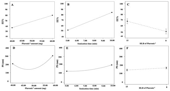
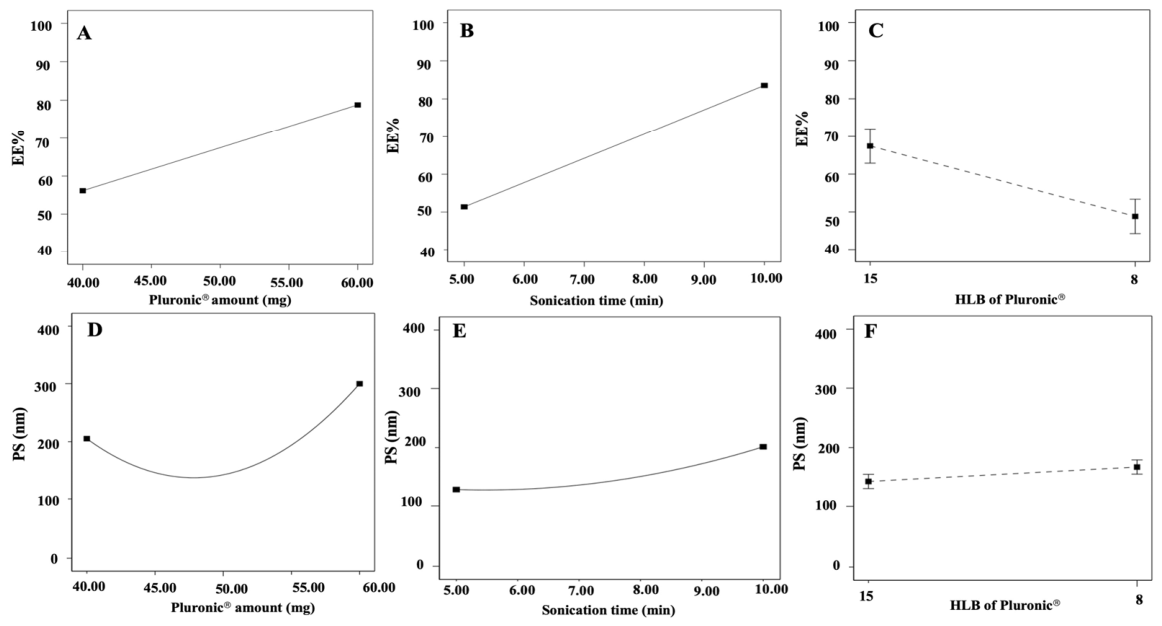
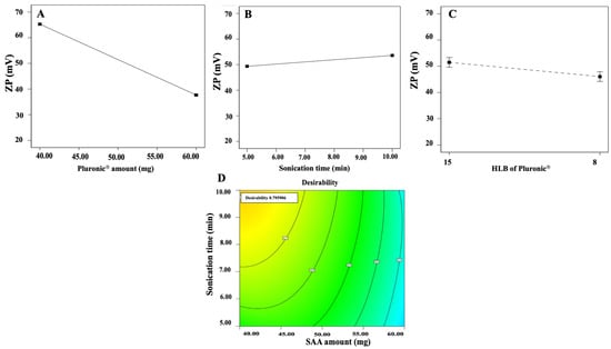
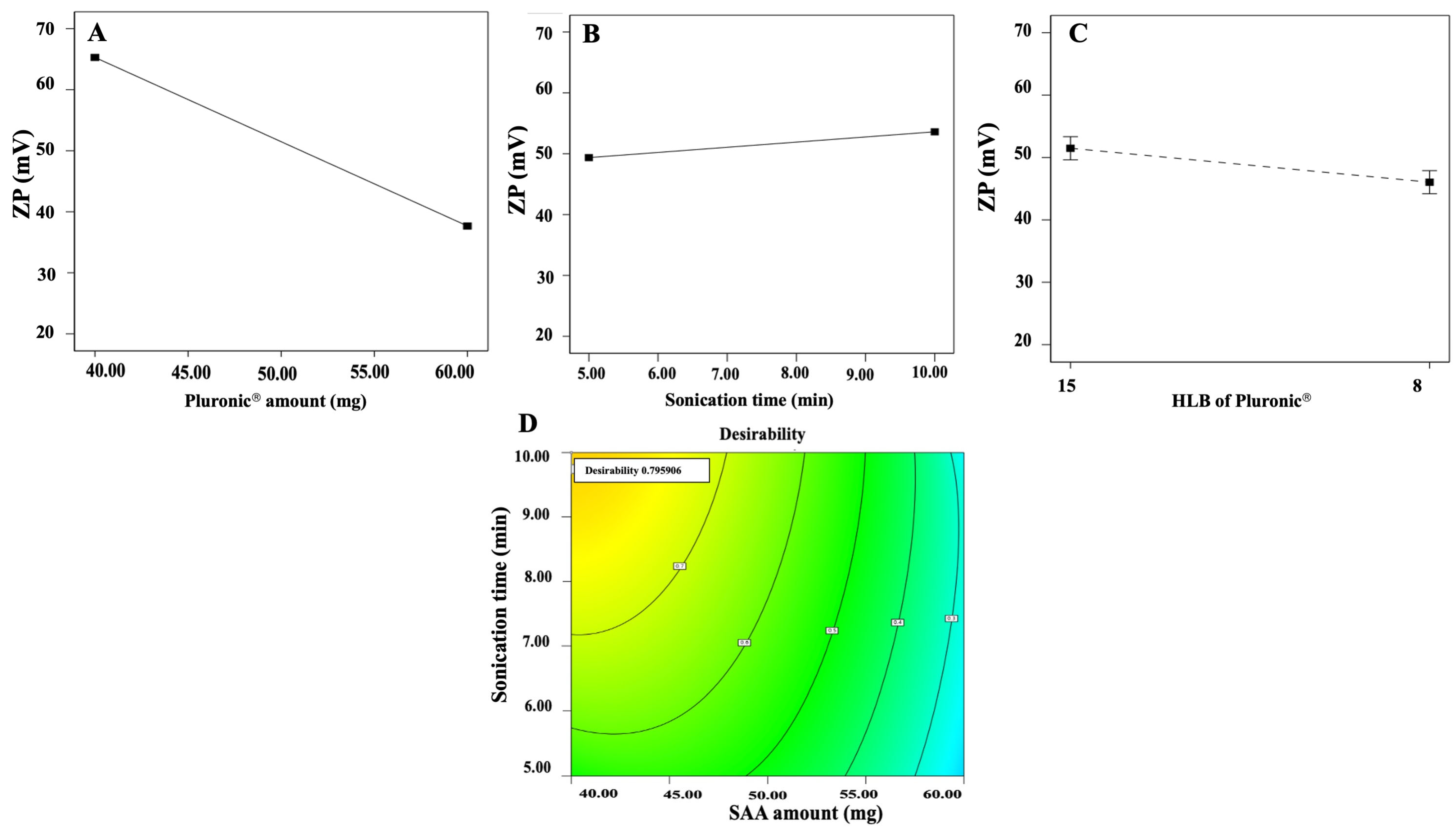
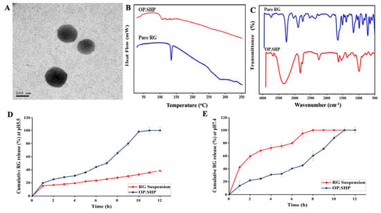
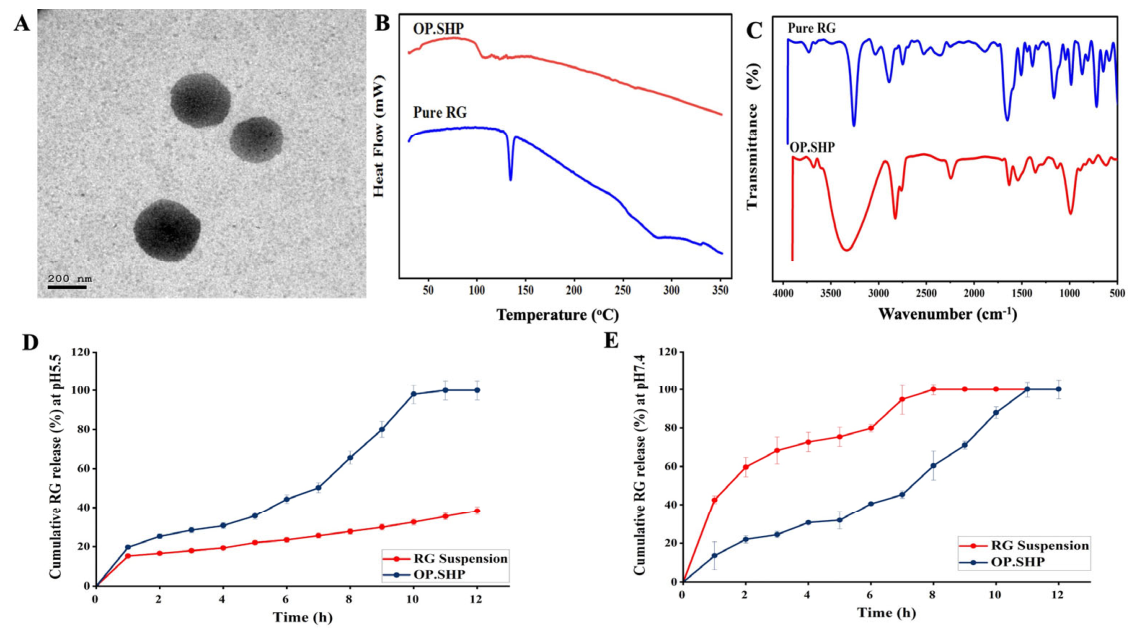
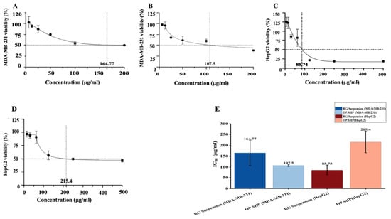
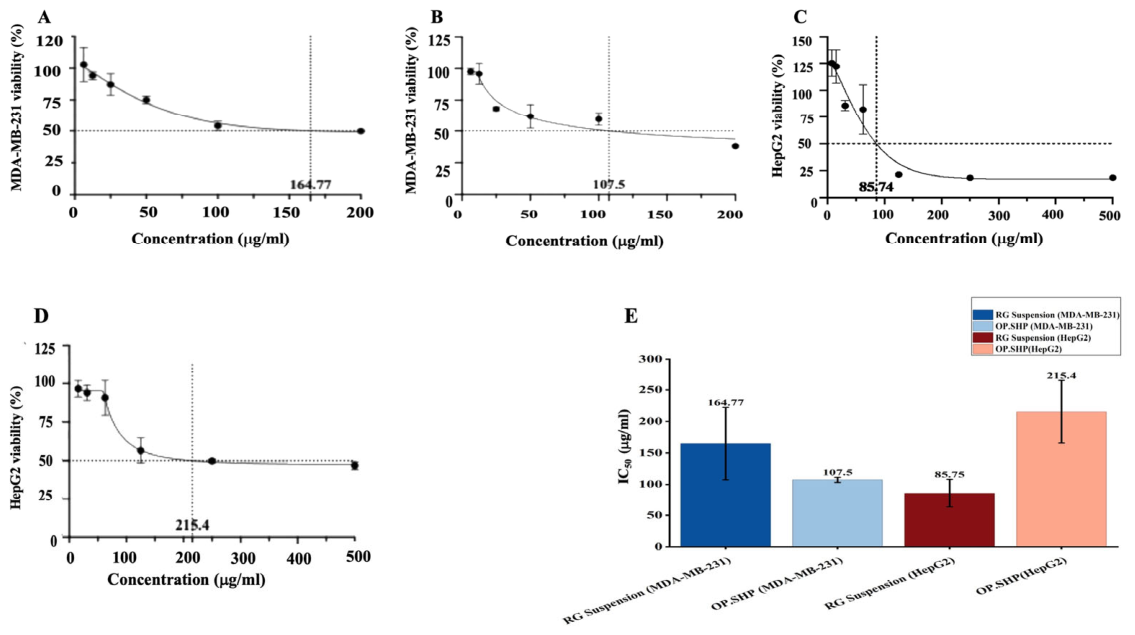
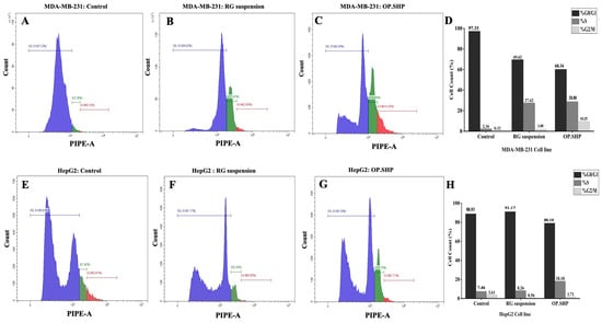
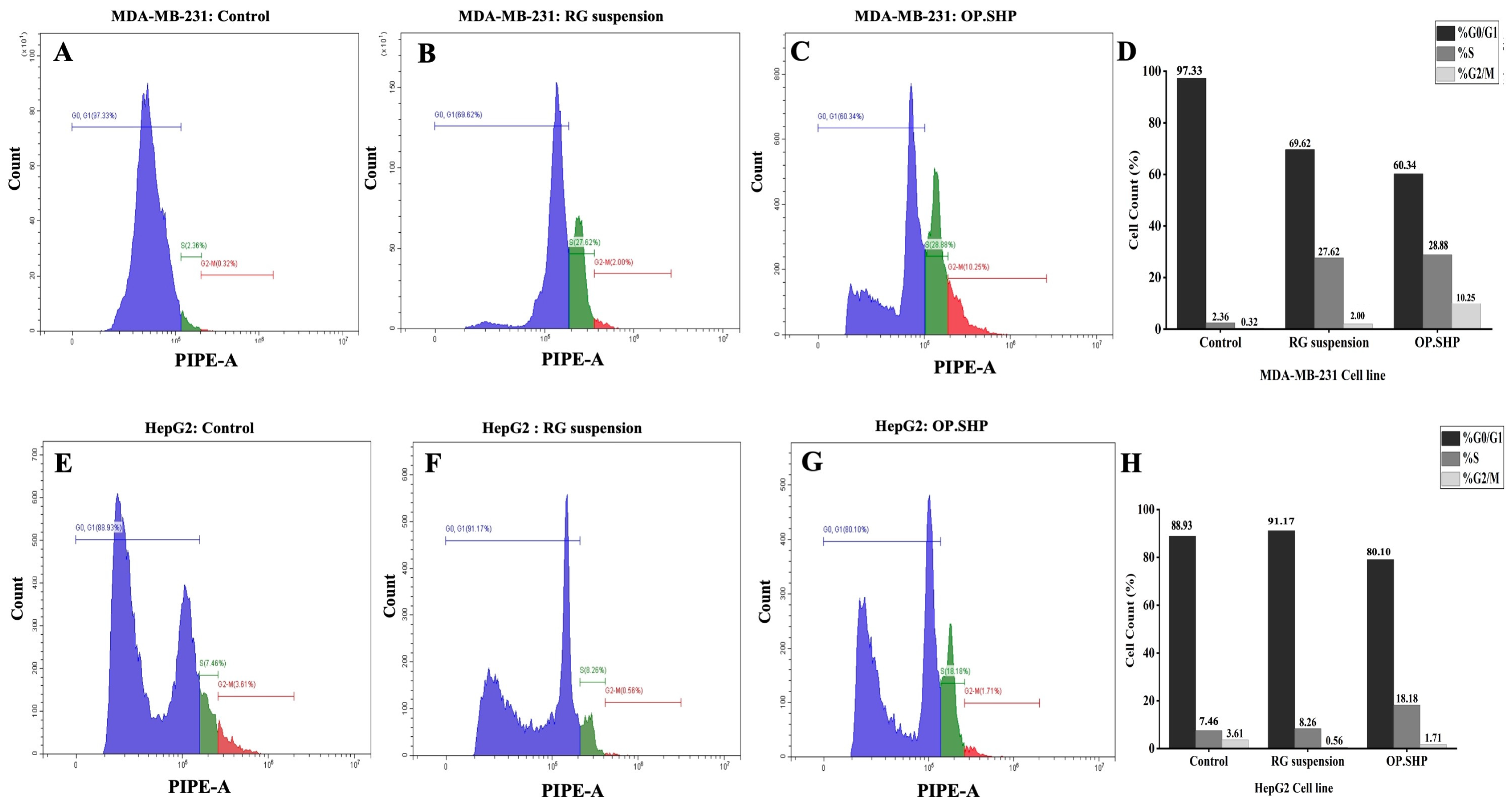
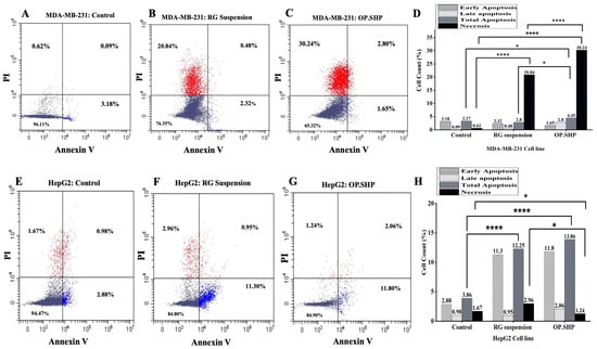
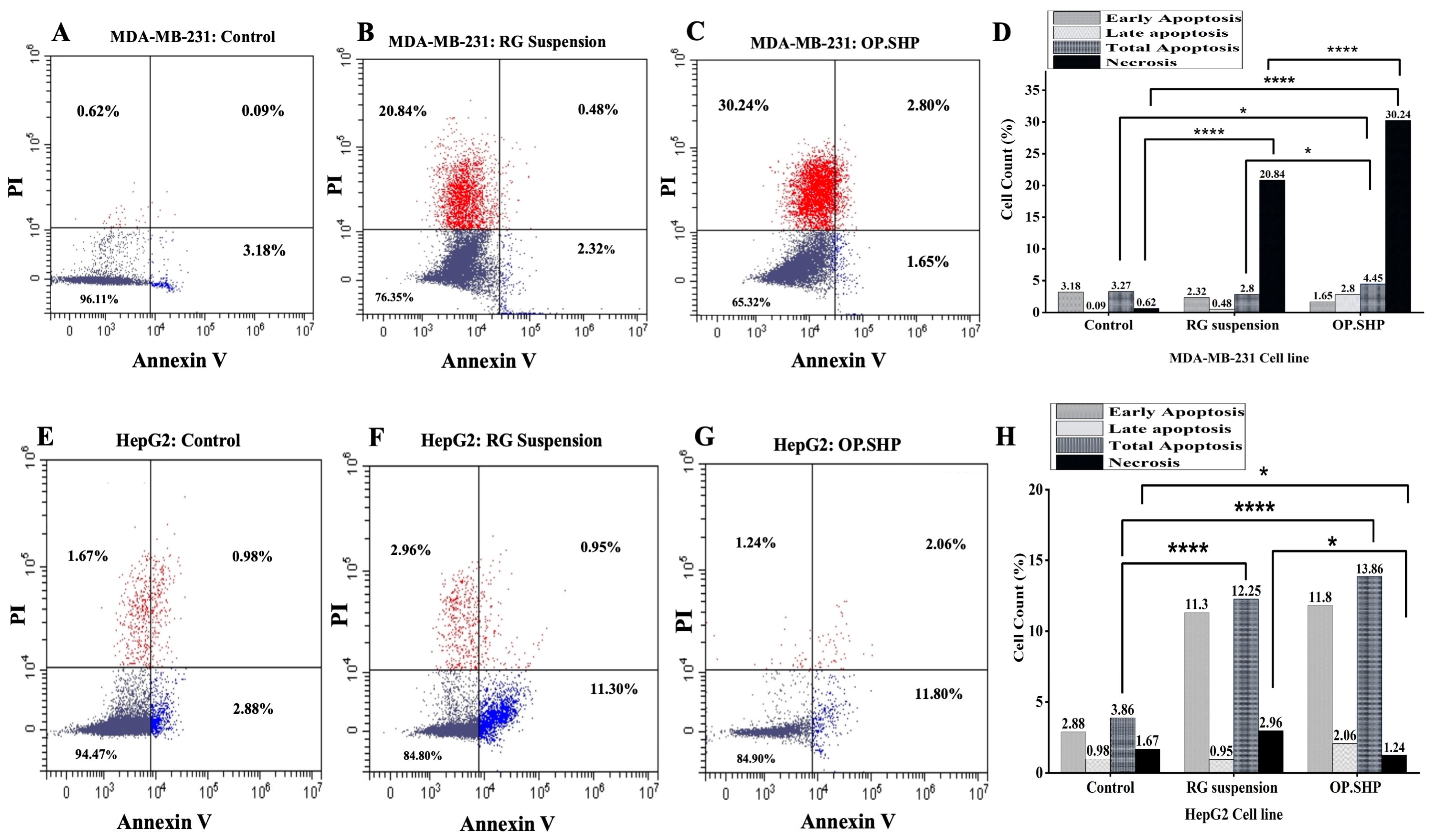
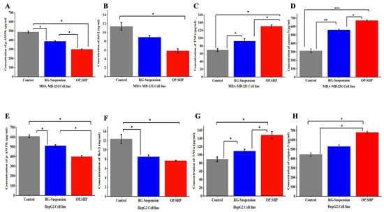
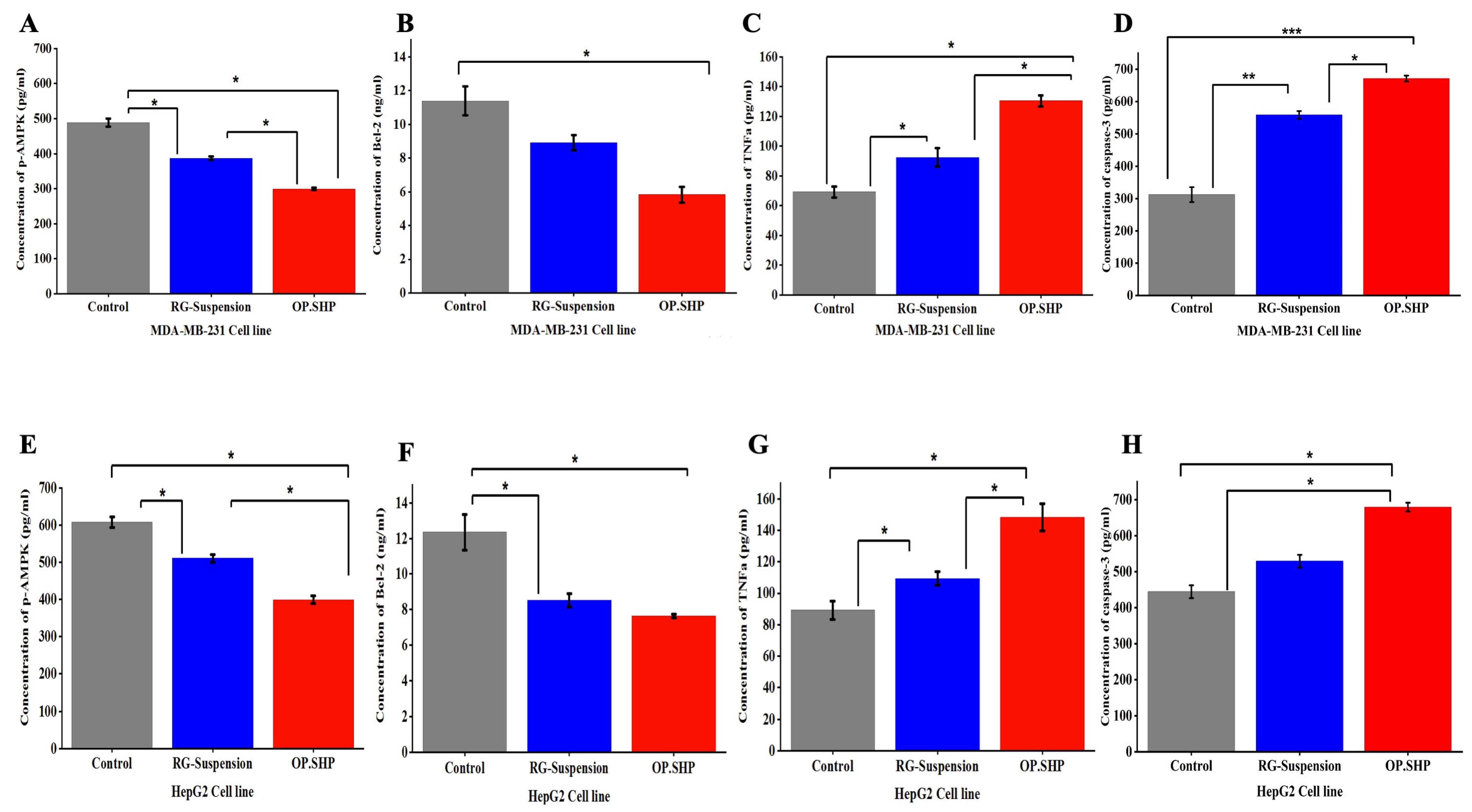
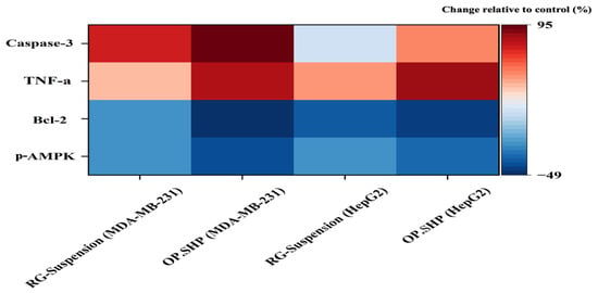
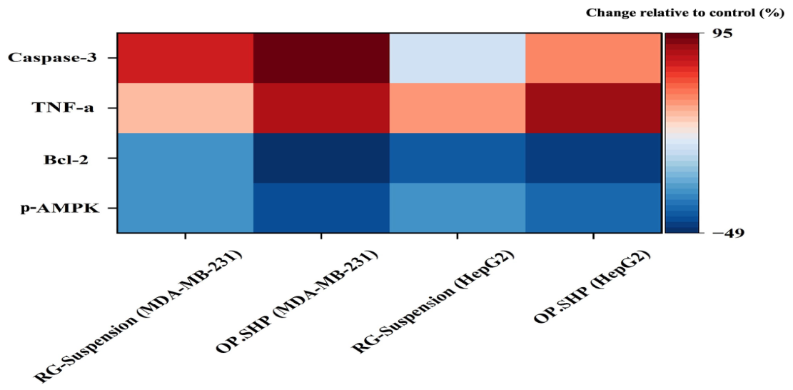
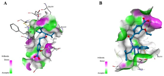
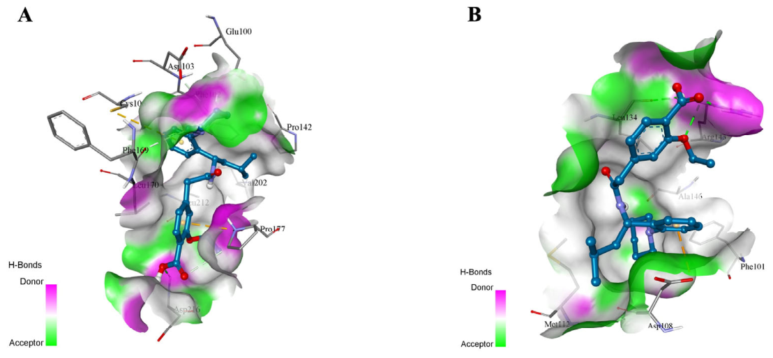

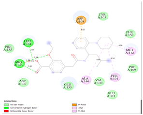
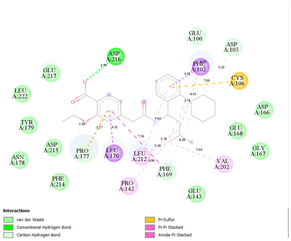
Disclaimer/Publisher’s Note: The statements, opinions and data contained in all publications are solely those of the individual author(s) and contributor(s) and not of MDPI and/or the editor(s). MDPI and/or the editor(s) disclaim responsibility for any injury to people or property resulting from any ideas, methods, instructions or products referred to in the content. |
© 2026 by the authors. Licensee MDPI, Basel, Switzerland. This article is an open access article distributed under the terms and conditions of the Creative Commons Attribution (CC BY) license.