


Abstract
Peptide–drug conjugates (PDCs) combine peptide-mediated targeting or transport with a pharmacologically active payload. This critical narrative review organizes oncology PDC development around release-competent exposure (RCE): the connection between conjugate integrity, formation of an identified active compound, delivery to its tumor compartment, pharmacodynamic effect, and therapeutic margin. Tumor uptake or accumulation is distinguished from the concentration–time profile of an identified active species in its relevant compartment. PubMed, ClinicalTrials.gov, and regulatory sources informed the review through 15 September 2026; the recorded retrieval procedures were re-executed and archived on 24 October 2026. We distinguish payload-releasing PDCs from enzyme-potentiated prodrugs and non-cleaving modalities, assemble established, adapted, and conceptual exposure measures, and integrate analytical requirements into a stage-gated development framework. Clinical examples include zelenectide pevedotin, PEN-221, zoptarelin doxorubicin, ANG1005, and melflufen. Five preclinical illustrations and a separate calculation from published tissue AUCs show how different observations inform the framework. ADC-derived principles are qualified by PDC-specific disposition, while sparse tissue sampling, imaging, and modeling are evaluated as translational strategies. Use of these exposure measures within RCE remains hypothesis-generating and requires construct-specific validation; established PK quantities are not thereby validated clinical surrogate endpoints.
1. Introduction
An estimated 20 million new cancer cases and 9.7 million deaths occurred in 2022 [1]. Treatment is constrained by injury to metabolically or proliferatively active normal cells. ADCs demonstrate targeted delivery of highly potent agents, but benefit depends on target expression, conjugate exposure, linker stability, released molecular species, and organ-specific toxicity [2,3]. PDCs occupy a related but kinetically distinct framework; smaller carriers support rapid tissue distribution, chemically defined attachment, and access to receptors or transport pathways difficult to reach with antibodies [4,5,6,7]. Their liabilities include proteolysis, renal clearance, brief intact-conjugate circulation, and premature deconjugation [8,9,10].
PDC reviews describe tumor-homing and cell-penetrating peptides, linkers, payloads, and clinical programs [11,12,13,14]; 2026 reviews extend coverage to development hurdles, hematological applications, imaging, and translation [15,16,17,18]. This critical narrative review focuses on the evidence linking conjugate integrity, active-compound formation, spatial tumor delivery, PD, and therapeutic margin. Its contribution is an author-defined framework integrating established PK, PDC-specific adaptations, and a conceptual kinetic index into testable development hypotheses.
PDCs are not smaller ADCs with transferable PK parameters. Circulating stability can coexist with efficient cleavage after tumor retention or uptake. Rapid plasma clearance can limit delivery while retained conjugate or active payload sustains activity. Linker selection must integrate systemic stability, productive tumor access, local release, and active-compound persistence [19,20,21]. Section 7.4 separates transferable principles from carrier-dependent assumptions and identifies measurement consequences.
2. Review Scope and Methods
2.1. Review Design
This is a critical narrative review, not a systematic review or meta-analysis. Its objective is to develop and critically examine a hypothesis-generating framework linking conjugate integrity, active-compound formation, spatial tumor delivery, PD, and therapeutic margin. This approach is appropriate because the question requires integration of chemical, analytical, preclinical, clinical, and regulatory evidence addressing different steps in a mechanistic chain. The synthesis evaluates what each evidence type establishes, which assumptions transfer between modalities, and where validation is missing; it does not seek exhaustive study identification or a pooled treatment-effect estimate. Article development was informed by the six quality domains of the Scale for the Assessment of Narrative Review Articles (SANRA): importance, aims, literature search, referencing, scientific reasoning, and relevant endpoint data [22]. SANRA is a narrative-review appraisal tool, not a systematic-review protocol or reporting guideline.
2.2. Search Strategy and Evidence Selection
The original narrative search used PubMed, ClinicalTrials.gov, regulatory sources, publisher searches, and citation chaining, with a 15 September 2026 cutoff. Exact historical queries and filter settings are unavailable. A written revision-stage record dated 9 October 2026 was available, but its corresponding raw-response archive was unavailable for proof-stage verification. The recorded PubMed and ClinicalTrials.gov query strings were therefore re-executed and archived on 24 October 2026. Supplementary Materials S1 provides the executed strings, interfaces, fields, date limits, filters, retrieval yields, and official-document lookup records; Table 1 summarizes their scope. Nine PubMed queries cover PDC terminology, carrier classes, platform aliases, and relevant mechanisms across PubMed, including records outside MEDLINE, with a publication-date upper limit of 15 September 2026. ClinicalTrials.gov retrieval used product aliases and the same cutoff for first posting; returned records reflect their versions at the new retrieval date. This proof-stage check documented retrieval procedures and tested a newly assembled benchmark of known citations. It neither reconstructed earlier database states nor comprehensively screened the retrieved records. PubMed search-field syntax and ClinicalTrials.gov API documentation are described in the official resources cited in Supplementary Materials S1 [23,24].
Table 1.
Search documentation and proof-stage retrieval verification for the critical narrative review. Complete strategies and dated records are provided in Supplementary Materials S1 and the companion archive.
Sources were selected for relevance to target or carrier selection; post-conjugation binding and trafficking; linker stability and release; active-compound identity, permeability, or mechanism; tumor or safety-organ distribution; multi-analyte PK or PD; clinical efficacy or safety; manufacturing and analytical control; or biomarker and regulatory strategy. Product claims prioritized primary clinical reports, trial records, labels, and assessments. Mechanistic claims are distinguished as direct PDC evidence in the reported construct and experimental setting; ADC evidence proposed to transfer only at a stated chemical or analytical level; or author extrapolation where PDC validation is absent. These evidence origins are distinct from the parameter classes in Section 3.2. Section 7.4 separates source observations, transfer limits, and our proposed measurement implications. Radioligand evidence informs target-access and organ-handling questions; radiation dosimetry remains modality-specific, and retained radiolabel does not establish cytotoxic-payload release (Section 8.6). Reviews supplied context and primary-literature leads, not primary outcome data.
The review scope excluded unconjugated peptides, nanoparticles with peptide decoration but no chemically defined payload relationship, and uptake-only reports lacking activation, retention, pharmacology, or safety information. English-language reports were prioritized, but non-English reports were not excluded solely because of language. No language filter was applied to the proof-stage PubMed or registry searches. Pre-2015 studies were retained for foundational carrier mechanisms or linker principles, clinical or quantitative-exposure benchmarks, or regulatory precedents. The evidence base contains 142 full peer-reviewed articles, 141 with DOIs; six DOI-bearing conference abstracts, one DOI-bearing corrigendum, and 24 regulatory, registry, sponsor, or technical-documentation records were retained as additional sources and excluded from the full-article total. Exhaustive study screening, independent dual-reviewer selection, protocol registration, and formal study-level risk-of-bias assessment were not undertaken. The cited-source totals describe the assembled bibliography, not a systematic screening flow; prevalence, comparative effect size, and literature completeness are not estimated.
The five preclinical illustrations in Section 10.1 are a purposive, availability-constrained sample with a convenience component, not a representative or systematic preclinical PDC sample. Selection combined relevance to distinct delivery or activation questions, numerical results extractable without image digitization, sufficient experimental context, and CC BY 4.0 licensing for adaptation. The five figures predate the proof-stage retrieval check and did not result from a systematic CC BY–only search for tabular endpoints. Licensing governed figure reuse, not review eligibility. Numerical extractability was not restricted to tables: some values came from published Results text and supplementary numerical tables.
These considerations describe the illustrative subset, not prospectively registered eligibility criteria. The five complementary studies do not provide balanced or maximum-diversity coverage of carriers, linkers, and payloads. No complete historical candidate inventory or study-by-study exclusion log is available. We therefore cannot identify all eligible unplotted studies, establish that these were the only studies meeting the presentation conditions, or assign retrospective exclusion reasons. Absence from these five illustrations means nonselection for illustration, not scientific ineligibility. Section 10.1 states each example’s contribution and inferential limits.
2.3. Critical Synthesis and Limits
Evidence was synthesized by position in the mechanistic evidence hierarchy defined in Section 3.2. Section 8 presents the authors’ narrative interpretations, informed by study design and comparator, population and evaluable denominators, endpoint and follow-up maturity, reporting completeness, and reported efficacy and safety findings. Randomized comparisons support treatment-effect conclusions that uncontrolled studies cannot establish; regulatory status and publication type provide context rather than independent grades of evidentiary certainty. No numerical score, formal evidence-grading system, or cross-agent ranking was applied. Conference abstracts are identified as abstract-level evidence, with 2026 updates explicitly treated as preliminary; publication in a journal supplement is not equivalent to a full peer-reviewed clinical report. Registry results are labeled separately. Non-peer-reviewed sponsor updates inform program status, not independent efficacy confirmation or agency decisions. Study design and reporting format are considered separately; these sources are not assigned the same evidentiary weight as full peer-reviewed randomized clinical reports.
3. Defining the PDC Modality
3.1. Operational Taxonomy
Here, a PDC comprises a peptide or peptidomimetic carrier covalently linked to a pharmacologically active small-molecule or macromolecular payload. The carrier mediates recognition, transport, or conditional uptake; the complete conjugate or a released compound produces the effect. An intact conjugate denotes the covalently linked carrier–linker–payload entity, not the carrier alone. The payload-releasing cytotoxic subset requires formation of an active payload or catabolite; Table 2 distinguishes this subset from other peptide-guided modalities.
For the authorization statement in Section 8, the subgroup is narrower: a peptide or peptidomimetic ligand must itself provide receptor or cell-surface recognition/transport and be covalently linked to a distinct, nonradioactive small-molecule cytotoxic payload. Classification follows the targeting component and effector, not peptide content alone; macromolecular cargo remains a separate category in Table 2. Authorization means marketing approval in the relevant jurisdiction at the cutoff, not trial registration, orphan designation or a pediatric waiver. These are scientific scope criteria, not regulatory definitions.
Peptide-activated prodrugs use enzyme and transport context rather than receptor-mediated tumor homing. Melflufen’s peptide-like promoiety promotes uptake and intracellular trapping of alkylating metabolites. Its placement in this mechanistic group does not imply an inactive parent; EMA describes it as a peptide-conjugated alkylating drug with intrinsic alkylating capacity [25].
Peptide radioligands used for PRRT deliver radiation without requiring linker cleavage; absorbed dose and microdistribution therefore replace active-catabolite exposure [26,27,28]. Photosensitizers require appropriate light and oxygen. Peptide-decorated nanoparticles, co-administered penetration enhancers, and unconjugated peptides fall outside the strict PDC boundary unless the peptide–payload relationship is chemically and experimentally defined.
Table 2 is an author-defined scientific framework for experimental comparison and regulatory dialogue. It neither creates a legal product class nor assigns an approval pathway or evidence standard; regulatory status concerns the complete product in its jurisdiction.
EMA’s synthetic-peptide guideline addresses conjugation quality, including cytotoxic-payload and imaging-agent delivery [29]. FDA’s ADC guidance concerns antibody- or antibody-fragment conjugates carrying cytotoxic small molecules [30]. Neither adopts this taxonomy. FDA’s ADC-specific drug–biologic combination-product description and preferred biologics license application route cannot automatically be extended to peptide-carrier conjugates [30]. Table 2 maps these scope and classification boundaries.
Table 2.
Operational boundaries, exposure variables, and regulatory relationships of peptide-guided payload modalities [10,11,26].
Figure 1 adapts Hoppenz et al.’s peptide–linker–drug architecture and uptake schemes [11] to distinguish payload-releasing conjugates, enzyme-potentiated prodrugs, and non-cleaving modalities, each with different activation and exposure requirements.
Figure 1.
Operational taxonomy of peptide-guided payload systems and the modality-specific meaning of release competence. (A) Payload-releasing PDCs, shown in teal, are evaluated through active-payload or active-catabolite exposure in the tumor. (B) Peptide-activated prodrugs, shown in orange, depend on intracellular active-metabolite formation. (C) Peptide radioligands, shown in purple, are evaluated through tumor absorbed dose and microdistribution. (D) Adjacent peptide-guided systems, shown in gray, require platform-specific evidence. Downward arrows link each modality to its architecture and productive-exposure variable; colors distinguish categories and do not encode quantitative values. Adapted from Figures 1 and 2 of Hoppenz et al. [11] under the Creative Commons Attribution 4.0 International License. Changes: completely redrawn, combined, simplified, recolored, and expanded with modality-specific decision variables.
3.2. Release Competent Exposure
Release-competent exposure links conjugate integrity, active-compound formation, spatial tumor delivery, pharmacodynamic effect, and therapeutic margin. For payload-releasing PDCs, its central quantity is the time-integrated exposure of an identified active payload or catabolite in the intended anatomical and subcellular compartment, with payload-target engagement or linked downstream PD and unbound systemic and dose-limiting-organ exposure compatible with the intended therapeutic window. Evaluation requires a pharmacologically relevant analyte, the correct compartment, appropriate timing, and organ-specific safety assessment. RCE is an evidence profile, not a universal score.
For internalizing, payload-releasing PDCs, the proposed causal sequence is as follows: administration → intact-PDC exposure → tumor access → cellular internalization/trafficking → active-species formation → active-site exposure → payload-target engagement → downstream PD → efficacy.
These are distinct evidence levels. Arrows denote mechanistic hypotheses to test, not proof supplied by the preceding measurement. Bulk tumor accumulation does not identify active species or their compartment; formation does not establish sufficient active-site exposure; exposure does not establish target engagement, and downstream PD does not guarantee efficacy. Carrier–receptor binding supports access/uptake and is distinct from engagement of the payload’s pharmacological target. Extracellular activation may precede active-product entry into cells, bypassing carrier internalization [33]. Active species may also reach the tumor from circulation; chemical identity alone does not prove local formation. Toxicity follows a parallel systemic/normal-organ exposure → tissue engagement or other injury → toxicity branch, modified by tissue susceptibility and possible carrier, linker or additional-product effects. Table 3 locates each measure within this hierarchy.
Implementation differs by development stage. Preclinical discovery and qualification can use time-resolved sampling across animal cohorts with chemically selective assays to estimate tumor and organ exposure, alongside spatial analysis and validated compartment-specific methods to examine localization. Cohort-derived tissue AUC, spatial distribution and subcellular access are distinct observations; homogenate measurements alone do not establish intracellular active-site exposure.
In clinical development, active-site AUC is a mechanistic reference, not a routine measurement requirement: repeated spatially resolved or subcellular tumor sampling sufficient for individual AUC is generally impractical. Realistically obtainable evidence includes serial plasma PK of quantifiable PDC-related species [34,35,36] and clinical efficacy/safety data, supplemented in selected substudies by ethically justified timed biopsies or planned resections [37,38], mechanism-linked PD and imaging. A biopsy provides a local concentration at one time; plasma, imaging and PD observations do not directly replace active-compound concentration–time data in the intended tumor compartment. Model-derived tumor exposure should be labeled as predicted, with assumptions and uncertainty reported; where data cannot identify exposure, they are reported as unmeasured or not estimable. Section 12 details these clinical strategies and limitations.
Throughout this review, tumor uptake denotes delivery into tumor tissue, whereas accumulation denotes material or signal present or retained there; neither alone establishes cellular internalization or active-payload identity. Tumor exposure specifies an analyte, compartment and temporal measure, such as a concentration–time profile, AUC or peak concentration. Active-site exposure additionally requires localization of the active species in its relevant compartment. Radioactivity, total drug after cleavage, and fluorescence can obscure molecular identity. A single tumor concentration establishes presence at the sampling time, not AUC, exposure duration, or spatial coverage.
ADC experiments show that linker-dependent catabolites differ in permeability and potency, so greater total tumor-metabolite AUC need not imply greater activity [39,40]. In separate ADC models, released MMAE concentrations correlated with cytotoxicity and tumor-growth inhibition [41]. These findings support chemical identification and exposure–PD testing; PDC-specific uptake, release kinetics, sampling, and thresholds require separate justification (Section 7.4).
Table 3 separates six core quantitative measures for payload-releasing PDCs (Part A, numbered 1–6) from supporting and modality-specific approaches (Part B), which are excluded from that count. The core measures are independently classified by methodological provenance: Class 1, an established PK/PD parameter; Class 2, an adaptation of an established parameter to PDCs; or Class 3, a new conceptual metric proposed by the authors. Active-compound AUC is established; dose-normalized tumor gain, apparent unbound tumor/plasma exposure, tumor-to-organ exposure ratios, and temporal coverage are adaptations; the kinetic-separation index is conceptual. Classification concerns methodological provenance, not measurement validity or clinical prediction. The selection and interpretation of these quantities within RCE remain hypothesis-generating. Interpretable estimates require analyte-selective assays, defined compartments, explicit assumptions and, for exposure comparisons, appropriate dose normalization and matched AUC intervals. These conditions do not establish a validated endpoint or universal cutoff.
Active-site exposure uses the established AUC parameter, , where P* is the chemically identified active compound and k is the intended anatomical or subcellular tumor compartment. The adapted dose-normalized tumor gain compares after the PDC with that after an equimolar free-payload comparator, following established relative-exposure comparisons [42]. For the adapted unbound exposure ratio [43], where unbound concentrations are measurable, describes the apparent unbound tumor-to-plasma exposure ratio. Local formation, trapping, and elimination can influence this ratio, so it is not necessarily an equilibrium partition coefficient.
The adapted tumor-to-organ exposure ratio for organ j is . An exposure ratio does not encode tissue susceptibility or establish therapeutic index [44]. Kidney, liver, marrow, peripheral nerve, skin, and gastrointestinal tissue are not interchangeable; each plausible dose-limiting organ should be reported separately where estimable, with unavailable human organ AUC recorded as not determined (Section 12). Temporal coverage adapts time-above-threshold PK/PD [45] as the fraction of the dosing interval during which tumor P* exceeds a mechanism-matched concentration such as an experimentally justified ECx. The author-proposed kinetic-separation index, Ksep = t1/2,plasma cleavage/t1/2,target matrix release, is an exploratory in vitro comparison, not a validated ranking rule or proof of in vivo selectivity. Its assumptions and restricted use are specified in Section 5.1.
These AUC ratios do not measure the fraction of payload released; distribution, binding, metabolism, and clearance prevent their interpretation as a molar mass balance. Spatial productive coverage and concordance between tumor P*, payload-target engagement, and downstream PD remain necessary to determine whether bulk exposure reaches viable payload-susceptible cells, including target-negative neighboring cells when bystander killing is intended.
Table 3.
Six core quantitative RCE measures (Part A) and supporting or modality-specific approaches (Part B): classification, definitions, and assumptions [26,27,28,41,42,43,44,45,46,47,48].
4. Peptide Carrier Selection
4.1. Addressability and Productive Trafficking
A useful carrier requires addressability and productive trafficking. Addressability is an accessible tumor-to-normal differential under the intended route and schedule; productive trafficking delivers or retains the final conjugate, including its linker and payload, in the pharmacological compartment. Peptide-receptor mapping established receptor-rich tumors as targets while revealing substantial intertumoral and normal-tissue variation [49]. High affinity can therefore fail through perivascular trapping, unproductive recycling, kidney accumulation, or loss of binding after conjugation.
In vivo phage display established accessible vascular and tumor molecular addresses and enabled tumor-homing motif discovery [50,51]. Examples include RGD, NGR-like vascular ligands, somatostatin analogues, gonadotropin-releasing hormone and melanocortin ligands, and library-derived peptides. RGD uptake can involve endothelium, tumor cells, or both [52]; LyP-1 linked selective tumor and lymphatic homing to internalization and growth inhibition [53]. Functional target selection should assess accessible density, internalization or retention, tumor distribution, shedding, normal-organ access, and deployable biomarkers.
Proteolytic exposure of a C-terminal CendR motif can engage neuropilin-1 and increase tissue penetration [54,55,56], potentially countering perivascular capture. Activity depends on the initiating receptor, proteases, perfusion, and neuropilin distribution; penetration must therefore be tested spatially for the final conjugate rather than inferred from the unconjugated peptide.
Cell-penetrating peptides can improve internalization of polar or macromolecular cargo but often lack tumor specificity and remain endosomally trapped [57,58,59]. TAT cargo was shown to enter through macropinocytosis, with a fusogenic element needed to enhance escape; cyclic and topologically constrained cationic peptides demonstrated that early-endosomal escape and cytosolic delivery can diverge from bulk uptake [60,61,62,63], as did an endosomolytic delivery study [64]. These uptake or escape modules need separate targeting or conditional activation. Cationic charge can increase nonspecific tissue and proximal-tubule interactions. Total fluorescence cannot resolve membrane binding, endosomal sequestration, degradation, and cytosolic delivery.
Dimeric RGD–paclitaxel achieved tumor uptake, while αvβ6-directed doxorubicin or paclitaxel constructs showed receptor-dependent internalization and cytotoxicity [65,66,67]. Nevertheless, αvβ6–paclitaxel produced efficacy similar to that of free paclitaxel in its xenograft experiment, showing that receptor selectivity need not improve therapeutic index.
4.2. Constrained Carriers and Kinetic Optimization
Cyclization, stapling, backbone modification, D-amino acids, N-methylation, and bicyclic architectures can improve protease resistance and binding while providing defined attachment sites [68,69,70], but may alter receptor conformation, internalization, solubility, or retention. Hydrophobic modification can prolong circulating peptide residence through albumin association while increasing aggregation or hepatic uptake. Protease resistance, permeability, filtration, and exposure extension remain distinct ADME problems [71]. Reversible transthyretin binding and a high-affinity albumin-binding peptide tag substantially extended half-life in experimental systems but also changed the distribution problem [72,73]. Each modification requires evaluation in the final conjugate.
Affinity must be balanced with transport: slow dissociation improves retention but can strengthen perivascular binding barriers; multivalency increases avidity and target-mediated disposition while reducing diffusion. Carrier panels should assess equilibrium affinity, binding kinetics, receptor competition, internalization and recycling, post-conjugation stability, penetration from vessels, tumor-to-organ ratios, renal handling, solubility, and synthetic scalability.
Figure 2 adapts Hoppenz et al.’s sequence of receptor binding, internalization, endolysosomal processing, and payload release [11], adding renal loss, perivascular retention, recycling, and lysosomal trapping as competing branches with decisive readouts. This compartmental itinerary distinguishes productive delivery from uptake followed by loss or sequestration. Table 4 summarizes carrier classes, principal failure modes, and candidate evidence.
Figure 2.
Carrier-dependent itinerary from vascular access to the pharmacological compartment. Adapted from Figure 2 of Hoppenz et al. [11] under the Creative Commons Attribution 4.0 International License, redrawn, simplified, recolored, and relabeled; competing loss pathways and decision readouts have been added. The depicted route represents receptor-mediated internalization; extracellular activation may bypass carrier internalization. Receptor binding denotes carrier–receptor interaction, while active-species identification, payload-target engagement and downstream PD require distinct evidence (Section 3.2). Horizontal arrows indicate the proposed sequence of delivery steps; downward arrows identify competing loss or retention pathways and productive intracellular delivery. Colored upper boxes distinguish stages, while red, orange, purple, and green branch arrows denote proteolysis or renal filtration, perivascular retention, recycling or lysosomal trapping, and productive cytosolic or nuclear delivery, respectively. The numbered dark-blue boxes identify the corresponding measurement readouts; color and arrow size are not quantitative.
Table 4.
Carrier classes and candidate translational evidence [8,56,71].
5. Linker and Payload Co Design
5.1. Linker Kinetics as a Therapeutic Index Switch
Systemic deconjugation must be slow enough for intact-PDC delivery, while local release or degradation must match carrier residence and payload action. Rapidly cleared constructs require delivery within the available vascular exposure window; subsequent activation depends on productive tumor retention and trafficking rather than plasma half-life alone. A single percentage-intact measurement can conceal biphasic degradation, labile subpopulations, and biological-species differences in cleavage.
For the kinetic-separation index, potentially informative comparisons are limited to a mechanistically comparable series with the same carrier–payload scaffold and matrix pair. Plasma bond cleavage and target-matrix formation of the active product may involve different enzymes or sequential cleavage, self-immolation, and catabolism; they do not measure one common reaction. ADC assays demonstrate enzyme-dependent differences in relative linker cleavage [74]; this informs assay design without validating PDC kinetics. Standardize each assay across constructs, documenting species, matrix source/fraction, substrate concentration, temperature, pH and trigger activity; differences between the paired matrices must be biologically justified. Verify product identity, recovery, stability and release yield. A single half-life requires sufficient sampling and an adequate kinetic model; incomplete, multiphasic or censored profiles may require reporting full curves and justified bounds or non-estimability, not an infinite ratio.
A value above unity means only that assayed plasma cleavage is slower than target-matrix active-product formation. It may help prioritize related constructs for further testing, with both absolute half-lives and uncertainty reported; it is not a universal ranking rule. Even a large ratio may accompany release too slow for productive tumor residence. The ratio does not establish delivery, cellular access, or normal-organ sparing. In vivo selectivity therefore requires construct-specific validation of intact-conjugate and active-product time courses in plasma, tumor and relevant normal tissues, with controls or validated modeling that distinguish local formation from redistribution of circulating payload. Until then, the index remains a Class 3 hypothesis, not an in vivo selectivity measure.
Cleavable linkers support near-parent payload release, permeability, or bystander killing—the death of neighboring target-negative cells exposed to released payload. Protease-cleavable linkers often use self-immolative spacers; PABC enabled rapid unmodified-doxorubicin release after dipeptide cleavage in model-linker and immunoconjugate studies [75]. Protease sensitivity is not tumor specificity; lysosomal enzymes occur in normal tissues, and circulating or extracellular enzymes vary across biological species and disease states [19,21]. In ADC studies, cathepsin-B deletion did not abolish nominally cathepsin-B-cleavable conjugate processing, indicating compensatory routes; added glutamate improved mouse-plasma Val–Cit stability while preserving activation [74,76]. Selectivity may depend on receptor-mediated uptake. We propose testing these candidate mechanisms in PDCs using human plasma and whole blood, relevant organ and lysosomal preparations, and efficacy and toxicology species.
Acid-labile and redox-sensitive linkers exploit pH or thiol gradients but face contextual limits; endolysosomal pH varies across cell states, extracellular tumor acidity is heterogeneous, disulfides can exchange with circulating thiols, and glutathione abundance does not establish cytosolic access. Bioorthogonal release reduces dependence on endogenous triggers but requires coordinated distribution, reaction kinetics, timing, and safety of two agents.
Non-cleavable linkers require carrier processing when activity depends on liberated products. ADC maytansinoid studies demonstrate linker-dependent catabolites with different permeability and extracellular potency [39,40]. We extrapolate the need to identify and characterize PDC-derived products, not antibody lysosomal processing or specific catabolite structures. Reduced bystander killing, improved systemic safety, and poorer coverage of heterogeneous tumors remain construct-specific PDC hypotheses. Processing site, product identity, potency, permeability, target access, and toxicity therefore require direct evaluation.
Conjugation chemistry and payload loading can change disposition before release. In ADC studies, hydrophobic loading accelerated clearance, hydrophilic design improved exposure, and higher loading did not proportionally improve in vivo efficacy or tolerability [77,78]. We propose transferring final-conjugate physicochemical characterization, not ADC clearance effects, loading optima, or antibody catabolism. Direct PDC evidence shows lower solubility of BT8009 than its precursor peptide [79]; this does not establish a PDC loading–clearance rule. PDC binding, filtration, proteolysis, and tissue distribution require direct measurement. ADC reviews identify conjugation site, payload and release chemistry as coupled variables [80,81]; their quantitative effects require PDC confirmation.
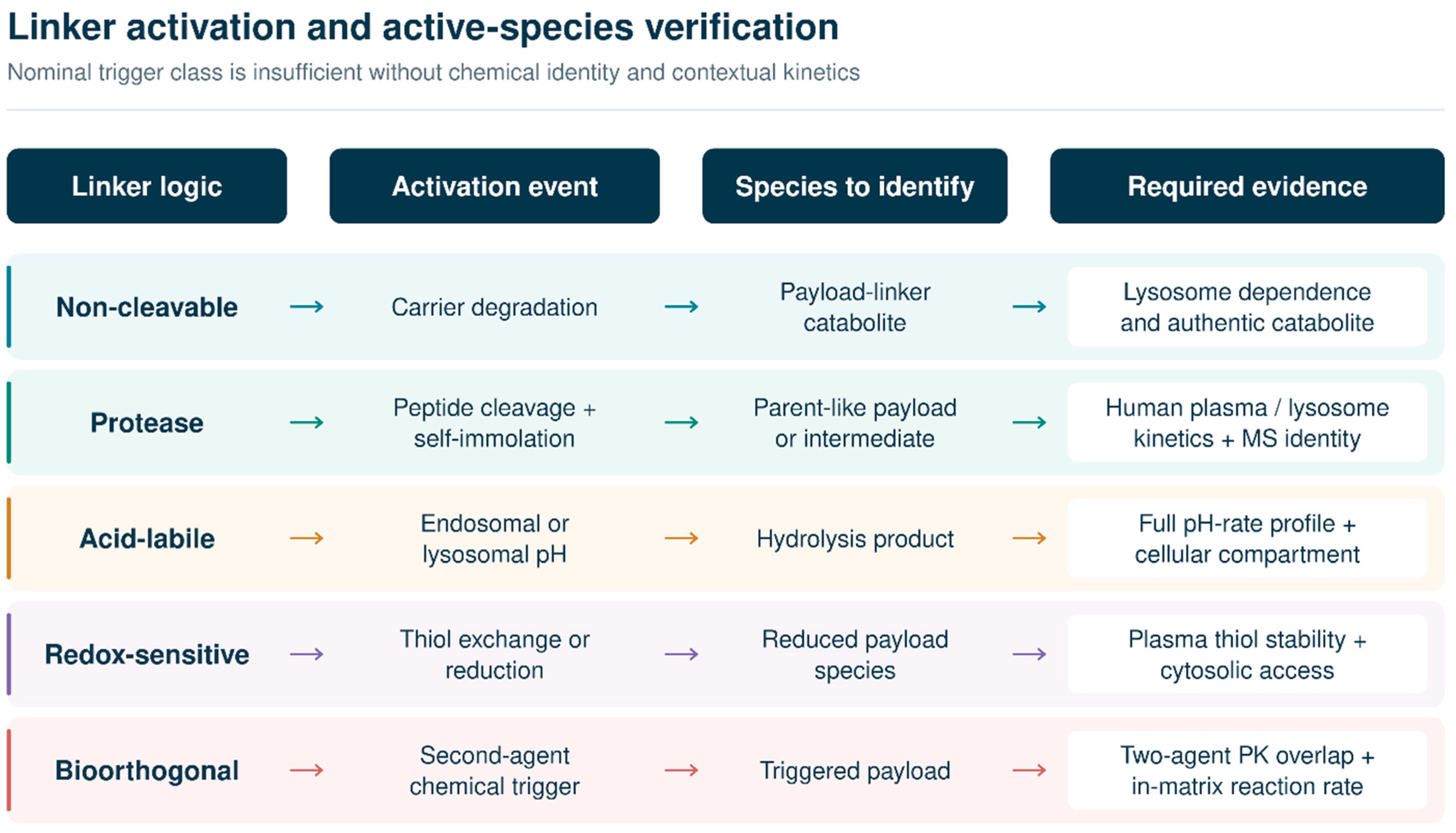
Figure 3 adapts Hoppenz et al.’s linker mechanisms and Vrettos et al.’s protease-triggered PABC self-immolation cascade [11,82], to compare five release mechanisms and their required analytical observations. The released molecular species, rather than the nominal trigger, is central to linker evaluation.
5.2. Identity of Released Payloads and Active Catabolites
Our proposed PDC release-characterization strategy applies established structural-analysis methods; it does not assert that ADC catabolites occur in PDCs. Released products and relevant intermediates are identified using LC–MS/MS or high-resolution MS/MS, authentic standards where available, and orthogonal characterization. Matrices are selected and sampled by activation route and development stage, distinguishing formation during sample processing from formation in vivo (Section 3.2 and Section 12).
We further propose characterizing each active PDC-derived species for potency, permeability, target access, protein binding, metabolic stability, and transporter interactions. The ADC literature describes extracellular-release and bystander mechanisms [83]; these are candidate mechanisms for PDC testing. Direct BT8009 experiments have measured intact conjugate and released MMAE [33], but these did not quantify the relative contributions of extracellular and intracellular release in vivo.
Figure 3.
Author-proposed linker activation routes and analytical tests. Lysosome dependence requires construct-specific confirmation and is not assumed for all non-cleavable PDCs. Adapted from Figure 5 of Hoppenz et al. [11] and Figure 5 of Vrettos et al. [82] under the Creative Commons Attribution 4.0 International License, redrawn, combined, simplified, recolored, and expanded to include contextual kinetics and active-compound verification. Rows are color-coded by linker logic: teal, non-cleavable; green, protease-sensitive; orange, acid-labile; purple, redox-sensitive; and red, bioorthogonal. Arrows connect each linker class with its proposed activation event, the molecular species to identify, and the required evidence; they do not indicate measured rates.
5.3. Payload Selection
Payload selection must match potency to the amount delivered while ensuring target access, sustained activity, activity against resistant tumors, and compatibility with conjugation and manufacturing processes. The desired bystander-killing profile should be specified before selection.
ADC experiments link payload permeability to bystander killing [41,84,85]; this mechanism is also discussed in the review by Giugliano et al. [86]. The resulting spatial coverage and normal-tissue risk require direct PDC evaluation. Maytansinoid analogue chemistry illustrates how side chains and disulfides connect conjugation with activation [87]. Microtubule-targeting payloads require control of toxicity, including neurotoxicity, whereas camptothecins act through TOP1–DNA cleavage complexes [88,89]. A HER2-targeted ADC carrying a topoisomerase I inhibitor provides a preclinical example of this payload class [90]. PBD dimers are potent, but their therapeutic application is particularly sensitive to toxicity [91]. Toxicity can originate from the carrier, linker, payload, or other released products [92].
Radionuclide selection must match half-life, emission, chelator stability, receptor retention, tumor size, heterogeneity, and organ dose. Beta emitters provide longer-range cross-fire; alpha emitters deliver high linear-energy-transfer radiation over shorter ranges. Photosensitizers require light delivery and management of oxygen limitation. Protein toxins, oligonucleotides, and degraders require intact cargo delivery and escape into the cytosol or nucleus. Table 5 summarizes the proposed linker and payload co-design considerations.
Table 5.
Author-proposed linker and payload co-design matrix. ADC precedents inform chemical-payload rows; validation is construct- and modality-specific [33,39,75,76,83].
6. Tumor and Intracellular Delivery Barriers
Small size does not overcome abnormal perfusion, interstitial pressure, dense matrix, stroma, hypoxia, necrosis, or heterogeneous receptors [93,94]. Short-circulating peptides cannot reach regions unperfused during exposure; strong perivascular binding can increase uptake without deeper delivery. Imaging mass cytometry, autoradiography, fluorescence with validated label stability, mass spectrometry imaging, ex vivo microscopy, and spatial PD should establish whether intact conjugate and active molecular species reach the same tumor regions. Three-dimensional spheroids and organoids support ranking but lack blood flow, filtration, and organ-specific processes; their results should be linked to in vivo exposure rather than substitute for it.
Trafficking must match activation and payload action: lysosomal-protease linkers require degradative-compartment access; extracellular activation does not. Non-cleavable conjugates may require carrier proteolysis into active payload–linker–amino-acid or peptide-bearing catabolites, whose identity and activity must be demonstrated. Disulfides require reducing-thiol access, potentially extracellularly; DNA-directed payloads require nuclear access. Uptake studies should resolve surface binding, internalization, recycling, degradation, and active-compound delivery to the pharmacological compartment [59,95].
Patient selection must account for TME differences in perfusion, stroma, macrophage uptake, extracellular proteases, hypoxia, and vascular permeability despite similar receptor expression. Receptor assessment should be matched to appropriate IHC methods; receptor imaging can help quantify the accessible target burden. Biomarkers should address the relevant uncertainty, using spatial pathology for heterogeneity and receptor imaging for accessibility.
SORT1-targeted TH1902 showed receptor-mediated uptake, apoptosis, xenograft activity, and tolerability in SORT1-positive breast cancer models, with subsequent studies extending investigation to ovarian and triple-negative breast cancer stem-like models [96,97]. Later work associated TH1902 treatment with cGAS–STING signaling and enhanced anti-PD-L1 activity; interpretation must use the corrected figures [98]. These findings support mechanisms and combination hypotheses, but they cannot replace analyte-resolved clinical evidence.
Figure 4 adapts Sheng et al.’s vascular, matrix, interstitial, and cellular barriers [99] to brief intact-PDC circulation, adding competing kidney and liver uptake and premature systemic release. These nanomedicine findings provide mechanistic analogies for evaluating spatial delivery in viable tumor regions.
Figure 4.
Sequential barriers and competing organ uptake that separate administered PDC dose from active-compound exposure in viable tumor cells. Adapted from Figure 1 of Sheng et al. [99] under the Creative Commons Attribution 4.0 International License, redrawn, simplified, recolored, and relabeled for peptide-conjugate kinetics, spatial pharmacology, and safety-organ exposure. This illustrates an intracellular-delivery route: “target binding and uptake” denotes carrier-mediated entry, not payload-target engagement. Extracellular-release mechanisms need not follow this route (Section 3.2). Horizontal arrows indicate sequential delivery barriers; colored downward arrows identify competing loss pathways: red, plasma deconjugation; orange, renal filtration and reabsorption; purple, hepatic uptake or metabolism; and gray, recycling, trapping, or inactive-catabolite formation. Colors of the upper boxes distinguish stages, with the dark-blue final box denoting intracellular active species; colors and arrow dimensions are schematic.
7. Multi Analyte Pharmacokinetics and Pharmacodynamics
7.1. Analyte Definition and Assay Architecture
For payload-releasing cytotoxic PDCs and peptide-activated prodrugs, we propose a mechanism-dependent analyte plan from the first in vivo study: intact conjugate or parent, total conjugated payload where meaningful, released payload, relevant active metabolites or catabolites, and dominant inactive or toxic degradants. ADC methodological literature motivates analyte separation and warns that biotransformation may complicate quantification [46,47,48]; the expanded PDC panel proposed here requires construct-specific justification. Intact-acting constructs and other modalities require a plan matched to their active entity (Section 10.2). Nonclinical tumor and safety-organ sampling should support AUC estimation or a justified model. Human tissue sampling belongs in justified substudies; serial plasma PK, imaging, and PD can support translation (Section 12). Total drug or radioactivity supports mass balance, not release competence. Sampling must capture early intact-PDC distribution and persistent active compounds, using common AUC intervals, stated integration and extrapolation methods, and adequate analyte-specific sensitivity.
Bioanalysis must control ex vivo deconjugation, adsorption, degradation, and poor tissue recovery through chemistry-appropriate stabilization, temperature control, matrix-specific recovery, incurred-sample reanalysis, and identity confirmation. Direct PDC clinical bioanalysis separates intact BT5528 from unconjugated MMAE [34] and quantifies intact zelenectide pevedotin [36]. The BT1718 assay instead measures total DM1-related material after reduction and derivatization; it does not selectively quantify DM1 released in vivo [35]. FDA bioanalytical and ADC clinical-pharmacology guidance supplies validation and analyte-selection principles requiring PDC-specific justification [30,100].
Joint intact-PDC, payload, and active-catabolite curves do not identify release location; tissue-generated payload can return to plasma. Estimating uptake, local formation, and elimination requires identifiable models supported by independent tissue, metabolism, or clearance observations. Unsupported processes should remain unestimated or sensitivity scenarios. ADC structures and Fc-dependent parameters cannot be imported unchanged (Section 7.4).
7.2. Renal and Hepatic Distribution and Clearance
Renal filtration can limit intact-PDC exposure, whereas proximal-tubule reabsorption and retention may increase kidney toxicity. Charge, hydrophobicity, protein binding, reabsorption, metabolic stability, and linker chemistry influence both processes. Figure 5 is our conceptual PDC adaptation of analyte separation in Glassman’s ADC modeling review [101], extending it to plasma, tumor, kidney, liver and other safety organs. Its validity and analytes remain construct-specific.
Figure 5.
Multi-analyte disposition of a peptide–drug conjugate with tumor active-compound formation and renal or hepatic uptake and clearance. Conceptually adapted from Figure 1 of Glassman [101] under the Creative Commons Attribution 4.0 International License. The ADC framework has been completely redrawn, simplified, recolored, relabeled in PDC terms, and expanded to include tumor and safety-organ matrices. The transferred element is analyte separation; antibody FcRn recycling, Fc-receptor uptake, clearance constants, and compartmental exchange rates are not assigned to PDCs (Section 7.4). Blue, green, orange, and purple boxes distinguish plasma, tumor, kidney, and liver or other organs, respectively. Colored arrows from plasma indicate delivery to the corresponding tissues; gray double-headed arrows denote comparisons of tissue exposure, not direct organ-to-organ transport. Arrows within boxes indicate local processing. The lower boxes identify the exposure measures; no arrow specifies a measured transfer rate.
For PDCs, extending intact-conjugate circulation may increase delivery to tumors but also prolong opportunities for systemic release. Albumin binding, lipidation, PEG-like polymers, and multivalency alter filtration, target access, binding, and distribution [102,103]. Reversible protein binding extended peptide half-life up to more than 20-fold in selected peptide-only systems, but unbound fraction and distribution require measurement [72,73]. Conjugation also changed clearance and hepatic distribution of structurally distinct anti-STEAP1 ADCs [104]. Decisions therefore require intact-conjugate and tumor active-compound exposure, absolute safety-organ exposure, schedule, and exposure–response; half-life or an unchanged distribution ratio alone is insufficient.
Direct PDC evidence from BT5528 and BT8009 preclinical studies shows brief intact-conjugate plasma persistence alongside sustained intratumoral MMAE concentrations [33,79,105]. First-in-human BT5528 showed intact-conjugate and released-MMAE plasma half-lives of approximately 0.4–0.7 h and 35–47 h, respectively, an objective response rate of 6.7%, and grade ≥ 3 neutropenia in 22.2% [106]. This kinetic divergence requires separate analyte measurement.
7.3. Exposure Response and Candidate Evidence
Exposure–response analysis should test mechanism-relevant AUC, peak concentration, or duration above a justified threshold; pulses and continuous exposure may differ, and PD damage may outlast exposure. Relate tumor active-compound exposure to payload-target engagement, downstream PD and efficacy, and systemic and organ exposure to toxicity. Radioligands require absorbed dose and dose rate. Construct-specific population PK can guide dosing without importing ADC thresholds. Section 10.2 proposes development questions and candidate evidence; Table 3 classifications do not supply mandatory thresholds or a requirement to obtain every measure.
7.4. Transferability of ADC Principles to PDC Metric Design
For two Nectin-4-directed MMAE conjugates, zelenectide pevedotin plasma half-life was 0.42–0.91 h across dose groups, vs. a population-PK estimate of 3.6 days for the enfortumab vedotin ADC analyte [107,108]. This approximately two-order difference is neither a class constant nor a controlled comparison; assays, doses, populations, and methods differ. Unconjugated-MMAE half-lives were approximately 37–50 h and 2.6 days, respectively. Short intact-conjugate circulation therefore does not justify truncating systemic payload AUC.
Disrupting FcRn binding with H435A altered ADC clearance and efficacy and tolerability despite similar in vitro cytotoxicity [109]. Fc-free peptides lack IgG salvage, although protein binding or size extension can modify disposition [72,73]. ADC elimination includes target-mediated and nontarget cellular uptake and catabolism opposed by FcRn salvage, not exclusively reticuloendothelial clearance. For proposed rapid renal elimination of small Bicycle conjugates [33,105], the cited imaging used a radiolabeled peptide analogue, not intact-PDC mass balance. Elimination of each final construct and released molecular species therefore requires evidence; neither universal renal PDC nor exclusively hepatic ADC clearance is justified [104].
Reduced kidney uptake in megalin-deficient mice demonstrated tubular reabsorption of radiolabeled peptides [110], supporting pathway testing in susceptible PDCs. It establishes neither universal megalin dependence nor active-payload AUC from retained radioactivity. Table 6 links these distinctions to Table 3; its sampling and modeling recommendations are author-proposed, not validated cross-platform thresholds.
Table 6.
Evidence provenance and transferability of ADC pharmacology to PDCs: observations, limits and author-proposed RCE implications.
8. Clinical Evidence and Mechanistic Reanalysis
Within the receptor-/cell-surface-targeted peptide–small-molecule cytotoxic subgroup defined in Section 3.1, the targeted narrative search identified no product with FDA approval or EU marketing authorization in force on 15 September 2026. This conclusion depends on the stated definition and search scope; it is not a claim that all products described as PDCs lack authorization.
Broader definitions include melflufen (Pepaxti), an EU-authorized peptide-conjugated alkylator with passive cellular entry and enzyme-potentiated activity rather than a receptor-binding carrier; its prior US approval (Pepaxto) was withdrawn [25,111]. Lutathera (lutetium-177 oxodotreotide) and the PSMA-directed small-molecule radioligand Pluvicto (lutetium-177 vipivotide tetraxetan) are EU-authorized, but their cytotoxic effect is mediated by radiation [112,113].
Broad polypeptide–toxin definitions would also encompass denileukin diftitox-cxdl (Lymphir; FDA-approved), an IL-2–diphtheria-toxin fusion, and tagraxofusp (Elzonris; EU-authorized), an IL-3–diphtheria-toxin fusion [114,115]. These receptor-directed cytotoxins are recombinant protein fusions, not peptide–small-molecule conjugates. ADCs remain antibody-carrier systems irrespective of peptide structures in their linkers or payloads [30]. These boundary cases are considered explicitly without treating their authorization as validation of the narrower cytotoxic PDC subgroup.
8.1. Zelenectide Pevedotin
Zelenectide pevedotin (BT8009, not PEN-221) couples a Nectin-4-targeting bicyclic peptide to MMAE. Duravelo-1 monotherapy dose escalation treated 49 patients with advanced solid tumors; among 42 efficacy-evaluable patients across tumor types, ORR was 24% (10/42; 95% CI 12–40), with median DOR of 11.1 months among 10 confirmed responders. In urothelial carcinoma patients, ORR was 38% (8/21; 95% CI 18–62), CBR 57% (12/21), median DOR 13.1 months among eight confirmed responders, and median PFS 7.4 months (95% CI 1.8–12.4). Grade ≥ 3 TRAEs occurred in 39%, including neutropenia in 16%; higher-schedule DLTs included grade 3 asthenia, fatigue, appetite loss, febrile neutropenia, and grade 4 sepsis [107].
Preliminary 2026 conference abstracts, rather than full peer-reviewed clinical reports, described confirmed ORRs of 32% (17/53) with monotherapy in pretreated enfortumab-vedotin-naive metastatic urothelial cancer patients and 50% (11/22) with pembrolizumab in first-line cisplatin-ineligible disease [116,117]. For zelenectide plus pembrolizumab in Duravelo-2, a further preliminary conference abstract reported blinded-independent-review ORRs of 55% (16/29; 95% CI 36–74) at 5 mg/m2 weekly and 58% (15/26; 95% CI 37–77) at 6 mg/m2 on days 1 and 8 every 21 days; the latter schedule was selected [118]. Survival remained immature without a standard-of-care efficacy comparison. In a non-peer-reviewed March 2026 corporate update, the sponsor reported that the prior phase II/III design was not an acceptable approval pathway and deprioritized internal development [119]. This was a sponsor account of development status, not a published agency decision or peer-reviewed efficacy assessment.
8.2. PEN 221
PEN-221 (nendratareotide uzatansine) is an SSTR2-targeted somatostatin-analogue–DM1 conjugate. A phase IIa conference abstract in GI midgut NET reported CBR of 88.5% (23/26; 95% CI 70–98), entirely stable disease, with ORR 0% and median PFS 9.0 months (95% CI 5.0–16.5) [120]. Posted registry results for pancreatic-NET and small-cell lung-cancer cohorts also showed ORR 0%, with median PFS 3.2 months (95% CI 2.0–5.9) and 1.4 months (95% CI 1.2–1.8), respectively [121]. Table 7 details phase I dose-limiting hepatotoxicity at 25 mg reported in a conference abstract [122]. Disease stabilization must remain distinct from tumor response.
SSTR2-binding maytansinoid optimization achieved selective uptake, tumor DM1 delivery, and regressions; subsequent small-cell lung-cancer experiments showed receptor-dependent internalization and complete regressions in selected xenografts [123,124]. The clinical ORR of 0% therefore exposes a translation gap despite preclinical delivery and activity.
8.3. Zoptarelin Doxorubicin
Zoptarelin doxorubicin (AEZS-108) tests whether receptor targeting improves therapeutic margin. A single-arm endometrial-cancer study reported ORR 23% and median OS 15 months [125]. In 511 patients, the randomized ZoptEC trial, reported in a conference abstract and registry results, did not demonstrate superiority over doxorubicin in OS (HR 1.06; 95% CI 0.87–1.30; p = 0.544) or PFS (HR 0.89; 95% CI 0.71–1.11; p = 0.309) [126,127]. Table 7 reports median survival, response, neutropenia, and serious-adverse-event results. Receptor expression and early activity did not establish improved therapeutic index.
In platinum-refractory/resistant ovarian cancer, phase II AEZS-108 yielded partial response in 14.3% and stable disease in 38% despite frequent LHRH-receptor positivity [128]. In castration- and taxane-resistant prostate cancer, phase I demonstrated circulating-tumor-cell internalization and a 210 mg/m2 maximum tolerated dose; phase II reported one confirmed partial response among 25 treated patients [129,130]. These findings did not predict randomized superiority.
8.4. Paclitaxel Trevatide
Paclitaxel trevatide (ANG1005) links three paclitaxel molecules to Angiopep-2 for LRP1-mediated blood–brain barrier transport. In recurrent breast-cancer brain metastases, phase II response and safety results, including investigator vs. independent response assessment, are detailed in Table 7; the independent-response criterion was not met. In leptomeningeal disease, independent intracranial disease control was 79% (19/24 response-evaluable patients), and median OS in the full 28-patient subgroup was 8.0 months (95% CI 5.4–9.4), without a control arm [131]. A subsequent high-grade-glioma study failed arm-specific response or PFS endpoints; overall median PFS was 1.4 months (95% CI 1.4–2.1) [132]. CNS delivery has not established comparative benefit.
Preclinical ANG1005 studies showed approximately 4.5-fold greater brain uptake than free paclitaxel, LRP1-sensitive uptake into brain metastases, and microenvironment-dependent LRP1 expression and transport [133,134,135]. Early trials established 650 mg/m2 every three weeks, reported responses in advanced solid tumors and recurrent glioma, and measured conjugate and paclitaxel concentrations in resected tumor [37,136]. Exploratory FLT-PET showed a median 20.9% decline in maximum standardized uptake value after one cycle, correlated with MRI response [137]. These observations support tissue access, without proving therapeutic-index improvement.
8.5. Melflufen as a Boundary Case
Melflufen is an enzyme-potentiated lipophilic peptide-conjugated alkylator, classified here as a peptide-activated prodrug (Section 3.1). Single-arm HORIZON reported ORR 29%, median PFS 4.2 months, and median OS 11.6 months; grade ≥ 3 neutropenia, thrombocytopenia, and anemia occurred in 79%, 76%, and 43% [138]. OCEAN reported median PFS 6.8 vs. 4.9 months (HR 0.79; 95% CI 0.64–0.98), although FDA’s prespecified analysis did not establish superiority; median OS was 19.8 vs. 25.0 months (HR 1.10; 95% CI 0.85–1.44) [139]. FDA withdrew Pepaxto on 23 February 2024, because confirmatory benefit was unverified and safety/effectiveness for its US indication unestablished; EU Pepaxti authorization remained under a narrower indication at cutoff [25,111]. Activation therefore requires interpretation alongside survival, subgroup risk, and regulatory context.
Melflufen studies demonstrated rapid intracellular melphalan accumulation, aminopeptidase-dependent activity, irreversible DNA damage, and activity in bortezomib-resistant and tumor-initiating myeloma cells [140,141,142], alongside endothelial and angiogenic effects [143]. Aminopeptidase associations with progression and ex vivo sensitivity remain an exploratory biomarker hypothesis [144].
After first-in-human solid-tumor testing, phase I–II myeloma development selected 40 mg, with concurrent responses and hematologic toxicity [145,146]. Longer follow-up retained a 31% response rate and median OS 20.7 months [147]. ANCHOR combination-cohort response rates exceeded 70%, but cohorts were small and nonrandomized; LIGHTHOUSE randomized only 54 of 240 planned patients, precluding definitive comparison [148,149].
In vitro whole-blood experiments showed blood-cell peaks of melflufen and melphalan at approximately 1 and 6 min, respectively; clinical PK yielded melflufen distribution and terminal half-lives of 1.24 and 26.7 min [150]. These mechanistic and PK findings could not override survival and safety findings.
8.6. Radioligand Success and Limits of Transfer to Cytotoxic PDCs
PRRT provides evidence of clinical benefit in selected receptor-positive tumors. NETTER-1 improved PFS in progressive midgut NET (HR 0.21; 95% CI 0.13–0.33; p < 0.001) [26]; final OS was nonsignificant with crossover [151]. In first-line higher-grade well-differentiated gastroenteropancreatic NET, NETTER-2 median PFS was 22.8 vs. 8.5 months (HR 0.276; 95% CI 0.182–0.418), with ORRs 43.0% vs. 9.3% [152]. Both compared 177Lu-DOTATATE plus octreotide with high-dose octreotide (Table 7). These results establish regimen- and population-specific benefit, not a general advantage of peptide-guided modalities.
PRRT combines imaging-based SSTR selection with a retained radiation source, accompanied by amino-acid renal protection and hematologic/renal monitoring [26,112,152]. A plausible mechanistic advantage is that beta irradiation does not require linker cleavage, formation of an active small-molecule catabolite, or cytosolic access. Finite-range cross-fire can irradiate neighboring cells without each internalizing conjugate, but it does not eliminate dependence on retention, spatial distribution, or radiosensitivity [112]. These features may reduce particular delivery barriers; their individual contributions to the contrast with cytotoxic programs were not established by these trials.
The transferable principles are functional patient/lesion selection, assessment of tumor residence and spatial coverage, normal-organ protection, and comparator-controlled confirmation of benefit. For cytotoxic PDCs, these require qualification of the final conjugate, chemically identified active-species exposure, linked PD and exposure–toxicity relationships. Radiation absorbed dose and cross-fire cannot substitute for chemical active-site exposure or bystander killing. PRRT uptake thresholds, dosing schedules, and amino-acid protocols require separate PDC validation. The NETTER trials tested fixed administered-activity regimens; their success does not validate personalized dosimetry or RCE metrics [26,112,152].
Zoptarelin and ANG1005 outcomes do not identify which additional requirement—active-species formation, access to the payload target, payload susceptibility, or tolerated exposure—limited benefit [126,127,131,132]. These remain mechanistic hypotheses. Differences in disease, selection, prior treatment, comparators, and endpoints prevent cross-trial attribution of success or failure to modality alone.
Figure 6 synthesizes five contrasting delivery hypotheses from the studies by Baldini, Halperin, Miller, Kumthekar, and Schjesvold and colleagues [107,120,126,131,139], identifying the clinically challenged step rather than treating development phase as evidence quality.
Figure 6.
Clinical programs interpreted as tests of distinct peptide-guided delivery hypotheses. This figure is an original synthesis by the authors from the studies cited in the preceding text; no source artwork was reproduced. PEN-221 and ZoptEC inputs include conference-abstract evidence [120,126]; source formats and study designs are distinguished in Table 7. Colors distinguish programs: orange, PEN-221; teal, ANG1005; green, zelenectide pevedotin; red, zoptarelin doxorubicin; and purple, melflufen. Each upward arrow links a program to the type or phase of evidence discussed; the labels do not describe its current development status or rank evidence quality. Regulatory statements reflect the review cutoff of 15 September 2026. The melflufen summary distinguishes withdrawal of US approval from retained EU authorization under its narrower indication.
Table 7.
Clinical maturity, efficacy, safety, and authors’ qualitative interpretations of representative peptide-targeted therapeutics.
9. Chemistry Manufacturing and Regulatory Strategy
9.1. Product Definition and Control Strategy
PDC quality control must connect molecular attributes to binding, stability, release, biodistribution, and potency. Relevant attributes include peptide sequence and stereochemistry, conjugation site, linker integrity, payload-related impurities, unconjugated peptide, free payload, residual reagents, counterions, aggregation, and degradation products.
Peptide synthesis introduces sequence errors, epimerization, oxidation, and aggregation; conjugation adds regioisomers, incomplete products, hydrolysis products, payload dimers, and potent residual reagents. Co-eluting molecular species can differ pharmacologically, requiring orthogonal chromatography and mass spectrometry, appropriate peptide mapping and stereochemical controls, residual-solvent and elemental-impurity assessment, and stability-indicating assays linked to biological function.
The EMA synthetic-peptide guideline, effective 1 June 2026, covers starting materials, synthesis, conjugation, characterization, specifications, and drug-product considerations [29]. Applying the scope distinctions in Section 3.1/Table 2, recombinant biological products and complete radiopharmaceuticals are excluded, while radiopharmaceutical synthetic-peptide components remain covered for synthesis and starting materials. ICH Q8(R2), Q9(R1), Q10, Q11, Q14, and M7(R2) complement development, risk management, lifecycle control, analytical development, and mutagenic-impurity assessment [153,154,155,156,157,158], subject to their own scope exclusions.
Payload-releasing PDC release testing requires free-payload limits, intact-conjugate quantification, related-substance profiles, binding or function, and degradation or release assays sensitive to clinically meaningful changes. Changes in the process affecting size, hydrophobicity, counterion, impurities, or conformation require comparability of binding, release kinetics, cell potency, and relevant PK.
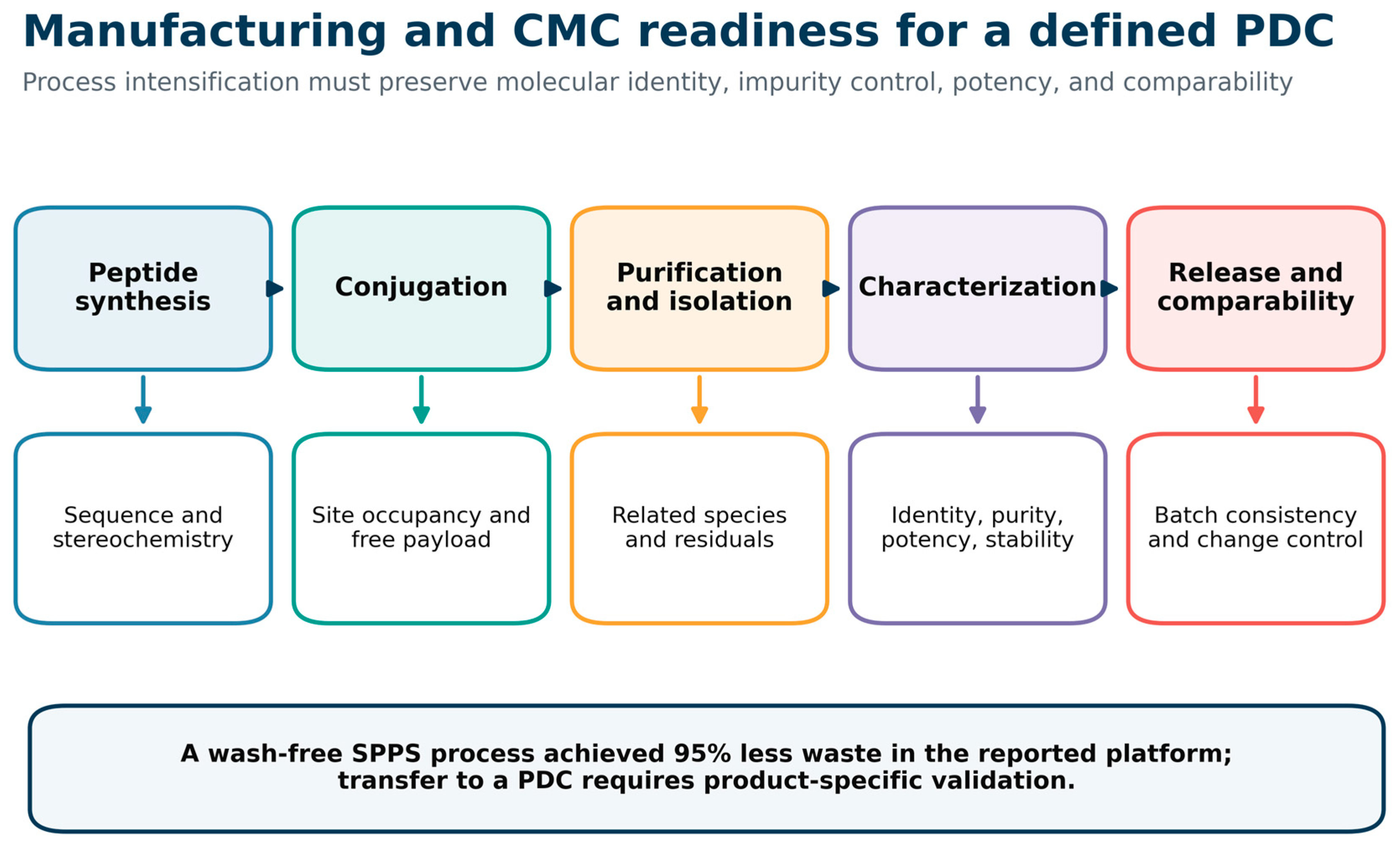
Collins et al. reported 95% less waste with wash-free solid-phase peptide synthesis in their studied platform [159]. Figure 7 adapts this process scheme to PDC manufacturing while retaining controls for molecular identity, conjugation, free payload, related substances, potency, stability, and comparability; the waste reduction is not a universal PDC result.
9.2. Bioanalysis and Clinical Pharmacology
We propose a construct-specific clinical assay plan for intact conjugate, released payload, and relevant catabolites identified nonclinically. Sampling should characterize their distinct concentration–time profiles; distinguishing release, binding, and elimination mechanisms requires independent observations and identifiable models.
FDA ADC guidance provides clinical-pharmacology precedent, not a PDC definition or default submission pathway (Section 3.1). Its analyte, exposure–response, intrinsic-factor, QT, interaction, and immunogenicity recommendations require construct-specific justification [30]. PDC adaptation must establish kinetics, elimination, relevant peptide immunogenicity risk, and active-catabolite identity. Expected low free-payload concentrations require testing, not assay omission. The proposed RCE measures and gates do not replace agency requirements or product-specific advice.
Figure 7.
Manufacturing and CMC readiness workflow for a chemically defined PDC. Adapted from Figure 1 of Collins et al. [159] under the Creative Commons Attribution 4.0 International License, redrawn, simplified, recolored, and expanded from peptide synthesis to PDC conjugation, purification, characterization, release testing, and comparability. Blue, green, orange, purple, and red distinguish peptide synthesis, conjugation, purification and isolation, characterization, and release testing and comparability, respectively. Horizontal arrows indicate the process sequence; downward arrows connect each stage to its associated quality attributes. Colors distinguish stages and do not indicate numerical values.
9.3. Biomarkers and Companion Diagnostics
Biomarkers should test the causative delivery bottleneck. Receptor IHC alone may miss trafficking, amplification, spatial heterogeneity, protease activity, perfusion, and resistance. Imaging can assess whole-body accessibility and organ accumulation; tissue assays resolve cellular localization and heterogeneity. Each readout needs a defined decision. FDA recommends contemporaneous approval or clearance of companion diagnostics and their corresponding therapeutics [160] and provides draft recommendations for their codevelopment [161]; early analytical validation, scoring, specimen requirements, prevalence, and turnaround assessment help separate drug failure from assay failure. Table 8 summarizes the CMC, bioanalytical, and regulatory readiness considerations.
Table 8.
CMC, bioanalytical, and regulatory readiness considerations [29,153,154,155,156,157,158].
10. Stage Gated Translational Roadmap
10.1. Quantitative Primary Research Examples
Figure 8, Figure 9, Figure 10, Figure 11 and Figure 12 illustrate the purposive subset described in Section 2.2. Published numerical summaries were replotted without digitization or individual-animal reanalysis; captions identify source locations, units, sample sizes, uncertainty, and normalized quantities. Each example addresses a distinct experimental question. A separate historical prodrug dataset supplies the tissue AUCs for the worked calculation and is outside the five CC BY 4.0 adaptations.
Currie et al. compared free docetaxel and SORT1-targeted TH1902 at matched docetaxel-content doses in ovarian and endometrial xenografts [162]. Figure 8 reproduces tabulated endpoint fold changes across three models, including high-dose TH1902 regression in SKOV3 and AN3-CA. This payload-equivalent comparison supports the carrier–payload hypothesis but identifies neither the active molecular species nor productive intratumoral exposure.
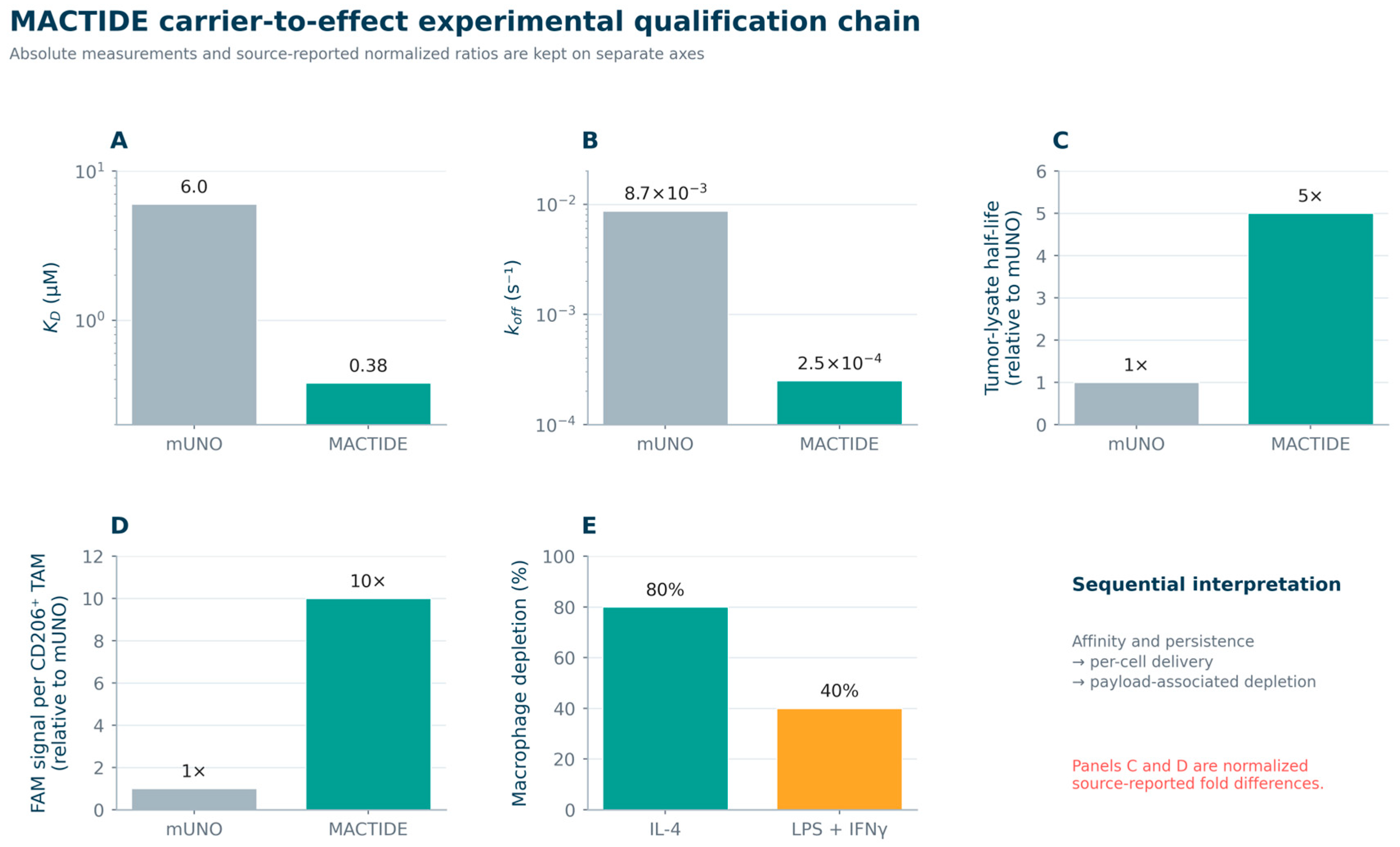
Lepland et al. constrained the CD206-binding mUNO motif into MACTIDE and coupled it to verteporfin [163]. Figure 9 uses results-text summaries to trace affinity, proteolytic persistence, delivery to tumor-associated macrophages, and phenotype-dependent payload action in a light-activated setting. It illustrates sequential carrier qualification beyond binding; peptide half-life and per-cell signal are normalized ratios, not absolute concentrations.
Figure 8.
Endpoint tumor-size progression after vehicle, free docetaxel, or SORT1-targeted TH1902 in ES-2, SKOV3, and AN3-CA xenografts. (A) ES-2 ovarian xenografts; (B) SKOV3 ovarian xenografts; (C) AN3-CA endometrial xenografts. Gray bars denote vehicle; light-blue and dark-blue bars denote docetaxel at 3.75 and 15 mg/kg; orange and green bars denote TH1902 at 8.75 and 35 mg/kg, respectively. Values are fold change from treatment Day 0 at the respective vehicle-group endpoint; the dashed line at 1 indicates no net change, and values below 1 indicate regression. Adapted from Figures 6 and 7 and Table 1 of Currie et al. [162] under the Creative Commons Attribution 4.0 International License. Changes have been made; exact tabulated values were reorganized into aligned quantitative panels and completely redrawn with standardized ordering, labels, colors, and scales; no error bars were added because the source table did not report them.
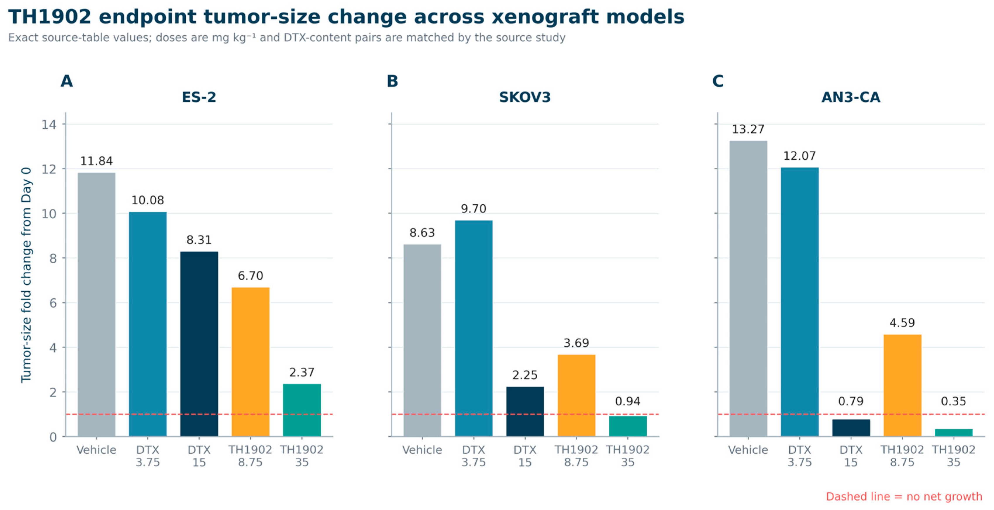
Figure 9.
Experimental qualification chain for the CD206-targeted MACTIDE platform. (A) Equilibrium dissociation constant; (B) dissociation rate constant; (C) peptide half-life in 4T1 tumor lysate relative to mUNO; (D) FAM signal per CD206-positive tumor-associated macrophage after intraperitoneal administration relative to mUNO; (E) depletion of IL-4-stimulated or LPS + IFNγ-stimulated macrophages after irradiated MACTIDE-V. Panels (A,B) use logarithmic axes. In panels (A–D), gray denotes mUNO and green denotes MACTIDE; in panel (E), green and orange denote IL-4- and LPS + IFNγ-stimulated macrophages, respectively. The arrows in the interpretation note link evidence domains rather than numerical quantities. Adapted from Figure 3A,B, 4B, and 5B of Lepland et al. [163] under the Creative Commons Attribution 4.0 International License. Changes have been made; reported numerical values and fold differences were extracted, normalized only where explicitly indicated, reorganized, and the figure has been completely redrawn in a unified multi-panel style.
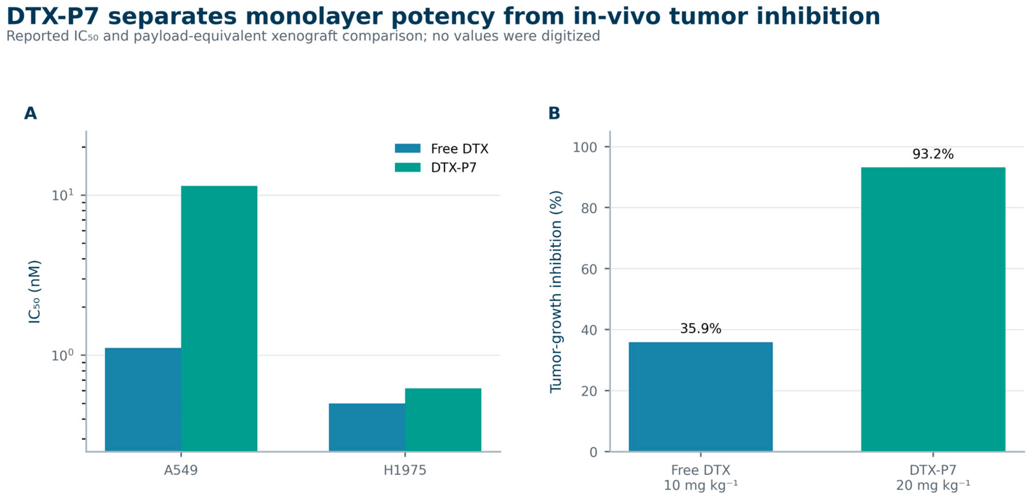
Jiang et al. illustrate discordance between intrinsic potency and delivery: DTX-P7 was approximately tenfold less potent than docetaxel in A549 monolayers yet achieved greater tumor-growth inhibition at payload-equivalent doses in A549 xenografts [164]. Figure 10 uses Results and supplementary values to show why in vitro potency cannot establish RCE; neither the mechanism nor magnitude of increased active-compound exposure was established.
Figure 10.
DTX-P7 activity in vitro and in vivo. Panel (A) shows reported IC50 values for free docetaxel and DTX-P7 in A549 and H1975 cells. Panel (B) shows reported tumor-growth inhibition after four weekly intraperitoneal treatments in A549 xenografts with n equals 5 mice per group. Adapted from Figures 1A,B and S1, and the accompanying Results of Jiang et al. [164] under the Creative Commons Attribution 4.0 International License. Changes have been made; reported values were extracted and replotted using unified axes, typography, ordering, and colors; no source artwork is reproduced.
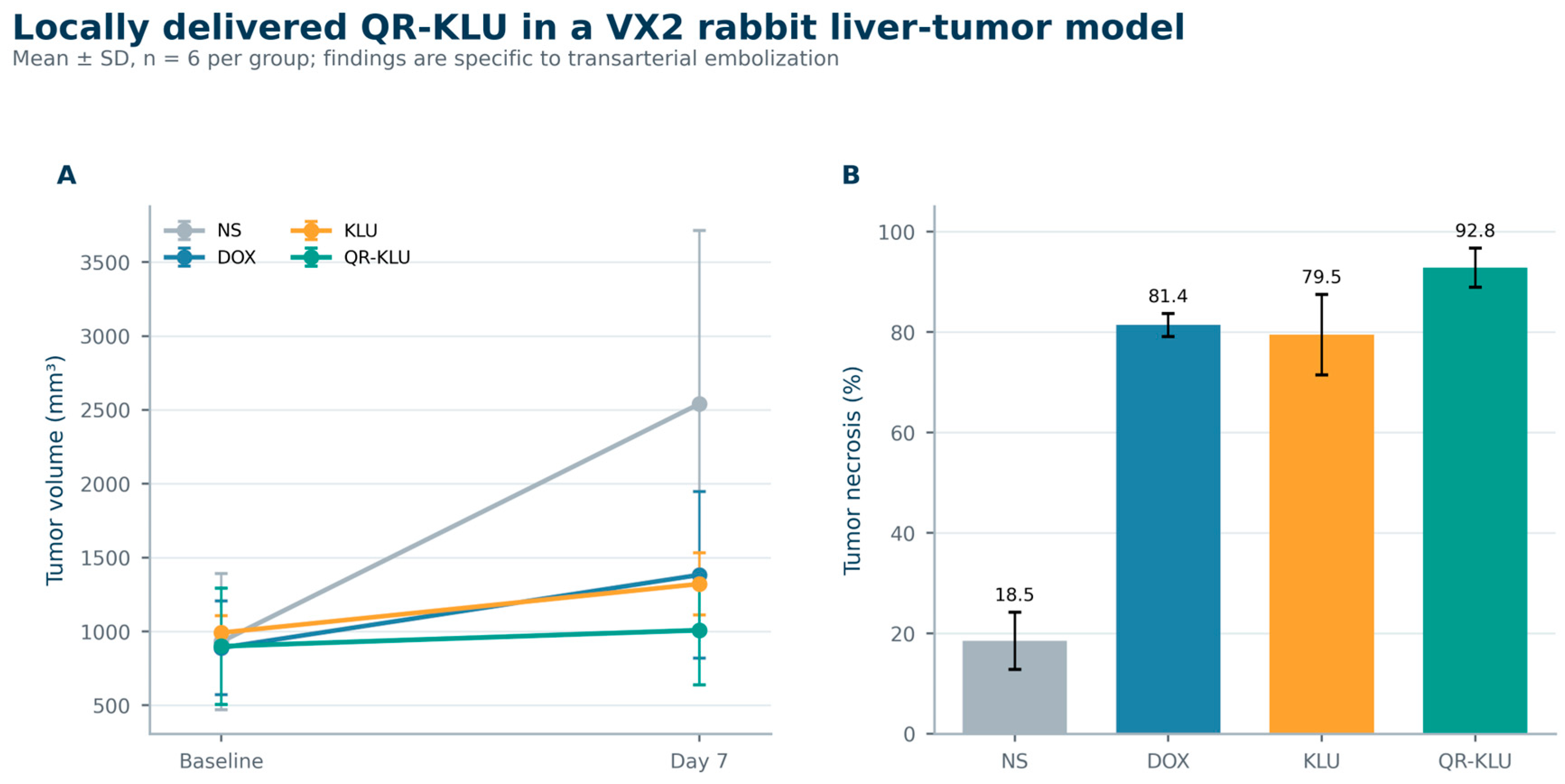
Wang et al. tested VEGFR-targeting lytic conjugate QR-KLU by transarterial delivery in VX2 rabbit liver tumors [165]. Figure 11 uses results-text summaries to show restrained enlargement and the highest measured necrosis fraction seven days after embolization. This local-delivery example demonstrates activity under those conditions, without quantifying intratumoral active-compound exposure or establishing systemic targeting.
Figure 11.
Tumor-volume change and necrosis after transarterial embolization with normal saline, doxorubicin, KLU, or QR-KLU in the VX2 rabbit liver-tumor model. (A) Tumor volumes at baseline and Day 7; (B) tumor necrosis at Day 7. Gray, blue, orange, and green denote normal saline (NS), doxorubicin (DOX), KLU, and QR-KLU, respectively. Overlapping baseline markers and error bars reflect similar group means and their reported variability; they have not been displaced. Values are mean plus or minus SD with n equals 6 per group. Adapted from Figures 4B and 5B and the accompanying Results of Wang et al. [165] under the Creative Commons Attribution 4.0 International License. Changes have been made: numerical results were extracted and replotted in a unified two-panel style; source magnetic-resonance and histology images are not reproduced.
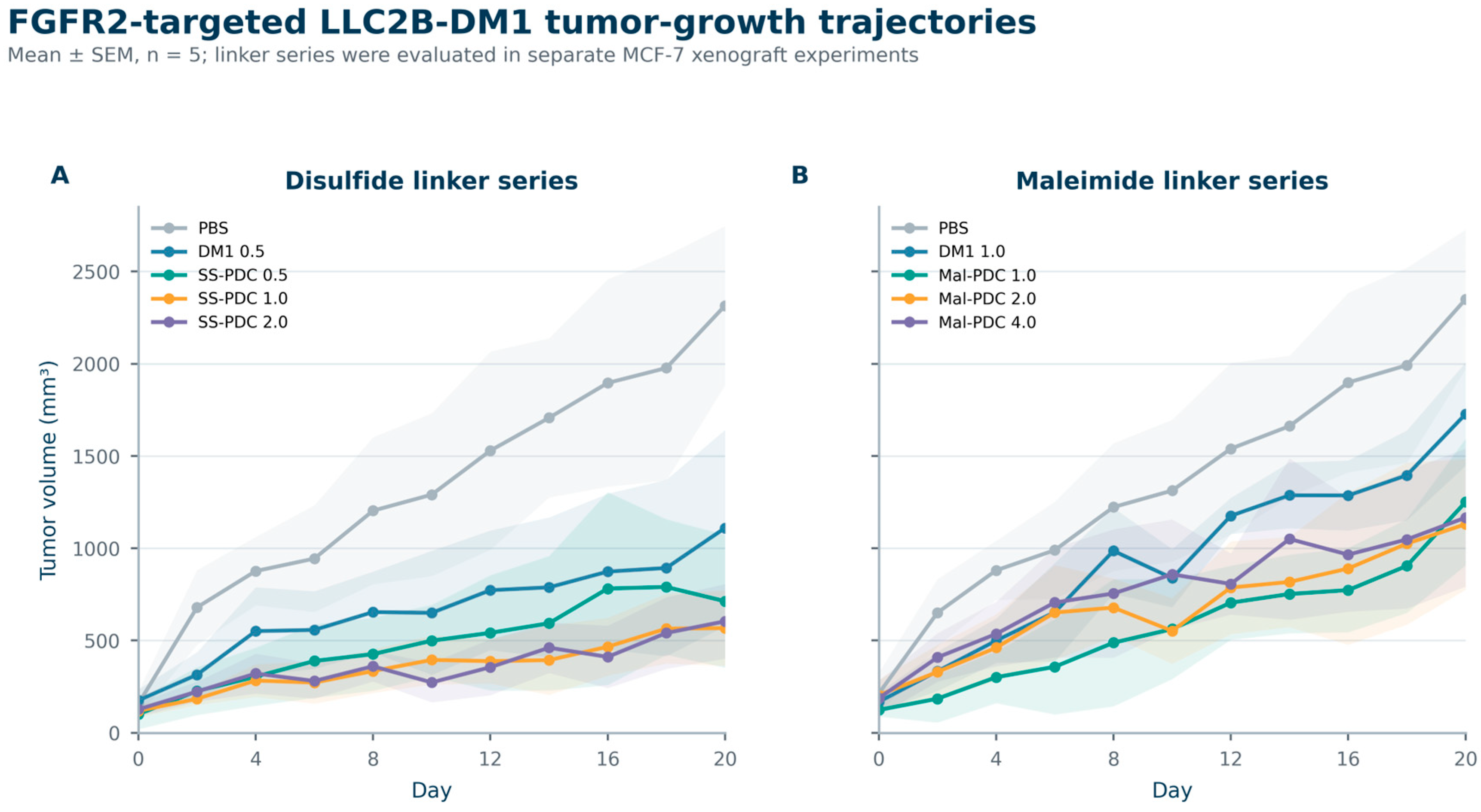
Wang et al. tested FGFR2-targeting LLC2B-DM1 with disulfide and maleimide linkers in separate MCF-7 xenograft experiments [166]. Figure 12 reproduces complete tumor-growth and uncertainty series from Supplementary Tables S2 and S3. Contemporaneous controls illustrate architecture- and dose-dependent performance; the separate experiments do not support a controlled head-to-head linker comparison.
Figure 12.
Tumor-growth trajectories for FGFR2-targeted LLC2B-DM1 conjugates in separate disulfide-linker and maleimide-linker MCF-7 xenograft experiments. (A) Disulfide-linker series (SS-PDC); (B) maleimide-linker series (Mal-PDC). Lines and markers show group means, and translucent bands show ±SEM. Colors identify the treatments and doses specified in each panel legend. Overlapping trajectories and uncertainty bands represent the reported measurements and are retained without displacement. Values are mean plus or minus SEM with n equals 5 per group; doses are expressed as DM1-equivalent mg per kg for conjugates. Adapted from Figure 6A,C and Supplementary Tables S2 and S3 of Wang et al. [166] under the Creative Commons Attribution 4.0 International License. Changes have been made; the full published mean and SEM series were replotted with standardized axes, labels, typography, and colors; no source artwork is reproduced.
Worked example using chemically resolved tissue exposure.
The five illustrations lack matched active-compound AUCs for the proposed ratios. For example, Figure 9 measures MACTIDE persistence, not verteporfin release, and cannot supply the kinetic-separation ratio’s target-release denominator [163]. Calculation therefore requires an additional dataset with the relevant analyte and comparators.
Dubois et al. reported such data for the peptide-activated doxorubicin prodrug CPI-0004Na (Section 3.1) [167]. MCF-7/6 xenograft-bearing BALB/c nu/nu mice received CPI-0004Na or doxorubicin hydrochloride by 2 h intravenous infusion at 86.2 μmol/kg; three animals per time point were sampled through 96 h. HPLC distinguished doxorubicin (Dox), intact prodrug, and peptide-bearing metabolites, correcting concentrations for extraction recovery and blood contamination. Table 9 uses source-reported trapezoidal Dox AUCs from zero to the last sample, without digitization or pooling distinct molecular species.
Table 9.
Published Dox tissue AUCs and calculated tumor-to-organ ratios for the CPI-0004Na worked example. AUC0–96 h units are pmol·h/mg tissue; inputs are from Table 3 of Dubois et al. [167].
As a Class 2 adaptation, dose-normalized tumor gain compares tumor Dox AUC per molar payload dose after prodrug vs. free drug. Each molecule contains one Dox unit; equal payload-equivalent doses therefore cancel, as follows:
Tumor gain = (2186/86.2)/(1138/86.2) = 1.92.
The tumor/heart ratio uses the same molecular species and tissue-mass units:
Tumor/heart ratio after CPI-0004Na = 2186/209.3 = 10.44.
The free-drug tumor/heart ratio is 1138/1933 = 0.589. However, the prodrug tumor/kidney ratio is 2186/4696 = 0.466; kidney Dox exposure exceeds tumor exposure. Renal exposure and toxicity therefore require separate evaluation despite favorable cardiac distribution. Neither ratio alone establishes overall safety, and unity is not a safety threshold.
This calculation implements two Class 2 adaptations in Part A of Table 3 in bulk tissue. Extractable homogenate Dox does not establish unbound intracellular or nuclear exposure, and thus complete RCE. The ratios exclude peptide-bearing metabolites and organ-specific pharmacological sensitivity; summary AUCs cannot support ratio confidence intervals. Unbound partitioning, mechanism-matched temporal coverage, paired cleavage/release half-lives, and spatial exposure–PD concordance remain unquantified. The example demonstrates calculability, not clinical-endpoint validation or a therapeutic-index claim.
10.2. Decision Gates
Table 10 proposes a sequence of development questions linking target access and conjugate integrity to the modality-appropriate active state, spatial delivery, PD, and therapeutic margin. The analyte, intended compartment, and mechanism determine which Table 3 quantities are informative and feasible. The suggested evidence supports hypothesis testing alongside manufacturing, biomarker, and comparator considerations; it does not constitute a validated scoring system, mandatory gate, or universal numerical rule for advancement.
Table 10.
Proposed development questions and modality-specific candidate evidence for PDCs [8,26,27,28,29,30,32,34,35,46,47,48].
Each claimed causal step needs a potentially disconfirming observation chosen for the mechanism; Table 10 makes modality-specific substitutions explicit. Clinical sampling must balance procedural risk and scientific value; advancement need not require all six core quantitative measures in every trial (Section 12).
11. Emerging Directions
Constrained and multicyclic peptides expand the range of binding targets while retaining chemically defined structures. Their value depends on active-compound tumor delivery, beyond plasma persistence or renal excretion. Screens should therefore assess post-conjugation binding, proteolysis, trafficking, and systemic distribution alongside affinity.
Tandem or AND-gate release can increase specificity through two biochemical conditions, but added chemistry may reduce yield and complicate product identification. Bioorthogonal activation adds external control while requiring compatible trigger distribution and reaction kinetics. Added complexity and controls should be justified by active-compound tumor exposure gains over simpler linkers.
Theranostic analogues can assess target access, whole-body distribution, and patient heterogeneity before cytotoxic treatment. Because linker, payload, chelator, charge, and albumin-binding changes can alter disposition, imaging must be validated against the therapeutic construct.
AI and machine learning support peptide generation, structure prediction, stability estimation, and multi-objective ranking [168]. PDC models should include negative results and binding, degradation, conjugation compatibility, release, trafficking, PK, and organ-uptake endpoints; affinity-only optimization repeats the single-variable selection problem addressed by RCE.
Solvent use, purification, low conjugation yields, and potent-payload handling can offset synthetic simplicity. Greener solid-phase synthesis, solvent reduction, convergent routes, and improved purification should be evaluated by product quality and scalability, beyond solvent substitution [159,169].
12. Limitations of the Framework
RCE is an author-proposed research framework, not a validated clinical surrogate or regulatory criterion. Established PK calculations retain their conventional meaning; their proposed predictive use within RCE requires validation. AUC-based estimates require concentration–time data for an identified active compound in a specified compartment. Lesion access, procedural risk, tissue yield, and patient burden often preclude dense biopsies. Research sampling requires a prespecified question, informed consent, independent ethical review, and proportionate risk [38]; routine biopsy-based individual tumor AUC reconstruction is not proposed. A single timed biopsy establishes one concentration, not individual AUC or exposure across lesions.
Microdialysis can serially measure diffusible, unbound extracellular analytes in accessible lesions, but it requires catheter placement and analyte-specific probe-recovery, stability, and adsorption assessment. Clinical carboplatin studies demonstrate feasibility alongside technical failures and variable recovery [170]. Extracellular profiles do not directly measure cytosolic or nuclear active-catabolite exposure; homogenates mix vascular, stromal, extracellular, and cellular material. Chemical identity, tissue localization, and subcellular access therefore require distinct evidence.
A feasible translational strategy could combine complementary evidence. Time-resolved tissue PK, identity of molecular species, and exposure–PD relationships can be established nonclinically; then, serial human plasma assays can be used to assess intact conjugate, released payload, and relevant catabolites. Separate intact-BT5528 and unconjugated-MMAE assays provide a direct PDC precedent [34], without establishing comprehensive catabolite coverage. Selected timed biopsies or planned resections can anchor tissue observations, with concurrent plasma and controls for ex vivo cleavage and residual blood. The GRN1005 phase I surgical cohort separately measured conjugate and paclitaxel in resected tumor with concurrent plasma [37], demonstrating tissue bioanalysis rather than individual tumor AUC, unbound exposure, or subcellular localization.
Molecular imaging can assess lesion accessibility, distribution, and heterogeneity less invasively. ZEPHIR combined radiolabeled trastuzumab PET and early metabolic imaging to examine heterogeneity and T-DM1 response [171], providing an ADC methodological precedent rather than validating PDC release. Carrier or diagnostic-analogue signals cannot locate or quantify released cytotoxic catabolites without validation. Label stability, labeled metabolites, and distribution effects of tracer mass, affinity, charge, and conjugation should be assessed. Uptake and metabolic response support interpretation but cannot directly supply active-catabolite AUC.
Population or physiologically based PK models could integrate nonclinical tissue kinetics, human plasma, and sparse tissue or imaging data to predict analyte-specific tumor concentration–time profiles and derived exposure measures. Antibody plasma/PET modeling provides a precedent and illustrates dependence on model assumptions [172]; PDC use requires construct-specific evaluation. Ethically justified sampling staggered across participants may constrain population profiles, not directly observed individual curves. Identifiability, sensitivity to release and partition assumptions, uncertainty intervals, and validation against independent tissue data should be reported where available. Plasma fit alone may leave tumor release unidentified; that quantity should be reported as not estimable. Transferred antibody models must replace carrier-specific recycling, tissue-exchange, and elimination assumptions with supported PDC processes (Section 7.4).
Payload-target engagement and downstream tissue PD, including damage responses, can support activity without full tumor PK but cannot uniquely determine active-compound concentration or duration above an effect threshold. Human safety-organ AUC is usually inaccessible; nonclinical distribution, clinical exposure–toxicity analysis, and organ monitoring provide safety evidence, not measured tumor/organ AUC ratios. Reporting should distinguish between unmeasured, below-quantification, and non-estimable quantities; missing denominators cannot imply favorable exposure ratios or safety. Plasma, imaging, or PD surrogates require prospective construct-specific calibration against selective tissue measurements of molecular species, then evaluation against benefit and toxicity. Explicit uncertainty should be retained to avoid generalizing thresholds across constructs, payloads, or biological species.
Many PDC programs remain preclinical or early-phase, clinical reporting is inconsistent, and negative programs are underreported. Target, payload, population, dose, and trial-design differences limit cross-study comparison. As a critical narrative review, this synthesis is susceptible to literature-selection and interpretive bias. Language prioritization may add selection bias, and unavailable historical search logs limit retrospective reproducibility. The documented retrieval check and qualitative appraisal do not establish completeness or eliminate bias; the proposed framework requires empirical validation.
The five adaptations also carry author-selection and availability bias. Permissive licensing and numerical accessibility constrain inclusion, and positive, null, and negative findings are unbalanced. Less accessible studies and unsuccessful constructs may be underrepresented. Mechanistic diversity does not establish representativeness or permit estimates of delivery success, platform rankings, or validation of RCE predictions and thresholds. The separate AUC example establishes calculability only.
13. Conclusions
RCE connects conjugate integrity, active-compound formation, active-species exposure in the relevant tumor compartment, PD, and therapeutic margin in a testable development framework. Its measures require defined analytes, compartments, intervals, and comparators, with uncertainty retained when direct measurements are infeasible. Programs should progressively strengthen the links between target access, active-species exposure, PD, and therapeutic margin before major development decisions. Early-stage advancement may proceed with documented uncertainties and a plan to address decision-relevant gaps. The predictive use of these established, adapted, and conceptual measures within RCE awaits construct-specific validation.
Supplementary Materials
The following supporting information can be downloaded at: https://www.mdpi.com/article/10.3390/pharmaceutics18111372/s1. Supplementary Materials S1 provides the search strategies and 24 October 2026 proof-stage retrieval verification. Supplementary_Search_Records_Proof_2026-10-24.zip contains the machine-readable queries, raw PubMed and ClinicalTrials.gov responses, identifier inventories, official-document lookup logs, reproducibility scripts, and file checksums. The earlier written record is retained separately as provenance; its unavailable raw-response archive has not been reconstructed.
Author Contributions
Conceptualization, methodology, investigation, writing—original draft preparation; A.A.K. Investigation, writing—review and editing, and visualization; F.C. Writing—review and editing, supervision; Ö.Y. All authors have read and agreed to the published version of the manuscript.
Funding
This research received no external funding.
Institutional Review Board Statement
Not applicable.
Informed Consent Statement
Not applicable.
Data Availability Statement
No new experimental data were generated. The worked ratios in Section 10.1 were calculated from the cited published AUC summaries. All sources used in the review are cited in the manuscript. Supplementary Materials S1 and Supplementary_Search_Records_Proof_2026-10-24.zip provide the proof-stage search requests, raw PubMed and ClinicalTrials.gov responses, identifier lists, and official-document lookup logs. These records were generated on 24 October 2026 and do not reconstruct the unavailable historical raw-response archive or constitute a screened study corpus. Figure provenance and reuse conditions are described in the article.
Acknowledgments
During preparation of this manuscript, the authors used ChatGPT (OpenAI) to draft visual layouts and prepare Supplementary Materials. The authors retain responsibility for source verification, scientific interpretation, and the final manuscript.
Conflicts of Interest
The authors declare no conflicts of interest.
References
- Bray, F.; Laversanne, M.; Sung, H.; Ferlay, J.; Siegel, R.L.; Soerjomataram, I.; Jemal, A. Global cancer statistics 2022: GLOBOCAN estimates of incidence and mortality worldwide for 36 cancers in 185 countries. CA Cancer J. Clin. 2024, 74, 229–263. [Google Scholar] [CrossRef] [Scilit] [PubMed]
- Beck, A.; Goetsch, L.; Dumontet, C.; Corvaïa, N. Strategies and challenges for the next generation of antibody–drug conjugates. Nat. Rev. Drug Discov. 2017, 16, 315–337. [Google Scholar] [CrossRef] [Scilit] [PubMed]
- Drago, J.Z.; Modi, S.; Chandarlapaty, S. Unlocking the potential of antibody–drug conjugates for cancer therapy. Nat. Rev. Clin. Oncol. 2021, 18, 327–344. [Google Scholar] [CrossRef] [Scilit] [PubMed]
- Muttenthaler, M.; King, G.F.; Adams, D.J.; Alewood, P.F. Trends in peptide drug discovery. Nat. Rev. Drug Discov. 2021, 20, 309–325. [Google Scholar] [CrossRef] [Scilit] [PubMed]
- Lau, J.L.; Dunn, M.K. Therapeutic peptides: Historical perspectives, current development trends, and future directions. Bioorg. Med. Chem. 2018, 26, 2700–2707. [Google Scholar] [CrossRef] [Scilit] [PubMed]
- Cooper, B.M.; Iegre, J.; O’ Donovan, D.H.; Ölwegård Halvarsson, M.; Spring, D.R. Peptides as a platform for targeted therapeutics for cancer: Peptide–drug conjugates (PDCs). Chem. Soc. Rev. 2021, 50, 1480–1494. [Google Scholar] [CrossRef] [Scilit] [PubMed]
- Zhu, Y.-S.; Tang, K.; Lv, J. Peptide–drug conjugate-based novel molecular drug delivery system in cancer. Trends Pharmacol. Sci. 2021, 42, 857–869. [Google Scholar] [CrossRef] [Scilit] [PubMed]
- Srinivasarao, M.; Galliford, C.V.; Low, P.S. Principles in the design of ligand-targeted cancer therapeutics and imaging agents. Nat. Rev. Drug Discov. 2015, 14, 203–219. [Google Scholar] [CrossRef] [Scilit] [PubMed]
- Aina, O.H.; Sroka, T.C.; Chen, M.L.; Lam, K.S. Therapeutic cancer targeting peptides. Biopolymers 2002, 66, 184–199. [Google Scholar] [CrossRef] [Scilit] [PubMed]
- Firer, M.A.; Gellerman, G. Targeted drug delivery for cancer therapy: The other side of antibodies. J. Hematol. Oncol. 2012, 5, 70. [Google Scholar] [CrossRef] [Scilit] [PubMed]
- Hoppenz, P.; Els-Heindl, S.; Beck-Sickinger, A.G. Peptide-Drug Conjugates and Their Targets in Advanced Cancer Therapies. Front. Chem. 2020, 8, 571. [Google Scholar] [CrossRef] [Scilit] [PubMed]
- Heh, E.; Allen, J.; Ramirez, F.; Lovasz, D.; Fernandez, L.; Hogg, T.; Riva, H.; Holland, N.; Chacon, J. Peptide Drug Conjugates and Their Role in Cancer Therapy. Int. J. Mol. Sci. 2023, 24, 829. [Google Scholar] [CrossRef] [Scilit] [PubMed]
- Jadhav, K.; Abhang, A.; Kole, E.B.; Gadade, D.; Dusane, A.; Iyer, A.; Sharma, A.; Rout, S.K.; Gholap, A.D.; Naik, J.; et al. Peptide–Drug Conjugates as Next-Generation Therapeutics: Exploring the Potential and Clinical Progress. Bioengineering 2025, 12, 481. [Google Scholar] [CrossRef] [Scilit] [PubMed]
- Wang, D.; Yin, F.; Li, Z.; Zhang, Y.; Shi, C. Current progress and remaining challenges of peptide–drug conjugates (PDCs): Next generation of antibody-drug conjugates (ADCs)? J. Nanobiotechnol. 2025, 23, 305. [Google Scholar] [CrossRef] [Scilit] [PubMed]
- Parang, K.; Do, T.; Dinh, C.; Davani-Davari, D.; Foroughi, M.; Khong, T.; Moazen, M.; Nasrolahi Shirazi, A. Tumor Targeting with Peptide-Drug Conjugates: Showcasing Key Progress and Hurdles. Drug Des. Dev. Ther. 2026, 20, 562135. [Google Scholar] [CrossRef] [Scilit] [PubMed]
- Jafari, K.N.; Chai, C.; Kim, S.; Kaur, K. Receptor-Mediated Drug Delivery: Redefining Targeted Drug Conjugates in Oncology. Pharmaceutics 2026, 18, 386. [Google Scholar] [CrossRef] [Scilit] [PubMed]
- Zhou, N.; Li, M.; Huang, Y.; Wang, Y.; Wang, J. Peptide-Drug Conjugates for Targeted Delivery in Hematological Malignancies: From Design Principles to Clinical Application. Pharmaceutics 2026, 18, 849. [Google Scholar] [CrossRef] [Scilit] [PubMed]
- Kumar, A.; Sharma, R.; Yadav, A.K. Engineering peptide-drug conjugates for targeted cancer therapy: Design principle, theranostic imaging, and translational challenges. Adv. Drug Deliv. Rev. 2026, 239, 115960. [Google Scholar] [CrossRef] [Scilit] [PubMed]
- Bargh, J.D.; Isidro-Llobet, A.; Parker, J.S.; Spring, D.R. Cleavable linkers in antibody–drug conjugates. Chem. Soc. Rev. 2019, 48, 4361–4374. [Google Scholar] [CrossRef] [Scilit] [PubMed]
- Tsuchikama, K.; An, Z. Antibody-drug conjugates: Recent advances in conjugation and linker chemistries. Protein Cell 2018, 9, 33–46. [Google Scholar] [CrossRef] [Scilit] [PubMed]
- Balamkundu, S.; Liu, C.F. Lysosomal-Cleavable Peptide Linkers in Antibody-Drug Conjugates. Biomedicines 2023, 11, 3080. [Google Scholar] [CrossRef] [Scilit] [PubMed]
- Baethge, C.; Goldbeck-Wood, S.; Mertens, S. SANRA-a scale for the quality assessment of narrative review articles. Res. Integr. Peer Rev. 2019, 4, 5. [Google Scholar] [CrossRef] [Scilit] [PubMed]
- National Library of Medicine. PubMed User Guide. Available online: https://pubmed.ncbi.nlm.nih.gov/help/ (accessed on 22 October 2026).
- ClinicalTrials.gov. API Documentation and Search Areas. Available online: https://clinicaltrials.gov/data-api/api (accessed on 22 October 2026).
- European Medicines Agency. Pepaxti: EPAR and product information. Available online: https://www.ema.europa.eu/en/medicines/human/EPAR/pepaxti (accessed on 15 September 2026).
- Strosberg, J.; El-Haddad, G.; Wolin, E.; Hendifar, A.; Yao, J.; Chasen, B.; Mittra, E.; Kunz, P.L.; Kulke, M.H.; Jacene, H.; et al. Phase 3 Trial of 177 Lu-Dotatate for Midgut Neuroendocrine Tumors. N. Engl. J. Med. 2017, 376, 125–135. [Google Scholar] [CrossRef] [Scilit] [PubMed]
- Hennrich, U.; Kopka, K. Lutathera®: The First FDA- and EMA-Approved Radiopharmaceutical for Peptide Receptor Radionuclide Therapy. Pharmaceuticals 2019, 12, 114. [Google Scholar] [CrossRef] [Scilit] [PubMed]
- Hofland, J.; Brabander, T.; Verburg, F.A.; Feelders, R.A.; de Herder, W.W. Peptide Receptor Radionuclide Therapy. J. Clin. Endocrinol. Metab. 2022, 107, 3199–3208. [Google Scholar] [CrossRef] [Scilit] [PubMed]
- European Medicines Agency. Guideline on the Development and Manufacture of Synthetic Peptides. EMA/CHMP/CVMP/QWP/367182/2025; EMA: Amsterdam, The Netherlands, 2025; Available online: https://www.ema.europa.eu/en/scientific-guidelines/development-manufacture-synthetic-peptides (accessed on 22 October 2026).
- U.S. Food and Drug Administration. Clinical Pharmacology Considerations for Antibody-Drug Conjugates: Guidance for Industry; U.S. Food and Drug Administration: Silver Spring, MD, USA, 2024. Available online: https://www.fda.gov/media/155997/download (accessed on 22 October 2026).
- European Parliament and Council of the European Union. Directive 2001/83/EC on the Community Code Relating to Medicinal Products for Human Use, as Amended. Article 1(6). Consolidated version of 1 January 2025. Available online: https://eur-lex.europa.eu/legal-content/EN/TXT/?uri=CELEX%3A02001L0083-20250101 (accessed on 22 October 2026).
- U.S. Food and Drug Administration. Combination Product Definition Combination Product Types. Available online: https://www.fda.gov/combination-products/about-combination-products/combination-product-definition-combination-product-types (accessed on 22 October 2026).
- Rigby, M.; Bennett, G.; Chen, L.; Mudd, G.E.; Harrison, H.; Beswick, P.J.; van Rietschoten, K.; Watcham, S.M.; Scott, H.S.; Brown, A.N.; et al. BT8009; a Nectin-4 targeting Bicycle toxin conjugate for treatment of solid tumors. Mol. Cancer Ther. 2022, 21, 1747–1756. [Google Scholar] [CrossRef] [Scilit] [PubMed]
- Tweed, J.A.; Adams-Dam, F.; Allanson, J.; Holmes, K.; Senior, R.; Xu, H.; Li, M.; Bennett, G.; Jeffrey, P. Bioanalysis of the Bicycle toxin conjugate BT5528 and released monomethyl auristatin E via liquid chromatography-tandem mass spectrometry. Bioanalysis 2024, 16, 155–169. [Google Scholar] [CrossRef] [Scilit] [PubMed]
- Gowland, C.; Berry, P.; Errington, J.; Jeffrey, P.; Bennett, G.; Godfrey, L.; Pittman, M.; Niewiarowski, A.; Symeonides, S.N.; Veal, G.J. Development of a LC-MS/MS method for the quantification of toxic payload DM1 cleaved from BT1718 in a phase I study. Bioanalysis 2021, 13, 101–113. [Google Scholar] [CrossRef] [Scilit] [PubMed]
- Tweed, J.A.; Adams-Dam, F.; Bader, J.; Zhang, Y.; Allanson, J.; Holmes, K.; Senior, R.; Rigby, M.; Jeffrey, P.; Xu, H. The validation and clinical bioanalysis of the Bicycle toxin conjugate zelenectide pevedotin in human plasma. Bioanalysis 2025, 17, 177–185. [Google Scholar] [CrossRef] [Scilit] [PubMed]
- Drappatz, J.; Brenner, A.; Wong, E.T.; Eichler, A.; Schiff, D.; Groves, M.D.; Mikkelsen, T.; Rosenfeld, S.; Sarantopoulos, J.; Meyers, C.A.; et al. Phase I study of GRN1005 in recurrent malignant glioma. Clin. Cancer Res. 2013, 19, 1567–1576. [Google Scholar] [CrossRef] [Scilit] [PubMed]
- Levit, L.A.; Peppercorn, J.M.; Tam, A.L.; Marron, J.M.; Mathews, D.J.H.; Levit, K.; Roach, N.; Ratain, M.J. Ethical Framework for Including Research Biopsies in Oncology Clinical Trials: American Society of Clinical Oncology Research Statement. J. Clin. Oncol. 2019, 37, 2368–2377. [Google Scholar] [CrossRef] [Scilit] [PubMed]
- Erickson, H.K.; Park, P.U.; Widdison, W.C.; Kovtun, Y.V.; Garrett, L.M.; Hoffman, K.; Lutz, R.J.; Goldmacher, V.S.; Blättler, W.A. Antibody-maytansinoid conjugates are activated in targeted cancer cells by lysosomal degradation and linker-dependent intracellular processing. Cancer Res. 2006, 66, 4426–4433. [Google Scholar] [CrossRef] [Scilit] [PubMed]
- Erickson, H.K.; Widdison, W.C.; Mayo, M.F.; Whiteman, K.; Audette, C.; Wilhelm, S.D.; Singh, R. Tumor delivery and in vivo processing of disulfide-linked and thioether-linked antibody-maytansinoid conjugates. Bioconjug. Chem. 2010, 21, 84–92. [Google Scholar] [CrossRef] [Scilit] [PubMed]
- Li, F.; Emmerton, K.K.; Jonas, M.; Zhang, X.; Miyamoto, J.B.; Setter, J.R.; Nicholas, N.D.; Okeley, N.M.; Lyon, R.P.; Benjamin, D.R.; et al. Intracellular released payload influences potency and bystander-killing effects of antibody-drug conjugates in preclinical models. Cancer Res. 2016, 76, 2710–2719. [Google Scholar] [CrossRef] [Scilit] [PubMed]
- Gallo, J.M.; Gupta, P.K.; Hung, C.T.; Perrier, D.G. Evaluation of drug delivery following the administration of magnetic albumin microspheres containing adriamycin to the rat. J. Pharm. Sci. 1989, 78, 190–194. [Google Scholar] [CrossRef] [Scilit] [PubMed]
- Gupta, A.; Chatelain, P.; Massingham, R.; Jonsson, E.N.; Hammarlund-Udenaes, M. Brain distribution of cetirizine enantiomers: Comparison of three different tissue-to-plasma partition coefficients: Kp, Kp,u, and Kp,uu. Drug Metab. Dispos. 2006, 34, 318–323. [Google Scholar] [CrossRef] [Scilit] [PubMed]
- Boddy, A.; Aarons, L.; Petrak, K. Efficiency of Drug Targeting: Steady-State Considerations Using a Three-Compartment Model. Pharm. Res. 1989, 6, 367–372. [Google Scholar] [CrossRef] [Scilit] [PubMed]
- Zhang, J.; Zhou, F.; Qi, H.; Ni, H.; Hu, Q.; Zhou, C.; Li, Y.; Baburina, I.; Courtney, J.; Salamone, S.J. Randomized study of individualized pharmacokinetically-guided dosing of paclitaxel compared with body-surface area dosing in Chinese patients with advanced non-small cell lung cancer. Br. J. Clin. Pharmacol. 2019, 85, 2292–2301. [Google Scholar] [CrossRef] [Scilit] [PubMed]
- Kamath, A.V.; Iyer, S. Challenges and advances in the assessment of the disposition of antibody-drug conjugates. Biopharm. Drug Dispos. 2016, 37, 66–74. [Google Scholar] [CrossRef] [Scilit] [PubMed]
- Gorovits, B.; Alley, S.C.; Bilic, S.; Booth, B.; Kaur, S.; Oldfield, P.; Purushothama, S.; Rao, C.; Shord, S.; Siguenza, P. Bioanalysis of antibody-drug conjugates: American Association of Pharmaceutical Scientists Antibody-Drug Conjugate Working Group position paper. Bioanalysis 2013, 5, 997–1006. [Google Scholar] [CrossRef] [Scilit] [PubMed]
- Kaur, S.; Xu, K.; Saad, O.M.; Dere, R.C.; Carrasco-Triguero, M. Bioanalytical assay strategies for the development of antibody-drug conjugate biotherapeutics. Bioanalysis 2013, 5, 201–226. [Google Scholar] [CrossRef] [Scilit] [PubMed]
- Reubi, J.C. Peptide receptors as molecular targets for cancer diagnosis and therapy. Endocr. Rev. 2003, 24, 389–427. [Google Scholar] [CrossRef] [Scilit] [PubMed]
- Pasqualini, R.; Ruoslahti, E. Organ targeting In vivo using phage display peptide libraries. Nature 1996, 380, 364–366. [Google Scholar] [CrossRef] [Scilit] [PubMed]
- Arap, W.; Pasqualini, R.; Ruoslahti, E. Cancer Treatment by Targeted Drug Delivery to Tumor Vasculature in a Mouse Model. Science 1998, 279, 377–380. [Google Scholar] [CrossRef] [Scilit] [PubMed]
- Danhier, F.; Le Breton, A.; Préat, V. RGD-based strategies to target alpha(v) beta(3) integrin in cancer therapy and diagnosis. Mol. Pharm. 2012, 9, 2961–2973. [Google Scholar] [CrossRef] [Scilit] [PubMed]
- Laakkonen, P.; Akerman, M.E.; Biliran, H.; Yang, M.; Ferrer, F.; Karpanen, T.; Hoffman, R.M.; Ruoslahti, E. Antitumor activity of a homing peptide that targets tumor lymphatics and tumor cells. Proc. Natl. Acad. Sci. USA 2004, 101, 9381–9386. [Google Scholar] [CrossRef] [Scilit] [PubMed]
- Teesalu, T.; Sugahara, K.N.; Kotamraju, V.R.; Ruoslahti, E. C-end rule peptides mediate neuropilin-1-dependent cell, vascular, and tissue penetration. Proc. Natl. Acad. Sci. USA 2009, 106, 16157–16162. [Google Scholar] [CrossRef] [Scilit] [PubMed]
- Sugahara, K.N.; Teesalu, T.; Karmali, P.P.; Kotamraju, V.R.; Agemy, L.; Greenwald, D.R.; Ruoslahti, E. Coadministration of a Tumor-Penetrating Peptide Enhances the Efficacy of Cancer Drugs. Science 2010, 328, 1031–1035. [Google Scholar] [CrossRef] [Scilit] [PubMed]
- Ruoslahti, E. Tumor penetrating peptides for improved drug delivery. Adv. Drug Deliv. Rev. 2017, 110–111, 3–12. [Google Scholar] [CrossRef] [Scilit] [PubMed]
- Guidotti, G.; Brambilla, L.; Rossi, D. Cell-Penetrating Peptides: From Basic Research to Clinics. Trends Pharmacol. Sci. 2017, 38, 406–424. [Google Scholar] [CrossRef] [Scilit] [PubMed]
- Bechara, C.; Sagan, S. Cell-penetrating peptides: 20 years later, where do we stand? FEBS Lett. 2013, 587, 1693–1702. [Google Scholar] [CrossRef] [Scilit] [PubMed]
- Pei, D.; Buyanova, M. Overcoming Endosomal Entrapment in Drug Delivery. Bioconjug. Chem. 2019, 30, 273–283. [Google Scholar] [CrossRef] [Scilit] [PubMed]
- Wadia, J.S.; Stan, R.V.; Dowdy, S.F. Transducible TAT-HA fusogenic peptide enhances escape of TAT-fusion proteins after lipid raft macropinocytosis. Nat. Med. 2004, 10, 310–315. [Google Scholar] [CrossRef] [Scilit] [PubMed]
- Appelbaum, J.S.; LaRochelle, J.R.; Smith, B.A.; Balkin, D.M.; Holub, J.M.; Schepartz, A. Arginine topology controls escape of minimally cationic proteins from early endosomes to the cytoplasm. Chem. Biol. 2012, 19, 819–830. [Google Scholar] [CrossRef] [Scilit] [PubMed]
- Qian, Z.; Liu, T.; Liu, Y.-Y.; Briesewitz, R.; Barrios, A.M.; Jhiang, S.M.; Pei, D. Efficient delivery of cyclic peptides into mammalian cells with short sequence motifs. ACS Chem. Biol. 2013, 8, 423–431. [Google Scholar] [CrossRef] [Scilit] [PubMed]
- Qian, Z.; LaRochelle, J.R.; Jiang, B.; Lian, W.; Hard, R.L.; Selner, N.G.; Luechapanichkul, R.; Barrios, A.M.; Pei, D. Early endosomal escape of a cyclic cell-penetrating peptide allows effective cytosolic cargo delivery. Biochemistry 2014, 53, 4034–4046. [Google Scholar] [CrossRef] [Scilit] [PubMed]
- Erazo-Oliveras, A.; Najjar, K.; Dayani, L.; Wang, T.-Y.; Johnson, G.A.; Pellois, J.-P. Protein delivery into live cells by incubation with an endosomolytic agent. Nat. Methods 2014, 11, 861–867. [Google Scholar] [CrossRef] [Scilit] [PubMed]
- Chen, X.; Plasencia, C.; Hou, Y.; Neamati, N. Synthesis and biological evaluation of dimeric RGD peptide-paclitaxel conjugate as a model for integrin-targeted drug delivery. J. Med. Chem. 2005, 48, 1098–1106. [Google Scholar] [CrossRef] [Scilit] [PubMed]
- Guan, H.; McGuire, M.J.; Li, S.; Brown, K.C. Peptide-targeted polyglutamic acid doxorubicin conjugates for the treatment of alpha(v)beta(6)-positive cancers. Bioconjug. Chem. 2008, 19, 1813–1821. [Google Scholar] [CrossRef] [Scilit] [PubMed]
- Li, S.; Gray, B.P.; McGuire, M.J.; Brown, K.C. Synthesis and biological evaluation of a peptide-paclitaxel conjugate which targets the integrin alpha(v)beta(6). Bioorg. Med. Chem. 2011, 19, 5480–5489. [Google Scholar] [CrossRef] [Scilit] [PubMed]
- Walensky, L.D.; Bird, G.H. Hydrocarbon-Stapled Peptides: Principles, Practice, and Progress. J. Med. Chem. 2014, 57, 6275–6288. [Google Scholar] [CrossRef] [Scilit] [PubMed]
- Heinis, C.; Rutherford, T.; Freund, S.; Winter, G. Phage-encoded combinatorial chemical libraries based on bicyclic peptides. Nat. Chem. Biol. 2009, 5, 502–507. [Google Scholar] [CrossRef] [Scilit] [PubMed]
- Ullrich, S.; Nitsche, C. Bicyclic peptides: Paving the road for therapeutics of the future. Pept. Sci. 2024, 116, e24326. [Google Scholar] [CrossRef] [Scilit]
- Di, L. Strategic approaches to optimizing peptide ADME properties. AAPS J. 2015, 17, 134–143. [Google Scholar] [CrossRef] [Scilit] [PubMed]
- Penchala, S.C.; Miller, M.R.; Pal, A.; Dong, J.; Madadi, N.R.; Xie, J.; Joo, H.; Tsai, J.; Batoon, P.; Samoshin, V.; et al. A biomimetic approach for enhancing the in vivo half-life of peptides. Nat. Chem. Biol. 2015, 11, 793–798. [Google Scholar] [CrossRef] [Scilit] [PubMed]
- Zorzi, A.; Middendorp, S.J.; Wilbs, J.; Deyle, K.; Heinis, C. Acylated heptapeptide binds albumin with high affinity and application as tag furnishes long-acting peptides. Nat. Commun. 2017, 8, 16092. [Google Scholar] [CrossRef] [Scilit] [PubMed]
- Anami, Y.; Yamazaki, C.M.; Xiong, W.; Gui, X.; Zhang, N.; An, Z.; Tsuchikama, K. Glutamic acid-valine-citrulline linkers ensure stability and efficacy of antibody-drug conjugates in mice. Nat. Commun. 2018, 9, 2512. [Google Scholar] [CrossRef] [Scilit] [PubMed]
- Dubowchik, G.M.; Firestone, R.A.; Padilla, L.; Willner, D.; Hofstead, S.J.; Mosure, K.; Knipe, J.O.; Lasch, S.J.; Trail, P.A. Cathepsin B-labile dipeptide linkers for lysosomal release of doxorubicin from internalizing immunoconjugates: Model studies of enzymatic drug release and antigen-specific in vitro anticancer activity. Bioconjug. Chem. 2002, 13, 855–869. [Google Scholar] [CrossRef] [Scilit] [PubMed]
- Caculitan, N.G.; Dela Cruz Chuh, J.; Ma, Y.; Zhang, D.; Kozak, K.R.; Liu, Y.; Pillow, T.H.; Sadowsky, J.; Cheung, T.K.; Phung, Q.; et al. Cathepsin B is dispensable for cellular processing of cathepsin B-cleavable antibody-drug conjugates. Cancer Res. 2017, 77, 7027–7037. [Google Scholar] [CrossRef] [Scilit] [PubMed]
- Lyon, R.P.; Bovee, T.D.; Doronina, S.O.; Burke, P.J.; Hunter, J.H.; Neff-LaFord, H.D.; Jonas, M.; Anderson, M.E.; Setter, J.R.; Senter, P.D. Reducing hydrophobicity of homogeneous antibody-drug conjugates improves pharmacokinetics and therapeutic index. Nat. Biotechnol. 2015, 33, 733–735. [Google Scholar] [CrossRef] [Scilit] [PubMed]
- Hamblett, K.J.; Senter, P.D.; Chace, D.F.; Sun, M.M.C.; Lenox, J.; Cerveny, C.G.; Kissler, K.M.; Bernhardt, S.X.; Kopcha, A.K.; Zabinski, R.F.; et al. Effects of drug loading on the antitumor activity of a monoclonal antibody drug conjugate. Clin. Cancer Res. 2004, 10, 7063–7070. [Google Scholar] [CrossRef] [Scilit] [PubMed]
- Mudd, G.E.; Scott, H.; Chen, L.; van Rietschoten, K.; Ivanova-Berndt, G.; Dzionek, K.; Brown, A.; Watcham, S.; White, L.; Park, P.U.; et al. Discovery of BT8009: A Nectin-4 targeting Bicycle toxin conjugate for the treatment of cancer. J. Med. Chem. 2022, 65, 14337–14347. [Google Scholar] [CrossRef] [Scilit] [PubMed]
- Jain, N.; Smith, S.W.; Ghone, S.; Tomczuk, B. Current ADC linker chemistry. Pharm. Res. 2015, 32, 3526–3540. [Google Scholar] [CrossRef] [Scilit] [PubMed]
- McCombs, J.R.; Owen, S.C. Antibody drug conjugates: Design and selection of linker, payload and conjugation chemistry. AAPS J. 2015, 17, 339–351. [Google Scholar] [CrossRef] [Scilit] [PubMed]
- Vrettos, E.I.; Mező, G.; Tzakos, A.G. On the design principles of peptide-drug conjugates for targeted drug delivery to the malignant tumor site. Beilstein J. Org. Chem. 2018, 14, 930–954. [Google Scholar] [CrossRef] [Scilit] [PubMed]
- Staudacher, A.H.; Brown, M.P. Antibody drug conjugates and bystander killing: Is antigen-dependent internalisation required? Br. J. Cancer 2017, 117, 1736–1742. [Google Scholar] [CrossRef] [Scilit] [PubMed]
- Doronina, S.O.; Toki, B.E.; Torgov, M.Y.; Mendelsohn, B.A.; Cerveny, C.G.; Chace, D.F.; DeBlanc, R.L.; Gearing, R.P.; Bovee, T.D.; Siegall, C.B.; et al. Development of potent monoclonal antibody auristatin conjugates for cancer therapy. Nat. Biotechnol. 2003, 21, 778–784. [Google Scholar] [CrossRef] [Scilit] [PubMed]
- Kovtun, Y.V.; Audette, C.A.; Ye, Y.; Xie, H.; Ruberti, M.F.; Phinney, S.J.; Leece, B.A.; Chittenden, T.; Blättler, W.A.; Goldmacher, V.S. Antibody-Drug Conjugates Designed to Eradicate Tumors with Homogeneous and Heterogeneous Expression of the Target Antigen. Cancer Res. 2006, 66, 3214–3221. [Google Scholar] [CrossRef] [Scilit] [PubMed]
- Giugliano, F.; Corti, C.; Tarantino, P.; Michelini, F.; Curigliano, G. Bystander effect of antibody–drug conjugates: Fact or fiction? Curr. Oncol. Rep. 2022, 24, 809–817. [Google Scholar] [CrossRef] [Scilit] [PubMed]
- Widdison, W.C.; Wilhelm, S.D.; Cavanagh, E.E.; Whiteman, K.R.; Leece, B.A.; Kovtun, Y.; Goldmacher, V.S.; Xie, H.; Steeves, R.M.; Lutz, R.J.; et al. Semisynthetic maytansine analogues for the targeted treatment of cancer. J. Med. Chem. 2006, 49, 4392–4408. [Google Scholar] [CrossRef] [Scilit] [PubMed]
- Dumontet, C.; Jordan, M.A. Microtubule-binding agents: A dynamic field of cancer therapeutics. Nat. Rev. Drug Discov. 2010, 9, 790–803. [Google Scholar] [CrossRef] [Scilit] [PubMed]
- Pommier, Y. Topoisomerase I inhibitors: Camptothecins and beyond. Nat. Rev. Cancer 2006, 6, 789–802. [Google Scholar] [CrossRef] [Scilit] [PubMed]
- Ogitani, Y.; Aida, T.; Hagihara, K.; Yamaguchi, J.; Ishii, C.; Harada, N.; Soma, M.; Okamoto, H.; Oitate, M.; Arakawa, S.; et al. DS-8201a, A Novel HER2-Targeting ADC with a Novel DNA Topoisomerase I Inhibitor, Demonstrates a Promising Antitumor Efficacy with Differentiation from T-DM1. Clin. Cancer Res. 2016, 22, 5097–5108. [Google Scholar] [CrossRef] [Scilit] [PubMed]
- Hartley, J.A. The development of pyrrolobenzodiazepines as antitumour agents. Expert. Opin. Investig. Drugs 2011, 20, 733–744. [Google Scholar] [CrossRef] [Scilit] [PubMed]
- Donaghy, H. Effects of antibody, drug and linker on the preclinical and clinical toxicities of antibody-drug conjugates. mAbs 2016, 8, 659–671. [Google Scholar] [CrossRef] [Scilit] [PubMed]
- Jain, R.K.; Stylianopoulos, T. Delivering nanomedicine to solid tumors. Nat. Rev. Clin. Oncol. 2010, 7, 653–664. [Google Scholar] [CrossRef] [Scilit] [PubMed]
- Harris, A.L.; Kerr, D.J.; Pezzella, F.; Ribatti, D. Accessing the vasculature in cancer: Revising an old hallmark. Trends Cancer 2024, 10, 1038–1051. [Google Scholar] [CrossRef] [Scilit] [PubMed]
- Banushi, B.; Joseph, S.R.; Lum, B.; Lee, J.J.; Simpson, F. Endocytosis in cancer and cancer therapy. Nat. Rev. Cancer 2023, 23, 450–473. [Google Scholar] [CrossRef] [Scilit] [PubMed]
- Demeule, M.; Charfi, C.; Currie, J.-C.; Larocque, A.; Zgheib, A.; Kozelko, S.; Béliveau, R.; Marsolais, C.; Annabi, B. TH1902, a new docetaxel-peptide conjugate for the treatment of sortilin-positive triple-negative breast cancer. Cancer Sci. 2021, 112, 4317–4334. [Google Scholar] [CrossRef] [Scilit] [PubMed]
- Demeule, M.; Charfi, C.; Currie, J.-C.; Zgheib, A.; Danalache, B.A.; Béliveau, R.; Marsolais, C.; Annabi, B. The TH1902 docetaxel peptide-drug conjugate inhibits xenografts growth of human SORT1-positive ovarian and triple-negative breast cancer stem-like cells. Pharmaceutics 2022, 14, 1910. [Google Scholar] [CrossRef] [Scilit] [PubMed]
- Demeule, M.; Currie, J.-C.; Charfi, C.; Zgheib, A.; Cousineau, I.; Lullier, V.; Béliveau, R.; Marsolais, C.; Annabi, B. Sudocetaxel zendusortide (TH1902) triggers the cGAS/STING pathway and potentiates anti-PD-L1 immune-mediated tumor cell killing. Front. Immunol. 2024, 15, 1355945, Corrigendum in Front. Immunol. 2024, 15, 1389603. https://doi.org/10.3389/fimmu.2024.1389603. [Google Scholar] [CrossRef] [Scilit] [PubMed]
- Sheng, J.; Yuan, W.; Zhang, M.; Chen, Z.; Qi, Y.; Zhao, Y.; Zhang, S. Overcoming tumor microenvironment barriers: Transformable and bioinspired nanomedicine strategies for deep tumor penetration. J. Nanobiotechnol. 2026, 24, 189. [Google Scholar] [CrossRef] [Scilit] [PubMed]
- U.S. Food and Drug Administration. Bioanalytical Method Validation: Guidance for Industry. 2018. Available online: https://www.fda.gov/files/drugs/published/Bioanalytical-Method-Validation-Guidance-for-Industry.pdf (accessed on 22 October 2026).
- Glassman, P.M. Pharmacokinetic and Pharmacodynamic Modeling of Antibody-Drug Conjugates. Cancers 2026, 18, 5. [Google Scholar] [CrossRef] [Scilit] [PubMed]
- Dennis, M.S.; Zhang, M.; Meng, Y.G.; Kadkhodayan, M.; Kirchhofer, D.; Combs, D.; Damico, L.A. Albumin Binding as a General Strategy for Improving the Pharmacokinetics of Proteins. J. Biol. Chem. 2002, 277, 35035–35043. [Google Scholar] [CrossRef] [Scilit] [PubMed]
- Sleep, D.; Cameron, J.; Evans, L.R. Albumin as a versatile platform for drug half-life extension. Biochim. Biophys. Acta (BBA)—Gen. Subj. 2013, 1830, 5526–5534. [Google Scholar] [CrossRef] [Scilit] [PubMed]
- Boswell, C.A.; Mundo, E.E.; Zhang, C.; Bumbaca, D.; Valle, N.R.; Kozak, K.R.; Fourie, A.; Chuh, J.; Koppada, N.; Saad, O.; et al. Impact of drug conjugation on pharmacokinetics and tissue distribution of anti-STEAP1 antibody-drug conjugates in rats. Bioconjug. Chem. 2011, 22, 1994–2004. [Google Scholar] [CrossRef] [Scilit] [PubMed]
- Bennett, G.; Brown, A.; Mudd, G.E.; Huxley, P.; van Rietschoten, K.; Pavan, S.; Chen, L.; Watcham, S.; Lahdenranta, J.; Keen, N. MMAE delivery using the Bicycle toxin conjugate BT5528. Mol. Cancer Ther. 2020, 19, 1385–1394. [Google Scholar] [CrossRef] [Scilit] [PubMed]
- Bashir, B.; Wang, J.S.; Falchook, G.; Fontana, E.; Arkenau, H.-T.; Carter, L.; Galot, R.; Basu, B.; Greystoke, A.; Subbiah, V.; et al. Results from first-in-human phase I dose-escalation study of a novel Bicycle toxin conjugate targeting EphA2 (BT5528) in patients with advanced solid tumors. J. Clin. Oncol. 2024, 42, 3443–3452. [Google Scholar] [CrossRef] [Scilit] [PubMed]
- Baldini, C.; Verlingue, L.; Goldschmidt, V.; Doger de Spéville, B.; Lostes, J.; Italiano, A.; Cousin, S.; Falchook, G.S.; Necchi, A.; Reig Torras, O.; et al. First-in-Human, Phase I/II Dose Escalation and Expansion Study of Zelenectide Pevedotin in Patients with Advanced Solid Tumors: Results From Monotherapy Dose Escalation. J. Clin. Oncol. 2025, 43, 3728–3738. [Google Scholar] [CrossRef] [Scilit] [PubMed]
- Zuo, P.; Bonate, P.; Garg, A.; Matsangou, M.; Tang, M. Population Pharmacokinetic Modeling and Exposure–Response Analysis for the Antibody–Drug Conjugate Enfortumab Vedotin in Locally Advanced or Metastatic Urothelial Carcinoma. Clin. Pharmacol. Ther. 2024, 116, 1278–1288. [Google Scholar] [CrossRef] [Scilit] [PubMed]
- Hamblett, K.J.; Le, T.; Rock, B.M.; Rock, D.A.; Siu, S.; Huard, J.N.; Conner, K.P.; Milburn, R.R.; O’Neill, J.W.; Tometsko, M.E.; et al. Altering Antibody-Drug Conjugate Binding to the Neonatal Fc Receptor Impacts Efficacy and Tolerability. Mol. Pharm. 2016, 13, 2387–2396. [Google Scholar] [CrossRef] [Scilit] [PubMed]
- Vegt, E.; Melis, M.; Eek, A.; de Visser, M.; Brom, M.; Oyen, W.J.G.; Gotthardt, M.; de Jong, M.; Boerman, O.C. Renal uptake of different radiolabelled peptides is mediated by megalin: SPECT and biodistribution studies in megalin-deficient mice. Eur. J. Nucl. Med. Mol. Imaging 2011, 38, 623–632. [Google Scholar] [CrossRef] [Scilit] [PubMed]
- U.S. Food and Drug Administration. Final Decision on the Proposal to Withdraw Approval of PEPAXTO (melphalan flufenamide) for Injection. Withdrawal Effective 23 February 2024. Available online: https://www.fda.gov/media/176510/download (accessed on 15 September 2026).
- European Medicines Agency. Lutathera: EPAR and Product Information. Available online: https://www.ema.europa.eu/en/medicines/human/EPAR/lutathera (accessed on 9 October 2026).
- European Medicines Agency. Pluvicto: EPAR and Product Information. Available online: https://www.ema.europa.eu/en/medicines/human/EPAR/pluvicto (accessed on 9 October 2026).
- U.S. Food and Drug Administration. LYMPHIR (Denileukin Diftitox-Cxdl): Prescribing Information. 2024. Available online: https://www.accessdata.fda.gov/drugsatfda_docs/label/2024/761312s000lbl.pdf (accessed on 9 October 2026).
- European Medicines Agency. Elzonris: EPAR and Product Information. Available online: https://www.ema.europa.eu/en/medicines/human/EPAR/elzonris (accessed on 9 October 2026).
- Reig Torras, O.; Crouzet, L.; Necchi, A.; Baldini, C.; Lostes-Bardaji, M.J.; Carter, L.; Doger de Spéville, B.G.; Italiano, A.; Verlingue, L.; Boni, V.; et al. Zelenectide pevedotin (BT8009) monotherapy in previously treated metastatic urothelial carcinoma (mUC): Update on Duravelo-1. J. Clin. Oncol. 2026, 44, 4563. [Google Scholar] [CrossRef] [Scilit]
- Duran, I.; Giannatempo, P.; Galsky, M.D.; Boni, V.; Necchi, A.; Italiano, A.; McKean, M.; Verlingue, L.; DeMars, L.; Brown, G.; et al. Zelenectide pevedotin (BT8009) plus pembrolizumab in 1L cisplatin-ineligible locally advanced/metastatic urothelial carcinoma: Update on Duravelo-1 B7. J. Clin. Oncol. 2026, 44, 4564. [Google Scholar] [CrossRef] [Scilit]
- Loriot, Y.; Reyes-Cosmelli, F.; Cutuli, H.J.; Fornarini, G.; Friedlander, T.W.; Garmezy, B.; Gómez De Liaño Lista, A.; Mainardi, N.; Duran, I.; Rausch, S.; et al. Interim analysis results from Duravelo-2: Zelenectide pevedotin (zele; BT8009) + pembrolizumab in patients (pts) with previously untreated locally advanced/metastatic urothelial carcinoma (la/mUC). J. Clin. Oncol. 2026, 44, 4516. [Google Scholar] [CrossRef] [Scilit]
- Bicycle Therapeutics plc. Bicycle Therapeutics Reports Recent Business Progress and Fourth Quarter and Full Year 2025 Financial Results. [Non-Peer-Reviewed Sponsor Corporate Update]. SEC-filed Exhibit 99.1. 17 March 2026. Available online: https://www.sec.gov/Archives/edgar/data/1761612/000110465926028954/tm268887d1_ex99-1.htm (accessed on 15 September 2026).
- Halperin, D.M.; Johnson, M.L.; Chan, J.A.; Hart, L.L.; Cook, N.; Patel, V.M.; Schlechter, B.L.; Cave, J.; Dowlati, A.; Blaszkowsky, L.S.; et al. The safety and efficacy of PEN-221 somatostatin analog (SSA)-DM1 conjugate in patients with advanced GI mid-gut neuroendocrine tumor (NET): Phase 2 results. J. Clin. Oncol. 2021, 39, 4110. [Google Scholar] [CrossRef] [Scilit]
- ClinicalTrials.gov. PEN-221 in Somatostatin Receptor 2 Expressing Advanced Cancers Including Neuroendocrine and Small Cell Lung Cancers. NCT02936323. Results Posted; Completed. Available online: https://clinicaltrials.gov/study/NCT02936323?tab=results (accessed on 15 September 2026).
- Johnson, M.L.; Meyer, T.; Halperin, D.M.; Fojo, A.T.; Cook, N.; Blaszkowsky, L.S.; Schlechter, B.L.; Yao, J.C.; Jemiai, Y.; Kriksciukaite, K.; et al. First in human phase 1/2a study of PEN-221 somatostatin analog (SSA)-DM1 conjugate for patients (PTS) with advanced neuroendocrine tumor (NET) or small cell lung cancer (SCLC): Phase 1 results. J. Clin. Oncol. 2018, 36, 4097. [Google Scholar] [CrossRef] [Scilit]
- White, B.H.; Whalen, K.; Kriksciukaite, K.; Alargova, R.; Au Yeung, T.; Bazinet, P.; Brockman, A.; DuPont, M.; Oller, H.; Lemelin, C.-A.; et al. Discovery of an SSTR2-targeting maytansinoid conjugate (PEN-221) with potent activity in vitro and in vivo. J. Med. Chem. 2019, 62, 2708–2719. [Google Scholar] [CrossRef] [Scilit] [PubMed]
- Whalen, K.A.; White, B.H.; Quinn, J.M.; Kriksciukaite, K.; Alargova, R.; Au Yeung, T.P.; Bazinet, P.; Brockman, A.; DuPont, M.M.; Oller, H.; et al. Targeting the somatostatin receptor 2 with the miniaturized drug conjugate, PEN-221: A potent and novel therapeutic for the treatment of small cell lung cancer. Mol. Cancer Ther. 2019, 18, 1926–1936. [Google Scholar] [CrossRef] [Scilit] [PubMed]
- Emons, G.; Gorchev, G.; Harter, P.; Wimberger, P.; Stähle, A.; Hanker, L.; Hilpert, F.; Beckmann, M.W.; Dall, P.; Gründker, C.; et al. Efficacy and safety of AEZS-108 (LHRH agonist linked to doxorubicin) in women with advanced or recurrent endometrial cancer expressing LHRH receptors: A multicenter phase 2 trial (AGO-GYN5). Int. J. Gynecol. Cancer 2014, 24, 260–265. [Google Scholar] [CrossRef] [Scilit] [PubMed]
- Miller, D.S.; Scambia, G.; Bondarenko, I.; Westermann, A.M.; Oaknin, A.; Oza, A.M.; Lisyanskaya, A.S.; Vergote, I.; Wenham, R.M.; Temkin, S.M.; et al. ZoptEC: Phase III randomized controlled study comparing zoptarelin with doxorubicin as second line therapy for locally advanced, recurrent, or metastatic endometrial cancer (NCT01767155). J. Clin. Oncol. 2018, 36, 5503. [Google Scholar] [CrossRef] [Scilit]
- ClinicalTrials.gov. Zoptarelin Doxorubicin (AEZS 108) as Second Line Therapy for Endometrial Cancer (ZoptEC). NCT01767155. Available online: https://clinicaltrials.gov/study/NCT01767155 (accessed on 15 September 2026).
- Emons, G.; Gorchev, G.; Sehouli, J.; Wimberger, P.; Stähle, A.; Hanker, L.; Hilpert, F.; Sindermann, H.; Gründker, C.; Harter, P. Efficacy and safety of AEZS-108 (INN: Zoptarelin doxorubicin acetate) an LHRH agonist linked to doxorubicin in women with platinum refractory or resistant ovarian cancer expressing LHRH receptors: A multicenter phase II trial of the ago-study group (AGO GYN 5). Gynecol. Oncol. 2014, 133, 427–432. [Google Scholar] [CrossRef] [Scilit] [PubMed]
- Liu, S.V.; Tsao-Wei, D.D.; Xiong, S.; Groshen, S.; Dorff, T.B.; Quinn, D.I.; Tai, Y.-C.; Engel, J.; Hawes, D.; Schally, A.V.; et al. Phase I, dose-escalation study of the targeted cytotoxic LHRH analog AEZS-108 in patients with castration- and taxane-resistant prostate cancer. Clin. Cancer Res. 2014, 20, 6277–6283. [Google Scholar] [CrossRef] [Scilit] [PubMed]
- Yu, S.S.; Athreya, K.; Liu, S.V.; Schally, A.V.; Tsao-Wei, D.; Groshen, S.; Quinn, D.I.; Dorff, T.B.; Xiong, S.; Engel, J.; et al. A phase II trial of AEZS-108 in castration- and taxane-resistant prostate cancer. Clin. Genitourin. Cancer 2017, 15, 742–749. [Google Scholar] [CrossRef] [Scilit] [PubMed]
- Kumthekar, P.; Tang, S.C.; Brenner, A.J.; Kesari, S.; Piccioni, D.E.; Anders, C.; Carrillo, J.; Chalasani, P.; Kabos, P.; Puhalla, S.; et al. ANG1005, a Brain-Penetrating Peptide-Drug Conjugate, Shows Activity in Patients with Breast Cancer with Leptomeningeal Carcinomatosis and Recurrent Brain Metastases. Clin. Cancer Res. 2020, 26, 2789–2799. [Google Scholar] [CrossRef] [Scilit] [PubMed]
- Dmello, C.; Brenner, A.; Piccioni, D.; Wen, P.Y.; Drappatz, J.; Mrugala, M.; Lewis, L.D.; Schiff, D.; Fadul, C.E.; Chamberlain, M.; et al. Phase II trial of blood-brain barrier permeable peptide-paclitaxel conjugate ANG1005 in patients with recurrent high-grade glioma. Neurooncol. Adv. 2024, 6, vdae186. [Google Scholar] [CrossRef] [Scilit] [PubMed]
- Régina, A.; Demeule, M.; Ché, C.; Lavallée, I.; Poirier, J.; Gabathuler, R.; Béliveau, R.; Castaigne, J.-P. Antitumour activity of ANG1005, a conjugate between paclitaxel and the new brain delivery vector Angiopep-2. Br. J. Pharmacol. 2008, 155, 185–197. [Google Scholar] [CrossRef] [Scilit] [PubMed]
- Thomas, F.C.; Taskar, K.; Rudraraju, V.; Goda, S.; Thorsheim, H.R.; Gaasch, J.A.; Mittapalli, R.K.; Palmieri, D.; Steeg, P.S.; Lockman, P.R.; et al. Uptake of ANG1005, a novel paclitaxel derivative, through the blood-brain barrier into brain and experimental brain metastases of breast cancer. Pharm. Res. 2009, 26, 2486–2494. [Google Scholar] [CrossRef] [Scilit] [PubMed]
- Bertrand, Y.; Currie, J.-C.; Poirier, J.; Demeule, M.; Abulrob, A.; Fatehi, D.; Stanimirovic, D.; Sartelet, H.; Castaigne, J.-P.; Béliveau, R. Influence of glioma tumour microenvironment on the transport of ANG1005 via low-density lipoprotein receptor-related protein 1. Br. J. Cancer 2011, 105, 1697–1707. [Google Scholar] [CrossRef] [Scilit] [PubMed]
- Kurzrock, R.; Gabrail, N.; Chandhasin, C.; Moulder, S.; Smith, C.; Brenner, A.; Sankhala, K.; Mita, A.; Elian, K.; Bouchard, D.; et al. Safety, pharmacokinetics, and activity of GRN1005, a novel conjugate of angiopep-2, a peptide facilitating brain penetration, and paclitaxel, in patients with advanced solid tumors. Mol. Cancer Ther. 2012, 11, 308–316. [Google Scholar] [CrossRef] [Scilit] [PubMed]
- O’Sullivan, C.C.; Lindenberg, M.; Bryla, C.; Patronas, N.; Peer, C.J.; Amiri-Kordestani, L.; Davarpanah, N.; Gonzalez, E.M.; Burotto, M.; Choyke, P.; et al. ANG1005 for breast cancer brain metastases: Correlation between 18F-FLT-PET after first cycle and MRI in response assessment. Breast Cancer Res. Treat. 2016, 160, 51–59. [Google Scholar] [CrossRef] [Scilit] [PubMed]
- Richardson, P.G.; Oriol, A.; Larocca, A.; Bladé, J.; Cavo, M.; Rodriguez-Otero, P.; Leleu, X.; Nadeem, O.; Hiemenz, J.W.; Hassoun, H.; et al. Melflufen and Dexamethasone in Heavily Pretreated Relapsed and Refractory Multiple Myeloma. J. Clin. Oncol. 2021, 39, 757–767. [Google Scholar] [CrossRef] [Scilit] [PubMed]
- Schjesvold, F.H.; Dimopoulos, M.-A.; Delimpasi, S.; Robak, P.; Coriu, D.; Legiec, W.; Pour, L.; Špička, I.; Masszi, T.; Doronin, V.; et al. Melflufen or pomalidomide plus dexamethasone for patients with multiple myeloma refractory to lenalidomide (OCEAN): A randomised, head-to-head, open-label, phase 3 study. Lancet Haematol. 2022, 9, e98–e110. [Google Scholar] [CrossRef] [Scilit] [PubMed]
- Chauhan, D.; Ray, A.; Viktorsson, K.; Spira, J.; Paba-Prada, C.; Munshi, N.; Richardson, P.; Lewensohn, R.; Anderson, K.C. In vitro and in vivo antitumor activity of a novel alkylating agent, melphalan-flufenamide, against multiple myeloma cells. Clin. Cancer Res. 2013, 19, 3019–3031. [Google Scholar] [CrossRef] [Scilit] [PubMed]
- Ray, A.; Ravillah, D.; Das, D.S.; Song, Y.; Nordström, E.; Gullbo, J.; Richardson, P.G.; Chauhan, D.; Anderson, K.C. A novel alkylating agent melflufen induces irreversible DNA damage and cytotoxicity in multiple myeloma cells. Br. J. Haematol. 2016, 174, 397–409. [Google Scholar] [CrossRef] [Scilit] [PubMed]
- Byrgazov, K.; Besse, A.; Kraus, M.; Slipicevic, A.; Lehmann, F.; Driessen, C.; Besse, L. Novel peptide-drug conjugate melflufen efficiently eradicates bortezomib-resistant multiple myeloma cells including tumor-initiating myeloma progenitor cells. HemaSphere 2021, 5, e602. [Google Scholar] [CrossRef] [Scilit] [PubMed]
- Strese, S.; Fryknäs, M.; Larsson, R.; Gullbo, J. The novel alkylating prodrug melflufen (J1) inhibits angiogenesis in vitro and in vivo. Biochem. Pharmacol. 2013, 86, 888–895. [Google Scholar] [CrossRef] [Scilit] [PubMed]
- Miettinen, J.J.; Kumari, R.; Traustadóttir, G.Á.; Huppunen, M.-E.; Sergeev, P.; Majumder, M.M.; Schepsky, A.; Gudjonsson, T.; Lievonen, J.; Bazou, D.; et al. Aminopeptidase expression in multiple myeloma associates with disease progression and sensitivity to melflufen. Cancers 2021, 13, 1527. [Google Scholar] [CrossRef] [Scilit] [PubMed]
- Berglund, Å.; Ullén, A.; Lisyanskaya, A.; Orlov, S.; Hagberg, H.; Tholander, B.; Lewensohn, R.; Nygren, P.; Spira, J.; Harmenberg, J.; et al. First-in-human, phase I/IIa clinical study of the peptidase-potentiated alkylator melflufen administered every three weeks to patients with advanced solid tumor malignancies. Investig. New Drugs 2015, 33, 1232–1241. [Google Scholar] [CrossRef] [Scilit] [PubMed]
- Richardson, P.G.; Bringhen, S.; Voorhees, P.; Plesner, T.; Mellqvist, U.-H.; Reeves, B.; Paba-Prada, C.; Zubair, H.; Byrne, C.; Chauhan, D.; et al. Melflufen plus dexamethasone in relapsed and refractory multiple myeloma (O-12-M1): A multicentre, international, open-label, phase 1-2 study. Lancet Haematol. 2020, 7, e395–e407. [Google Scholar] [CrossRef] [Scilit] [PubMed]
- Bringhen, S.; Voorhees, P.M.; Plesner, T.; Mellqvist, U.-H.; Reeves, B.; Sonneveld, P.; Byrne, C.; Nordström, E.; Harmenberg, J.; Obermüller, J.; et al. Melflufen plus dexamethasone in relapsed/refractory multiple myeloma: Long-term survival follow-up from the phase II study O-12-M1. Br. J. Haematol. 2021, 193, 1105–1109. [Google Scholar] [CrossRef] [Scilit] [PubMed]
- Ocio, E.M.; Efebera, Y.A.; Hájek, R.; Straub, J.; Maisnar, V.; Eveillard, J.-R.; Karlin, L.; Mateos, M.-V.; Oriol, A.; Ribrag, V.; et al. ANCHOR: Melflufen plus dexamethasone and daratumumab or bortezomib in relapsed/refractory multiple myeloma: Final results of a phase I/IIa study. Haematologica 2024, 109, 867–876. [Google Scholar] [CrossRef] [Scilit] [PubMed]
- Pour, L.; Szarejko, M.; Bila, J.; Schjesvold, F.H.; Spicka, I.; Maisnar, V.; Jurczyszyn, A.; Grudeva-Popova, Z.; Hájek, R.; Usenko, G.; et al. Efficacy and safety of melflufen plus daratumumab and dexamethasone in relapsed/refractory multiple myeloma: Results from the randomized, open-label, phase III LIGHTHOUSE study. Haematologica 2024, 109, 895–905. [Google Scholar] [CrossRef] [Scilit] [PubMed]
- Huledal, G.; Ruiz-Garcia, A.; Kawakatsu, S.; Wang, X.; Sjöberg, P.; Gullbo, J.; Pekar, D.; Norin, S.; Jerling, M. Pharmacokinetics and metabolism of melflufen, an alkylating peptide-drug conjugate, in patients with relapsed refractory multiple myeloma. J. Clin. Pharmacol. 2024, 64, 240–252. [Google Scholar] [CrossRef] [Scilit] [PubMed]
- Strosberg, J.R.; Caplin, M.E.; Kunz, P.L.; Ruszniewski, P.B.; Bodei, L.; Hendifar, A.; Mittra, E.; Wolin, E.M.; Yao, J.C.; Pavel, M.E.; et al. 177Lu-Dotatate plus long-acting octreotide versus high-dose long-acting octreotide in patients with midgut neuroendocrine tumours (NETTER-1): Final overall survival and long-term safety results from an open-label, randomised, controlled, phase 3 trial. Lancet Oncol. 2021, 22, 1752–1763. [Google Scholar] [CrossRef] [Scilit] [PubMed]
- Singh, S.; Halperin, D.; Myrehaug, S.; Herrmann, K.; Pavel, M.; Kunz, P.L.; Chasen, B.; Tafuto, S.; Lastoria, S.; Capdevila, J.; et al. [177Lu]Lu-DOTA-TATE plus long-acting octreotide versus high-dose long-acting octreotide for the treatment of newly diagnosed, advanced grade 2-3, well-differentiated, gastroenteropancreatic neuroendocrine tumours (NETTER-2): An open-label, randomised, phase 3 study. Lancet 2024, 403, 2807–2817. [Google Scholar] [CrossRef] [Scilit] [PubMed]
- International Council for Harmonisation. ICH Q8(R2): Pharmaceutical Development; ICH: Geneva, Switzerland, 2009; Available online: https://database.ich.org/sites/default/files/Q8_R2_Guideline.pdf (accessed on 22 October 2026).
- International Council for Harmonisation. ICH Q9(R1): Quality Risk Management; ICH: Geneva, Switzerland, 2023; Available online: https://database.ich.org/sites/default/files/ICH_Q9%28R1%29_Guideline_Step4_2025_0115.pdf (accessed on 22 October 2026).
- International Council for Harmonisation. ICH Q10: Pharmaceutical Quality System; ICH: Geneva, Switzerland, 2008; Available online: https://database.ich.org/sites/default/files/Q10%20Guideline.pdf (accessed on 22 October 2026).
- International Council for Harmonisation. ICH Q11: Development and Manufacture of Drug Substances (Chemical Entities and Biotechnological/Biological Entities); ICH: Geneva, Switzerland, 2012; Available online: https://database.ich.org/sites/default/files/Q11%20Guideline.pdf (accessed on 22 October 2026).
- International Council for Harmonisation. ICH Q14: Analytical Procedure Development; ICH: Geneva, Switzerland, 2023; Available online: https://database.ich.org/sites/default/files/ICH_Q14_Guideline_2023_1130_ErrorCorrection_2025.pdf (accessed on 22 October 2026).
- International Council for Harmonisation. ICH M7(R2): Assessment and Control of DNA Reactive (Mutagenic) Impurities in Pharmaceuticals to Limit Potential Carcinogenic Risk; ICH: Geneva, Switzerland, 2023; Available online: https://database.ich.org/sites/default/files/ICH_M7%28R2%29_Guideline_Step4_2023_0216_0.pdf (accessed on 22 October 2026).
- Collins, J.M.; Singh, S.K.; White, T.A.; Cesta, D.J.; Simpson, C.L.; Tubb, L.J.; Houser, C.L. Total wash elimination for solid phase peptide synthesis. Nat. Commun. 2023, 14, 8168. [Google Scholar] [CrossRef] [Scilit] [PubMed]
- U.S. Food and Drug Administration. In Vitro Companion Diagnostic Devices: Guidance for Industry and Food and Drug Administration Staff. 2014. Available online: https://www.fda.gov/regulatory-information/search-fda-guidance-documents/in-vitro-companion-diagnostic-devices (accessed on 22 October 2026).
- U.S. Food and Drug Administration. Principles for Codevelopment of an In Vitro Companion Diagnostic Device with a Therapeutic Product: Draft Guidance for Industry and Food and Drug Administration Staff. 2016. Available online: https://www.fda.gov/regulatory-information/search-fda-guidance-documents/principles-codevelopment-in-vitro-companion-diagnostic-device-therapeutic-product (accessed on 22 October 2026).
- Currie, J.-C.; Demeule, M.; Charfi, C.; Zgheib, A.; Larocque, A.; Danalache, B.A.; Ouanouki, A.; Béliveau, R.; Marsolais, C.; Annabi, B. The peptide-drug conjugate TH1902: A new sortilin receptor-mediated cancer therapeutic against ovarian and endometrial cancers. Cancers 2022, 14, 1877. [Google Scholar] [CrossRef] [Scilit] [PubMed]
- Lepland, A.; Peranzoni, E.; Haljasorg, U.; Asciutto, E.K.; Crespí-Amer, M.; Modesti, L.; Kilk, K.; Lombardia, M.; Acosta, G.; Royo, M.; et al. Peptide-drug conjugate for therapeutic reprogramming of tumor-associated macrophages in breast cancer. Adv. Sci. 2025, 12, 2410288. [Google Scholar] [CrossRef] [Scilit] [PubMed]
- Jiang, Y.; Huang, W.; Sun, X.; Yang, X.; Wu, Y.; Shi, J.; Zheng, J.; Fan, S.; Liu, J.; Wang, J.; et al. DTX-P7, a peptide-drug conjugate, is highly effective for non-small cell lung cancer. J. Hematol. Oncol. 2022, 15, 73. [Google Scholar] [CrossRef] [Scilit] [PubMed]
- Wang, D.; Liu, J.; Li, T.; Wang, Y.; Liu, X.; Bai, Y.; Wang, C.; Ju, S.; Huang, S.; Yang, C.; et al. A VEGFR targeting peptide-drug conjugate (PDC) suppresses tumor angiogenesis in a TACE model for hepatocellular carcinoma therapy. Cell Death Discov. 2022, 8, 411. [Google Scholar] [CrossRef] [Scilit] [PubMed]
- Wang, Y.; Li, Y.; Cao, J.; Meng, Q.; Li, X.; Zhang, Y.; Lam, K.S.; Hong, A.; Liu, R.; Chen, X. Development and characterization of a novel peptide-drug conjugate with DM1 for treatment of FGFR2-positive tumors. Biomedicines 2021, 9, 849. [Google Scholar] [CrossRef] [Scilit] [PubMed]
- Dubois, V.; Dasnois, L.; Lebtahi, K.; Collot, F.; Heylen, N.; Havaux, N.; Fernandez, A.-M.; Lobl, T.J.; Oliyai, C.; Nieder, M.; et al. CPI-0004Na, a new extracellularly tumor-activated prodrug of doxorubicin: In vivo toxicity, activity, and tissue distribution confirm tumor cell selectivity. Cancer Res. 2002, 62, 2327–2331. Available online: https://pubmed.ncbi.nlm.nih.gov/11956091/ (accessed on 22 October 2026). [PubMed]
- Zhai, S.; Liu, T.; Lin, S.; Li, D.; Liu, H.; Yao, X.; Hou, T. Artificial intelligence in peptide-based drug design. Drug Discov. Today 2025, 30, 104300. [Google Scholar] [CrossRef] [Scilit] [PubMed]
- Mthethwa, N.; Nandhini, K.P.; Kumar, A.; Sharma, A.; de la Torre, B.G.; Albericio, F. Toward sustainable solid-phase peptide synthesis strategy—In situ Fmoc removal. Green. Chem. Lett. Rev. 2024, 17, 2325993. [Google Scholar] [CrossRef] [Scilit]
- Konings, I.R.H.M.; Engels, F.K.; Sleijfer, S.; Verweij, J.; Wiemer, E.A.C.; Loos, W.J. Application of prolonged microdialysis sampling in carboplatin-treated cancer patients. Cancer Chemother. Pharmacol. 2009, 64, 509–516. [Google Scholar] [CrossRef] [Scilit] [PubMed]
- Mileva, M.; de Vries, E.G.E.; Guiot, T.; Wimana, Z.; Deleu, A.-L.; Schröder, C.P.; Lefebvre, Y.; Paesmans, M.; Stroobants, S.; Huizing, M.; et al. Molecular imaging predicts lack of T-DM1 response in advanced HER2-positive breast cancer (final results of ZEPHIR trial). npj Breast Cancer 2024, 10, 4. [Google Scholar] [CrossRef] [Scilit] [PubMed]
- Michelet, R.; Petersson, K.; Huisman, M.C.; Menke-van der Houven van Oordt, C.W.; Miedema, I.H.C.; Thiele, A.; Montaseri, G.; Pérez-Pitarch, A.; Busse, D. A minimal physiologically-based pharmacokinetic modeling platform to predict intratumor exposure and receptor occupancy of an anti-LAG-3 monoclonal antibody. CPT Pharmacomet. Syst. Pharmacol. 2025, 14, 460–473. [Google Scholar] [CrossRef] [Scilit] [PubMed]
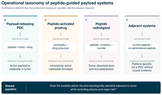
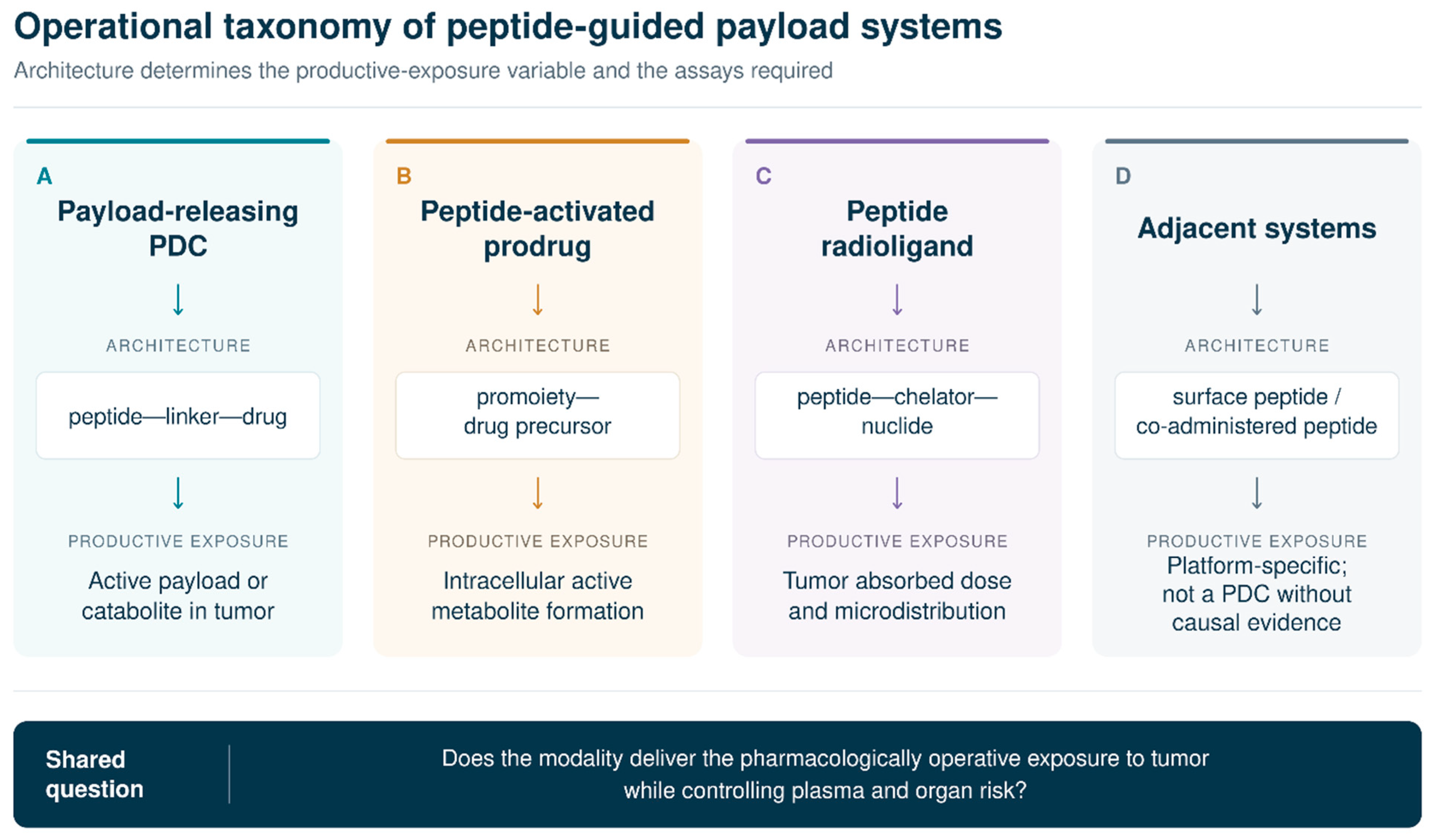
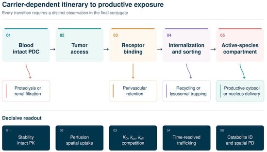
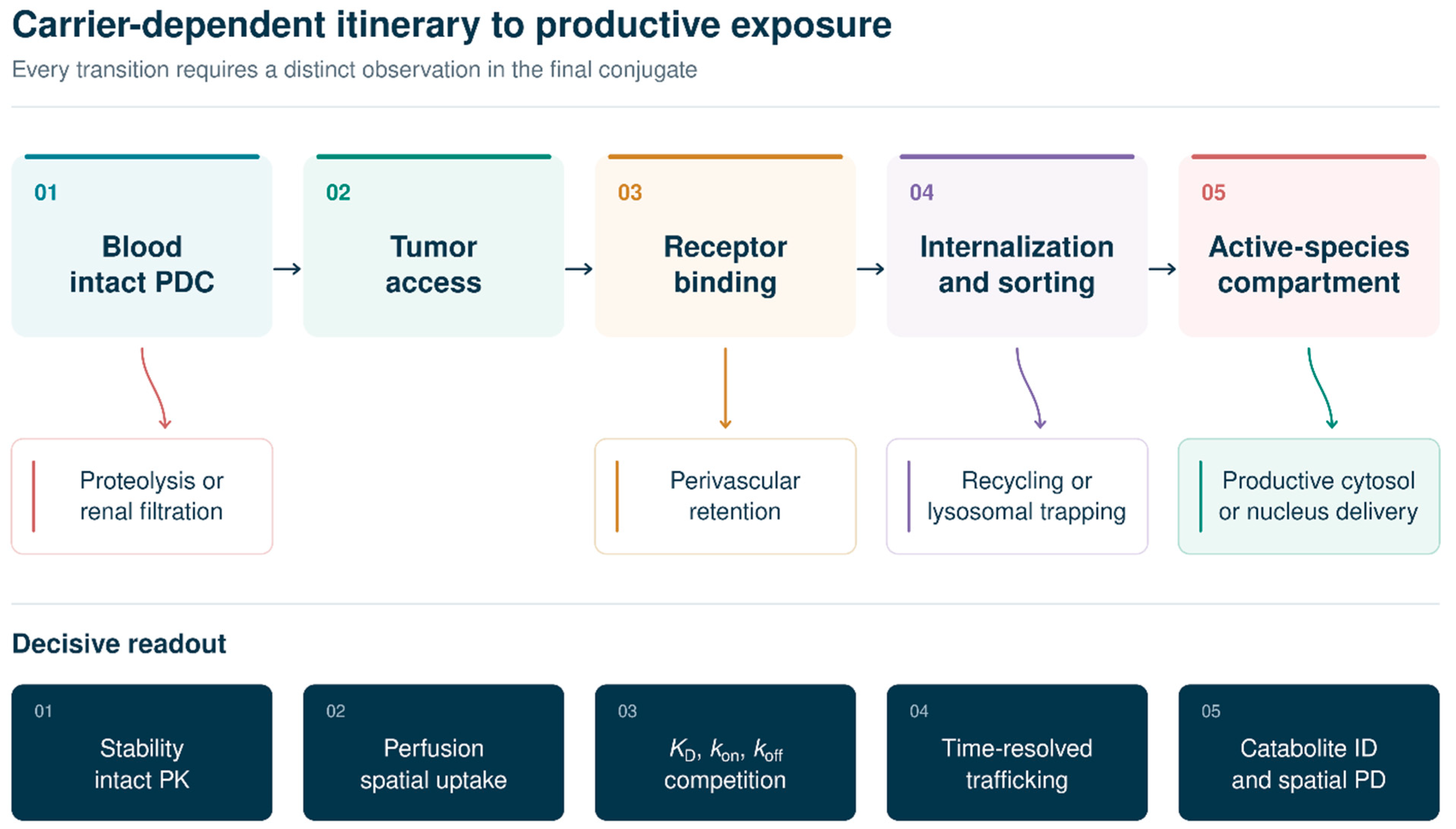
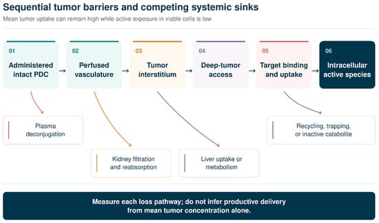
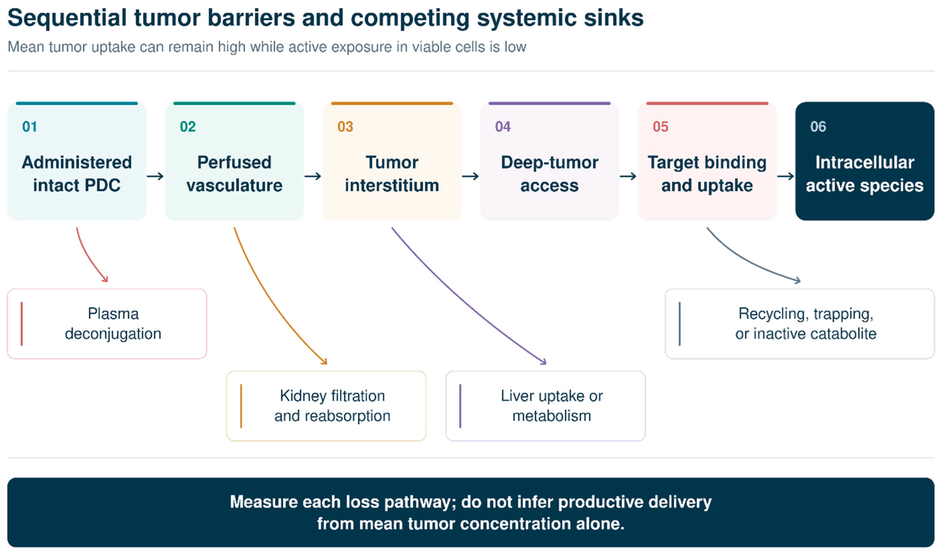
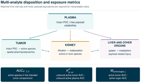
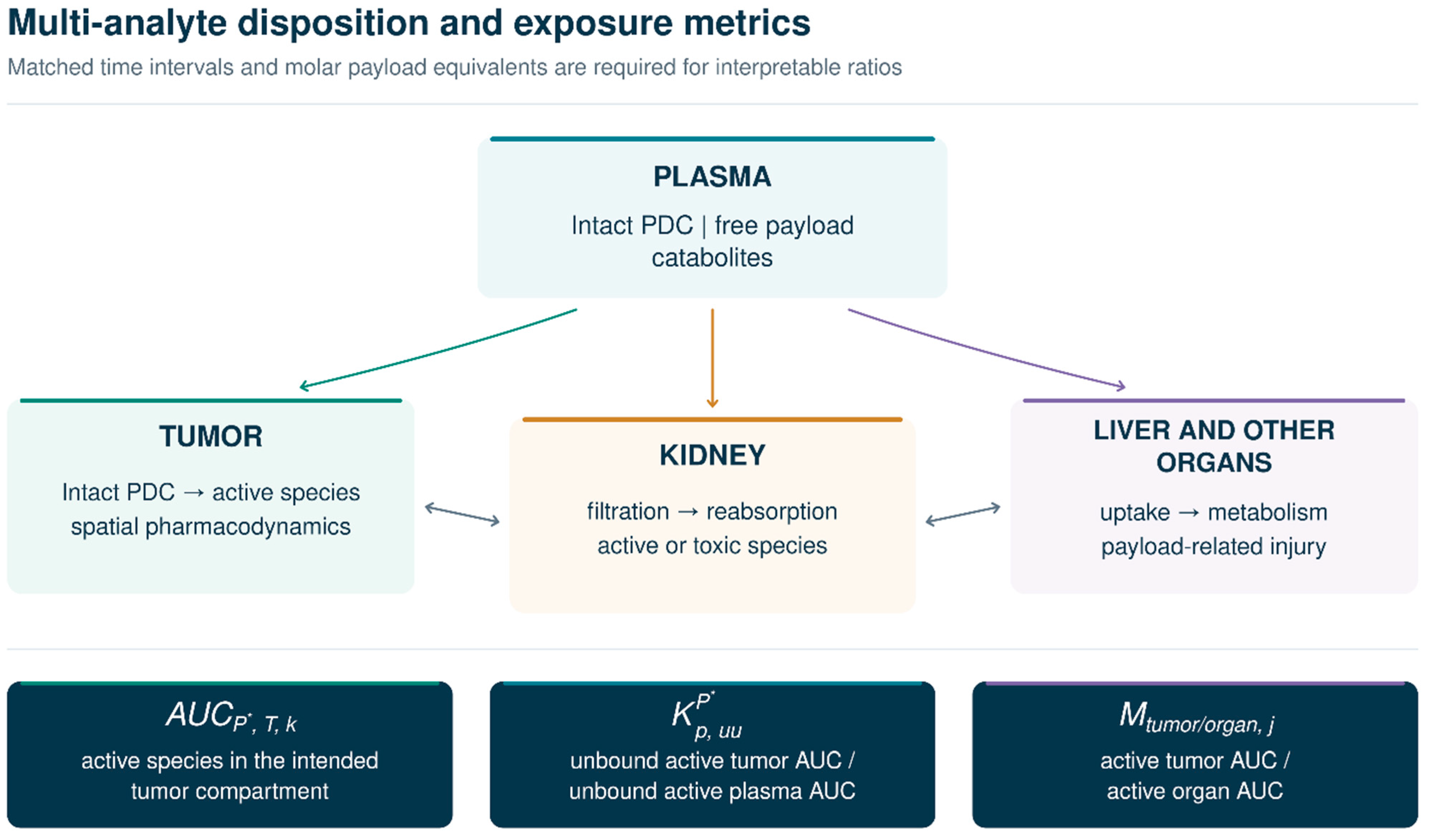
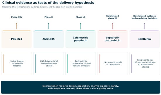
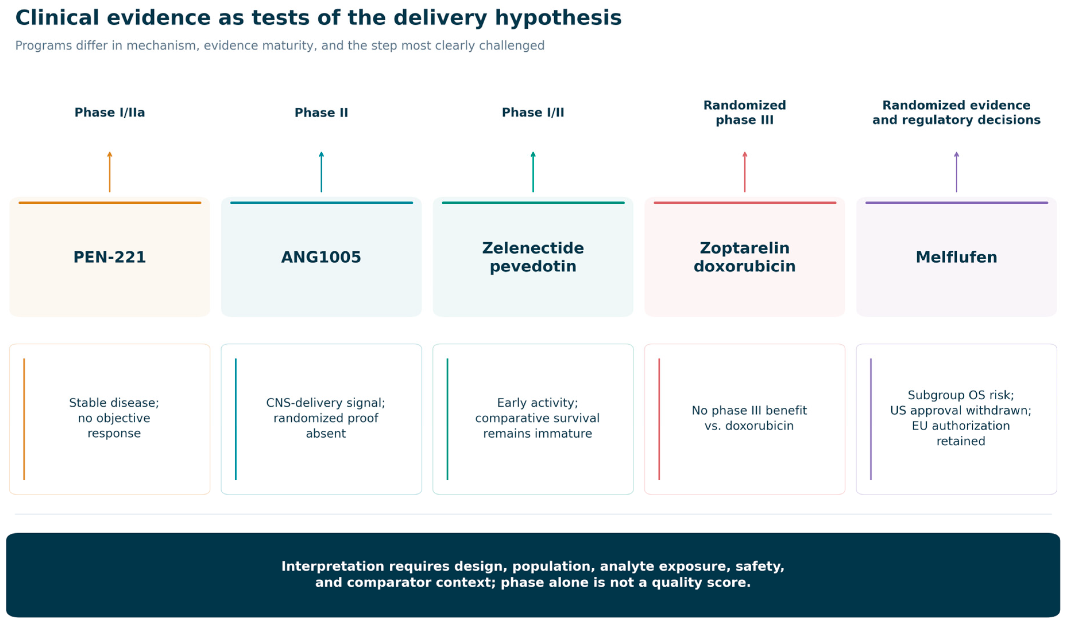

Disclaimer/Publisher’s Note: The statements, opinions and data contained in all publications are solely those of the individual author(s) and contributor(s) and not of MDPI and/or the editor(s). MDPI and/or the editor(s) disclaim responsibility for any injury to people or property resulting from any ideas, methods, instructions or products referred to in the content. |
© 2026 by the authors. Licensee MDPI, Basel, Switzerland. This article is an open access article distributed under the terms and conditions of the Creative Commons Attribution (CC BY) license.