


Abstract
Background: Chlorotonil A, a keto-macrolide produced by the myxobacterium Sorangium cellulosum, has potent antibiotic and antimalarial activities. Notably, chlorotonil A is active against both asexual and sexual blood-stage malaria parasites, making it a promising candidate for the development of an antimalarial treatment that also reduces parasite transmission to mosquitoes. Objectives: Here, we characterize the gametocytocidal and transmission-reducing activity of chlorotonil A. Methods and results: Using direct membrane feeding assays with Plasmodium falciparum isolates from a malaria-endemic area in Gabon, we show that chlorotonil A reduces oocyst infection prevalence and intensity in Anopheles gambiae (Kisumu) mosquitoes. Furthermore, we demonstrate that chlorotonil A is a particularly fast-acting gametocytocidal compound in vitro. Fluorescence and transmission electron microscopy revealed that chlorotonil A impairs membrane integrity and induces intracellular vacuolization after 6 h of drug exposure, with accumulation of reactive oxygen species (ROS) and intracellular acidification after 24 h. Conclusions: Our findings show that chlorotonil A has the potential for development as a dual-active antimalarial.
1. Introduction
Despite substantial control and elimination efforts, malaria remains a major cause of morbidity and mortality, with 282 million cases resulting in 610,000 deaths in 2024 alone [1]. The greatest burden occurs in Sub-Saharan Africa and is largely attributable to Plasmodium falciparum, the most virulent species causing malaria in humans [2]. P. falciparum sporozoites are injected into the bloodstream of the human host by a mosquito, then migrate to the liver and invade the hepatocytes, where they undergo asexual replication [3,4]. Afterwards, the parasitophorous vacuole ruptures, infected liver cells detach and merozoite-filled vesicles (merosomes) form via budding, releasing the parasites into the bloodstream via the liver sinusoids [5]. Merozoites then invade erythrocytes to initiate successive cycles of asexual replication, causing the symptoms of malaria [6]. During this process, a small proportion of parasites commit to sexual development [6]. Male and female gametocytes undergo five distinct developmental stages, of which stages I-IV are sequestered mainly in the bone marrow and the spleen [7]. After 10–12 days of maturation, the transmissive stage V gametocytes are released into the bloodstream where they can be ingested by another mosquito. In the mosquito midgut, male and female gametocytes differentiate into micro- and macrogametes, which fuse and form the zygote. Next, an ookinete is formed, which penetrates the mosquito midgut epithelium and positions itself near the basal lamina [6]. Finally, oocysts are formed that in turn produce sporozoites that migrate to the mosquito’s salivary glands, thus completing the parasite life cycle [6].
The current treatment of uncomplicated malaria in endemic regions relies on six artemisinin-based combination therapies (ACTs) [8], which rapidly kill asexual blood stages, but only have a limited effect against immature (stage I–III) gametocytes and no direct activity against mature (stage IV/V) gametocytes [9]. The only available interventions against mature gametocytes are primaquine and tafenoquine [10]. Currently, the World Health Organization (WHO) recommends addition of a single dose of 0.25 mg/kg body weight of primaquine after a standard ACT regimen, to reduce transmission while minimizing the risk of acute anemia in glucose-6-phosphate dehydrogenase-deficient patients [8,11]. However, this recommendation is not yet routinely implemented [12]. After initially appearing in Southeast Asia [13,14,15], partial artemisinin resistance has emerged independently in Eastern Africa in recent years [16,17,18] while, simultaneously, markers of reduced lumefantrine susceptibility surfaced [19]. Currently, most ACTs remain above the WHO-defined threshold of 90% PCR-corrected efficacy [16,20], meaning that current implemented drug regimens remain adequate for the treatment of malaria. Nevertheless, intervention strategies that have a low propensity to elicit resistant parasite lines as well as interrupt parasite transmission will be crucial to ensure successful antimalarial treatment in the future [21]. The discovery of gametocytocidal compounds is difficult, as gametocytes pose a challenging target due to their low metabolic activity, lack of multiplication and differences in uptake of antimalarials compared to asexual blood stages [22,23].
An especially promising novel compound in this regard is chlorotonil A, a keto-macrolide antibiotic produced by the soil-dwelling myxobacterium Sorangium cellulosum [24].
Chlorotonil A has served as a starting point for the development of semisynthetic derivatives, including dehalogenil [25,26], a lead candidate currently undergoing preclinical testings to support its advancement towards clinical development. In addition to its strong antibacterial activity [26], chlorotonil A acts potently against asexual blood stages of P. falciparum in vitro and ex vivo using clinical isolates, as well as in vivo against Plasmodium berghei in mice [27]. Chlorotonil A’s speed of activity as measured by the parasite reduction ratio was second only to artesunate, the fastest-acting antimalarial currently in use [28]. Of special interest is the high activity of chlorotonil A against mature gametocytes in vitro [27]. It has so far not been assessed whether the gametocytocidal activity of chlorotonils, represented by chlorotonil A, also translates into a transmission-blocking effect, and how chlorotonils exert their gametocytocidal activity. Closing this knowledge gap would allow for the positioning of chlorotonils as a transmission-blocking drug in the Target Candidate profile (TCP5) established by the Medicines for Malaria Venture [29]. The main goal of this study was therefore to further characterize the gametocytocidal and putative transmission-blocking activity of chlorotonil A. We performed direct membrane feeding assays (DMFAs) using blood meals containing P. falciparum gametocytes from infected individuals sampled in Gabon, a malaria-endemic country, to evaluate whether the gametocytocidal as well as sporontocidal activity and potential insecticidal effects of chlorotonil A translate into a reduction in parasite transmission. We determined the onset of gametocytocidal activity of chlorotonil A and assessed structural and metabolic changes in gametocytes in response to treatment with chlorotonil A in vitro using fluorescence and transmission electron microscopy.
2. Materials and Methods
2.1. Chemicals and Reagents
O+ erythrocytes and human AB serum were obtained from the blood bank of the University Hospital Tübingen (Tübingen, Germany). Chlorotonil A (M = 479.4 g/mol) was prepared as described previously [24,26]. The gametocytocidal compounds methylene blue (M = 319.85 g/mol, Sigma-Aldrich, St. Louis, MO, USA, M9140) and epoxomicin (M = 554.72 g/mol, MedChemExpress, Monmouth Junction, NJ, USA, HY-13821) were used as positive controls. Stock solutions of chlorotonil A and epoxomicin were prepared in DMSO; methylene blue was dissolved in double-distilled water and sterile-filtered.
2.2. Clinical Isolates for Direct Membrane Feeding Assays
P. falciparum isolates were collected from participants aged 5 years or older during November 2024–May 2025 at the Centre de Recherches Médicales de Lambaréné (CERMEL). Inclusion criteria were age of participant ≥ 5 years, symptomatic or asymptomatic monoinfection with P. falciparum (determined via Giemsa-stained thick blood smear), informed consent (in the case of participants younger than 18 years from parents or guardians) and assent from adolescents between 11 and 18 years of age. Exclusion criteria were age < 5 years, hemoglobin < 6 g/dL, the use of antimalarial medication in the last 30 days, co-infection with non-falciparum species of Plasmodium and refusal of informed consent/assent. The study protocol and all related documents were approved by the Comité d’Éthique du Centre de Recherches Médicales de Lambaréné, Gabon (CEI014/2018). Four individuals fulfilling the inclusion criteria were enrolled and another Giemsa-stained thick blood smear was prepared to quantify the parasitaemia and gametocytaemia before blood samples were collected. Afterwards, participants were treated with artemether-lumefantrine according to local guidelines.
2.3. Mosquito Maintenance
A colony of Anopheles gambiae s.s. (Kisumu strain) was maintained as described previously [30]. Briefly, larvae were kept at 29 °C and fed fish flakes (Vitakraft, Germany). Adult mosquitoes were maintained at 26 °C and 80% relative humidity under a 12 h:12 h light:dark cycle and provided with a 10% sugar solution. Once per week, mosquitoes were fed with uninfected blood of healthy volunteers not undergoing chemoprophylaxis for malaria.
2.4. Preparations for Direct Membrane Feeding Assays with Clinical Isolates
Blood samples containing gametocytes were prepared as described previously [30,31]. In total, 10 mL of heparinized blood collected from participants was centrifuged immediately after drawing at 2000 rpm at 37 °C for 5 min to remove plasma. Erythrocytes were washed once with RPMI-1640 warmed to 37 °C.
2.5. Direct Membrane Feeding Assays with or Without Pre-Feeding Incubation
For the direct membrane feeding assays (DMFAs), packed erythrocytes containing gametocytes were added to malaria-naïve human AB serum with final concentrations of 5 µM methylene blue, 50 nM chlorotonil A, 500 nM chlorotonil A, 1 µM of chlorotonil A, 0.1% DMSO alone or only serum at a haematocrit of 40%. Drug concentrations were aligned with previous publications for comparability [31,32]. The final concentration of DMSO was adjusted to 0.1% in all chlorotonil A and DMSO control samples. In total, 300 µL of the prepared blood meals containing the drugs were each added to a glass feeder at 37 °C and 50 starved mosquitoes (3–6 days old) were allowed to feed through Parafilm.
For the DMFA with pre-feeding incubation, one additional step was performed: packed erythrocytes containing gametocytes were added to RPMI-1640 medium supplemented with 25 mM sodium bicarbonate and 10% human AB serum, spiked with the respective compounds, to a final haematocrit of 40% and incubated for 24 h at 37 °C, 5% O2 and 5% CO2. After the incubation, samples were centrifuged, supernatant was removed, and infected erythrocytes were resuspended in malaria-naïve human AB serum to achieve a haematocrit of 40%. In total, 300 µL of the blood meals were then each added to a glass feeder and 50 starved mosquitoes (3–6 days old) per assay condition were allowed to feed through Parafilm.
After feeding, the mosquitoes were maintained under standard insectary conditions. After 6–9 days, 30 mosquitoes per condition were dissected, the midguts isolated, stained with 1.2% mercurochrome and examined using a Leica DM750 light microscope (Leica Microsystems, Wetzlar, Germany) with 20× magnification. The number of oocysts was determined for each mosquito.
2.6. Determining the Mortality of Mosquitoes After DMFA Without Pre-Feeding Incubation
After feeding as described in Section 2.5, fed mosquitoes were kept under standard insectary conditions and dead mosquitoes were counted each day to detect possible insecticidal effects of the tested compounds. The mosquito mortality rate was calculated by dividing the number of dead mosquitoes until dissection by the number of mosquitoes who fed on the blood meal.
2.7. P. falciparum Culture
P. falciparum strain NF54 (a vigorous gametocyte producer) was obtained from Sanaria, Inc. as cryopreserved sporozoites, and asexual blood-stage parasites were isolated from an infected participant following a controlled human malaria infection through venous inoculation [33]. Parasites were kept in complete medium (RPMI-1640 medium supplemented with 2 mM L-glutamine, 25 mM HEPES, 50 μg/mL gentamicin, and 0.45% (w/v) AlbuMax II) at 37 °C in 5% CO2 and 5% O2. O+ erythrocytes for parasite culture were obtained from the blood bank of the University Hospital Tübingen (Tübingen, Germany). If not stated otherwise, parasites were cultured at a haematocrit of 2.5% with medium replacement and dilution using fresh erythrocytes three times per week to keep parasitaemia < 3% [34].
P. falciparum mature gametocytes for subsequent assays were generated as described previously [35]. In brief, gametocyte culture was initiated with synchronized ring-stage NF54 parasites at 6% haematocrit and 0.3% parasitaemia (25 mL total culture volume). Complete medium (as described above) was supplemented with 5% (v/v) complement-depleted human AB serum and replaced daily without parasite dilution for 15 days. Once parasitaemia reached 3%, an additional 25 mL of complete medium was added, reducing the haematocrit from 6% to 3% (total culture volume 50 mL). From days 11 to 14, N-acetyl-D-glucosamine hydrochloride (Sigma-Aldrich, USA, A3286) was added to a final concentration of 50 mM to remove remaining asexual parasites. On day 15, mature gametocytes were enriched by density gradient centrifugation using a 27.6% NycoDenz solution (MW = 821 g/mol, Serumwerk Bernburg, Bernburg, Germany, 18003) and magnetic-activated cell sorting (MACS) with LD-columns (Miltenyi Biotec, Bergisch Gladbach, Germany), and subsequently quantified using a Neubauer counting chamber [36].
2.8. Assessment of the Onset of Chlorotonil A Activity Against Mature Gametocytes
To assess the kinetics of the gametocytocidal activity of chlorotonil A, drug sensitivity assays with mature gametocytes were performed following different incubation periods (2, 6, 12, 24 and 48 h) as described previously [35,36]. Briefly, stock solutions of the compounds in either DMSO (chlorotonil A and epoxomicin) or double-distilled water (methylene blue) were further diluted in complete medium to reach the desired concentrations. Final solvent concentrations in these assays did not interfere with parasite viability and were maintained below 0.11% (v/v) (dilution of at least 1:900). Chlorotonil A, epoxomicin, and methylene blue (positive controls) were dispensed into 96-well flat-bottom plates as 3-fold serial dilutions comprising seven concentration steps with a medium-only control. Purified mature gametocytes were seeded into the plates (50,000 gametocytes/well) and incubated at 37 °C in 5% CO2 and 5% O2 for the respective times. Gametocyte ATP levels, used as a proxy for viability, were then measured using the luciferase-based BacTiter-GLO kit (Promega, Fitchburg, WI, USA, G8232) according to the manufacturer’s instructions. Luminescence was quantified using either a LUmo microplate luminometer (Autobio, Zhengzhou, China) or a CLARIOStar microplate reader (BMG Labtech, Ortenberg, Germany).
2.9. Evaluation of Structural and Metabolic Effects of Chlorotonil A on Mature Gametocytes Using Fluorescent Dyes
To investigate the mechanism underlying the gametocytocidal activity of chlorotonil A, we examined whether chlorotonil A influenced membrane and nuclear integrity as well as reactive oxygen species (ROS) and changes in intracellular pH. To do so, purified gametocytes were exposed to 60 nM chlorotonil A (~5× IC50 at 48 h) for 5 h and 24 h and afterwards incubated with different fluorescent markers (Table 1). CM-H2DCFDA was an exception and was loaded prior to drug treatment. The gametocytes were then mounted on a glass slide, covered with a coverslip and visualized using a Leica DM LM fluorescence microscope (Leica Microsystems, Wetzlar, Germany), and images were taken by a JENOPTIK ProgRes C10 camera (Jenoptic, Jena, Germany).
Table 1.
Fluorescent dyes used to investigate cellular effects of chlorotonil A on mature gametocytes.
2.10. Transmission Electron Microscopy of Chlorotonil A-Treated Mature Gametocytes
Mature gametocytes were treated with 170 nM or 850 nM chlorotonil A for 6 h (~5× and 25× IC50 at this timepoint, respectively) or 10 nM chlorotonil A for 48 h (~1× IC50 at this timepoint). Samples were then washed once with PBS (pH 7.4, 37 °C) and centrifuged at 1800 rpm at ambient temperature. The supernatant was discarded and the cells were fixed in Karnovsky‘s solution containing 3% formaldehyde and 2.5% glutaraldehyde in 0.1 M phosphate buffer for 2 h at ambient temperature and stored at 4 °C. Samples were centrifuged at 2000 g for 5 min, supernatant was discarded, and pellets were resuspended in approximately 20 µL agarose (3.9%) at 37 °C, cooled to room temperature, and cut into small pieces. Samples were post-fixed in 1% osmium tetroxide containing 2.5% potassium ferrocyanide (Morphisto, Offenbach am Main, Germany) for 2 h. Then, the samples were washed with double-distilled water and stained with a 2% aqueous uranyl acetate solution for 45 min at ambient temperature in the dark. Samples were then washed again with double-distilled water, dehydrated with graded ethanol (50%, 75%, 95%, 100%), embedded in glycidyl ether and sectioned using an ultramicrotome (Ultracut E, Reichert, Depew, NY, USA). Ultra-thin sections (30 nm) were mounted on copper grids and analyzed using a Zeiss LIBRA 120 transmission electron microscope (Carl Zeiss, Jena, Germany) operating at 120 kV. Images represent one biological replicate, for which at least two gametocytes were observed (except for 850 nM for 6 h, where only one intact gametocyte was found).
2.11. Data Analysis
For the DMFAs, data were analyzed with Microsoft Excel version 2412. To determine the prevalence, the proportion of infected mosquitoes was calculated for each clinical isolate, and the median and interquartile range (IQR) of these proportions were calculated afterwards. For the infection intensity, the median and IQR of oocysts/mosquito was calculated; once over all mosquitoes together and once separately for each clinical isolate. Four clinical isolates with 30 mosquitoes each were used for the DMFA without pre-feeding incubation and three clinical isolates with 30 mosquitoes each were used for the DMFA with 24 h pre-feeding incubation. Given the small number of clinical isolates (three or four), no formal statistical testing was performed.
To assess the onset of activity on mature gametocytes, drug sensitivity assays were performed with at least two biological replicates per condition. From the luminescence data, dose–response curves and IC50 values were generated using R version 4.3.2 and drc-package version 3.0-1.
Transmission electron micrographs and fluorescence micrographs were contrast-enhanced, and transmission electron micrographs were pseudocolored using the GNU Image Manipulation Program (GIMP) version 3.0.4. Besides that, no changes were made to the micrographs. All charts were prepared using GraphPad Prism version 10.4.3 or 11.02.
3. Results
3.1. Chlorotonil A Does Not Influence Mosquito Viability
The transmission-blocking potential of chlorotonil A was evaluated using an ex vivo direct membrane feeding assay (DMFA). Fifty A. gambiae mosquitoes were allowed to feed on blood meals containing either serum-only, 50 nM, 500 nM or 1 µM chlorotonil A, 0.1% DMSO (negative control) or 5 µM methylene blue (positive control). Neither treatment impacted mosquito viability (Figure S1) when inspected on day of dissection (6–9 days after feeding).
3.2. Chlorotonil A Has a Potent Transmission-Blocking Effect
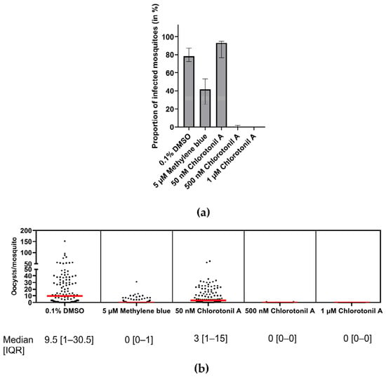
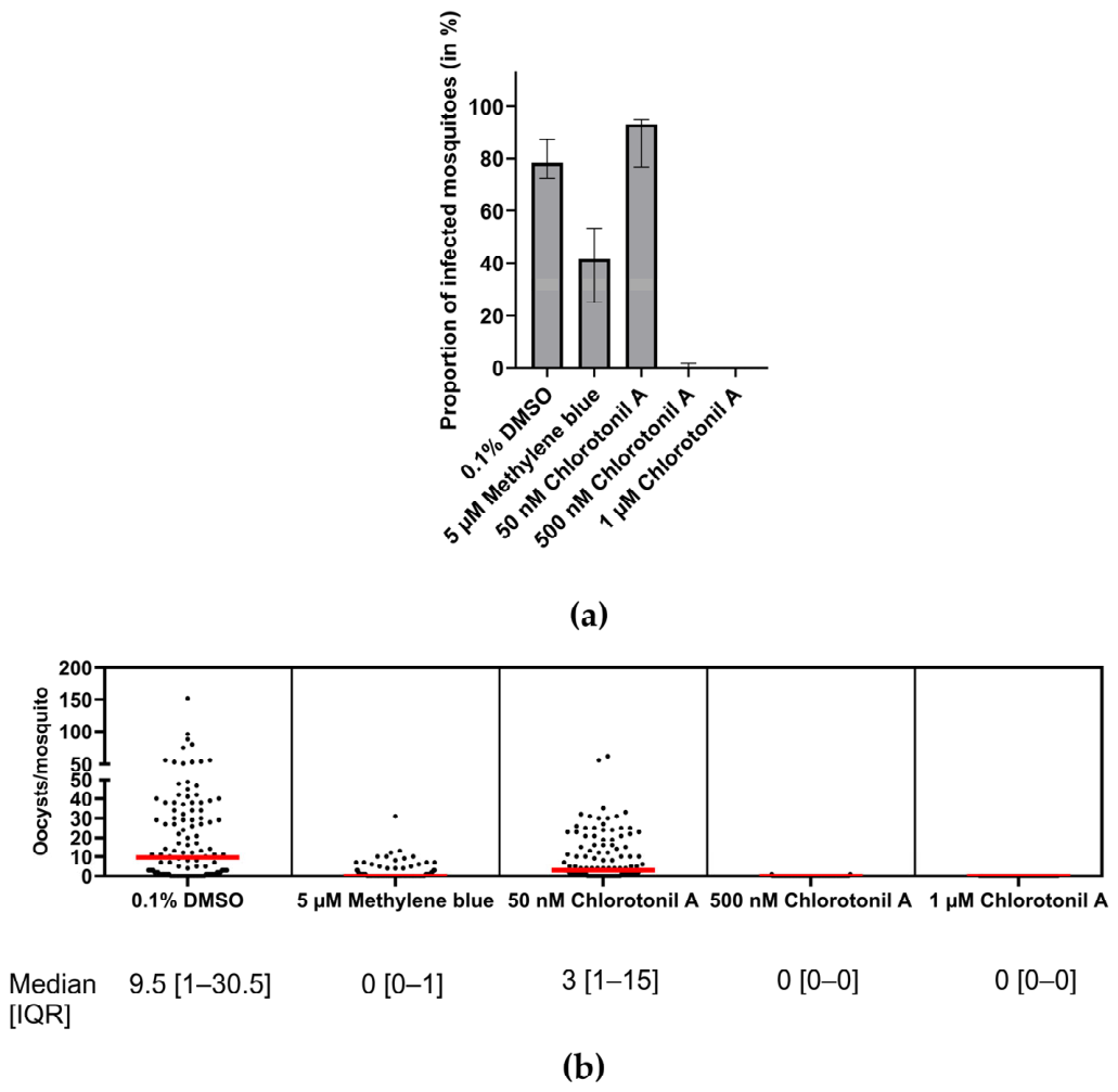
Chlorotonil A showed a dose-dependent activity (Figure 1, Table S1 and Figure S2). At 1 µM, chlorotonil A completely prevented transmission as none of the dissected mosquitoes was infected, while 500 nM chlorotonil A reduced the median prevalence to 0% [IQR: 0–1.7%] (Figure 1a). Treatment with 50 nM chlorotonil A yielded a median prevalence of infected mosquitoes of 93.3% [IQR: 76.7–95%], comparable to the DMSO-only control (median prevalence of 78.3% [IQR: 72.5–87.5%]). At 5 µM, methylene blue reduced median prevalence to 41.7% [IQR: 25–53.3%]. A similar pattern was observed for infection intensity (Figure 1b, four clinical isolates with 120 mosquitoes pooled together). At 1 µM, chlorotonil A completely prevented oocyst formation, while chlorotonil A at 500 nM resulted in 0 [IQR: 0–0] median oocysts per mosquito with a total of two infected mosquitoes. Chlorotonil A at 50 nM led to a median number of three [IQR: 1–15] oocysts per mosquito, lower than the DMSO control (9.5 [IQR: 1–30.5] median oocysts per mosquito). Treatment with 5 µM methylene blue reduced median infection intensity to 0 [IQR: 0–1] median oocysts per mosquito. The infection intensities of the single experiments of the four clinical isolates can be seen in Figure S2.
Figure 1.
DMFA with four clinical isolates of P. falciparum gametocytes. Blood meals were spiked with either 0.1% DMSO (negative control), 5 µM methylene blue (positive control) or 50 nM, 500 nM or 1 µM of chlorotonil A. For each condition 50 A. gambiae mosquitoes were allowed to feed for a maximum of. 5 min. After 6–9 days, 30 mosquitoes per condition were dissected, and oocysts were counted. (a) Median proportion of infected mosquitoes with IQR. (b) Oocysts per mosquito midgut (four clinical isolates, 120 mosquitoes pooled together). Each dot represents one mosquito, each square a separate treatment. IQR = Interquartile range.
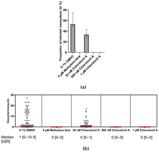
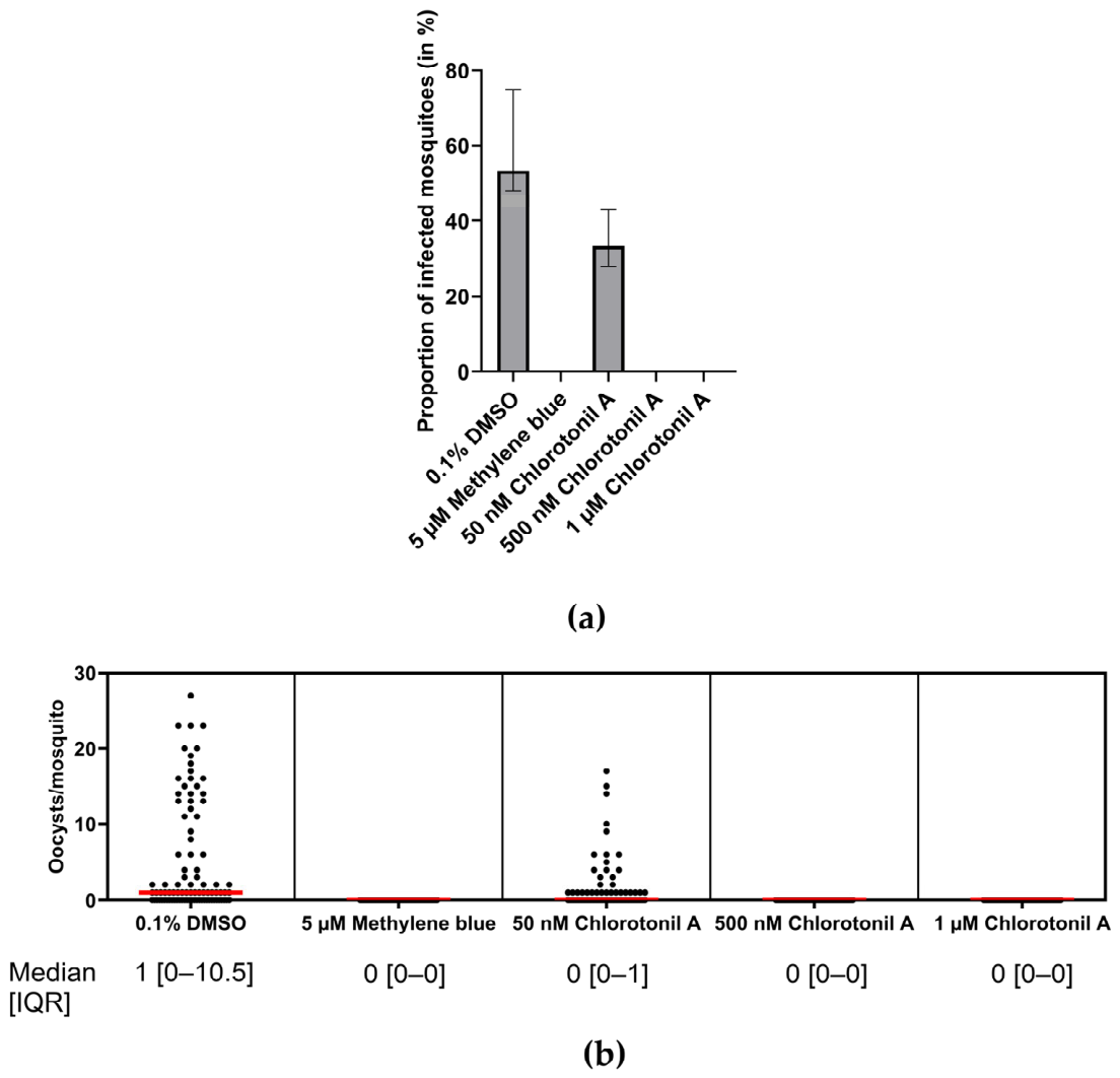
In addition, DMFAs were performed with an additional 24 h incubation time of the participants’ blood with the drugs to more closely reflect the natural treatment setting. Clinical isolate 3 failed to establish an infection after this additional incubation time even in the absence of drug pressure and was excluded from the analysis. Treatment with chlorotonil A at 1 µM and 500 nM completely prevented transmission (Figure 2a) as none of the dissected mosquitoes were infected. Treatment with 50 nM chlorotonil A reduced median infection prevalence to 33.3% [IQR: 28.3–43.3%] compared to 53.3% [IQR: 48.3–75%] for 0.1% DMSO-only. Regarding infection intensity, chlorotonil A at 1 µM and 500 nM completely prevented oocyst development, while 50 nM chlorotonil A reduced median oocyst count to 0 [IQR: 0–1] compared to a median oocyst count of 1 [IQR: 0–10.5] for 0.1% DMSO-only (Figure 2b, three clinical isolates with 90 mosquitoes pooled together). The positive control 5 µM methylene blue resulted in complete prevention of transmission.
Figure 2.
DMFA with 24 h pre-feeding incubation performed with three clinical isolates of P. falciparum gametocytes. Blood meals were spiked with either 0.1% DMSO (negative control), 5 µM methylene blue (positive control) and 50 nM, 500 nM or 1 µM of chlorotonil A and incubated for 24 h at 37 °C, 5% O2 and 5% CO2 before 50 A. gambiae mosquitoes per condition were allowed to feed on them. After 6–9 days, 30 mosquitoes per condition were dissected, and oocysts were counted. (a) Median proportion of infected mosquitoes with IQR. (b) Oocysts per mosquito midgut (three clinical isolates, 90 mosquitoes pooled together). Each dot represents one mosquito, each square a separate treatment. IQR = Interquartile range.
For both experimental approaches, there was variability between the different clinical isolates in terms of infection intensity (see Table S1, Figures S2 and S3, which show each individual feed separately). At 0.1%, DMSO did not impact infection intensity or prevalence compared to an untreated control (Figure S4).
3.3. Chlorotonil A Shows Rapid Onset of Activity Against Mature P. falciparum Gametocytes
To determine the onset of chlorotonil A activity, we treated mature gametocytes with chlorotonil A, methylene blue, and epoxomicin (control drugs) and determined the IC50 after different incubation times with the drugs (2 h, 6 h, 12 h, and 24 h, compared to the commonly used incubation time of 48 h [36]). Both methylene blue and epoxomicin showed a slow onset of activity in comparison to chlorotonil A. Epoxomicin showed pronounced activity only at 48 h, while methylene blue showed low activity after 24 h and the expected three-digit nanomolar IC50 after 48 h (see Table 2 and Figure S5 for dose–response curves). Chlorotonil A had a notably fast speed of action, with a double-digit, nanomolar IC50 at 6 h, thus already exhibiting its pronounced activity after this short incubation time.
Table 2.
Speed of action of chlorotonil A and the positive controls epoxomicin or methylene blue against mature P. falciparum gametocytes (at least two biological replicates). Chlorotonil A showed a rapid onset of activity after 6 h, while epoxomicin and methylene blue achieved high activity only after 48 h.
3.4. Chlorotonil A Impairs Membrane Integrity and Induces Intracellular Acidification and ROS Production
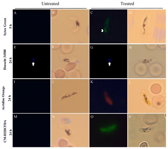
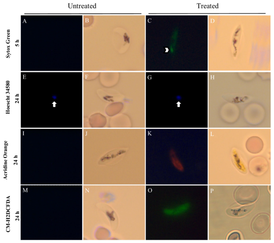
In addition, we aimed to qualitatively assess how treatment with chlorotonil A impacts different structural and metabolic parameters of gametocytes. To do so, mature gametocytes were treated with 60 nM chlorotonil A for 5 h and 24 h (~5× IC50 at 48 h), stained with different fluorescent dyes, and observed under a fluorescence microscope (one biological replicate). Plasma membrane integrity was assessed by staining with the membrane-impermeable nucleic acid dye Sytox Green. Untreated gametocytes showed no intracellular fluorescence, indicating an intact plasma membrane (Figure 3A,B). In contrast, nuclear staining with Sytox Green was already observed after a short treatment period of 5 h with chlorotonil A, suggesting damage to the plasma membrane (Figure 3C,D). Among the effects observed by fluorescence microscopy, only damage to the membrane could be observed at the shortest time assessed. The DNA-binding dye Hoechst 34580 was used to assess nuclear integrity. As expected, untreated parasites showed a non-fragmented nucleus, indicated by the single dot (Figure 3E,F). After 24 h of treatment with chlorotonil A, the gametocytes still presented non-fragmented nuclei comparable to the untreated ones. The dye acridine orange, which accumulates in membrane-bound compartments with a low pH, was used to track intracellular acidification. While for the untreated parasites (Figure 3I,J) no detectable signal was observed, an intense intracellular acidification could be observed at 24 h of treatment with chlorotonil A (Figure 3K,L). Finally, the dye CM-H2DCFDA was used to detect the intracellular production of reactive oxygen species (ROS). Similar to acridine orange, a marked fluorescence and thus production of ROS could only be observed after the longer incubation time of 24 h (Figure 3O,P), indicating a time-dependent effect.
Figure 3.
Light and fluorescence micrographs of untreated P. falciparum mature gametocytes or gametocytes treated with 60 nM chlorotonil A (~5× IC50 at 48 h) for 5 h or 24 h (1 biological replicate). (A–D): Staining with Sytox Green to visualize membrane integrity. Arrow indicates fluorescence focus. (E–H): Staining with Hoechst 34580 to examine nuclear integrity. Arrow indicates nucleus. (I–L): Staining with acridine orange to detect intracellular acidification. (M–P): Staining with CM-H2DCFDA to assess intracellular ROS production. While a short treatment of 5 h with chlorotonil A was already enough to induce membrane damage (C), intracellular acidification (K) and accumulation of ROS (O) were observed after 24 h only.
3.5. Chlorotonil A Induces Ultrastructural Damage to Mature Gametocytes
Subsequently, we complemented these phenotypic data by qualitatively examining whether chlorotonil A damages specific subcellular compartments of gametocytes. For this purpose, mature gametocytes were treated with chlorotonil A at 170 nM for 6 h (~5× IC50 at this timepoint), the timepoint when a clear gametocytocidal effect became visible, and at 10 nM for 48 h (endpoint measurement, ~1× IC50 at this timepoint). Afterwards, they were visualized using transmission electron microscopy to qualitatively analyze their ultrastructure (one biological replicate).
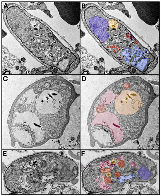
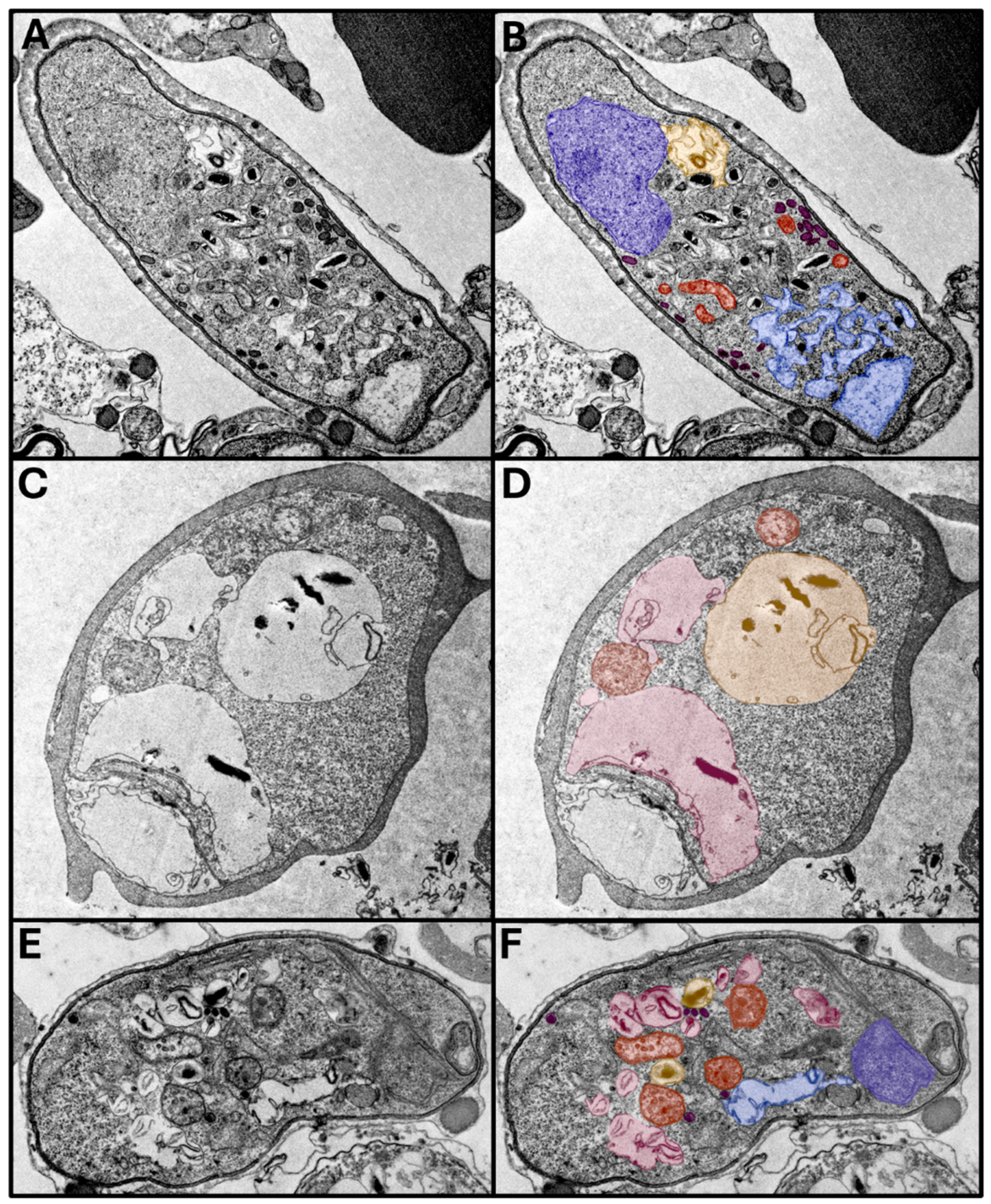
After 48 h of incubation with medium only, gametocytes showed their typical morphology (Figure 4A,B). The gametocyte, contained within the parasitophorous vacuole membrane, occupies almost the entire volume of the infected erythrocyte. An intact nucleus (marked in dark blue) and a food vacuole containing electron-dense haemozoin crystals (marked in yellow) were visible. In addition, the large and convoluted mitochondrion with cristae can be seen as round structures in the cross-section (marked in orange). The endoplasmic reticulum (in light blue) presented as several separated and bulging membrane vacuoles, while osmiophilic bodies (purple) appeared as electron-dense vesicles clustering next to the plasma membrane. After 6 h of treatment with chlorotonil A (Figure 4C,D), gametocyte ultrastructure was severely damaged, with most of the cytosol occupied by enlarged, malformed food vacuoles and large vacuoles (marked in pink). While the mitochondrion appears to preserve its morphology, the finer cristae structure is impacted by chlorotonil A treatment. After 48 h of treatment (Figure 4E,F), similar intracellular vacuolization was observed. Atypically, multiple small food vacuoles containing haemozoin crystals could be seen, while nuclear integrity was not affected. Furthermore, the endoplasmic reticulum has lost its fine structure, while some osmiophilic bodies were scattered throughout the cytosol. Further micrographs with a similar phenotype are shown in Figure S6.
Figure 4.
Transmission electron micrographs of mature P. falciparum gametocytes (one biological replicate). (A,B): Untreated gametocyte. (C,D): Gametocyte treated with 170 nM chlorotonil A for 6 h. (E,F): Gametocyte treated with 10 nM chlorotonil A for 48 h. (A,C,E) show the original, gray-scaled images. In (B,D,F), important subcellular structures were false-colored (blue: nucleus, yellow: food vacuole, orange: mitochondrion, pink: intracellular vacuolization, light-blue: endoplasmic reticulum, and purple: osmiophilic bodies).
4. Discussion
To advance towards malaria elimination, the next generation of antimalarial treatment combinations should include dual-active compounds targeting both asexual blood-stage parasites and sexual stage gametocytes, allowing for the simultaneous treatment of malaria and transmission reduction [21,37]. Chlorotonil A is a promising candidate in this regard, possessing favorable properties such as rapid and potent schizonticidal and gametocytocidal activity in vitro and ex vivo, with no indication of cross-resistance to known antimalarials [27]. In our study, we showed for the first time that chlorotonil A has a potent ex vivo transmission-blocking effect and further analyzed its gametocytocidal activity in vitro regarding cellular effects and onset of activity.
The gold-standard methodology to assess the transmission-blocking activity of antimalarial candidates is the membrane feeding assay [38], which allows for detection of gametocytocidal, gametocyte-sterilizing and sporontocidal effects in the blood meal and inside the mosquitoes, as well as insecticidal activity against the mosquitoes themselves [31]. Furthermore, we used clinical isolates from a malaria-endemic area, which differ from culture-adapted laboratory strains by multiplicity of infection, high genetic diversity and gametocyte density, all modulating parasite transmission to the mosquito [39,40,41]. Furthermore, resistance to conventional antimalarials in clinical isolates has been shown to modulate the efficacy of novel antimalarials against gametocytes [42]. Therefore, the use of clinical isolates allowed us to assess transmission-blocking activity under close-to-natural conditions.
We found that at a concentration of 1 µM, chlorotonil A completely prevented oocyst development. These levels of inhibition are comparable to published results for cipargamin (complete blocking of transmission at 500 nM) and ganaplacide (>90% reduction in oocysts/mosquito at 500 nM), two novel antimalarial candidates for treatment of uncomplicated malaria and transmission disruption [43,44]. Methylene blue, which is known for its potent and rapid transmission-blocking activity [30,45], had a weaker effect on oocyst formation at 5 µM than chlorotonil A at 1 µM. Compared to previous studies, we observed a stronger reduction in the proportion of infected mosquitoes by 46.7% compared to 21.8% for clinical isolates [31] and a weaker reduction in the proportion of infected mosquitoes than in a study using laboratory strains (78.7%) [30]. These differences might be attributed to the abovementioned higher variability of clinical gametocyte isolates in terms of gametocyte density, and probably also polyclonality and genetic diversity. As expected, infection intensity followed a similar pattern since the oocyst count is strongly correlated to the proportion of infected mosquitoes [31,38].
One limitation of this experimental approach is that it cannot differentiate between a gametocytocidal and downstream sporontocidal effect. For this, a different experimental setup needs to be selected, only exposing later stages to the respective drugs. Furthermore, the small sample size precludes the use of statistical significance tests, making these findings preliminary evidence for a potent transmission-blocking effect. We also observed variability between the isolates regarding both infection prevalence and intensity, possibly due to different gametocyte densities and multiplicities of infection, strain-specific genetic variations or differences in gametocyte viability before feeding. However, while lower variability is expected with a P. falciparum lab strain, clinical isolates give a better estimate of the activity in the natural/real-world setting. In addition, the concentrations where we see clear activity do not show such variability, showing the robustness of the compound’s activity at these concentrations.
These findings were complemented by DMFA with a 24 h pre-feeding incubation with the respective compounds, which is closer to the natural setting where long-circulating gametocytes [46] are likely to encounter drugs before being taken up by the mosquito. We determined that chlorotonil A at concentrations of 500 nM and 1 µM, as well as methylene blue at 5 µM, completely prevented transmission to the mosquito, while the low dose of 50 nM chlorotonil A reduced both the proportion of infected mosquitoes and median oocyst count per mosquito, further underpinning the potency of the transmission-blocking effect of chlorotonil A. The same limitations outlined above also applied for these experiments.
In summary, chlorotonil A not only has gametocytocidal activity in vitro, but also strong transmission-blocking activity ex vivo, supporting the suitability of chlorotonils as next-generation transmission-blocking antimalarials.
We further characterized the exceptional gametocytocidal activity of chlorotonil A regarding its onset of activity and possible induced metabolic and ultrastructural alterations in vitro. Gametocytocidal compounds are often classified as fast-acting when activity is observed at 24 h, and slow-acting when activity is only detectable after 48 h of incubation [47]. Rapid onset of activity is an especially valuable property for novel antimalarials to quickly alleviate symptoms and narrow the window during which individuals are infective to mosquitoes. Therefore, we assessed the onset of chlorotonil A activity against mature gametocytes in vitro. These experiments revealed chlorotonil A as a fast-acting gametocytocidal compound with an IC50 at roughly 35 nM after only 6 h of incubation. In contrast, both methylene blue and epoxomicin only showed potent activity after 48 h of incubation, positioning chlorotonil A as the faster compound with high activity.
Furthermore, parasites from a single biological replicate were treated with the IC50 concentrations determined for each treatment duration and subsequently examined by fluorescence microscopy and transmission electron microscopy (TEM). The images shown are representative of the predominant effects observed in a large number of parasites examined. The assessment of mature gametocytes’ metabolic and structural changes suggested membrane damage after 5 h of chlorotonil A treatment, which matches the sharp decrease in IC50 of chlorotonil A after 6 h of incubation. Therefore, the activity of chlorotonil A may be linked to membrane permeabilization, either by direct interaction or via a membrane-related protein target. After 24 h of treatment, generalized intracellular acidification and ROS production were clearly observed. Several other novel antimalarial compounds have been described to act against plasmodia by interfering with pH regulation in the parasitic cytosol, for instance MMV007839 and MMV000972, which cause acidification by inhibiting lactate transporters [48]. Regarding ROS production, antimalarials currently in clinical use, such as artemisinins or 4-aminoquinolines, exert their activity by inducing increased ROS production, either directly from the pharmacophore as for endoperoxides or indirectly, e.g., by interfering with heme detoxification [49]. However, as both effects appear only with a delay during incubation with chlorotonil A, they are more likely secondary effects, resulting from cellular damage interfering with physiological pH homeostasis and ROS detoxification. Using transmission electron microscopy, we observed generalized ultrastructural damage in chlorotonil A-treated gametocytes, mainly in the form of intracellular vacuolization. However, these findings do not incriminate a specific mode of action, as previous studies described similar findings for a broad range of compounds with antimalarial activity, relating it to an atypical form of non-regulated, drug-induced cell death [50].
Limitations of our study include the modest number of clinical isolates used for DMFAs from only one area, which does not capture the diversity of circulating strains on the African continent. Since fluorescence and transmission electron microscopy experiments were conducted with only one biological replicate and the number of observed gametocytes per cut was not systematically quantified, these results should be treated as a qualitative observation of the cellular effects of chlorotonil A exposure on mature gametocytes. Furthermore, we could not provide conclusive evidence pointing at a definitive antiplasmodial mode of action of chlorotonil A.
In recent years, chlorotonil A underwent derivatization, yielding the lead compound dehalogenil, which was shown to act against Gram-positive bacteria of high concern, including methicillin-resistant Staphylococcus aureus and vancomycin-resistant Enterococcus faecium, with improved activity against P. falciparum as well as improved pharmacological properties, especially better solubility [25,26]. Furthermore, dehalogenil has been shown to kill bacteria mainly through direct interaction with the lipids of the plasma membrane, leading to influx of potassium ions, in addition to inhibition of peptidoglycan synthesis and methionine aminopeptidase [51]. Further research now needs to be dedicated to elucidating the antiplasmodial mode of action of chlorotonils and the current lead compound dehalogenil in detail, especially their putative interaction with plasmodial membranes to drive the development of chlorotonils as antibiotics and antimalarials for clinical use.
In conclusion, chlorotonil A proved to be promising for the future development of a dual-active antimalarial to treat the symptoms of malaria caused by asexual blood stages and to prevent transmission back to the mosquito to potentially reduce malaria cases on a population level.
Supplementary Materials
The following supporting information can be downloaded at https://www.mdpi.com/article/10.3390/pharmaceutics18111358/s1, Figure S1: Comparison of the mortality rate of mosquitoes after feeding with a gametocyte-containing blood meal spiked with either serum only, 0.1% DMSO, 5 µM methylene blue and 50 nM, 500 nM or 1 µM of chlorotonil A until mosquito dissection. 50 mosquitoes were allowed to feed on the blood meal and mortality rate was calculated by dividing the number of dead mosquitoes on the day of dissection (after 6–9 days) by the number of mosquitoes that fed on the blood meal. Treatment did not have any clear effect on mosquito viability (two clinical isolates). Figure S2: Graphical representation of the infection intensities for the DMFA. Each feed is shown separately: each dot represents one mosquito, each column one treatment, and each square one clinical isolate. The red line indicates the median. There was a marked difference in infection intensity between the individual feeds. Figure S3: Graphical representation of the infection intensities for the DMFA with 24 h pre-feeding incubation. Each feed is shown separately: each dot represents one mosquito, each column one treatment, and each square one clinical isolate. The red line indicates the median. There was a marked difference in infection intensity between the individual feeds. Figure S4: Comparison of infectivity of blood meals with serum only (untreated) and treated with 0.1% DMSO (vehicle control) for three clinical isolates. Prevalence and median oocysts/mosquito were calculated separately for each isolate. There was no consistent difference between the untreated samples and the vehicle control. (a): Proportion of infected mosquitoes for direct feeding. (b): Proportion of infected mosquitoes for 24 h pre-feeding incubation. (c): Median oocysts/mosquito for direct feeding. (d): Median oocysts/mosquito with 24 h pre-feeding incubation. Figure S5: Exemplary dose–response curves for the measurement of the onset of activity of chlorotonil A, epoxomicin and methylene blue. (a): 6 h, (b): 12 h, (c): 24 h, and (d): 48 h of incubation before measurement of ATP production. Since there was no growth inhibition for epoxomicin and methylene blue before 24 h, only the dose–response curve for chlorotonil A is shown in (a) and (b). Figure S6: Further electron micrographs of untreated and chlorotonil A-treated mature gametocytes. A: Untreated gametocyte after 48 h incubation showing healthy morphology. B: Gametocyte treated with 850 nM chlorotonil A for 6 h. At this high concentration, intracellular vacuolization as well as membrane damage is visible. C: Gametocyte treated with 170 nM for 6 h. Most of the cytosol is taken up by empty vacuoles. D: Gametocyte in a vertical cut after treatment with 10 nM chlorotonil A for 48 h. While subcellular structures seem largely intact, intracellular vacuolization is visible. Table S1: Overview of the four clinical isolates used in this study with gametocyte densities (treatment with 0.1% DMSO-only). The proportion of infected mosquitoes and median number of oocysts/mosquitoes with the interquartile range (IQR) was calculated separately for each clinical isolate. Initial gametocytaemia did not show a clear influence on either metric of mosquito infection, which might be due to the small sample size. DF: DMFA without incubation, 24 h-DF: DMFA with 24 h pre-feeding incubation.
Author Contributions
Conceptualization, J.H. (Jana Held) and L.P.d.C.; validation, J.H. (Jana Held), S.B., L.P.d.C. and P.F.B.; formal analysis, T.M.S. and L.K.; investigation, T.M.S., L.K., L.P.d.C., O.A.J.B., B.N. and M.F.A.; resources, A.M.K., J.H. (Jana Held), R.M. and J.H. (Jennifer Herrmann); data curation, T.M.S. and L.K.; writing—original draft preparation, T.M.S.; writing—review and editing, L.P.d.C., L.K., A.S., O.A.J.B., B.N., M.F.A., C.A., P.F.B., A.M.K., J.H. (Jennifer Herrmann), R.M., A.A.A., P.G.K., S.B. and J.H. (Jana Held); visualization, T.M.S. and L.P.d.C.; supervision, J.H. (Jana Held), S.B., L.P.d.C. and P.F.B.; project administration, J.H. (Jana Held) and L.P.d.C.; funding acquisition, J.H. (Jana Held) and R.M. All authors have read and agreed to the published version of the manuscript.
Funding
This research was funded by the German Federal Ministry of Research, Technology and Space as part of the ChloroClinaria grant (16GW0204K and 16GW0206) as well as the Open Access Publishing Fund of the University of Tübingen.
Institutional Review Board Statement
The study was conducted in accordance with the Declaration of Helsinki and approved by the Ethics Committee of the Centre de Recherches Médicales de Lambaréné, Gabon (CEI014/2018).
Informed Consent Statement
Informed consent was obtained from all subjects involved in the study. In the case of underage children, informed consent was obtained from parents or legal guardians in addition to informed assent in adolescents aged 11 to 18.
Data Availability Statement
The analyzed drug response and membrane feeding data as well as original micrographs can be found in the publication and the Supplementary Material. Raw drug response and membrane feeding data are available upon request.
Acknowledgments
Preliminary transmission-blocking assays were conducted on the IBMC INSECTA platform, member of the European Infrastructure Erinha, of the National Infrastructure EMERGE28099IN and of the IBISA and CORTECS networks (https://ibmc.cnrs.fr/laboratoire/insectarium/) (accessed on 18 October 2026). We thank the Electron Microscopy Core Facility of the University Hospital Tübingen for conducting the transmission electron microscopy measurements, especially Birgit Fehrenbacher and Anneli Yilmaz. The graphical abstract was created using Biorender (“Graphical Abstract” created in BioRender. Schäfer, T. M. (https://BioRender.com/kxxq8kz) (accessed on 18 October 2026) is licenced under CC BY 4.0). Claude (Model Opus 5, Anthropic, San Francisco, CA, USA) was used to identify grammatical and typographical mistakes (no text was generated). We also thank the Open Access Publishing Fund of the University of Tübingen for their financial support of the publication.
Conflicts of Interest
The authors declare no conflicts of interest. The funders had no role in the design of the study; in the collection, analyses, or interpretation of data; in the writing of the manuscript; or in the decision to publish the results.
Abbreviations
The following abbreviations are used in this manuscript:
| ACT | Artemisinin-based combination therapy |
| ATP | Adenosine triphosphate |
| CERMEL | Centre de Recherches Médicales de Lambaréné |
| CM-H2DCFDA | Chloromethyl-2′,7′-dichlorodihydrofluorescein diacetate |
| DMFA | Direct membrane feeding assay |
| DMSO | Dimethyl sulfoxide |
| HEPES | 4-(2-hydroxyethyl)-1-piperazineethanesulfonic acid |
| IC50 | Inhibitory concentration 50 |
| IQR | Interquartile range |
| MACS | Magnetic-activated cell sorting |
| ROS | Reactive oxygen species |
| WHO | World Health Organization |
References
- WHO. World Malaria Report 2025; World Health Organization: Geneva, Switzerland, 2025. [Google Scholar]
- Miller, L.H.; Baruch, D.I.; Marsh, K.; Doumbo, O.K. The pathogenic basis of malaria. Nature 2002, 415, 673–679. [Google Scholar] [CrossRef] [Scilit] [PubMed]
- Ouologuem, D.T.; Dara, A.; Kone, A.; Ouattara, A.; Djimde, A.A. Plasmodium falciparum Development from Gametocyte to Oocyst: Insight from Functional Studies. Microorganisms 2023, 11, 1966. [Google Scholar] [CrossRef] [Scilit] [PubMed]
- Sato, S. Plasmodium-a brief introduction to the parasites causing human malaria and their basic biology. J. Physiol. Anthr. 2021, 40, 1. [Google Scholar] [CrossRef] [Scilit] [PubMed]
- Sturm, A.; Amino, R.; van de Sand, C.; Regen, T.; Retzlaff, S.; Rennenberg, A.; Krueger, A.; Pollok, J.M.; Menard, R.; Heussler, V.T. Manipulation of host hepatocytes by the malaria parasite for delivery into liver sinusoids. Science 2006, 313, 1287–1290. [Google Scholar] [CrossRef] [Scilit] [PubMed]
- Fikadu, M.; Ashenafi, E. Malaria: An Overview. Infect. Drug Resist. 2023, 16, 3339–3347. [Google Scholar] [CrossRef] [Scilit] [PubMed]
- Ngotho, P.; Soares, A.B.; Hentzschel, F.; Achcar, F.; Bertuccini, L.; Marti, M. Revisiting gametocyte biology in malaria parasites. FEMS Microbiol. Rev. 2019, 43, 401–414. [Google Scholar] [CrossRef] [Scilit] [PubMed]
- WHO. WHO Guidelines for Malaria, 30 November 2024; World Health Organization: Geneva, Switzerland, 2024. [Google Scholar]
- Reader, J.; van der Watt, M.E.; Birkholtz, L.M. Streamlined and Robust Stage-Specific Profiling of Gametocytocidal Compounds Against Plasmodium falciparum. Front. Cell Infect. Microbiol. 2022, 12, 926460. [Google Scholar] [CrossRef] [Scilit] [PubMed]
- Llanos-Cuentas, A.; Lacerda, M.V.G.; Hien, T.T.; Velez, I.D.; Namaik-Larp, C.; Chu, C.S.; Villegas, M.F.; Val, F.; Monteiro, W.M.; Brito, M.A.M.; et al. Tafenoquine versus Primaquine to Prevent Relapse of Plasmodium vivax Malaria. N. Engl. J. Med. 2019, 380, 229–241. [Google Scholar] [CrossRef] [Scilit] [PubMed]
- Yilma, D.; Stepniewska, K.; Bousema, T.; Drakeley, C.; Eachempati, P.; Guerin, P.J.; Martensson, A.; Mwaiswelo, R.; Taylor, W.R.; Barnes, K.I.; et al. Safety and efficacy of single-dose primaquine to interrupt Plasmodium falciparum malaria transmission in children compared with adults: A systematic review and individual patient data meta-analysis. Lancet Infect. Dis. 2025, 25, 965–976. [Google Scholar] [CrossRef] [Scilit] [PubMed]
- Mwaiswelo, R.O.; Kabuga, H.; Kweka, E.J.; Baraka, V. Is it time for Africa to adopt primaquine in the era of malaria control and elimination? Trop. Med. Health 2022, 50, 17. [Google Scholar] [CrossRef] [Scilit] [PubMed]
- Dondorp, A.M.; Nosten, F.; Yi, P.; Das, D.; Phyo, A.P.; Tarning, J.; Lwin, K.M.; Ariey, F.; Hanpithakpong, W.; Lee, S.J.; et al. Artemisinin Resistance in Plasmodium falciparum Malaria. N. Engl. J. Med. 2009, 361, 455–467. [Google Scholar] [CrossRef] [Scilit] [PubMed]
- Ashley, E.A.; Dhorda, M.; Fairhurst, R.M.; Amaratunga, C.; Lim, P.; Suon, S.; Sreng, S.; Anderson, J.M.; Mao, S.; Sam, B.; et al. Spread of artemisinin resistance in Plasmodium falciparum malaria. N. Engl. J. Med. 2014, 371, 411–423. [Google Scholar] [CrossRef] [Scilit] [PubMed]
- Balmer, A.J.; White, N.F.D.; Unlu, E.S.; Lee, C.; Pearson, R.D.; Almagro-Garcia, J.; Ariani, C. Understanding the global rise of artemisinin resistance: Insights from over 100,000 Plasmodium falciparum samples. eLife 2025, 14, 105544. [Google Scholar] [CrossRef] [Scilit] [PubMed]
- Balikagala, B.; Fukuda, N.; Ikeda, M.; Katuro, O.T.; Tachibana, S.I.; Yamauchi, M.; Opio, W.; Emoto, S.; Anywar, D.A.; Kimura, E.; et al. Evidence of Artemisinin-Resistant Malaria in Africa. N. Engl. J. Med. 2021, 385, 1163–1171. [Google Scholar] [CrossRef] [Scilit] [PubMed]
- Fola, A.A.; Feleke, S.M.; Mohammed, H.; Brhane, B.G.; Hennelly, C.M.; Assefa, A.; Crudal, R.M.; Reichert, E.; Juliano, J.J.; Cunningham, J.; et al. Plasmodium falciparum resistant to artemisinin and diagnostics have emerged in Ethiopia. Nat. Microbiol. 2023, 8, 1911–1919. [Google Scholar] [CrossRef] [Scilit] [PubMed]
- Osoti, V.; Wamae, K.; Musau, M.M.; Magudha, J.B.; Ndwiga, L.; Gichuki, P.M.; Okoyo, C.; Rosebella, K.; Mahugu, S.; Aricha, S.; et al. Serial cross-sectional school surveys identifies C469Y, P553L, R561H and A675V kelch 13 mutations associated with artemisinin resistance in Western Kenya. Sci. Rep. 2025, 15, 38303. [Google Scholar] [CrossRef] [Scilit] [PubMed]
- Okitwi, M.; Orena, S.; Tumwebaze, P.K.; Katairo, T.; Taremwa, Y.; Byaruhanga, O.; Tukwasibwe, S.; Nsobya, S.L.; Legac, J.; Bailey, J.A.; et al. Changes in susceptibility of Plasmodium falciparum to antimalarial drugs in Uganda over time: 2019–2024. Nat. Commun. 2025, 16, 7353. [Google Scholar] [CrossRef] [Scilit] [PubMed]
- Bohissou, F.E.T.; Nassa, G.J.W.; Sondo, P.; Rouamba, T.; Inoue, J.; Kabore, B.; Asua, V.; Held, J.; Tinto, H. Spatio-temporal trends of artemisinin-based combination therapy efficacy from 2010 to 2024 in sub-Saharan Africa: A systematic review and meta-analysis. BMC Infect. Dis. 2025, 25, 1684. [Google Scholar] [CrossRef] [Scilit] [PubMed]
- Schäfer, T.M.; Pessanha de Carvalho, L.; Inoue, J.; Kreidenweiss, A.; Held, J. The problem of antimalarial resistance and its implications for drug discovery. Expert. Opin. Drug Discov. 2024, 19, 209–224. [Google Scholar] [CrossRef] [Scilit] [PubMed]
- Chawla, J.; Oberstaller, J.; Adams, J.H. Targeting Gametocytes of the Malaria Parasite Plasmodium falciparum in a Functional Genomics Era: Next Steps. Pathogens 2021, 10, 346. [Google Scholar] [CrossRef] [Scilit] [PubMed]
- Naude, M.; van Heerden, A.; Reader, J.; van der Watt, M.; Niemand, J.; Joubert, D.; Siciliano, G.; Alano, P.; Njoroge, M.; Chibale, K.; et al. Eliminating malaria transmission requires targeting immature and mature gametocytes through lipoidal uptake of antimalarials. Nat. Commun. 2024, 15, 9896. [Google Scholar] [CrossRef] [Scilit] [PubMed]
- Gerth, K.; Steinmetz, H.; Hofle, G.; Jansen, R. Chlorotonil A, a macrolide with a unique gem-dichloro-1,3-dione functionality from Sorangium cellulosum, So ce1525. Angew. Chem. Int. Ed. Engl. 2008, 47, 600–602. [Google Scholar] [CrossRef] [Scilit] [PubMed]
- Hofer, W.; Deschner, F.; Jezequel, G.; Pessanha de Carvalho, L.; Abdel-Wadood, N.; Patzold, L.; Bernecker, S.; Morgenstern, B.; Kany, A.M.; Grosse, M.; et al. Functionalization of Chlorotonils: Dehalogenil as Promising Lead Compound for In Vivo Application. Angew. Chem. Int. Ed. Engl. 2024, 63, e202319765. [Google Scholar] [CrossRef] [Scilit] [PubMed]
- Hofer, W.; Oueis, E.; Fayad, A.A.; Deschner, F.; Andreas, A.; de Carvalho, L.P.; Huttel, S.; Bernecker, S.; Patzold, L.; Morgenstern, B.; et al. Regio- and Stereoselective Epoxidation and Acidic Epoxide Opening of Antibacterial and Antiplasmodial Chlorotonils Yield Highly Potent Derivatives. Angew. Chem. Int. Ed. Engl. 2022, 61, e202202816. [Google Scholar] [CrossRef] [Scilit] [PubMed]
- Held, J.; Gebru, T.; Kalesse, M.; Jansen, R.; Gerth, K.; Muller, R.; Mordmuller, B. Antimalarial activity of the myxobacterial macrolide chlorotonil a. Antimicrob. Agents Chemother. 2014, 58, 6378–6384. [Google Scholar] [CrossRef] [Scilit] [PubMed]
- de Carvalho, L.P.; Niepoth, E.; Mavraj-Husejni, A.; Kreidenweiss, A.; Herrmann, J.; Muller, R.; Knaab, T.; Burckhardt, B.B.; Kurz, T.; Held, J. Quantification of Plasmodium falciparum HRP-2 as an alternative method to [(3)H]hypoxanthine incorporation to measure the parasite reduction ratio in vitro. Int. J. Antimicrob. Agents 2023, 62, 106894. [Google Scholar] [CrossRef] [Scilit] [PubMed]
- Burrows, J.N.; Duparc, S.; Gutteridge, W.E.; Hooft van Huijsduijnen, R.; Kaszubska, W.; Macintyre, F.; Mazzuri, S.; Mohrle, J.J.; Wells, T.N.C. New developments in anti-malarial target candidate and product profiles. Malar. J. 2017, 16, 26. [Google Scholar] [CrossRef] [Scilit] [PubMed]
- Saison, N.; Franetich, J.F.; Pinilla, Y.T.; Hoffmann, A.; Boussougou-Sambe, S.T.; Ngossanga, B.; Tefit, M.; Ashraf, K.; Amanzougaghene, N.; Tajeri, S.; et al. Rapid and Specific Action of Methylene Blue against Plasmodium Transmission Stages. Pharmaceutics 2022, 14, 2794. [Google Scholar] [CrossRef] [Scilit] [PubMed]
- Henry, N.B.; Soulama, I.; Serme, S.S.; Bolscher, J.M.; Huijs, T.T.G.; Coulibaly, A.S.; Sombie, S.; Ouedraogo, N.; Diarra, A.; Zongo, S.; et al. Assessment of the transmission blocking activity of antimalarial compounds by membrane feeding assays using natural Plasmodium falciparum gametocyte isolates from West-Africa. PLoS ONE 2023, 18, e0284751, Correction in PLoS ONE 2024, 19, e0315144. https://doi.org/10.1371/journal.pone.0315144. [Google Scholar] [CrossRef] [Scilit] [PubMed]
- Bolscher, J.M.; Koolen, K.M.; van Gemert, G.J.; van de Vegte-Bolmer, M.G.; Bousema, T.; Leroy, D.; Sauerwein, R.W.; Dechering, K.J. A combination of new screening assays for prioritization of transmission-blocking antimalarials reveals distinct dynamics of marketed and experimental drugs. J. Antimicrob. Chemother. 2015, 70, 1357–1366. [Google Scholar] [CrossRef] [Scilit] [PubMed]
- Mordmuller, B.; Supan, C.; Sim, K.L.; Gomez-Perez, G.P.; Ospina Salazar, C.L.; Held, J.; Bolte, S.; Esen, M.; Tschan, S.; Joanny, F.; et al. Direct venous inoculation of Plasmodium falciparum sporozoites for controlled human malaria infection: A dose-finding trial in two centres. Malar. J. 2015, 14, 117. [Google Scholar] [CrossRef] [Scilit] [PubMed]
- de Carvalho, L.P.; Sandri, T.L.; Jose Tenorio de Melo, E.; Fendel, R.; Kremsner, P.G.; Mordmuller, B.; Held, J. Ivermectin Impairs the Development of Sexual and Asexual Stages of Plasmodium falciparum In Vitro. Antimicrob. Agents Chemother. 2019, 63, e00085-19. [Google Scholar] [CrossRef] [Scilit] [PubMed]
- de Carvalho, L.P.; Groeger-Otero, S.; Kreidenweiss, A.; Kremsner, P.G.; Mordmuller, B.; Held, J. Boromycin has Rapid-Onset Antibiotic Activity Against Asexual and Sexual Blood Stages of Plasmodium falciparum. Front. Cell Infect. Microbiol. 2021, 11, 802294. [Google Scholar] [CrossRef] [Scilit] [PubMed]
- Lelievre, J.; Almela, M.J.; Lozano, S.; Miguel, C.; Franco, V.; Leroy, D.; Herreros, E. Activity of clinically relevant antimalarial drugs on Plasmodium falciparum mature gametocytes in an ATP bioluminescence “transmission blocking” assay. PLoS ONE 2012, 7, e35019. [Google Scholar] [CrossRef] [Scilit] [PubMed]
- Siqueira-Neto, J.L.; Wicht, K.J.; Chibale, K.; Burrows, J.N.; Fidock, D.A.; Winzeler, E.A. Antimalarial drug discovery: Progress and approaches. Nat. Rev. Drug Discov. 2023, 22, 807–826. [Google Scholar] [CrossRef] [Scilit] [PubMed]
- Churcher, T.S.; Blagborough, A.M.; Delves, M.; Ramakrishnan, C.; Kapulu, M.C.; Williams, A.R.; Biswas, S.; Da, D.F.; Cohuet, A.; Sinden, R.E. Measuring the blockade of malaria transmission—An analysis of the Standard Membrane Feeding Assay. Int. J. Parasitol. 2012, 42, 1037–1044. [Google Scholar] [CrossRef] [Scilit] [PubMed]
- Touray, A.O.; Mobegi, V.A.; Wamunyokoli, F.; Herren, J.K. Diversity and Multiplicity of P. falciparum infections among asymptomatic school children in Mbita, Western Kenya. Sci. Rep. 2020, 10, 5924. [Google Scholar] [CrossRef] [Scilit] [PubMed]
- Andolina, C.; Ramjith, J.; Rek, J.; Lanke, K.; Okoth, J.; Grignard, L.; Arinaitwe, E.; Briggs, J.; Bailey, J.; Aydemir, O.; et al. Plasmodium falciparum gametocyte carriage in longitudinally monitored incident infections is associated with duration of infection and human host factors. Sci. Rep. 2023, 13, 7072. [Google Scholar] [CrossRef] [Scilit] [PubMed]
- Nsango, S.E.; Abate, L.; Thoma, M.; Pompon, J.; Fraiture, M.; Rademacher, A.; Berry, A.; Awono-Ambene, P.H.; Levashina, E.A.; Morlais, I. Genetic clonality of Plasmodium falciparum affects the outcome of infection in Anopheles gambiae. Int. J. Parasitol. 2012, 42, 589–595. [Google Scholar] [CrossRef] [Scilit] [PubMed]
- Greyling, N.; van der Watt, M.; Gwarinda, H.; van Heerden, A.; Greenhouse, B.; Leroy, D.; Niemand, J.; Birkholtz, L.M. Genetic complexity alters drug susceptibility of asexual and gametocyte stages of Plasmodium falciparum to antimalarial candidates. Antimicrob. Agents Chemother. 2024, 68, e0129123. [Google Scholar] [CrossRef] [Scilit] [PubMed]
- van Pelt-Koops, J.C.; Pett, H.E.; Graumans, W.; van der Vegte-Bolmer, M.; van Gemert, G.J.; Rottmann, M.; Yeung, B.K.; Diagana, T.T.; Sauerwein, R.W. The spiroindolone drug candidate NITD609 potently inhibits gametocytogenesis and blocks Plasmodium falciparum transmission to anopheles mosquito vector. Antimicrob. Agents Chemother. 2012, 56, 3544–3548. [Google Scholar] [CrossRef] [Scilit] [PubMed]
- Kuhen, K.L.; Chatterjee, A.K.; Rottmann, M.; Gagaring, K.; Borboa, R.; Buenviaje, J.; Chen, Z.; Francek, C.; Wu, T.; Nagle, A.; et al. KAF156 is an antimalarial clinical candidate with potential for use in prophylaxis, treatment, and prevention of disease transmission. Antimicrob. Agents Chemother. 2014, 58, 5060–5067. [Google Scholar] [CrossRef] [Scilit] [PubMed]
- Adjalley, S.H.; Johnston, G.L.; Li, T.; Eastman, R.T.; Ekland, E.H.; Eappen, A.G.; Richman, A.; Sim, B.K.; Lee, M.C.; Hoffman, S.L.; et al. Quantitative assessment of Plasmodium falciparum sexual development reveals potent transmission-blocking activity by methylene blue. Proc. Natl. Acad. Sci. USA 2011, 108, E1214–E1223. [Google Scholar] [CrossRef] [Scilit] [PubMed]
- Gebru, T.; Lalremruata, A.; Kremsner, P.G.; Mordmuller, B.; Held, J. Life-span of in vitro differentiated Plasmodium falciparum gametocytes. Malar. J. 2017, 16, 330. [Google Scholar] [CrossRef] [Scilit] [PubMed]
- Miguel-Blanco, C.; Molina, I.; Bardera, A.I.; Diaz, B.; de Las Heras, L.; Lozano, S.; Gonzalez, C.; Rodrigues, J.; Delves, M.J.; Ruecker, A.; et al. Hundreds of dual-stage antimalarial molecules discovered by a functional gametocyte screen. Nat. Commun. 2017, 8, 15160. [Google Scholar] [CrossRef] [Scilit] [PubMed]
- Hapuarachchi, S.V.; Cobbold, S.A.; Shafik, S.H.; Dennis, A.S.; McConville, M.J.; Martin, R.E.; Kirk, K.; Lehane, A.M. The Malaria Parasite’s Lactate Transporter PfFNT Is the Target of Antiplasmodial Compounds Identified in Whole Cell Phenotypic Screens. PLoS Pathog. 2017, 13, e1006180. [Google Scholar] [CrossRef] [Scilit] [PubMed]
- Egwu, C.O.; Augereau, J.M.; Reybier, K.; Benoit-Vical, F. Reactive Oxygen Species as the Brainbox in Malaria Treatment. Antioxidants 2021, 10, 1872. [Google Scholar] [CrossRef] [Scilit] [PubMed]
- Totino, P.R.; Daniel-Ribeiro, C.T.; Corte-Real, S.; de Fatima Ferreira-da-Cruz, M. Plasmodium falciparum: Erythrocytic stages die by autophagic-like cell death under drug pressure. Exp. Parasitol. 2008, 118, 478–486. [Google Scholar] [CrossRef] [Scilit] [PubMed]
- Deschner, F.; Mostert, D.; Daniel, J.M.; Voltz, A.; Schneider, D.C.; Khangholi, N.; Bartel, J.; Pessanha de Carvalho, L.; Brauer, M.; Gorelik, T.E.; et al. Natural products chlorotonils exert a complex antibacterial mechanism and address multiple targets. Cell Chem. Biol. 2025, 32, 586–602.e515. [Google Scholar] [CrossRef] [Scilit] [PubMed]

Disclaimer/Publisher’s Note: The statements, opinions and data contained in all publications are solely those of the individual author(s) and contributor(s) and not of MDPI and/or the editor(s). MDPI and/or the editor(s) disclaim responsibility for any injury to people or property resulting from any ideas, methods, instructions or products referred to in the content. |
© 2026 by the authors. Licensee MDPI, Basel, Switzerland. This article is an open access article distributed under the terms and conditions of the Creative Commons Attribution (CC BY) license.