


Abstract
Members of the Picornaviridae family of viruses have single-stranded positive-sense RNA genomes, and are conventionally considered to contain a single open reading frame (ORF) preceded by a long 5′ untranslated region (UTR). The 5′UTR contains an internal ribosomal entry site (IRES) that mediates end-independent initiation of translation of ORF1, resulting in synthesis of a polyprotein that is proteolytically processed to yield the individual structural and nonstructural proteins. However, recent studies indicate that the dogma that picornavirus genomes contain a single ORF (ORF1) is an oversimplification, and that members of many picornavirus genera contain an additional ORF that is translated to yield an accessory protein that may influence pathogenicity and tissue specificity. This review summarizes the structural organization of picornavirus genomes that contain a second ORF. Most commonly, these ORFs overlap the 5′-end of ORF1, but they may also be wholly distinct from ORF1 or located entirely within it in a different reading frame. Ribosomal access to these alternate ORFs has been shown or is hypothesized to depend on a remarkable variety of non-canonical translation mechanisms, including ribosomal recruitment by a second IRES, leaky scanning after IRES-mediated internal ribosomal entry, cap-independent initiation, frame-shifting and termination–reinitiation.
1. Introduction
The size and structure of the genomes of positive sense RNA viruses are constrained by the packaging limit of virions and the potentially lethal combination of the low fidelity of replication coupled with a lack of proofreading. A further limitation is imposed by the canonical mechanism of protein synthesis in eukaryotes, which restricts translation to a single open reading frame (ORF). These constraints favor genome compression for RNA viruses. To maximize utilization of genomic coding potential, many viruses have evolved the ability to exploit non-canonical translation mechanisms that enable ribosomes to access additional ORFs and overlapping reading frames. These mechanisms include internal initiation, leaky scanning, reinitiation, programmed ribosomal frame-shifting and stop codon readthrough [1,2,3,4].
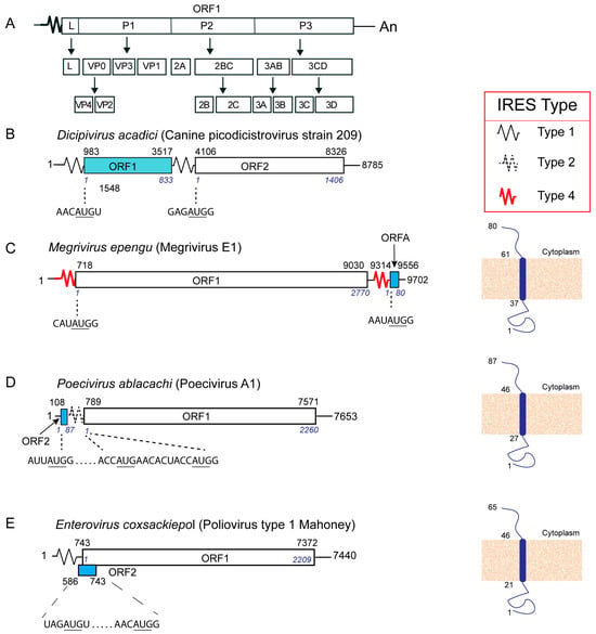
The Picornaviridae currently comprise 68 genera and numerous unclassified members. Here, species are named using the binomial nomenclature, together with common names if appropriate. Picornavirus genomes are conventionally described as mono-cistronic, encoding a single polyprotein that is cleaved into individual structural and nonstructural proteins by virus-encoded proteases [5] (Figure 1A). The capsid proteins VP4, VP2, VP3 and VP1 are derived from the N-terminal P1 region of the polyprotein, which in some viruses is preceded by a leader (L) protein. Nonstructural proteins 2A, 2B and 2C are cleaved from the central P2 region of the polyprotein, and the 3A, 3B, 3C (protease) and 3D (polymerase) proteins are derived from the C-terminal P3 region. The 2A protein is a chymotrypsin-like protease in many (but not all) picornaviruses. However, accumulating evidence indicates that the dogma that picornavirus genomes are monocistronic is an oversimplification, and that picornaviruses belonging to many genera utilize diverse mechanisms to enable translation of alternate open reading frames (altORFs). Translation of a single polyprotein limits the possibilities for control over the timing and stoichiometry of synthesis of proteins, whereas the presence of altORFs allows greater control over these aspects of gene expression. Here, I review this evolving topic, describe validated altORFs and suggest additional possible examples of picornavirus altORF translation.
Figure 1.
Dicistronic genomes in picornaviruses. Schematic representations of (A) a generic monocistronic picornavirus genome, showing the polyprotein-encoding ORF1, the individual proteins and their precursors derived by proteolytic cleavage of the polyprotein. A leader (L) protein is encoded by only a subset of picornaviruses. The L and the 2A proteins have proteolytic activity in only a subset of picornaviruses. (B, left panel) Dicipivirus acadici (Canine picodicistrovirus strain 209) (NCBI Ref. seq.: NC_021178.1), (C, left panel) Megrivirus enpengu (Megrivirus E1 isolate KGI-Bel-P5/2015) (GenBank: MF405436.1), (D) Poecivirus ablacachi (Poecivirus A1) (GenBank: KU977108.1) and (E, left panel) Enterovirus coxsackiepol (poliovirus type 1 Mahoney) (NCBI Ref. seq.: NC_002058.3), showing the relative locations of the polyprotein-encoding ORF1, in the case of Dicipivirus acadici, of ORF2, and (C–E) of the altORF ORF2, and the nucleotide context of the corresponding initiation codons (underlined). (C–E) Nucleotide and amino acid residues are numbered (plain text and italic, respectively), and different types of IRESs are represented by zigzag lines (as indicated in the inset key at top right). The structures of ORF2-encoded polypeptides (B–D, right-hand panels) are illustrated to show the trans-membrane domain, cytoplasmic element and non-cytoplasmic element, predicted (C,D) using the Phobius webserver [6], (E) as described [7] and numbered to indicate amino acid residues.
2. The Canonical Mechanism of Translation in Eukaryotes
The cyclical process of eukaryotic translation consists of four stages: initiation, elongation, termination and post-termination recycling. The canonical initiation process [8] begins with separated 40S and 60S ribosomal subunits, derived by recycling of post-termination ribosomes or splitting of vacant 80S ribosomes. Aminoacylated initiator tRNA (Met-tRNAiMet) forms a ternary complex with eukaryotic initiation factor (eIF) 2 and GTP that binds to the 40S subunit with eIF1, eIF1A and the multi-subunit eIF3 to form a 43S preinitiation complex. Cellular mRNAs have a 5′-terminal m7G cap and 43S complexes are recruited to it by the cap-binding complex eIF4F, a heterotrimer that consists of eIF4E (the cap-binding subunit), eIF4A (an RNA helicase), and eIF4G (which binds to eIF4E, eIF4A, and eIF3). The 43S complex then scans downstream in a process that integrates ribosomal movement, unwinding of secondary structure in the 5′ untranslated region (5′UTR) of the mRNA and monitoring of the 5′UTR to identify the initiation codon, which is the first AUG triplet in a ‘good’ context (e.g., ACCAUGG), defined by the presence of purine residues at −3 and +4 positions (bold) relative to the AUG codon (underlined). AUG codons in a suboptimal context are predominantly bypassed by ‘leaky scanning’, leading to initiation further downstream. Scanning on structured 5′UTRs also requires the DExH-box helicase DHX29 [9]. Recognition of context nucleotides involves establishment of specific interactions between them and eIF1A, eIF2 and the 18S ribosomal RNA (rRNA) constituent of the 40S subunit [10,11]. When the scanning complex encounters a favorable AUG codon, hydrolysis of eIF2-bound GTP is induced by eIF5, eIF1 is displaced and initiator tRNA is released into the ribosomal peptidyl (P) site, resulting in formation of a 48S complex. The scanning-competence of 43S complexes is dependent on eIF1, which also enforces the fidelity of initiation codon selection [8,12,13]. eIF1 induces a scanning-permissive conformation of the 43S complex and maintains initiator tRNA in a metastable state that is permissive for scanning and initiation codon inspection and that is adjusted following displacement of eIF1, resulting in formation of the 48S complex with established codon–anticodon base-pairing [14]. eIF5B induces reorientation of initiator tRNA on the 40S subunit and the joining of a 60S subunit, thereby promoting dissociation of factors from the interface surface of the 40S subunit and formation of an 80S ribosome [8].
Elongation involves cycles of delivery of cognate aminoacyl-tRNA to the ribosome by eukaryotic elongation factor (eEF) 1A•GTP to decode the mRNA codon in the ribosomal aminoacyl (A) site, release of eEF1A•GDP, peptide bond formation, and translocation promoted by eEF2 of the base-paired mRNA and peptidyl-tRNA from A to P sites [15]. Eukaryote-specific elements of the ribosome, tRNAs and eEF2 prevent slippage of the translational reading frame [16]. However, specific sequence motifs and structures in the coding region can induce programmed frame-shifting so that ribosomes continue translation in a different reading frame [4]. The 3′-end of an ORF is marked by a stop codon, and its entry into the A site signals the termination of translation by eukaryotic release factor (eRF) 1 and eRF3•GTP [17]. eRF1 recognizes the stop codon, which triggers hydrolysis of eRF3-bound GTP and release of eRF3•GDP, followed by conformational changes in eRF1 that enable it to enter the ribosomal peptidyl transferase center, triggering cleavage of peptidyl-tRNA and release of the completed polypeptide [18]. In the final stage of translation, the post-termination ribosomal complex is split by ABCE1, releasing the 60S subunit [19]. The 40S subunit remains bound to mRNA and deacylated tRNA, which are released via redundant mechanisms involving the factors eIF2D, or MCTS1/DENR, or eIF1, eIF1A and eIF3.
Three of these four stages in the translation process are or have the potential to be subverted to enable translation of altORFs in picornavirus genomes in addition to the principal ORF.
5. End-Dependent Initiation of an Alternative ORF on Poecivirus Genomic mRNA?
Poecivirus genomes encode a single large polyprotein [81,82], preceded by a type 2 IRES that is related to those of members of the genera Avisivirus and Sicinivirus of Picornaviridae and of several avian caliciviruses [67,83]. A 272–312 nt long ORF2 located near the 5′ end of the genome upstream of the IRES (Figure 1D, left panel) encodes a highly conserved putative FAST Protein (Figure 1D, right panel) [78]. It is possible that this ORF is translated following cap-independent but end-dependent initiation: initiation complexes can scan through stable secondary structure (e.g., [9]) and could therefore potentially scan from the 5′-end of poecivirus genomes through the first ~100 nt of the 5′UTR to the ORF2 initiation codons (AUG108 in Poecivirus ablacachi (strain BCCH-449: GenBank NC_055108.1) and AUG107 in strain RBNU-929 (GenBank: MN944596.1)). In light of a report that IRESs can stimulate translation of upstream ORFs [84], one could speculate that the type 2 IRES downstream of ORF2 might stimulate translation of this protein.
6. Translation of Overlapping ORFs Following IRES-Mediated Initiation
The AUG triplet of the Yn-Xm-AUG motif at the 3′-border of type 1 IRESs (poliovirus AUG586 and its equivalent in other enteroviruses) is an important determinant of IRES function [7,26], but it also supports robust initiation of translation in vitro if its weak context is improved, leading to translation of a 65-codon-long ORF2 that overlaps AUG743, the initiation codon for ORF1, which encodes the poliovirus polyprotein [25,27] (Figure 1E, left panel). Ribosomal entry onto the viral mRNA likely occurs on the Yn-Xm-AUG motif [30] and ribosomes then scan downstream, either arresting at this AUG codon or scanning past it to the ORF1 initiation codon. Many enterovirus genomes contain ORF2 sequences that encode related 56–76-a.a.-long polypeptides that are predicted to contain a trans-membrane domain (Figure 1E, right panel) and that similarly overlap ORF1 [7,25,30,85]. Notably, ORF2 occurs in enteric but not in respiratory enteroviruses [25,84]. Although early studies using cell-free translation extracts had suggested that initiation occurred on two AUG codons in the poliovirus genome, leading to synthesis of an unidentified 5–10 kDa polypeptide and to the structural protein precursor [86,87], the ORF2-encoded polypeptide had escaped detection in enterovirus-infected cells, possibly due to its small size. However, it was observed by Western blotting of native or tagged forms in cells infected by echovirus 7, enterovirus 71 and poliovirus [7,85]. Ablation of ORF2 compromised replication of echovirus 7 in human intestinal cells [7] and replication of enterovirus 71 in human intestinal and murine neuronal cells [85]. Ectopically expressed ORF2 from heterologous enteroviruses enabled the replication in human intestinal cells of respiratory enteroviruses that naturally lack ORF2 (e.g., Enterovirus D68) or in which it has been inactivated, and this activity was dependent on the putative trans-membrane domain [85]. The ORF2 product promotes virus release from enteric cells [7,85].
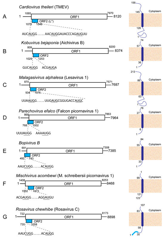
The DA strain of Theiler’s murine encephalomyelitis virus (TMEV; Cardiovirus theileri) is a member of the genus Cardiovirus. Its type 2 IRES promotes translation of the ~18 kDa L* protein from the good-context AUG1078 and from AUG1090, both located downstream of AUG1065, the initiation codon for the polyprotein (Figure 2A, left panel), likely as a result of leaky scanning after ribosomal entry onto the Yn-Xm-AUG motif [88,89]. The L* ORF is present in other TMEV strains [90], although in some, initiation occurs at near-cognate ACG codons located at equivalent positions rather than at AUG codons [91,92]. It is predicted to encode a trans-membrane protein (Figure 2A, right panel), and related proteins are translated from an overlapping ORF in the genomes of other Cardiovirus species, including C. ranori, C. rudhira and C. dhusarah [7]. A similar L* polypeptide is encoded by some Saffold viruses (which are also members of C. theileri (e.g., NC_009448.1)), and its translation likely also initiates at an ACG (Thr) codon, but in most of them, this altORF is interrupted by a stop codon that truncates the encoded protein before the predicted trans-membrane domain is translated [93]. L* determines the ability of the DA and related strains to establish a persistent infection in the central nervous system of mice [91,94]. It accumulates in the mitochondrial outer membrane [95], and binds to RNAse L, an important anti-viral innate immune effector, thereby preventing its interaction with and activation by 2′, 5′-oligoadenylates [96,97]. L* is therefore a determinant of pathogenesis, and remarkably, of species specificity, since L* from the (murine) DA strain of TMEV does not inhibit RNAse L from chickens, humans and numerous other mammals, even including rats [96].
Figure 2.
Overlapping ORFs in picornavirus genomes that encode putative single-pass trans-membrane proteins. Schematic representations of the genomes of (A) Cardiovirus theileri (Theiler’s murine encephalomyelitis virus strain DA; GenBank: JX443418), (B) Kobuvirus bejaponia (Aichivirus B strain U-1; NCBI Ref seq. NC_004421.1), (C) Malagasivirus alphalesa (Lesavirus 1 isolate Mis 101308/2012; GenBank: KM396707.1), (D) Parechovirus efalco (Falcon picornavirus 1; NCBI Ref. seq. NC_035779.1), (E) Bopivirus B (Bopivirus p. strain ovine/TB14.2010-Hun; GenBank: MW298057.1), (F) Mischivirus acombewi (Miniopterus schreibersii picornavirus 1; GenBank: JQ814851.1) and (G) Rosavirus chewhibe (Rosavirus C strain RASK8F; NCBI Ref. seq. NC_075425.1) showing the relative locations of the polyprotein-encoding ORF1 and the overlapping ORF2, the 5′UTRs and 3′UTRs (solid lines), and the nucleotide context of initiation codons. Dashed lines indicate the positions of ORF1 and ORF2 initiation codons. (A–F, right-hand panels) ORF2-encoded polypeptides, illustrated to show signal sequences (light blue), the trans-membrane domain (dark blue), cytoplasmic element and non-cytoplasmic element, predicted using the Phobius webserver [6].
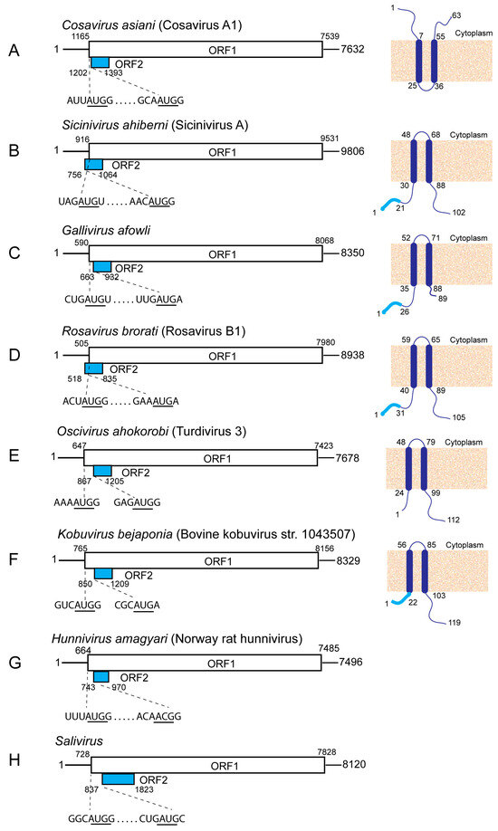
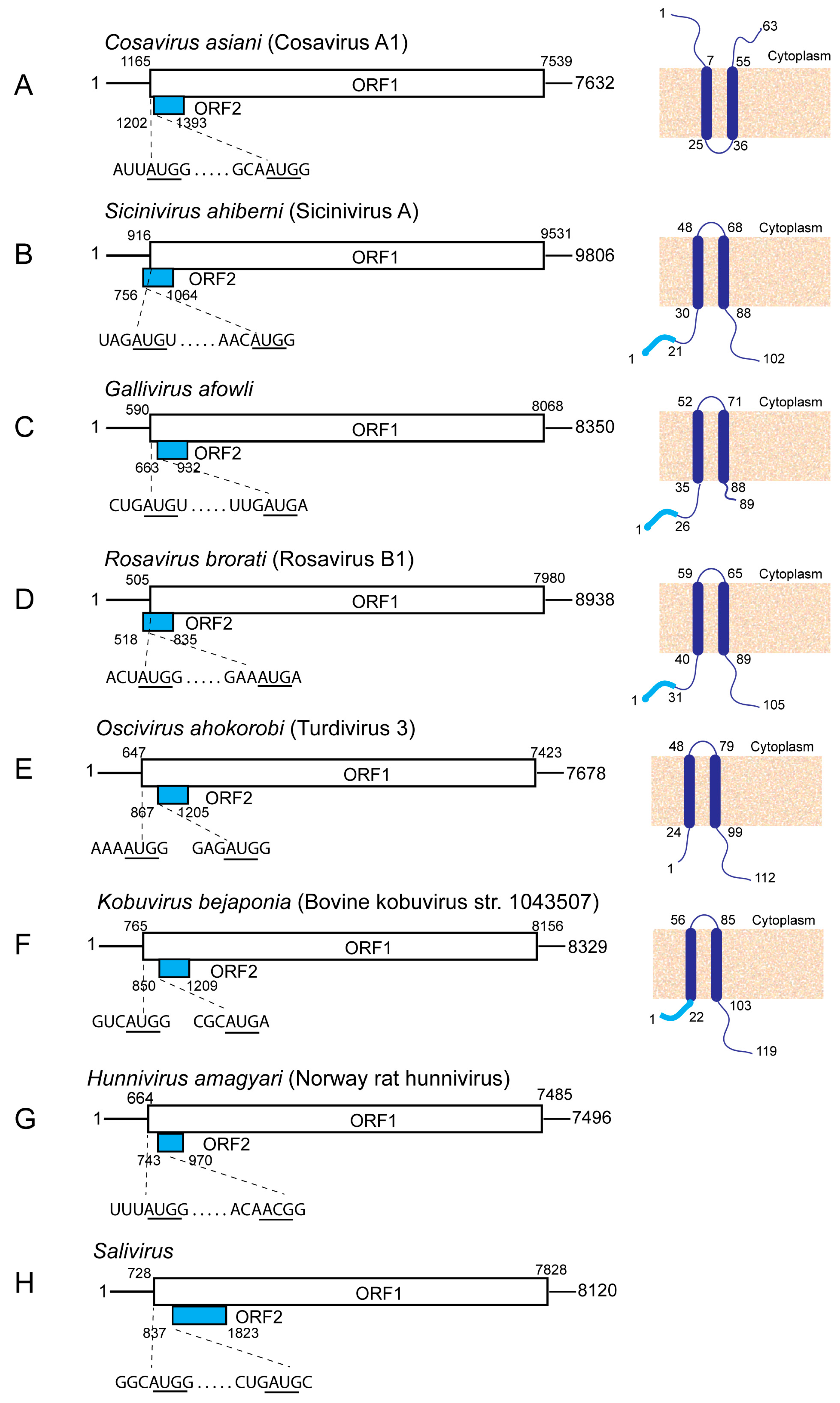
Analysis of the coding potential of members of numerous other picornavirus genera indicates that they also contain altORFs that overlap the 5′-terminal region of the polyprotein-encoding ORF1. These genera include Cosavirus, Hunnivirus, Malagasivirus, Mischivirus, Oscivirus, Rosavirus, Sicinivirus [7], Bopivirus, Gallivirus, Parechovirus, Kobuvirus and Salivirus (Figure 2 and Figure 3). The polypeptides encoded by these altORFs fall into various categories, including proteins with predicted single- and multiple-pass trans-membrane domains as well as unclassified proteins (Figure 2 and Figure 3). Expression of these altORFs in the context of viral infection has not been confirmed, but several have been found to be subject to strong purifying selection, indicating that they encode functional proteins [7]. The level of expression of these altORFs in infected cells may be low, because their initiation codons are often in a suboptimal nucleotide context, and some lie downstream of the ORF1 initiation codon. Moreover, as noted previously, the altORFs in genomes of Hunnivirus amagyari [7] and Bopivirus B [98] initiate at conserved near-cognate ACG and CUG initiation codons respectively (Figure 2E and Figure 3G).
Figure 3.
Overlapping ORFs in picornavirus genomes. Schematic representations of the genomes of (A) Cosavirus asiani (Cosavirus A1 strain HCoSV-A1; GenBank: FJ438902.1), (B) Sicinivirus ahiberni (Sicinivirus A isolate GA/1479/2004; GenBank: MN873048.1), (C) Gallivirus afowli (Gallivirus sp. isolate GALL/PB18-HII34/Switzerland/2019; GenBank: OM469266.1), (D) Rosavirus brorati (Rosavirus B1 strain RNCW0602091R; GenBank: KX783423.1), (E) Oscivirus ahorkorobi (Turdivirus 3 strain 10878; GenBank GU182410.1), (F) Kobuvirus bejaponia (Bovine kobuvirus strain 1043507 (GenBank: MW605074.1)), (G) Hunnivirus amagyari (Norway rat hunnivirus isolate NrHuV/NYC-E21) (GenBank: KJ950971.1) and (H) Salivirus FHB (Genbank: NC_025114.1), showing the relative locations of the polyprotein-encoding ORF1 and the overlapping ORF2, the 5′UTRs and 3′UTRs (solid lines), the nucleotide context of initiation codons. Dashed lines indicate the positions of ORF1 and ORF2 initiation codons. (A–E, right-hand panels) ORF2-encoded polypeptides, illustrated to show signal sequences (light blue), the trans-membrane domain (dark blue), cytoplasmic element and non-cytoplasmic element, predicted using the Phobius webserver [6].
The presence of altORFs in members of some virus species is sporadic. For example, the 123-a.a.-long polypeptide encoded by the altORF in Bopivirus sp. strain deer/VIC82-2020/AUS (Genbank: MZ436972.1) shares 51–63% identity with polypeptides encoded by similarly positioned sequences in other bopivirus genomes (Genbank: NC_026249.1 and MW298059.1) which are, however, interrupted by one and two stop codons, respectively. Similarly, a 210 nt long element in parechovirus sp. isolate SQ/N.fuscus/Pare (Genbank PX095822.1) that is positioned similarly to the Parechovirus falco ORF2 (Figure 2D) would encode a related polypeptide to this altORF product, but is interrupted by three stop codons. Whereas Kobuvirus bejaponia (Bovine kobuvirus strain 1043507 (GenBank: MW605074.1)) encodes a 119-a.a.-long polypeptide with two predicted trans-membrane domains (Figure 3F), there are 5′-terminal substitutions in the equivalent sequence in Kobuvirus bejaponia (Aichivirus B strain U-1; NCBI Ref seq. NC_004421.1) that lead to loss of the initiation codon and gain of a stop codon, so that the residual ORF encodes a truncated 74-a.a.-long polypeptide that is nevertheless 80% identical to the longer equivalent (Figure 2B). Notably, these altORFs all occupy a genomic location that is a known hotspot for recombination [21,99]. Their sporadic appearance and apparent mutational inactivation in related viruses are consistent with a dynamic process of repeated acquisition of altORFs by recombination, followed either by retention and optimization or, if they do not confer any selective advantage on these viruses, by degeneration and loss.
ORF2 in many these viral genomes occurs downstream of type 2 or type 5 IRESs. As described above, initiation mediated by type 2 IRESs can involve scanning after ribosomal attachment, and as a result, some initiation complexes bypass an initiation codon near the landing site and scan downstream at a level that depends on the identity of the IRES, the nucleotide context of the initiation codon and the intracellular environment [41,43,100]. Type 5 IRESs are commonly separated from the polyprotein initiation codon by large variable spacers, suggesting that initiation mediated by them also involves scanning [64]. An IRES-initiated leaky scanning mechanism would allow regulation of ORF2 translation both with respect to the stoichiometry and the time of appearance of its product relative to the ORF1-encoded polyprotein. The level of ORF2 translation in genomes in which it overlaps ORF1 may be low early infection, because the initiation codon is usually in a poor nucleotide context, is commonly positioned downstream of the ORF1 polyprotein initiation codon, and in the case of genus Oscivirus, Hunnivirus, Bopivirus and some Cardiovirus genomes, initiates at near-cognate ACG or CUG codons [91,92] or is likely to do so [7]. However, the cytoplasmic environment is progressively altered during picornavirus infection in a manner that could affect ORF2 translation, for example by viral protease-mediated cleavage of some initiation factors (including eIF4A, eIF4G, eIF5B and the poly(A)-binding protein) and ITAFs (e.g., PCBP2), altered subcellular localization of initiation factors and ITAFs, activation of translational regulators and induced changes in intracellular ionic conditions [101,102,103].
7. Programmed Ribosomal Frame-Shifting During the Elongation Phase of Cardiovirus Translation
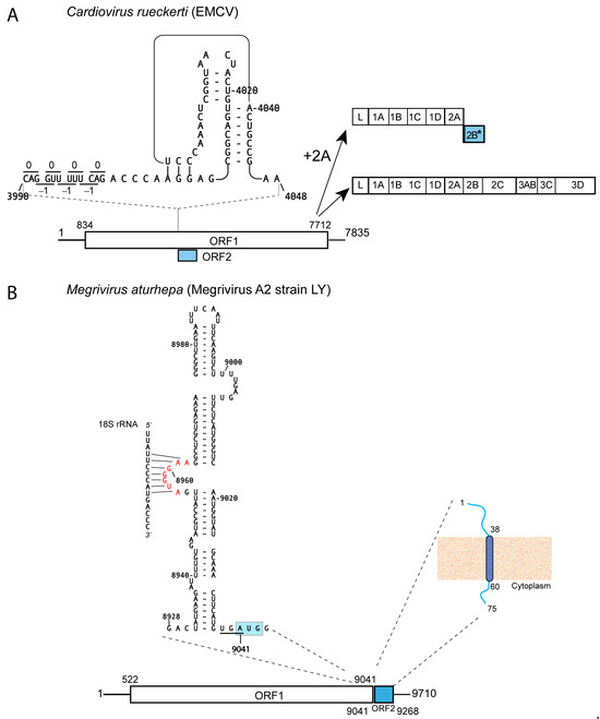
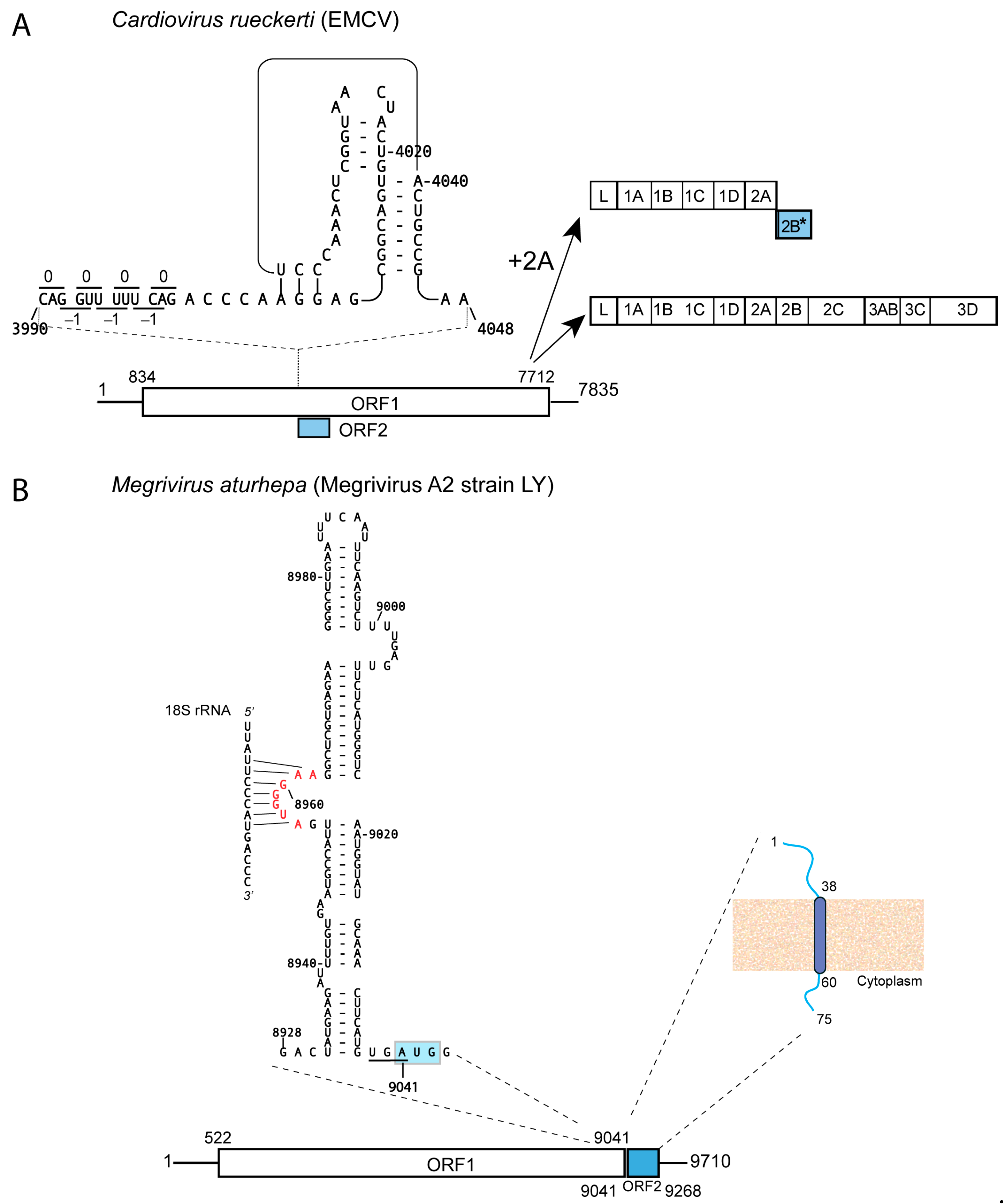
The genomes of Cardiovirus rueckerti (EMCV) and other members of the Cardiovirus genus contain a ~120-codon-long region of elevated synonymous site conservation immediately after the 2A/2B junction in the −1 reading frame relative to the polyprotein ORF1 [104]. Frame-shifting occurs at a G-GUU-GUU ‘slippery’ sequence and is promoted with high (~70%) efficiency by an RNP complex that is formed by binding of the viral 2A protein to an adjacent downstream RNA structure (Figure 4A), likely resulting in a conformational change in it from a hairpin to a pseudoknot [105,106]. Frame-shifting on the EMCV genomic mRNA results in synthesis of a novel 129-a.a.-long trans-frame protein (2B*) [104], after which translation terminates. 2B* enhances virus release via caspase-3-mediated and caspase-3-independent cell lysis pathways [107] and binds to members of the 14-3-3 family of scaffold proteins, likely disrupting innate immune signaling [108]. Loss of 2B* leads to reduced viral plaque size and impaired lytic virus release [104,109]. The requirement of the 2A protein for effective frame-shifting leads to temporal control over translation: the viral polyprotein, including the nonstructural proteins that are derived by proteolytic cleavage of the P2 and P3 regions of the polyprotein, are translated early in infection, and as 2A accumulates, it promotes translation of 2B* followed by termination of translation. Therefore, 2A acts as a switch that downregulates translation of replication proteins so that the translation apparatus becomes dedicated to synthesis of structural proteins [109].
Figure 4.
RNA structure-mediated initiation/reinitiation of translation of picornavirus altORFs. (A) The cardiovirus ribosomal frame-shifting element, showing the sequence and structure of this element in the EMCV genome (GenBank: NC_001479.1), its position relative to the G_GUU_UUU slippery sequence and the codons in the 0 and −1 reading frames. Binding of the viral 2A protein to this pseudoknot structure shifts ribosomes from translation of the polyprotein-encoding ORF1 to translation of the 2B* protein in the −1 reading frame followed by termination of translation, as indicated b the arrows. (B) The sequence and structure of the putative termination upstream ribosome binding site (TURBS) element in the genome of Megrivirus aturhepa (NCBI Ref. seq.: NC_024120.1) and its position immediately upstream of the overlapping ORF1 termination codon and the ORF2 initiation codon (indicated by a light blue shaded box). The conserved motif 1 sequence AUGGGAA (red font) can base-pair to an element of helix 26 of 18S rRNA, anchoring the post-termination ribosome prior to reinitiation. The ORF2-encoded polypeptide is illustrated to show the trans-membrane domain (dark blue), cytoplasmic element and non-cytoplasmic element, predicted using the Phobius webserver [6]. Dashed lines indicate (A,B) the positions of RNA elements and (B) link ORF2 to the FAST protein that it is predicted to encode.
Frame-shifting elements of the same type occur in Cardiovirus theileri (TMEV), in C. rudhira and in C. saffoldi (e.g., [104]). In C. theileri, this element promotes frame-shifting at up to 82% efficiency [110]. The 2B* transframe polypeptides derived from these genomes would be only 14 a.a. long and are therefore likely nonfunctional. However, the 2A-mediated induction of translation termination before the synthesis of replicative proteins is important, because it enforces a switch to preferential translation of structural proteins late in infection. Impairment of frame-shifting resulted in TMEV having a small plaque phenotype and reduced growth kinetics, indicating the likely importance of this switch for viral replication [110,111].
8. Termination–Reinitiation Elements in Megrivirus Genomes
Numerous megrivirus genomic sequences that contain a FAST protein-encoding ORF2 [7,76,77] lack obvious IRES-like sequences between it and the polyprotein-encoding ORF1, and therefore likely exploit alternative mechanisms for initiation of ORF2 translation. In two groups of these megrivirus genomes, the ORF1 termination codon (underlined) overlaps the ORF2 initiation codon (bold) (UAAUG or UGAUG) or is separated from it by two or three nucleotides (UAA-2/3nt-AUG (Figure 4B) or, less commonly, UAG-2/3nt-AUG). This configuration of stop and start codons suggests that these ORF2 entities could be translated by a termination–reinitiation mechanism. The best-characterized mechanism of this type occurs on bicistronic subgenomic calicivirus mRNAs: translation of the minor capsid protein from the second ORF is mediated by a ~40–80 nt long TURBS (termination upstream ribosome binding site) immediately upstream of the restart AUG [112]. This structured RNA contains an essential conserved motif 1, whose AUGGGA core is complementary to the apical loop of helix 26 of 18S rRNA and which tethers the post-termination 40S ribosomal subunit to the stop/restart region, and the species-specific motif 2, which base-pairs to the complementary motif 2*, immediately preceding motif 1 [112,113,114,115]. Motif 1 is commonly flanked by basal and distal helices, but recent analysis identified a TURBS that lacks the downstream helical element [116]. The TURBS also interacts with eIF3, which enhances reinitiation [117], possibly by stabilizing the TURBS–ribosome interaction and/or by promoting recruitment of the eIF2-GTP/initiator tRNA ternary complex [118].
Remarkably, analysis of Megrivirus aturhepa sequences revealed a 113 nt long TURBS that formed a single extended domain with five helical elements and a canonical UGGGA motif 1 in a centrally located loop that has the potential to base-pair with the apex of helix 26 of 18S rRNA (Figure 4B). With the caveat that this putative megrivirus TURBS has not been experimentally validated, this element is longer than the 40–80 nt long calicivirus TURBS and thus has the potential to interact more extensively with eIF3.
A second group of related megrivirus genomes that contain ORF2 elements that encode FAST proteins is epitomized by Megrivirus chigalli (e.g., Megrivirus C2 isolate 27C (NCBI Ref. Seq. NC_024769.1)). It contains a conserved sequence upstream of the conserved stop/start sequence (AUGUAAAUUAUG, in which the stop codon is underlined and the initiation codon is bold). However, it lacks a motif 1 (UGGGA) sequence and does not appear to form a conventional TURBS. If this ORF2 is translated, it remains to be determined whether translation results from a divergent termination–reinitiation mechanism or is mediated by an IRES embedded within the 3′-terminal region of ORF1.
9. Conclusions
A rapidly expanding number of picornavirus genomes has been found to contain a second ORF in addition to the principal polyprotein-encoding ORF1. ORF2 occurs at various positions: overlapping the 5′-terminal region of ORF1, upstream of ORF1, downstream of it or contained entirely within it. Although several ORF2 candidates reported here reman to be validated, it is notable that many have been found to be subject to strong purifying selection [25], which is suggestive of their functional importance. A systematic bioinformatic search would almost certainly identify additional candidates for experimental verification. In light of the identification of validated and potential altORFs near the 5′ and 3′ extremities of some picornavirus genomes [76,77,78,79,81,82], it is possible that many reported genomic sequences that contain a full polyprotein-encoding ORF may nevertheless be incomplete and that this may have resulted in altORFs having been overlooked.
This diversity in ORF positioning is matched by the diversity of function of the encoded polypeptides. The various second cistrons encode different classes of protein, including the TMEV L* protein (an RNAse L inhibitor) [96], the EMCV 2B* protein (which promotes cell lysis to facilitate viral release and binds to 14-3-3 proteins to impair innate immune signaling [107,108]), and the enterovirus ORF2 product, a putative trans-membrane protein that facilitates viral release from cells [7,85]. Several other picornavirus altORFs are predicted to encode trans-membrane polypeptides, including megrivirus and poecivirus ORF2-encoded polypeptides that belong to the fusion-associated trans-membrane (FAST) class of proteins [78], whereas others are predicted to contain trans-membrane domains but lack other characteristics of FAST proteins. The functions of FAST-related and other potential trans-membrane proteins in the infection cycle of picornaviruses remain to be determined.
Investigation of picornavirus translation has focused almost exclusively on IRES-mediated initiation, and translation of many of the confirmed and hypothesized ORF2 moieties described here occurs as a result of this process, either due to IRES duplication (which allows initiation to occur at independent sites) or IRES-mediated initiation followed by leaky scanning. Notably, at least three other processes may be responsible for ORF2 translation, namely end-dependent but cap-independent initiation, programmed ribosomal frame-shifting and TURBS-mediated termination–reinitiation. Exploitation of these alternative mechanisms would not only enable ribosomes to gain access to alternate ORFs, but would also allow the level of synthesis of the encoded products and of the timing of their formation to be regulated during the infection cycle.
Funding
This work was in part supported by NIH grant 1 R21 AI188505.
Data Availability Statement
All novel data presented in this work are contained within the manuscript figures.
Conflicts of Interest
The author declares no conflicts of interest.
Abbreviations
The following abbreviations are used in this manuscript:
| altORF | alternative open reading frame |
| ABCE1 | ATP-binding cassette subfamily E member 1 |
| CPDV | canine picodicistrovirus |
| DHX29 | DExH-box helicase 29 |
| eEF | eukaryotic elongation factor |
| eIF | eukaryotic initiation factor |
| EMCV | encephalomyocarditis virus |
| eRF | eukaryotic release factor |
| FAST | fusion-associated small transmembrane |
| FMDV | Foot-and-mouth disease virus |
| GDP | Guanosine diphosphate |
| GTP | Guanosine triphosphate |
| HAV | Hepatitis A virus |
| IGR | intergenic region |
| IRES | Internal ribosomal entry site |
| ITAF | IRES trans-acting factor |
| NCBI | National Center for Biotechnology Information |
| ORF | Open reading frame |
| PCBP2 | Poly(rC)-Binding Protein 2 |
| RNA | Ribonucleic acid |
| RNAse | ribonuclease |
| RNP | ribonucleoprotein |
| rRNA | ribosomal RNA |
| TMEV | Theiler’s murine encephalomyelitis virus |
| tRNA | transfer RNA |
| TURBS | termination upstream ribosome binding site |
| UTR | Untranslated region |
| VPg | viral protein genome-linked |
References
- Firth, A.E.; Brierley, I. Non-canonical translation in RNA viruses. J. Gen. Virol. 2012, 93, 1385–1409. [Google Scholar] [CrossRef] [Scilit] [PubMed]
- Mailliot, J.; Martin, F. Viral internal ribosomal entry sites: Four classes for one goal. Wiley Interdiscip. Rev. RNA 2018, 9, e1458. [Google Scholar] [CrossRef] [Scilit] [PubMed]
- Sherlock, M.E.; Baquero Galvis, L.; Vicens, Q.; Kieft, J.S.; Jagannathan, S. Principles, mechanisms, and biological implications of translation termination-reinitiation. RNA 2023, 29, 865–884. [Google Scholar] [CrossRef] [Scilit] [PubMed]
- Hill, C.H.; Brierley, I. Structural and functional insights into viral programmed ribosomal frameshifting. Annu. Rev. Virol. 2023, 10, 217–242. [Google Scholar] [CrossRef] [Scilit] [PubMed]
- Zell, R.; Delwart, E.; Gorbalenya, A.E.; Hovi, T.; King, A.M.Q.; Knowles, N.J.; Lindberg, A.M.; Pallansch, M.A.; Palmenberg, A.C.; Reuter, G.; et al. ICTV Virus Taxonomy Profile: Picornaviridae. J. Gen. Virol. 2017, 98, 2421–2422. [Google Scholar] [CrossRef] [Scilit] [PubMed]
- Käll, L.; Krogh, A.; Sonnhammer, E.L. Advantages of combined transmembrane topology and signal peptide prediction--the Phobius web server. Nucleic Acids Res. 2007, 35, W429–W432. [Google Scholar] [CrossRef] [Scilit] [PubMed]
- Lulla, V.; Dinan, A.M.; Hosmillo, M.; Chaudhry, Y.; Sherry, L.; Irigoyen, N.; Nayak, K.M.; Stonehouse, N.J.; Zilbauer, M.; Goodfellow, I.; et al. An upstream protein-coding region in enteroviruses modulates virus infection in gut epithelial cells. Nat. Microbiol. 2019, 4, 280–292. [Google Scholar] [CrossRef] [Scilit] [PubMed]
- Jackson, R.J.; Hellen, C.U.; Pestova, T.V. The mechanism of eukaryotic translation initiation and principles of its regulation. Nat. Rev. Mol. Cell Biol. 2010, 11, 113–127. [Google Scholar] [CrossRef] [Scilit] [PubMed]
- Pisareva, V.P.; Pisarev, A.V.; Komar, A.A.; Hellen, C.U.; Pestova, T.V. Translation initiation on mammalian mRNAs with structured 5′UTRs requires DExH-box protein DHX29. Cell 2008, 135, 1237–1250. [Google Scholar] [CrossRef] [Scilit] [PubMed]
- Pisarev, A.V.; Kolupaeva, V.G.; Pisareva, V.P.; Merrick, W.C.; Hellen, C.U.; Pestova, T.V. Specific functional interactions of nucleotides at key −3 and +4 positions flanking the initiation codon with components of the mammalian 48S translation initiation complex. Genes Dev. 2006, 20, 624–636. [Google Scholar] [CrossRef] [Scilit] [PubMed]
- Petrychenko, V.; Yi, S.H.; Liedtke, D.; Peng, B.Z.; Rodnina, M.V.; Fischer, N. Structural basis for translational control by the human 48S initiation complex. Nat. Struct. Mol. Biol. 2025, 32, 62–72. [Google Scholar] [CrossRef] [Scilit] [PubMed]
- Pestova, T.V.; Borukhov, S.I.; Hellen, C.U. Eukaryotic ribosomes require initiation factors 1 and 1A to locate initiation codons. Nature 1998, 394, 854–859. [Google Scholar] [CrossRef] [Scilit] [PubMed]
- Pestova, T.V.; Kolupaeva, V.G. The roles of individual eukaryotic translation initiation factors in ribosomal scanning and initiation codon selection. Genes Dev. 2002, 16, 2906–2922. [Google Scholar] [CrossRef] [Scilit] [PubMed]
- Brito Querido, J.; Díaz-López, I.; Ramakrishnan, V. The molecular basis of translation initiation and its regulation in eukaryotes. Nat. Rev. Mol. Cell Biol. 2024, 25, 168–186. [Google Scholar] [CrossRef] [Scilit] [PubMed]
- Dever, T.E.; Dinman, J.D.; Green, R. Translation elongation and recoding in eukaryotes. Cold Spring Harb. Perspect. Biol. 2018, 10, a032649. [Google Scholar] [CrossRef] [Scilit] [PubMed]
- Djumagulov, M.; Demeshkina, N.; Jenner, L.; Rozov, A.; Yusupov, M.; Yusupova, G. Accuracy mechanism of eukaryotic ribosome translocation. Nature 2021, 600, 543–546. [Google Scholar] [CrossRef] [Scilit] [PubMed]
- Alkalaeva, E.Z.; Pisarev, A.V.; Frolova, L.Y.; Kisselev, L.L.; Pestova, T.V. In vitro reconstitution of eukaryotic translation reveals cooperativity between release factors eRF1 and eRF3. Cell 2006, 125, 1125–1136. [Google Scholar] [CrossRef] [Scilit] [PubMed]
- Hellen, C.U.T. Translation termination and ribosome recycling in eukaryotes. Cold Spring Harb. Perspect. Biol. 2018, 10, a032656. [Google Scholar] [CrossRef] [Scilit] [PubMed]
- Pisarev, A.V.; Skabkin, M.A.; Pisareva, V.P.; Skabkina, O.V.; Rakotondrafara, A.M.; Hentze, M.W.; Hellen, C.U.; Pestova, T.V. The role of ABCE1 in eukaryotic posttermination ribosomal recycling. Mol. Cell 2010, 37, 196–210. [Google Scholar] [CrossRef] [Scilit] [PubMed]
- Francisco-Velilla, R.; Embarc-Buh, A.; Abellan, S.; Martinez-Salas, E. Picornavirus translation strategies. FEBS Open Bio 2022, 12, 1125–1141. [Google Scholar] [CrossRef] [Scilit] [PubMed]
- Arhab, Y.; Bulakhov, A.G.; Pestova, T.V.; Hellen, C.U.T. Dissemination of internal ribosomal entry sites (IRES) between viruses by horizontal gene transfer. Viruses 2020, 12, 612. [Google Scholar] [CrossRef] [Scilit] [PubMed]
- Pelletier, J.; Sonenberg, N. Internal initiation of translation of eukaryotic mRNA directed by a sequence derived from poliovirus RNA. Nature 1988, 334, 320–325. [Google Scholar] [CrossRef] [Scilit] [PubMed]
- Woo, P.C.; Lau, S.K.; Choi, G.K.; Huang, Y.; Teng, J.L.; Tsoi, H.W.; Tse, H.; Yeung, M.L.; Chan, K.H.; Jin, D.Y.; et al. Natural occurrence and characterization of two internal ribosome entry site elements in a novel virus, canine picodicistrovirus, in the picornavirus-like superfamily. J. Virol. 2012, 86, 2797–2808. [Google Scholar] [CrossRef] [Scilit] [PubMed]
- Boros, Á.; Pankovics, P.; Simmonds, P.; Pollák, E.; Mátics, R.; Phan, T.G.; Delwart, E.; Reuter, G. Genome analysis of a novel, highly divergent picornavirus from common kestrel (Falco tinnunculus): The first non-enteroviral picornavirus with type-I-like IRES. Infect. Genet. Evol. 2015, 32, 425–431. [Google Scholar] [CrossRef] [Scilit] [PubMed]
- Pestova, T.V.; Hellen, C.U.; Wimmer, E. Translation of poliovirus RNA: Role of an essential cis-acting oligopyrimidine element within the 5′ nontranslated region and involvement of a cellular 57-kilodalton protein. J. Virol. 1991, 65, 6194–6204. [Google Scholar] [CrossRef] [Scilit] [PubMed]
- Pelletier, J.; Flynn, M.E.; Kaplan, G.; Racaniello, V.; Sonenberg, N. Mutational analysis of upstream AUG codons of poliovirus RNA. J. Virol. 1988, 62, 4486–4492. [Google Scholar] [CrossRef] [Scilit] [PubMed]
- Sweeney, T.R.; Abaeva, I.S.; Pestova, T.V.; Hellen, C.U. The mechanism of translation initiation on Type 1 picornavirus IRESs. EMBO J. 2014, 33, 76–92. [Google Scholar] [CrossRef] [Scilit] [PubMed]
- de Breyne, S.; Yu, Y.; Unbehaun, A.; Pestova, T.V.; Hellen, C.U. Direct functional interaction of initiation factor eIF4G with type 1 internal ribosomal entry sites. Proc. Natl. Acad. Sci. USA 2009, 106, 9197–9202. [Google Scholar] [CrossRef] [Scilit] [PubMed]
- Pestova, T.V.; Hellen, C.U.; Wimmer, E. A conserved AUG triplet in the 5′ nontranslated region of poliovirus can function as an initiation codon in vitro and in vivo. Virology 1994, 204, 729–737. [Google Scholar] [CrossRef] [Scilit] [PubMed]
- O’Connor, R.L.; Cook, G.M.; Hankinson, J.; Fominykh, K.; Cheng, S.H.; Nash, D.A.; Cenier, A.; Nayak, K.M.; Graham, S.C.; Deane, J.E.; et al. Flexibility and modulation of translation initiation in enterovirus genomes. PLoS Pathog. 2026, 22, e1013967. [Google Scholar] [CrossRef] [Scilit] [PubMed]
- Jang, S.K.; Kräusslich, H.G.; Nicklin, M.J.; Duke, G.M.; Palmenberg, A.C.; Wimmer, E. A segment of the 5′ nontranslated region of encephalomyocarditis virus RNA directs internal entry of ribosomes during in vitro translation. J. Virol. 1998, 62, 2636–2643. [Google Scholar] [CrossRef] [Scilit] [PubMed]
- Kühn, R.; Luz, N.; Beck, E. Functional analysis of the internal translation initiation site of foot-and-mouth disease virus. J. Virol. 1990, 64, 4625–4631. [Google Scholar] [CrossRef] [Scilit] [PubMed]
- Pestova, T.V.; Hellen, C.U.; Shatsky, I.N. Canonical eukaryotic initiation factors determine initiation of translation by internal ribosomal entry. Mol. Cell. Biol. 1996, 16, 6859–6869. [Google Scholar] [CrossRef] [Scilit] [PubMed]
- Pestova, T.V.; Shatsky, I.N.; Hellen, C.U. Functional dissection of eukaryotic initiation factor 4F: The 4A subunit and the central domain of the 4G subunit are sufficient to mediate internal entry of 43S preinitiation complexes. Mol. Cell. Biol. 1996, 16, 6870–6878. [Google Scholar] [CrossRef] [Scilit] [PubMed]
- Imai, S.; Kumar, P.; Hellen, C.U.; D’Souza, V.M.; Wagner, G. An accurately preorganized IRES RNA structure enables eIF4G capture for initiation of viral translation. Nat. Struct. Mol. Biol. 2016, 23, 859–864. [Google Scholar] [CrossRef] [Scilit] [PubMed]
- Imai, S.; Suzuki, H.; Fujiyoshi, Y.; Shimada, I. Dynamically regulated two-site interaction of viral RNA to capture host translation initiation factor. Nat. Commun. 2023, 14, 4977. [Google Scholar] [CrossRef] [Scilit] [PubMed]
- Bhattacharjee, S.; Abaeva, I.S.; Brown, Z.P.; Arhab, Y.; Fallah, H.; Hellen, C.U.T.; Frank, J.; Pestova, T.V. The mechanism of ribosomal recruitment during translation initiation on the Type 2 encephalomyocarditis virus IRES. EMBO J. 2026, 45, 2666–2693. [Google Scholar] [CrossRef] [Scilit] [PubMed]
- Kaminski, A.; Howell, M.T.; Jackson, R.J. Initiation of encephalomyocarditis virus RNA translation: The authentic initiation site is not selected by a scanning mechanism. EMBO J. 1990, 9, 3753–3759. [Google Scholar] [CrossRef] [Scilit] [PubMed]
- Dmitriev, S.E.; Pisarev, A.V.; Rubtsova, M.P.; Dunaevsky, Y.E.; Shatsky, I.N. Conversion of 48S translation preinitiation complexes into 80S initiation complexes as revealed by toeprinting. FEBS Lett. 2003, 533, 99–104. [Google Scholar] [CrossRef] [Scilit] [PubMed]
- Abaeva, I.S.; Arhab, Y.; Miścicka, A.; Hellen, C.U.T.; Pestova, T.V. In vitro reconstitution of SARS-CoV-2 Nsp1-induced mRNA cleavage reveals the key roles of the N-terminal domain of Nsp1 and the RRM domain of eIF3g. Genes Dev. 2023, 37, 844–860. [Google Scholar] [CrossRef] [Scilit] [PubMed]
- Clarke, B.E.; Sangar, D.V.; Burroughs, J.N.; Newton, S.E.; Carroll, A.R.; Rowlands, D.J. Two initiation sites for foot-and-mouth disease virus polyprotein in vivo. J. Gen. Virol. 1985, 66, 2615–2626. [Google Scholar] [CrossRef] [Scilit] [PubMed]
- Sangar, D.V.; Newton, S.E.; Rowlands, D.J.; Clarke, B.E. All foot and mouth disease virus serotypes initiate protein synthesis at two separate AUGs. Nucleic Acids Res. 1987, 15, 3305–3315. [Google Scholar] [CrossRef] [Scilit] [PubMed]
- Belsham, G.J. Dual initiation sites of protein synthesis on foot-and-mouth disease virus RNA are selected following internal entry and scanning of ribosomes in vivo. EMBO J. 1992, 11, 1105–1110. [Google Scholar] [CrossRef] [Scilit] [PubMed]
- Andreev, D.E.; Fernandez-Miragall, O.; Ramajo, J.; Dmitriev, S.E.; Terenin, I.M.; Martinez-Salas, E.; Shatsky, I.N. Differential factor requirement to assemble translation initiation complexes at the alternative start codons of foot-and-mouth disease virus RNA. RNA 2007, 13, 1366–1374. [Google Scholar] [CrossRef] [Scilit] [PubMed]
- Hinton, T.M.; Li, F.; Crabb, B.S. Internal ribosomal entry site-mediated translation initiation in equine rhinitis A virus: Similarities to and differences from that of foot-and-mouth disease virus. J. Virol. 2000, 74, 11708–11716. [Google Scholar] [CrossRef] [Scilit] [PubMed]
- Hinton, T.M.; Crabb, B.S. The novel picornavirus Equine rhinitis B virus contains a strong type II internal ribosomal entry site which functions similarly to that of Encephalomyocarditis virus. J. Gen. Virol. 2001, 82, 2257–2269. [Google Scholar] [CrossRef] [Scilit] [PubMed]
- Pankovics, P.; Boros, Á.; Mátics, R.; Kapusinszky, B.; Delwart, E.; Reuter, G. Ljungan/Sebokele-like picornavirus in birds of prey, common kestrel (Falco tinnunculus) and red-footed falcon (F. vespertinus). Infect. Genet. Evol. 2017, 55, 14–19. [Google Scholar] [CrossRef] [Scilit] [PubMed]
- Brown, E.A.; Day, S.P.; Jansen, R.W.; Lemon, S.M. The 5′ nontranslated region of hepatitis A virus RNA: Secondary structure and elements required for translation in vitro. J. Virol. 1991, 65, 5828–5838. [Google Scholar] [CrossRef] [Scilit] [PubMed]
- Balázs, B.; Boros, Á.; Pankovics, P.; Mátics, R.; Urbán, P.; Herczeg, R.; Knowles, N.J.; Reuter, G. Genetic characterization of two novel picornaviruses from birds, white-tailed eagle (Haliaeetus albicilla) and pied avocet (Recurvirostra avosetta) in Hungary. Sci. Rep. 2026, 16, 9816. [Google Scholar] [CrossRef] [Scilit] [PubMed]
- Tesar, M.; Harmon, S.A.; Summers, D.F.; Ehrenfeld, E. Hepatitis A virus polyprotein synthesis initiates from two alternative AUG codons. Virology 1992, 186, 609–618. [Google Scholar] [CrossRef] [Scilit] [PubMed]
- Shirasaki, T.; Lenarcic, E.; Misumi, I.; Xie, L.; Fusco, W.G.; Yonish, B.; Das, A.; Kim, H.; Cameron, C.E.; Léger-Abraham, M.; et al. Hepatovirus translation requires PDGFA-associated protein 1, an eIF4E-binding protein regulating endoplasmic reticulum stress responses. Sci. Adv. 2024, 10, eadq6342. [Google Scholar] [CrossRef] [Scilit] [PubMed]
- Ali, I.K.; McKendrick, L.; Morley, S.J.; Jackson, R.J. Activity of the hepatitis A virus IRES requires association between the cap-binding translation initiation factor (eIF4E) and eIF4G. J. Virol. 2001, 75, 7854–7863. [Google Scholar] [CrossRef] [Scilit] [PubMed]
- Borman, A.M.; Michel, Y.M.; Kean, K.M. Detailed analysis of the requirements of hepatitis A virus internal ribosome entry segment for the eukaryotic initiation factor complex eIF4F. J. Virol. 2001, 75, 7864–7871. [Google Scholar] [CrossRef] [Scilit] [PubMed]
- Koirala, D.; Shao, Y.; Koldobskaya, Y.; Fuller, J.R.; Watkins, A.M.; Shelke, S.A.; Pilipenko, E.V.; Das, R.; Rice, P.A.; Piccirilli, J.A. A conserved RNA structural motif for organizing topology within picornaviral internal ribosome entry sites. Nat. Commun. 2019, 10, 3629. [Google Scholar] [CrossRef] [Scilit] [PubMed]
- Avanzino, B.C.; Fuchs, G.; Fraser, C.S. Cellular cap-binding protein, eIF4E, promotes picornavirus genome restructuring and translation. Proc. Natl. Acad. Sci. USA 2017, 114, 9611–9616. [Google Scholar] [CrossRef] [Scilit] [PubMed]
- Pisarev, A.V.; Chard, L.S.; Kaku, Y.; Johns, H.L.; Shatsky, I.N.; Belsham, G.J. Functional and structural similarities between the internal ribosome entry sites of hepatitis C virus and porcine teschovirus, a picornavirus. J. Virol. 2004, 78, 4487–4497. [Google Scholar] [CrossRef] [Scilit] [PubMed]
- Chard, L.S.; Bordeleau, M.E.; Pelletier, J.; Tanaka, J.; Belsham, G.J. Hepatitis C virus-related internal ribosome entry sites are found in multiple genera of the family Picornaviridae. J. Gen. Virol. 2006, 87, 927–936. [Google Scholar] [CrossRef] [Scilit] [PubMed]
- Hellen, C.U.; de Breyne, S. A distinct group of hepacivirus/pestivirus-like internal ribosomal entry sites in members of diverse picornavirus genera: Evidence for modular exchange of functional noncoding RNA elements by recombination. J. Virol. 2007, 81, 5850–5863. [Google Scholar] [CrossRef] [Scilit] [PubMed]
- Asnani, M.; Kumar, P.; Hellen, C.U. Widespread distribution and structural diversity of Type IV IRESs in members of Picornaviridae. Virology 2015, 478, 61–74. [Google Scholar] [CrossRef] [Scilit] [PubMed]
- Segar, K.E.; Sherlock, M.E.; Kieft, J.S. Distribution and structural diversity of Type IV internal ribosome entry sites. RNA 2026, 35, 622–634. [Google Scholar] [CrossRef] [Scilit] [PubMed]
- Pestova, T.V.; Shatsky, I.N.; Fletcher, S.P.; Jackson, R.J.; Hellen, C.U. A prokaryotic-like mode of cytoplasmic eukaryotic ribosome binding to the initiation codon during internal translation initiation of hepatitis C and classical swine fever virus RNAs. Genes Dev. 1998, 12, 67–83. [Google Scholar] [CrossRef] [Scilit] [PubMed]
- de Breyne, S.; Yu, Y.; Pestova, T.V.; Hellen, C. U Factor requirements for translation initiation on the Simian picornavirus internal ribosomal entry site. RNA 2008, 14, 367–380. [Google Scholar] [CrossRef] [Scilit] [PubMed]
- Brown, Z.P.; Abaeva, I.S.; De, S.; Hellen, C.U.T.; Pestova, T.V.; Frank, J. Molecular architecture of 40S translation initiation complexes on the hepatitis C virus IRES. EMBO J. 2022, 41, e110581. [Google Scholar] [CrossRef] [Scilit] [PubMed]
- Yu, Y.; Sweeney, T.R.; Kafasla, P.; Jackson, R.J.; Pestova, T.V.; Hellen, C.U. The mechanism of translation initiation on Aichivirus RNA mediated by a novel type of picornavirus IRES. EMBO J. 2011, 30, 4423–4436. [Google Scholar] [CrossRef] [Scilit] [PubMed]
- Sweeney, T.R.; Dhote, V.; Yu, Y.; Hellen, C.U. A distinct class of internal ribosomal entry site in members of the Kobuvirus and proposed Salivirus and Paraturdivirus genera of the Picornaviridae. J. Virol. 2012, 86, 1468–1486. [Google Scholar] [CrossRef] [Scilit] [PubMed]
- Pankovics, P.; Boros, Á.; Phan, T.G.; Delwart, E.; Reuter, G. A novel passerivirus (family Picornaviridae) in an outbreak of enteritis with high mortality in estrildid finches (Uraeginthus sp.). Arch. Virol. 2018, 163, 1063–1071. [Google Scholar] [CrossRef] [Scilit] [PubMed]
- Arhab, Y.; Miścicka, A.; Pestova, T.V.; Hellen, C.U.T. Horizontal gene transfer as a mechanism for the promiscuous acquisition of distinct classes of IRES by avian caliciviruses. Nucleic Acids Res. 2022, 50, 1052–1068. [Google Scholar] [CrossRef] [Scilit] [PubMed]
- Reuter, G.; Boros, Á.; Földvári, G.; Szekeres, S.; Mátics, R.; Kapusinszky, B.; Delwart, E.; Pankovics, P. Dicipivirus (family Picornaviridae) in wild Northern white-breasted hedgehog (Erinaceus roumanicus). Arch. Virol. 2018, 163, 175–181. [Google Scholar] [CrossRef] [Scilit] [PubMed]
- Fehér, E.; Kemenesi, G.; Görföl, T.; Wazzani, Y.; Bodó, K.; Lanszki, J.; Máté, D.; Kaszab, E.; Domán, M.; Zádori, Z.; et al. Occurrence of recombinant canine coronavirus, picodicistrovirus, and circovirus in red foxes (Vulpes vulpes) implies frequent virus transmission events among carnivores. Transbound. Emerg. Dis. 2025, 2025, 6681119. [Google Scholar] [CrossRef] [Scilit] [PubMed]
- Chen, Y.M.; Hu, S.J.; Lin, X.D.; Tian, J.H.; Lv, J.X.; Wang, M.R.; Luo, X.Q.; Pei, Y.Y.; Hu, R.X.; Song, Z.G.; et al. Host traits shape virome composition and virus transmission in wild small mammals. Cell 2023, 186, 4662–4675.e12. [Google Scholar] [CrossRef] [Scilit] [PubMed]
- Asnani, M.; Pestova, T.V.; Hellen, C.U. Initiation on the divergent Type I cadicivirus IRES: Factor requirements and interactions with the translation apparatus. Nucleic Acids Res. 2016, 44, 3390–3407. [Google Scholar] [CrossRef] [Scilit] [PubMed]
- Asnani, M.; Pestova, T.V.; Hellen, C.U. PCBP2 enables the cadicivirus IRES to exploit the function of a conserved GRNA tetraloop to enhance ribosomal initiation complex formation. Nucleic Acids Res. 2016, 44, 9902–9917. [Google Scholar] [CrossRef] [Scilit] [PubMed]
- Wierenga, J.R.; Grimwood, R.M.; Taylor, H.S.; Hunter, S.; Argilla, L.S.; Webster, T.; Lim, L.; French, R.; Schultz, H.; Jorge, F.; et al. Total infectome investigation of diphtheritic stomatitis in yellow-eyed penguins (Megadyptes antipodes) reveals a novel and abundant megrivirus. Vet. Microbiol. 2023, 286, 109895. [Google Scholar] [CrossRef] [Scilit] [PubMed]
- Waller, S.J.; Wierenga, J.R.; Heremia, L.; Darnley, J.A.; de Vries, I.; Dubrulle, J.; Robinson, Z.; Miller, A.K.; Niebuhr, C.N.; Melville, D.S.; et al. Foraging ecology drives viral community structure in New Zealand’s aquatic birds. bioRxiv 2025, 12.09.693119. [Google Scholar] [CrossRef] [Scilit]
- Yinda, C.K.; Esefeld, J.; Peter, H.U.; Matthijnssens, J.; Zell, R. Penguin megrivirus, a novel picornavirus from an Adélie penguin (Pygoscelis adeliae). Arch. Virol. 2019, 164, 2887–2890. [Google Scholar] [CrossRef] [Scilit] [PubMed]
- Sherlock, M.E.; Segar, K.E.; Kieft, J.S. A bicistronic viral genome uses a compact type IV IRES near its 3′ end to express a transmembrane protein. Cell Rep. 2026, 45, 116982. [Google Scholar] [CrossRef] [Scilit] [PubMed]
- Voichek, M.; Bernhard, A.; Novatchkova, M.; Handler, D.; Möseneder, P.; Rafanel, B.; Duchek, P.; Senti, K.A.; Brennecke, J. Direct cell-to-cell transmission of retrotransposons. bioRxiv 2025, 03.14.642691. [Google Scholar] [CrossRef] [Scilit] [PubMed]
- Farkas, T.; Fey, B.; Hargitt, E., III; Parcells, M.; Ladman, B.; Murgia, M.; Saif, Y. Molecular detection of novel picornaviruses in chickens and turkeys. Virus Genes 2012, 44, 262–272. [Google Scholar] [CrossRef] [Scilit] [PubMed]
- Boros, Á.; Pankovics, P.; Knowles, N.J.; Nemes, C.; Delwart, E.; Reuter, G. Comparative complete genome analysis of chicken and Turkey megriviruses (family Picornaviridae): Long 3′ untranslated regions with a potential second open reading frame and evidence for possible recombination. J. Virol. 2014, 88, 6434–6443. [Google Scholar] [CrossRef] [Scilit] [PubMed]
- Duncan, R. Fusogenic reoviruses and their fusion-associated small transmembrane (FAST) proteins. Annu. Rev. Virol. 2019, 6, 341–363. [Google Scholar] [CrossRef] [Scilit] [PubMed]
- Zylberberg, M.; Van Hemert, C.; Dumbacher, J.P.; Handel, C.M.; Tihan, T.; DeRisi, J.L. Novel picornavirus associated with avian keratin disorder in Alaskan birds. mBio 2016, 7, e00874-16. [Google Scholar] [CrossRef] [Scilit] [PubMed]
- Zylberberg, M.; Van Hemert, C.; Handel, C.M.; Liu, R.M.; DeRisi, J.L. Poecivirus is present in individuals with beak deformities in seven species of north american birds. J. Wildl. Dis. 2021, 57, 273–281. [Google Scholar] [CrossRef] [Scilit] [PubMed]
- Arhab, Y.; Pestova, T.V.; Hellen, C.U.T. Translation of overlapping open reading frames promoted by type 2 IRESs in avian calicivirus genomes. Viruses 2024, 16, 1413. [Google Scholar] [CrossRef] [Scilit] [PubMed]
- Jünemann, C.; Song, Y.; Bassili, G.; Goergen, D.; Henke, J.; Niepmann, M. Picornavirus internal ribosome entry site elements can stimulate translation of upstream genes. J. Biol. Chem. 2007, 282, 132–141. [Google Scholar] [CrossRef] [Scilit] [PubMed]
- Guo, H.; Li, Y.; Liu, G.; Jiang, Y.; Shen, S.; Bi, R.; Huang, H.; Cheng, T.; Wang, C.; Wei, W. A second open reading frame in human enterovirus determines viral replication in intestinal epithelial cells. Nat. Commun. 2019, 10, 4066. [Google Scholar] [CrossRef] [Scilit] [PubMed]
- Celma, M.; Ehrenfeld, E. Translation of poliovirus RNA in vitro: Detection of two different initiation sites. J. Mol. Biol. 1975, 98, 761–780. [Google Scholar] [CrossRef] [Scilit] [PubMed]
- Humphries, S.; Knauert, F.; Ehrenfeld, E. Capsid protein precursor is one of two initiated products of translation of poliovirus RNA in vitro. J. Virol. 1979, 30, 481–488. [Google Scholar] [CrossRef] [Scilit] [PubMed]
- Kong, W.P.; Roos, R.P. Alternative translation initiation site in the DA strain of Theiler’s murine encephalomyelitis virus. J. Virol. 1991, 65, 3395–3399. [Google Scholar] [CrossRef] [Scilit] [PubMed]
- Yamasaki, K.; Weihl, C.C.; Roos, R.P. Alternative translation initiation of Theiler’s murine encephalomyelitis virus. J. Virol. 1999, 73, 8519–8526. [Google Scholar] [CrossRef] [Scilit] [PubMed]
- Buckwalter, M.R.; Nga, P.T.; Gouilh, M.A.; Fiette, L.; Bureau, J.F.; Laird, M.E.; Buchrieser, J.; Ozden, S.; Cheval, J.; Eloit, M.; et al. Identification of a novel neuropathogenic Theiler’s murine encephalomyelitis virus. J. Virol. 2011, 85, 6893–6905. [Google Scholar] [CrossRef] [Scilit] [PubMed]
- van Eyll, O.; Michiels, T. Non-AUG-initiated internal translation of the L* protein of Theiler’s virus and importance of this protein for viral persistence. J. Virol. 2002, 76, 10665–10673. [Google Scholar] [CrossRef] [Scilit] [PubMed]
- Stavrou, S.; Baida, G.; Viktorova, E.; Ghadge, G.; Agol, V.I.; Roos, R.P. Theiler’s murine encephalomyelitis virus L* amino acid position 93 is important for virus persistence and virus-induced demyelination. J. Virol. 2010, 84, 1348–1354. [Google Scholar] [CrossRef] [Scilit] [PubMed]
- Chiu, C.Y.; Greninger, A.L.; Kanada, K.; Kwok, T.; Fischer, K.F.; Runckel, C.; Louie, J.K.; Glaser, C.A.; Yagi, S.; Schnurr, D.P.; et al. Identification of cardioviruses related to Theiler’s murine encephalomyelitis virus in human infections. Proc. Natl. Acad. Sci. USA 2008, 105, 14124–14129. [Google Scholar] [CrossRef] [Scilit] [PubMed]
- Ghadge, G.D.; Ma, L.; Sato, S.; Kim, J.; Roos, R.P. A protein critical for a Theiler’s virus-induced immune system-mediated demyelinating disease has a cell type-specific antiapoptotic effect and a key role in virus persistence. J. Virol. 1998, 72, 8605–8612. [Google Scholar] [CrossRef] [Scilit] [PubMed]
- Sorgeloos, F.; Vertommen, D.; Rider, M.H.; Michiels, T. Theiler’s virus L protein is targeted to the mitochondrial outer membrane. J. Virol. 2011, 85, 3690–3694. [Google Scholar] [CrossRef] [Scilit] [PubMed]
- Sorgeloos, F.; Jha, B.K.; Silverman, R.H.; Michiels, T. Evasion of antiviral innate immunity by Theiler’s virus L* protein through direct inhibition of RNase L. PLoS Pathog. 2013, 9, e1003474. [Google Scholar] [CrossRef] [Scilit] [PubMed]
- Drappier, M.; Jha, B.K.; Stone, S.; Elliott, R.; Zhang, R.; Vertommen, D.; Weiss, S.R.; Silverman, R.H.; Michiels, T. A novel mechanism of RNase L inhibition: Theiler’s virus L* protein prevents 2-5A from binding to RNase L. PLoS Pathog. 2018, 14, e1006989. [Google Scholar] [CrossRef] [Scilit] [PubMed]
- László, Z.; Pankovics, P.; Reuter, G.; Cságola, A.; Bálint, Á.; Albert, M.; Boros, Á. Multiple types of novel enteric Bopiviruses (Picornaviridae) with the possibility of interspecies transmission identified from cloven-hoofed domestic livestock (ovine, caprine and bovine) in Hungary. Viruses 2021, 13, 66. [Google Scholar] [CrossRef] [Scilit] [PubMed]
- Muslin, C.; Joffret, M.L.; Pelletier, I.; Blondel, B.; Delpeyroux, F. Evolution and emergence of enteroviruses through intra- and inter-species recombination: Plasticity and phenotypic impact of modular genetic exchanges in the 5′ untranslated region. PLoS Pathog. 2015, 11, e1005266. [Google Scholar] [CrossRef] [Scilit] [PubMed]
- Ohlmann, T.; Jackson, R.J. The properties of chimeric picornavirus IRESes show that discrimination between internal translation initiation sites is influenced by the identity of the IRES and not just the context of the AUG codon. RNA 1999, 5, 764–778. [Google Scholar] [CrossRef] [Scilit] [PubMed]
- Egberts, E.; Hackett, P.B.; Traub, P. Alteration of the intracellular energetic and ionic conditions by mengovirus infection of Ehrlich ascites tumor cells and its influence on protein synthesis in the midphase of infection. J. Virol. 1977, 22, 591–597. [Google Scholar] [CrossRef] [Scilit] [PubMed]
- Fitzgerald, K.D.; Semler, B.L. Re-localization of cellular protein SRp20 during poliovirus infection: Bridging a viral IRES to the host cell translation apparatus. PLoS Pathog. 2011, 7, e1002127. [Google Scholar] [CrossRef] [Scilit] [PubMed]
- Abedeera, S.M.; Davila-Calderon, J.; Haddad, C.; Henry, B.; King, J.; Penumutchu, S.; Tolbert, B.S. The repurposing of cellular proteins during enterovirus A71 infection. Viruses 2023, 16, 75. [Google Scholar] [CrossRef] [Scilit] [PubMed]
- Loughran, G.; Firth, A.E.; Atkins, J.F. Ribosomal frameshifting into an overlapping gene in the 2B-encoding region of the cardiovirus genome. Proc. Natl. Acad. Sci. USA 2011, 108, E1111–E1119. [Google Scholar] [CrossRef] [Scilit] [PubMed]
- Betts, J.K.; Jeffries, C.M.; Passchier, T.C.; Kung, H.C.Y.; Graham, S.P.; Abdelhamid, M.A.S.; Howard, J.A.L.; Craggs, T.D.; Graham, S.C.; Brierley, I.; et al. A new protein-dependent riboswitch activates ribosomal frameshifting. bioRxiv 2025, 07.17.665365. [Google Scholar] [CrossRef] [Scilit]
- Hill, C.H.; Pekarek, L.; Napthine, S.; Kibe, A.; Firth, A.E.; Graham, S.C.; Caliskan, N.; Brierley, I. Structural and molecular basis for Cardiovirus 2A protein as a viral gene expression switch. Nat. Commun. 2021, 12, 7166. [Google Scholar] [CrossRef] [Scilit] [PubMed]
- Nguyen, S.K.; Long, E.; Edgar, J.R.; Firth, A.E.; Stewart, H. The EMCV protein 2B* is required for efficient cell lysis via both caspase-3-dependent and -independent pathways during infection. J. Gen. Virol. 2025, 106, 002075. [Google Scholar] [CrossRef] [Scilit] [PubMed]
- Nguyen, S.K.; Anders, N.; Holmes, S.; Barrow, H.G.; Lukhovitskaya, N.; Jahun, A.S.; Georgana, I.; Caller, L.G.; Edgar, J.R.; Graham, S.C.; et al. Encephalomyocarditis virus protein 2B* interacts with 14-3-3 proteins through a phosphorylated C-terminal binding motif. mBio 2025, 16, e0100825. [Google Scholar] [CrossRef] [Scilit] [PubMed]
- Napthine, S.; Ling, R.; Finch, L.K.; Jones, J.D.; Bell, S.; Brierley, I.; Firth, A.E. Protein-directed ribosomal frameshifting temporally regulates gene expression. Nat. Commun. 2017, 8, 15582. [Google Scholar] [CrossRef] [Scilit] [PubMed]
- Finch, L.K.; Ling, R.; Napthine, S.; Olspert, A.; Michiels, T.; Lardinois, C.; Bell, S.; Loughran, G.; Brierley, I.; Firth, A.E. Characterization of ribosomal frameshifting in Theiler’s murine encephalomyelitis virus. J. Virol. 2015, 89, 8580–8589. [Google Scholar] [CrossRef] [Scilit] [PubMed]
- Napthine, S.; Bell, S.; Hill, C.H.; Brierley, I.; Firth, A.E. Characterization of the stimulators of protein-directed ribosomal frameshifting in Theiler’s murine encephalomyelitis virus. Nucleic Acids Res. 2019, 47, 8207–8223. [Google Scholar] [CrossRef] [Scilit] [PubMed]
- Meyers, G. Translation of the minor capsid protein of a calicivirus is initiated by a novel termination-dependent reinitiation mechanism. J. Biol. Chem. 2003, 278, 34051–34060. [Google Scholar] [CrossRef] [Scilit] [PubMed]
- Luttermann, C.; Meyers, G. The importance of inter- and intramolecular base pairing for translation reinitiation on a eukaryotic bicistronic mRNA. Genes Dev. 2009, 23, 331–344. [Google Scholar] [CrossRef] [Scilit] [PubMed]
- McCormick, C.J.; Salim, O.; Lambden, P.R.; Clarke, I.N. Translation termination reinitiation between open reading frame 1 (ORF1) and ORF2 enables capsid expression in a bovine norovirus without the need for production of viral subgenomic RNA. J. Virol. 2008, 82, 8917–8921. [Google Scholar] [CrossRef] [Scilit] [PubMed]
- Meyers, G. Characterization of the sequence element directing translation reinitiation in RNA of the calicivirus rabbit hemorrhagic disease virus. J. Virol. 2007, 81, 9623–9632. [Google Scholar] [CrossRef] [Scilit] [PubMed]
- Sherlock, M.E.; Langeberg, C.J.; Segar, K.E.; Kieft, J.S. A conserved class of viral RNA structures regulates translation reinitiation through dynamic ribosome interactions. Cell Rep. 2025, 44, 115236. [Google Scholar] [CrossRef] [Scilit] [PubMed]
- Pöyry, T.A.; Kaminski, A.; Connell, E.J.; Fraser, C.S.; Jackson, R.J. The mechanism of an exceptional case of reinitiation after translation of a long ORF reveals why such events do not generally occur in mammalian mRNA translation. Genes Dev. 2007, 21, 3149–3162. [Google Scholar] [CrossRef] [Scilit] [PubMed]
- Zinoviev, A.; Hellen, C.U.T.; Pestova, T.V. Multiple mechanisms of reinitiation on bicistronic calicivirus mRNAs. Mol. Cell 2015, 57, 1059–1073. [Google Scholar] [CrossRef] [Scilit] [PubMed]
Disclaimer/Publisher’s Note: The statements, opinions and data contained in all publications are solely those of the individual author(s) and contributor(s) and not of MDPI and/or the editor(s). MDPI and/or the editor(s) disclaim responsibility for any injury to people or property resulting from any ideas, methods, instructions or products referred to in the content. |
© 2026 by the author. Licensee MDPI, Basel, Switzerland. This article is an open access article distributed under the terms and conditions of the Creative Commons Attribution (CC BY) license.