


Abstract
The Oropouche virus (OROV), an orthobunyavirus primarily transmitted by the biting midge Culicoides paraensis, is the causative agent of Oropouche fever, a re-emerging arboviral disease associated with significant morbidity in Central and South America. The increasing frequency of outbreaks, including cases of sustained transmission in non-endemic regions and reports of vertical transmission, highlights the growing public health concern posed by OROV. Currently, there are no specific antiviral therapies or licensed vaccines available, underscoring the urgent need for effective therapeutic and preventive strategies. Recent advances in antiviral research have identified promising candidates, including repurposed drugs and bioactive compounds that target key stages of the viral replication cycle. In parallel, vaccine development has progressed through modern platforms, including viral vector-based and nucleic-acid-based technologies, enabling rapid responses to emerging outbreaks. However, major challenges remain, particularly due to the limited understanding of OROV pathogenesis, virus–host interactions, and the correlates of protective immunity. Furthermore, the ongoing evolution of OROV, including the genetic diversity and potential genomic rearrangements observed among circulating strains, represents an additional challenge that may influence viral characteristics and potentially affect the long-term efficacy of antiviral interventions and vaccine-induced protection. This review summarizes recent advances in the discovery of antiviral candidates and the development of vaccine approaches against OROV, both of which are essential for reducing the impact of OROV infections and strengthening preparedness for future outbreaks.
1. Introduction
Oropouche virus (OROV) is an enveloped, tripartite RNA virus belonging to the Orthobunyavirus genus within the Peribunyaviridae family and has recently emerged as a significant public health threat across the Americas. The virus is primarily transmitted to humans through the bite of the midge Culicoides paraensis, although certain mosquito species have also been implicated in its complex transmission cycles [1,2]. Clinically, Oropouche fever manifests as an acute febrile illness characterized by the sudden onset of high fever, severe headache, arthralgia, and myalgia, frequently leading to misdiagnosis with other co-circulating arboviral diseases such as dengue, zika, and chikungunya [3]. Rare complications include severe neurological manifestations, such as meningitis and encephalitis, with a recent increase in reported neuroinvasive cases, as well as an association with Guillain–Barré syndrome, reinforcing the need for enhanced surveillance and a better understanding of contemporary OROV strains [4,5].
Historically considered a neglected tropical pathogen confined to the Amazon Basin, OROV has been associated with an estimated 336,000 infections during periods of continuous endemic transmission and approximately 9.4 million infections during major outbreaks between 1960 and 2025 [6]. However, a significant expansion in the geographic distribution of OROV occurred between 2024 and 2025, with cases reported across multiple countries in Latin America and the Caribbean [7,8,9,10,11]. The actual burden of Oropouche fever is likely underestimated because of underreporting, particularly in these regions. In addition, travel-related cases have been documented in North America and Europe [12,13].
This recent epidemiological shift is underscored by alarming reports of the first documented OROV-related fatalities in two previously healthy young adults from a non-endemic region of Brazil outside the Amazon Basin, followed by two additional confirmed deaths, according to the 2024 epidemiological update for the Region of the Americas. Furthermore, as of August 2025, five deaths had been reported in Brazilian states, with two additional cases still under investigation, while one death was reported in Darién Province, Panama [14,15,16].
In July 2024, the Pan American Health Organization issued an Epidemiological Alert regarding the association between OROV infection and vertical transmission. OROV has been detected in multiple fetal compartments, including blood, cerebrospinal fluid, placenta, and umbilical cord tissue, as well as in internal organs such as the brain, heart, lungs, liver, kidneys, and spleen. Perinatal autopsy findings have provided evidence of significant central nervous system involvement in infants with congenital OROV infection, revealing severe abnormalities including microcephaly, ventriculomegaly, neuronal necrosis, and agenesis of the corpus callosum. According to recent reports from Brazil, there are currently three confirmed cases of vertical transmission, resulting in two fetal deaths and one congenital malformation, while investigations remain ongoing regarding OROV’s potential involvement in an additional 15 fetal deaths, three congenital malformations, and five spontaneous miscarriages [17,18,19]. Currently, the primary preventive measures against Oropouche fever remain vector control and personal protective strategies, including the use of insect repellents and window screens. However, the emergence of resistance in Culicoides paraensis populations to commonly used pyrethroid insecticides represents a substantial challenge to these traditional control strategies [1,20].
Despite the increasing clinical severity and epidemic potential of OROV, there are currently no approved antiviral therapies or vaccines, and patient management remains limited to supportive care [21]. Nevertheless, recent research has identified promising antiviral candidates currently under evaluation [22], while advances in vaccine development have generated encouraging results [1,23]. Due to its tripartite negative-sense RNA genome, OROV exhibits considerable genetic plasticity, facilitating evolution through mechanisms such as genetic reassortment, a hallmark of segmented orthobunyaviruses. Reassortment occurs when two distinct viral strains co-infect the same host-cell, enabling the exchange of genome segments and potentially generating novel lineages with altered biological properties [24,25]. The unprecedented 2023–2024 outbreak in the Americas has been associated with the emergence of a novel reassortant lineage containing an M segment derived from eastern Amazonian isolates and L and S segments originating from viruses previously circulating in Peru, Colombia, and Ecuador [26]. In addition to reassortment, OROV undergoes antigenic drift through the gradual accumulation of point mutations, further enhancing its capacity to adapt to new hosts and changing environmental conditions [27,28].
These genetic changes may have important phenotypic consequences, affecting viral replication efficiency, host immune responses, and pathogenic potential, while also posing significant challenges for the development of effective antiviral therapies and vaccines [24,25]. Contemporary outbreak isolates, including OROV-240023 and AM0088, exhibit enhanced replication kinetics in both human cell lines and insect vectors when compared with the historical Lineage I prototype strain BeAn19991. This increased fitness may be associated with sequence variations in the RNA-dependent RNA polymerase (RdRp), as well as polymorphisms within untranslated regions capable of modulating viral transcription, replication efficiency, and polymerase activity. These evolutionary adaptations are particularly concerning because they coincide with an expanded clinical spectrum observed during recent outbreaks, including the first reported human fatalities, severe neurological manifestations, and adverse pregnancy outcomes associated with vertical transmission [29,30,31].
Another major challenge is the historical lack of robust animal models capable of accurately reproducing the complexity of human OROV infection and disease progression. Consequently, in vitro experimental systems remain essential for studies of viral propagation, replication, and antiviral evaluation. Mammalian cell lines, including Vero cells (derived from African green monkey kidney epithelial cells) and Huh7 cells (derived from human hepatocellular carcinoma), are widely used because of their high permissiveness to OROV infection. Notably, Huh7 cells exhibit greater viral replication efficiency and faster growth kinetics than Vero cells, highlighting their value as a complementary in vitro model for investigating OROV biology and evaluating antiviral candidates, particularly given their relevance to OROV hepatotropism [32,33]. Concerning in vivo models, neonatal mice and immunocompromised strains, such as IFNAR−/− and IRF3/7/5−/− mice, are commonly used to investigate neuroinvasion and lethal disease because adult immunocompetent mice are naturally resistant to severe manifestations [33,34]. Syrian hamsters provide a naturally susceptible alternative that recapitulates systemic dissemination and overt clinical symptoms, including hind-limb paralysis [35], whereas non-human primates, including rhesus and pigtail macaques, support high-titer viremia and robust immune responses but generally remain asymptomatic [36].
However, each of these models presents important limitations. Immortalized cell lines may lack physiological relevance because of long-term adaptation to in vitro growth conditions, whereas immunocompromised mice fail to fully reproduce an intact human immune response. Furthermore, the availability of immunological reagents for hamster models remains limited, and the absence of clinical disease in non-human primates, combined with high costs and ethical constraints, reduces their usefulness as pathogenicity models. Importantly, most studies have focused on the historical BeAn19991 prototype strain, which may be laboratory-adapted and therefore display phenotypic characteristics distinct from those of recently emerged epidemic reassortant strains [22,32].
Therefore, this review aims to provide an updated overview of potential therapeutic strategies against OROV, focusing on the identification of promising candidates through drug-repurposing approaches and the evaluation of antiviral compounds targeting critical stages of the viral replication cycle. In parallel, advances in preventive strategies, including vaccine development, are also discussed. Ultimately, the successful control of OROV will depend on coordinated efforts to strengthen surveillance systems and expand research into antiviral and vaccine development.
2. Structure and Replication Cycle of OROV
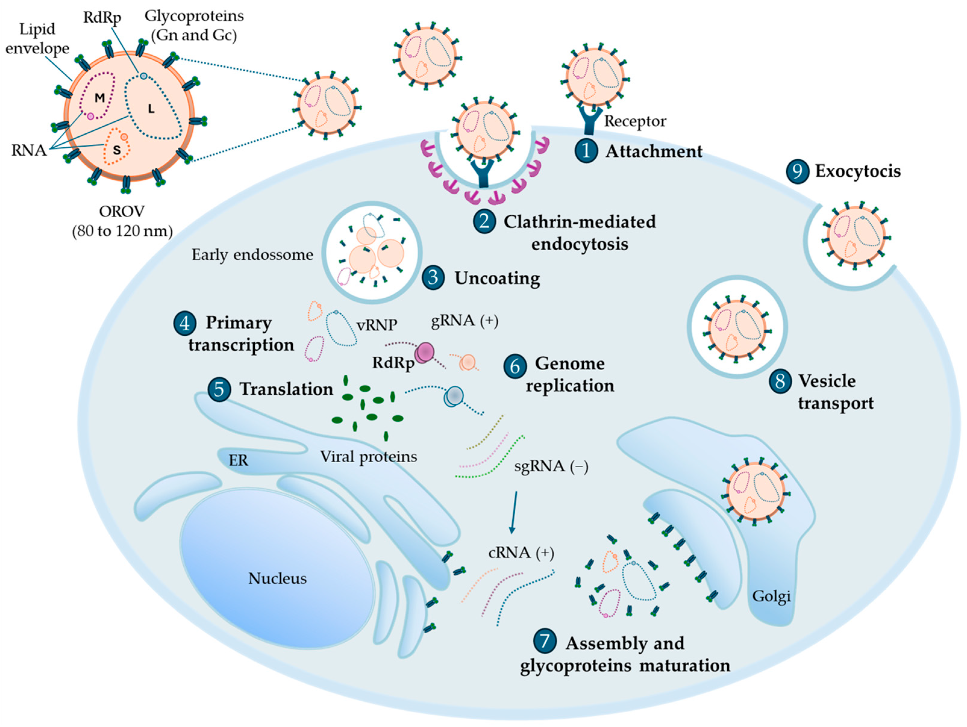
OROV is a segmented negative-sense RNA virus belonging to the genus Orthobunyavirus and the family Peribunyaviridae. This enveloped virus, with a diameter ranging from 80 to 120 nm, possesses a tripartite genome consisting of three single-stranded RNA segments: Large (L), Medium (M), and Small (S). The L segment is approximately 6852 nucleotides long and encodes the RdRp, while the M segment is approximately 4385 nucleotides long and encodes the envelope glycoproteins Gn and Gc, as well as the non-structural protein of the M segment (NSm), which is thought to contribute to viral assembly and budding. Furthermore, the S segment is approximately 946 nucleotides in length and encodes the nucleocapsid (N) protein and the non-structural protein of the S segment (NSs), a key factor involved in immune evasion and viral pathogenesis [1,37,38].
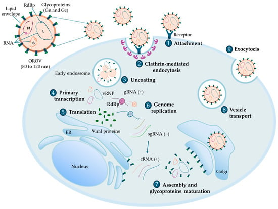
The OROV replication cycle depends on endocytic entry and membrane fusion within acidified intracellular compartments. It begins with the binding of the viral surface Gn and Gc to specific host-cell receptors, including the low-density lipoprotein receptor-related protein 1 (LRP1) [39]. This interaction triggers clathrin-mediated endocytosis and subsequent formation of early endosomes. As the endosomal environment acidifies, the viral envelope fuses with the endosomal membrane, a process mediated by Gc, which functions as a class II viral fusion protein. Consequently, the viral ribonucleoprotein complexes (vRNPs) are released into the host-cell cytoplasm [40].
Intracellularly, RdRp initiates the primary transcription of the negative-sense genomic RNA (gRNA) to generate subgenomic mRNAs (sgRNA), which are subsequently translated by the host-cell machinery into viral proteins. RdRp employs a “cap-snatching” mechanism, whereby capped fragments derived from host mRNAs are used to prime viral mRNA synthesis [41]. Simultaneously, RdRp synthesizes full-length complementary RNA (cRNA) intermediates from the genomic RNA segments, which subsequently serve as templates to produce progeny genomic RNA [20].
The final stages of the replication cycle involve the maturation of Gn and Gc within the endoplasmic reticulum–Golgi intermediate compartment and the Golgi apparatus. Newly synthesized genomic RNA associates with the N protein to form new vRNP complexes, which are then assembled into mature virions through Golgi-dependent budding. These mature particles are transported through the secretory vesicular trafficking pathway to the plasma membrane and released into the extracellular environment via calcium-dependent exocytosis (Figure 1) [20].
Figure 1.
OROV replication cycle. (1) OROV attaches to specific receptors on the host-cell surface through its glycoproteins Gn and Gc, promoting viral entry. (2) The virus enters the host-cell through clathrin-mediated endocytosis. (3) The viral envelope subsequently fuses with the endosomal membrane, resulting in viral uncoating and the release of viral ribonucleoprotein complexes (vRNPs) into the host-cell cytoplasm. (4) The RNA-dependent RNA polymerase (RdRp) initiates the primary transcription of the negative-sense genomic RNA (gRNA), generating subgenomic viral mRNAs. (5) These viral mRNAs are subsequently translated by the host-cell machinery into viral proteins. (6) During viral mRNA synthesis, RdRp employs a “cap-snatching” mechanism, hijacking capped host mRNA fragments to prime viral transcription. The negative-sense genomic RNA is subsequently replicated through a positive-sense antigenomic RNA intermediate, which serves as a template for the synthesis of new genomic RNA segments. (7) Newly synthesized genomic RNAs and structural proteins are transported to the Golgi apparatus, where virion assembly occurs. (8) Newly formed viral particles are transported from the Golgi apparatus in secretory vesicles toward the plasma membrane. (9) Finally, mature virions are released from the host-cell via calcium-dependent exocytosis, enabling OROV dissemination and the infection of neighboring cells.
6. Potential Vaccines in Development for OROV and Associated Challenges
Developing safe, effective, and accessible vaccines against OROV represents a critical public health priority for addressing this neglected emerging threat. The absence of licensed vaccines continues to place a substantial burden on healthcare systems in affected countries. Accordingly, several innovative vaccine strategies have recently emerged to address this unmet public health need.
Pioneering studies established the first reverse genetics system for OROV, successfully generating recombinant viruses lacking either the NSm or NSs proteins. These viral proteins play important roles in OROV pathogenesis. NSm has been implicated in viral morphogenesis and budding, whereas NSs functions as a major virulence factor by antagonizing interferon-mediated antiviral responses and suppressing host-cell transcription, thereby promoting immune evasion [38,79]. Similar attenuation strategies have been successfully applied to other orthobunyaviruses, including Rift Valley fever virus, in which deletion of NSs and NSm generated genetically stable, attenuated, and immunogenic vaccine candidates capable of inducing protective immunity without causing clinical disease [80,81]. Therefore, the generation of OROV mutants lacking these virulence-associated factors provides a promising platform for the development of live-attenuated vaccines with improved safety profiles and the potential to induce robust and long-lasting immune responses.
In parallel with reverse genetics approaches, immunoinformatic-based strategies have emerged as valuable tools for rational vaccine design. Computational analyses have been employed to identify highly antigenic regions within the OROV polyprotein, including potential B-cell and T-cell epitopes with strong immunogenic potential. These approaches have also enabled the prediction of three-dimensional protein structures and the identification of potential ligand-binding sites through molecular docking analyses [82].
Such in silico approaches provide a rapid and cost-effective framework for antigen selection, particularly for neglected viral diseases, in which conventional vaccine development is often constrained by financial and technological limitations. However, despite these advantages, epitope-based vaccine candidates still require extensive experimental validation to confirm their immunogenicity, safety, and protective efficacy [82,83].
Among the most advanced preclinical vaccine platforms evaluated against OROV is a replication-competent chimeric vesicular stomatitis virus (VSV)-based vaccine expressing the OROV glycoprotein complex. The VSV-OROV chimera induced robust neutralizing antibody responses, including antibodies directed against the N-terminal region of the Gc. Importantly, vaccination significantly reduced viral replication and tissue viral loads in mice challenged with wild-type OROV, providing the first experimental evidence that targeting OROV surface glycoproteins may represent an effective vaccination strategy [84].
The VSV vector platform offers several advantages, including low levels of pre-existing vector immunity, the absence of genomic integration into host-cells, and a high capacity for the incorporation of foreign genetic sequences. Furthermore, this platform has already demonstrated clinical success through the rVSV-ZEBOV (Ervebo) vaccine against Ebola virus disease, which was approved by the European Medicine Agency in November 2019 and by the U.S. Food and Drug Administration in December 2019. This vaccine demonstrated very high efficacy in a Phase III ring vaccination trial conducted during the West Africa outbreak. Moreover, a single dose of the rVSV-ZEBOV (Ervebo) induces durable antibody responses for up to five years, supporting its utility for outbreak control despite gradual decline in antibody levels over time [85,86].
More recently, human codon-optimized messenger RNA–lipid nanoparticle (mRNA-LNP) vaccines encoding the OROV envelope glycoproteins from both a historical prototype lineage (BeAn19991, Brazil) and a recently circulating outbreak lineage (AM0059, Brazil) have emerged as highly promising vaccine candidates. Preclinical studies demonstrated that these vaccines provided complete protection against lethal OROV challenge in BALB/c and A129 mice by inducing robust humoral and cellular immune responses, including high titers of OROV-specific IgG antibodies and potent CD4+ and CD8+ T-cell responses. Notably, vaccines based on contemporary outbreak-associated sequences also protected against prototype viral strains, suggesting that incorporating updated antigenic sequences may enhance cross-lineage protection [87]. The flexibility of mRNA-LNP platforms, which enables the rapid modification in antigen sequences according to circulating viral variants, represents a major advantage for addressing the genetic diversity and evolutionary dynamics of OROV [23,87].
Despite these advances, several challenges remain for the successful development of an effective OROV vaccine. One important limitation is the lack of high-resolution structural information on the viral surface Gn and Gc, which restricts the rational identification of optimal antigenic targets and raises questions regarding whether future vaccines should target the glycoproteins, the nucleocapsid, or combinations of multiple viral antigens [23].
Genomic surveillance has identified amino acid substitutions in surface-exposed regions of the Gc among contemporary OROV strains, which may contribute to immune escape from antibodies elicited against ancestral lineages and underscore the need for lineage-matched vaccines or broadly neutralizing antibody-based approaches. Importantly, recent studies have identified antigenic differences between the historical prototype strain BeAn19991 and contemporary outbreak-associated lineages, such as AM0059. These findings suggest that vaccines based exclusively on ancestral strains may exhibit reduced neutralizing activity against currently circulating viruses, emphasizing the importance of incorporating updated antigenic sequences or developing broadly protective vaccine strategies [87].
Animal models remain essential for understanding OROV pathogenesis and evaluating vaccine efficacy. Although immunocompetent adult mice generally do not develop severe disease following infection, IFNAR−/− mice and Syrian hamsters are widely used as relevant models for investigating viral replication, disease mechanisms, and vaccine-induced protection [33,35]. Furthermore, non-human primates support efficient viral replication and mount immune responses that more closely resemble those observed in humans, providing an important translational model. An important research priority is determining whether maternal vaccination can prevent vertical transmission and fetal complications, particularly in light of recent evidence demonstrating OROV infection in human placental explants and trophoblast organoid models [22].
Additionally, vaccine candidates must demonstrate favorable safety profiles across diverse populations, including pregnant individuals, given previous concerns associated with vaccine development against other orthobunyaviruses [81]. Another critical consideration is the establishment of scalable, affordable, and sustainable manufacturing platforms to ensure vaccine accessibility in endemic regions with limited resources. Although several vaccine candidates have demonstrated promising immunogenicity and protective efficacy in animal models, none has yet advanced to clinical evaluation in humans [23].
The clinical diagnosis of OROV infection remains challenging because its symptoms frequently overlap with those of other co-circulating arboviral diseases in endemic regions, particularly dengue and yellow fever, making clinical diagnosis difficult [88]. Consequently, misdiagnosis may lead to inappropriate clinical management, delays in public health interventions, and underestimation of viral circulation.
Accurate and rapid identification of OROV infection is essential for mapping outbreak areas, monitoring viral dissemination, and implementing targeted control strategies, including vector-control measures and public health awareness campaigns. Recently, mouse monoclonal antibodies developed for OROV detection in indirect immunofluorescence assays and immunohistochemistry have shown considerable promise as diagnostic tools [89]. Although the two anti-OROV monoclonal antibodies exhibited promising reactivity patterns, they have not yet been evaluated against all four recognized OROV genotypes. Therefore, it remains unclear whether these antibodies can detect all currently circulating OROV lineages in South America. Nevertheless, the monoclonal antibody 63B3E7 recognizes a linear epitope within the nucleocapsid protein, one of the most conserved viral proteins among orthobunyaviruses, suggesting its potential broad applicability for OROV detection. Overall, these monoclonal antibodies represent valuable and versatile tools for OROV research, with applications in differential diagnosis, epidemiological surveillance, and studies of viral biology and pathogenesis [89].
The development of an effective OROV vaccine is essential to control future outbreaks and reduce the impact of this emerging arboviral disease. The combination of arthropod-borne transmission, increasing human mobility, and the expanding geographic distribution of OROV creates a significant risk of further dissemination beyond traditionally endemic regions [84]. A licensed vaccine would therefore represent a valuable preventive strategy, with the potential to reduce disease burden, prevent severe clinical manifestations, and protect vulnerable populations [74,90].
Vaccination strategies would be particularly relevant for communities in endemic regions of South and Central America, where recurrent outbreaks place considerable pressure on healthcare systems and contribute to substantial social and economic burdens [23,72]. In parallel with vaccine development, strengthening genomic surveillance, improving early diagnostic capacity, and enhancing epidemiological surveillance will remain essential components of integrated public health strategies aimed at controlling OROV transmission and limiting its future geographic spread. Collectively, these efforts will be critical for reducing the global impact of OROV infection and strengthening preparedness against future outbreaks of this re-emerging arbovirus.
7. Conclusions and Future Directions
In conclusion, OROV has emerged as an increasingly important public health concern in the Americas, driven by its expanding geographic distribution, the widespread distribution of competent vectors, and the large proportion of susceptible individuals. In this context, the development of effective antivirals and safe, effective vaccines is essential. Strategies such as drug repurpose and the identification of viral and host therapeutic targets offer substantial reductions in development time and cost, lower the risk of toxicity-related failures, and enable more rapid therapeutic responses, which are particularly important during outbreaks.
At the same time, important limitations must be acknowledged. Our current understanding of OROV biology, including its pathogenesis and the immune mechanisms underlying protective immunity, remains incomplete, directly limiting the rational design of both antiviral agents and vaccine candidates. Furthermore, the impact of viral genetic diversity on antiviral efficacy and vaccine performance has not yet been comprehensively evaluated.
OROV possesses a tripartite segmented RNA genome, which facilitates frequent genetic reassortment, as demonstrated by the 2023–2024 outbreak lineage containing an eastern Amazonian M segment combined with L and S segments derived from viruses circulating in Peru, Colombia, and Ecuador. This novel reassorting lineage exhibits enhanced replicative fitness and increased virulence compared with the historical Lineage I prototype strain BeAn19991. Such genetic variation represents a major challenge for therapeutic and vaccine development, as sequence divergence within the Gc may promote immune escape and reduce susceptibility to neutralizing antibodies elicited against ancestral strains.
Furthermore, although several antiviral candidates have shown promising activity, their mechanisms of action remain incompletely characterized in many cases. Among the compounds evaluated, 4′-fluorouridine (4′-FlU) stands out as one of the most promising antiviral candidates against OROV, combining sub nanomolar-to-low-nanomolar antiviral potency, high selectivity, and complete protection in animal models. In contrast, remdesivir and molnupiravir exhibit potent in vitro antiviral activity but still require validation in in vivo models of OROV infection. Although favipiravir and ribavirin demonstrated protective efficacy in animal models, their substantially higher inhibitory concentrations suggest lower intrinsic antiviral potency. Importantly, in vitro antiviral potency alone does not necessarily predict therapeutic efficacy in vivo. When in vivo efficacy is considered, the relative performance of these compounds changes substantially. Despite its comparatively modest in vitro activity, favipiravir significantly protected Syrian hamsters by limiting viral dissemination to peripheral organs and the central nervous system. Likewise, ribavirin reduced viral loads and provided partial protection, although it failed to achieve complete viral clearance. Despite these encouraging findings, further preclinical investigation remains necessary before clinical translation can be considered. Although high viral replication has been demonstrated in human placental explants and three-dimensional brain organoids, correlating with manifestations such as vertical transmission and neuroinvasive disease, these experimental systems cannot fully recapitulate the complex circulatory, immunological, and multi-organ interactions that occur during human infection.
This limitation is further compounded by shortcomings in currently available animal models. Immunocompetent adult mice are naturally resistant to severe OROV disease, necessitating the use of immunocompromised strains, such as IFNAR−/− mice, which do not fully reproduce intact human immune responses. Syrian hamsters provide a naturally susceptible alternative but remain limited by the availability of immunological reagents for detailed immune characterization. Non-human primates, including rhesus and pigtail macaques, support efficient viral replication and develop immune responses that closely resemble those observed in humans. However, they generally remain clinically asymptomatic, limiting their utility for studies of disease pathogenesis. Furthermore, their use is constrained by high costs and important ethical considerations, despite their value for evaluating vaccine safety and efficacy.
Several vaccine platforms are currently under development, including mRNA-LNP vaccines, VSV-based chimeric vaccines, and live-attenuated NSs/NSm-deficient viruses. Recent studies have demonstrated that lineage-matched mRNA-LNP vaccines encoding OROV glycoproteins provide complete protection in murine models. Nevertheless, prototype-based vaccines derived from BeAn19991 may provide only partial cross-protection against recently emerged reassorting epidemic strains because of subtle but immunologically relevant antigenic differences within the Gc. These findings underscore the urgent need for flexible vaccine platforms that can be rapidly updated based on real-time genomic surveillance.
Similarly, the contributions of innate and adaptive immune responses, particularly those mediated by natural killer cells and T lymphocytes, to protection against OROV infection remain poorly understood. Future studies should investigate the potential occurrence of antibody-dependent enhancement through in vivo reinfection models and cross-neutralization assays involving phylogenetically related orthobunyaviruses. A more comprehensive understanding of the immune mechanisms underlying OROV infection will be essential for guiding the development of vaccines capable of inducing durable, broad, and cross-protective immunity against genetically diverse OROV lineages.
Taken together, these challenges underscore the urgent need for integrated, multidisciplinary, and sustained research efforts focused on OROV. Progress in this field will depend not only on continued scientific and technological advances but also on interdisciplinary collaboration and international cooperation to mitigate the geographic expansion of OROV. Strengthening surveillance systems, investing in adaptable and scalable technological platforms, and ensuring equitable access to future antiviral therapies and vaccines will be fundamental to reducing the public health burden of this neglected emerging arbovirus.
Author Contributions
Conceptualization, V.C.S. and S.S.G.D.; writing—original draft preparation, V.C.S. and S.S.G.D.; writing—review and editing, V.C.S. and S.S.G.D.; supervision, S.S.G.D. All authors have read and agreed to the published version of the manuscript.
Funding
This research received no external funding.
Data Availability Statement
No new data were created or analyzed in this study. Data sharing is not applicable to this article.
Acknowledgments
The figures were partly generated using Servier Medical Art (https://smart.servier.com/, accessed on 28 May 2026), licensed under CC BY 4.0 (https://creativecommons.org/licenses/by/4.0/, accessed on 28 May 2026). The authors have reviewed and edited the output and take full responsibility for the content of this publication. The authors would like to thank Coordenação de Aperfeiçoamento de Pessoal de Nível Superior (CAPES)—Finance Code 001—Process 88887.078169/2024-00, and Fundação Carlos Chagas Filho de Amparo à Pesquisa do Estado do Rio de Janeiro (FAPERJ)—Process E-26/200.351/2025 for the research fellowships awarded to the authors.
Conflicts of Interest
The authors declare no conflicts of interest.
Abbreviations
The following abbreviations are used in this manuscript:
| 4′-FlU | 4′-fluorouridine |
| CPE | Cytopathic effect |
| cRNA | Complementary RNA |
| dpi | Days post-infection |
| dsRNA | Double-stranded RNA |
| FASLG | Fas Ligand |
| FCCR3A | Fc Gamma Receptor IIIa |
| Gc | Glycoprotein C |
| Gn | Glycoprotein N |
| gRNA | Genomic RNA |
| HCV | Hepatitis C virus |
| hpi | Hours post-infection |
| IF16 | Interferon Alpha Inducible Protein 6 |
| IFIT1 | Interferon-Induced Protein with Tetratricopeptide Repeats 1 |
| IFN-I | Type I interferon |
| IFN-α | Interferon-alpha |
| IFN-β | Interferon-beta |
| IL-10 | Interleukin 10 |
| ISG15 | Interferon-Stimulated Gene 15 |
| ISGs | Interferon-stimulated genes |
| Large | L segment |
| LLOQ | Lower limit of quantification |
| Lrp1 | Low-density lipoprotein receptor-related protein 1 |
| Medium | M segment |
| mRNA-LNP | mRNA-lipid nanoparticle |
| MX1 | MX Dynamin-Like GTPase 1 |
| N | Nucleocapsid |
| NHC | β-D-N4-hydroxycytidine triphosphate |
| NSm | Non-structural protein of the M Segment |
| NSs | Non-structural protein of the S Segment |
| OROV | Oropouche virus |
| PTPRC | Protein Tyrosine Phosphatase Receptor Type C |
| RBV | Ribavirin |
| RdRp | RNA-dependent RNA polymerase |
| RMP | Remdesivir monophosphate |
| RTR | Triphosphate form |
| SARS-CoV-2 | Severe acute respiratory syndrome coronavirus 2 |
| SFTS | Severe Fever with Thrombocytopenia Syndrome |
| Small | S segment |
| SgRNA | Subgenomic mRNAs |
| vRNP | Viral ribonucleoprotein complex |
| VSV | Vesicular stomatitis virus |
| WDL | Wedelolactone |
References
- Files, M.A.; Hansen, C.A.; Herrera, V.C.; Schindewolf, C.; Barrett, A.D.T.; Beasley, D.W.C.; Bourne, N.; Milligan, G.N. Baseline mapping of Oropouche virology, epidemiology, therapeutics, and vaccine research and development. NPJ Vaccines 2022, 7, 38. [Google Scholar] [CrossRef] [Scilit] [PubMed]
- Guagliardo, S.A.J.; Connelly, C.R.; Lyons, S.; Martin, S.W.; Sutter, R.; Hughes, H.R.; Brault, A.C.; Lambert, A.J.; Gould, C.V.; Staples, J.E. Reemergence of Oropouche Virus in the Americas and Risk for Spread in the United States and Its Territories, 2024. Emerg. Infect. Dis. 2024, 30, 2241–2249. [Google Scholar] [CrossRef] [Scilit] [PubMed]
- Riccò, M.; Corrado, S.; Bottazzoli, M.; Marchesi, F.; Gili, R.; Bianchi, F.P.; Frisicale, E.M.; Guicciardi, S.; Fiacchini, D.; Tafuri, S.; et al. (Re-)Emergence of Oropouche Virus (OROV) Infections: Systematic Review and Meta-Analysis of Observational Studies. Viruses 2024, 16, 1498. [Google Scholar] [CrossRef] [Scilit] [PubMed]
- Vernal, S.; Martini, C.C.R.; da Fonseca, B.A.L. Oropouche Virus-Associated Aseptic Meningoencephalitis, Southeastern Brazil. Emerg. Infect. Dis. 2019, 25, 380–382. [Google Scholar] [CrossRef] [Scilit] [PubMed]
- Martos-Benítez, F.D.; Betancourt-Plaza, I.; Osorio-Carmenates, I.; González-Martínez, N.J.; Moráles-Suárez, I.; Peña-García, C.E.; Pérez-Matos, Y.L.; Lestayo-O’Farrill, Z.; de Armas-Fernández, J.R.; Cárdenas-González, R.C.; et al. Neurological Performance and Clinical Outcomes Related to Patients with Oropouche-Associated Guillain-Barré Syndrome. J. Peripher. Nerv. Syst. 2025, 30, e12683. [Google Scholar] [CrossRef] [Scilit] [PubMed]
- Manuli, E.R.; Hua, X.; Scachetti, G.C.; Forato, J.; Claro, I.M.; Pereira, G.M.; Sacchetto, L.; Cortes-Azuero, O.; Bernardo, A.C.; Resende, C.F.; et al. Transmission dynamics of Oropouche virus in Latin America and the Caribbean. Nat. Med. 2026, 32, 1383–1392. [Google Scholar] [CrossRef] [Scilit] [PubMed]
- de Melo Iani, F.C.; Pereira, F.M.; de Oliveira, E.C.; Rodrigues, J.T.N.; Machado, M.H.; Fonseca, V.; Adelino, T.E.R.; Guimarães, N.R.; Tomé, L.M.R.; Gómez, M.K.A.; et al. Travel-associated international spread of Oropouche virus beyond the Amazon. J. Travel Med. 2025, 32, taaf018. [Google Scholar] [CrossRef] [Scilit] [PubMed]
- Srivastava, S.; Sah, R.; Babu, M.R.; Sharma, D.; Kumar, S.; Sridhar, S.B.; Wadhwa, T.; Shareef, J.; Rao, G.S.N.K.; Feehan, J.; et al. The emergence of oropouche fever: A potential new threat? New Microbes New Infect. 2025, 65, 101596. [Google Scholar] [CrossRef] [Scilit] [PubMed]
- Rodriguez-Morales, A.J.; Navarro, J.C.; Romero-Alvarez, D.; Paniz-Mondolfi, A.; Forero-Peña, D.A.; Naranjo-Lara, L.; Suárez, J.A. Reemergence of Oropouche virus infection in Ecuador, 2024: Vectors and cases. J. Infect. 2025, 91, 106544. [Google Scholar] [CrossRef] [Scilit] [PubMed]
- Bello-Rodríguez, B.M.; Vega-Jiménez, J.; Cañete, R.; Rodriguez-Morales, A.J. Emergence of Oropouche virus infection in Matanzas, Cuba, 2024. J. Infect. 2025, 90, 106470. [Google Scholar] [CrossRef] [Scilit] [PubMed]
- Usuga, J.; Limonta, D.; Perez-Restrepo, L.S.; Ciuoderis, K.A.; Moreno, I.; Arevalo, A.; Vargas, V.; Berg, M.G.; Cloherty, G.A.; Hernandez-Ortiz, J.P.; et al. Co-Circulation of 2 Oropouche Virus Lineages, Amazon Basin, Colombia, 2024. Emerg. Infect. Dis. 2024, 30, 2375–2380. [Google Scholar] [CrossRef] [Scilit] [PubMed]
- Scroggs, S.L.P.; Gutierrez, J.; Reister-Hendricks, L.M.; Gunter, K.B.; Tilston, N.L.; McGregor, B.L. Enhanced infection and transmission of the 2022-2024 Oropouche virus strain in the North American biting midge Culicoides sonorensis. Sci. Rep. 2025, 15, 27368. [Google Scholar] [CrossRef] [Scilit] [PubMed]
- Capobianchi, M.R.; Castilletti, C.; Gobbi, F.G. Potential risks of Oropouche virus importation into Europe. J. Travel Med. 2024, 31, taae109. [Google Scholar] [CrossRef] [Scilit] [PubMed]
- Tilston-Lunel, N.L. Oropouche Virus: An Emerging Orthobunyavirus. J. Gen. Virol. 2024, 105, 002027. [Google Scholar] [CrossRef] [Scilit] [PubMed]
- Bandeira, A.C.; Pereira, F.M.; Leal, A.; Santos, S.P.O.; Barbosa, A.C.; Souza, M.S.P.L.; de Souza, D.R.; Guimaraes, N.; Fonseca, V.; Giovanetti, M.; et al. Fatal Oropouche Virus Infections in Nonendemic Region, Brazil, 2024. Emerg. Infect. Dis. 2024, 30, 2370–2374. [Google Scholar] [CrossRef] [Scilit] [PubMed]
- Pan American Health Organization. Epidemiological Update: Oropouche in the Region of the Americas, 13 August 2025; PAHO: Washington, DC, USA, 2025. Available online: https://www.paho.org/en/documents/epidemiological-update-oropouche-region-americas-13-august-2025 (accessed on 19 June 2026).
- Schwartz, D.A.; Dashraath, P.; Baud, D. Oropouche Virus (OROV) in Pregnancy: An Emerging Cause of Placental and Fetal Infection Associated with Stillbirth and Microcephaly following Vertical Transmission. Viruses 2024, 16, 1435. [Google Scholar] [CrossRef] [Scilit] [PubMed]
- Schwartz, D.A. Novel Reassortants of Oropouche Virus (OROV) Are Causing Maternal-Fetal Infection During Pregnancy, Stillbirth, Congenital Microcephaly and Malformation Syndromes. Genes 2025, 16, 87. [Google Scholar] [CrossRef] [Scilit] [PubMed]
- Martins-Filho, P.R.; Carvalho, T.A.; Dos Santos, C.A. Oropouche fever: Reports of vertical transmission and deaths in Brazil. Lancet Infect. Dis. 2024, 24, e662–e663. [Google Scholar] [CrossRef] [Scilit] [PubMed]
- Lin, S.; Wang, Q.; Wang, C.; Liu, Q. Oropouche virus: Molecular virological characteristics and research progress of a neglected arbovirus. Virol. J. 2025, 22, 393. [Google Scholar] [CrossRef] [Scilit] [PubMed]
- Centers for Disease Control and Prevention (CDC). Clinical Overview of Oropouche Virus Disease. 2025. Available online: https://www.cdc.gov/oropouche/hcp/clinical-overview/index.html# (accessed on 14 May 2026).
- Morley, D.; Kennedy, E.; Dowall, S. Preclinical Models of Oropouche Virus Infection and Disease. Pathogens 2025, 14, 1272. [Google Scholar] [CrossRef] [Scilit] [PubMed]
- Wu, W.; Kam, Y.W. Oropouche Virus (OROV) Vaccine Development for Vulnerable Populations: Epidemiological Context, Challenges and Future Directions. Vaccines 2026, 14, 267. [Google Scholar] [CrossRef] [Scilit] [PubMed]
- Li, P.; Zhao, D.; Fu, J.; Zhang, Y.; Cai, S.; Deng, M.; Li, C.; Huang, J.; Qin, L.; Gao, H.; et al. Orthobunyaviruses: Epidemiology, innate immune escape, and intervention strategies. Virulence 2025, 16, 2585624. [Google Scholar] [CrossRef] [Scilit] [PubMed]
- Sankhe, S.; Dieng, I.; Kane, M.; Diallo, A.; Ndiaye, N.A.; Top, N.M.; Dia, M.; Faye, O.; Sall, A.A.; Sembene, P.M.; et al. Genomic Insights into Neglected Orthobunyaviruses: Molecular Characterization and Phylogenetic Analysis. Viruses 2025, 17, 406. [Google Scholar] [CrossRef] [Scilit] [PubMed]
- Naveca, F.G.; Almeida, T.A.P.; Souza, V.; Nascimento, V.; Silva, D.; Nascimento, F.; Mejía, M.; Oliveira, Y.S.; Rocha, L.; Xavier, N.; et al. Human outbreaks of a novel reassortant Oropouche virus in the Brazilian Amazon region. Nat. Med. 2024, 30, 3509–3521. [Google Scholar] [CrossRef] [Scilit] [PubMed]
- de Souza, W.M.; Calisher, C.H.; Carrera, J.P.; Hughes, H.R.; Nunes, M.R.T.; Russell, B.; Tilson-Lunel, N.L.; Venter, M.; Xia, H. ICTV Virus Taxonomy Profile: Peribunyaviridae 2024. J. Gen. Virol. 2024, 105, 002034. [Google Scholar] [CrossRef] [Scilit] [PubMed]
- Elliott, R.M. Orthobunyaviruses: Recent genetic and structural insights. Nat. Rev. Microbiol. 2014, 12, 673–685. [Google Scholar] [CrossRef] [Scilit] [PubMed]
- Gunter, K.B.; Bowen, J.M.; Clarke, A.T.; McFarlane, M.; Omoga, D.C.A.; Pozuelos, S.; Rogers, L.M.; Aronoff, D.M.; Vornhagen, J.; Brennan, B.; et al. From prototype to outbreak: Conserved pathogenesis of Oropouche virus in a novel murine pregnancy model highlights its public health implications. bioRxiv 2025. [Google Scholar] [CrossRef] [Scilit] [PubMed]
- Scachetti, G.C.; Forato, J.; Claro, I.M.; Hua, X.; Salgado, B.B.; Vieira, A.; Simeoni, C.L.; Barbosa, A.R.C.; Rosa, I.L.; de Souza, G.F.; et al. Re-emergence of Oropouche virus between 2023 and 2024 in Brazil: An observational epidemiological study. Lancet Infect. Dis. 2025, 25, 166–175. [Google Scholar] [CrossRef] [Scilit] [PubMed]
- Okesanya, O.J.; Amisu, B.O.; Adigun, O.A.; Ahmed, M.M.; Agboola, A.O.; Kab, T.; Eshun, G.; Ukoaka, B.M.; Oso, T.A.; Ogaya, J.B.; et al. Correction: Addressing the emerging threat of Oropouche virus: Implications and public health responses for healthcare systems. Trop. Dis. Travel Med. Vaccines 2026, 12, 1. [Google Scholar] [CrossRef] [Scilit] [PubMed]
- Yoshikawa, R.; Ishii, Y.; Sano, N.; Yasuda, J. Remdesivir as a potent antiviral against prototype and current epidemic Oropouche virus strains (BeAn19991 and PE-IAM4637). Virus Res. 2026, 363, 199680. [Google Scholar] [CrossRef] [Scilit] [PubMed]
- Proenca-Modena, J.L.; Sesti-Costa, R.; Pinto, A.K.; Richner, J.M.; Lazear, H.M.; Lucas, T.; Hyde, J.L.; Diamond, M.S. Oropouche virus infection and pathogenesis are restricted by MAVS, IRF-3, IRF-7, and type I interferon signaling pathways in nonmyeloid cells. J. Virol. 2015, 89, 4720–4737. [Google Scholar] [CrossRef] [Scilit] [PubMed]
- Gunter, K.; Omoga, D.; Bowen, J.M.; Gonzalez, L.R.; Severt, S.; Davis, M.; Szymanski, M.; Sandusky, G.; Duprex, W.P.; Tilston-Lunel, N.L. A reporter Oropouche virus expressing ZsGreen from the M segment enables pathogenesis studies in mice. J. Virol. 2024, 98, e0089324. [Google Scholar] [CrossRef] [Scilit] [PubMed]
- Rodrigues, A.H.; Santos, R.I.; Arisi, G.M.; Bernardes, E.S.; Silva, M.L.; Rossi, M.A.; Lopes, M.B.; Arruda, E. Oropouche virus experimental infection in the golden hamster (Mesocrisetus auratus). Virus Res. 2011, 155, 35–41. [Google Scholar] [CrossRef] [Scilit] [PubMed]
- Yee, D.S.; Davies, J.P.; VanBlargan, L.A.; Rahmberg, A.R.; Burgomaster, K.E.; Brooks, K.; Flynn, J.K.; Shue, B.; Ortiz, A.M.; Schaughency, P.; et al. Oropouche virus efficiently replicates and is immunostimulatory in vivo in nonhuman primate species. Sci. Adv. 2025, 11, eadx9405. [Google Scholar] [CrossRef] [Scilit] [PubMed]
- Thomas, S. Charting the Proteins of Oropouche Virus. Viruses 2025, 17, 1434. [Google Scholar] [CrossRef] [Scilit] [PubMed]
- Tilston-Lunel, N.L.; Acrani, G.O.; Randall, R.E.; Elliott, R.M. Generation of Recombinant Oropouche Viruses Lacking the Nonstructural Protein NSm or NSs. J. Virol. 2015, 90, 2616–2627. [Google Scholar] [CrossRef] [Scilit] [PubMed]
- Schwarz, M.M.; Price, D.A.; Ganaie, S.S.; Feng, A.; Mishra, N.; Hoehl, R.M.; Fatma, F.; Stubbs, S.H.; Whelan, S.P.J.; Cui, X.; et al. Oropouche orthobunyavirus infection is mediated by the cellular host factor Lrp1. Proc. Natl. Acad. Sci. USA 2022, 119, e2204706119. [Google Scholar] [CrossRef] [Scilit] [PubMed]
- Xin, Q.; Lozach, P.Y. Orthobunyavirus entry: Receptors, endocytosis, and fusion cues. Curr. Opin. Virol. 2026, 75, 101523. [Google Scholar] [CrossRef] [Scilit] [PubMed]
- Olschewski, S.; Cusack, S.; Rosenthal, M. The Cap-Snatching Mechanism of Bunyaviruses. Trends Microbiol. 2020, 28, 293–303. [Google Scholar] [CrossRef] [Scilit] [PubMed]
- Du, S.; Hu, X.; Li, P.; Xu, S.; Kim, M.; Liu, X.; Zhan, P. Antiviral drug discovery and development: Challenges and future directions. Signal Transduct. Target. Ther. 2026, 11, 69. [Google Scholar] [CrossRef] [Scilit] [PubMed]
- Sousa Moraes, C.; Gonzalez, G.; Sato, A.; Miki, S.; Inoue, A.; Tabata, K.; Kranrod, J.W.; Kabamba, C.F.; Ohnuma, A.; Matsuno, K.; et al. Integrated in vivo and transcriptomic analyses of lethal Oropouche virus infection reveal suppression of pathogenic host responses by antiviral therapy. bioRxiv 2026. [Google Scholar] [CrossRef] [Scilit]
- Ferrié, M.; Damuzey, M.; Tubiana, T.; Khan, M.; Roskams, T.; Weynand, B.; Cremers, N.; Hendrickx, S.; Donckers, K.; Portal, T.M.; et al. Oral 4’-fluorouridine completely suppresses epidemic Oropouche virus replication in mice and confers protection against virus-induced disease. bioRxiv 2025. [Google Scholar] [CrossRef] [Scilit]
- Westover, J.B.; Jung, K.H.; Rojas, I.; Bailey, K.W.; Landinez-Aponte, J.; Blumeling, G.R.; Mao, S.; Kolykhalov, A.A.; Natchus, M.G.; Painter, G.R.; et al. Effective treatment of advanced Oropouche virus, Rift Valley fever virus, and Dabie bandavirus infections with 4′-fluorouridine. mBio 2025, 16, e0146725. [Google Scholar] [CrossRef] [Scilit] [PubMed]
- Li, J.; Wang, X.; Ding, Y.; Qin, F.; de Lima, S.T.S.; Papamichail, L.; Schraauwen, R.; Forato, J.; Claro, I.M.; Hua, X.; et al. Human liver-derived organoids recapitulate Oropouche virus infection and manifestation, enabling antiviral drug discovery. Cell Rep. Med. 2026, 7, 102646. [Google Scholar] [CrossRef] [Scilit] [PubMed]
- Saivish, M.V.; Menezes, G.L.; da Silva, R.A.; de Assis, L.R.; Teixeira, I.D.S.; Fulco, U.L.; Avilla, C.M.S.; Eberle, R.J.; Santos, I.A.; Korostov, K.; et al. Acridones as promising drug candidates against Oropouche virus. Curr. Res. Microb. Sci. 2024, 6, 100217. [Google Scholar] [CrossRef] [Scilit] [PubMed]
- Shiraki, K.; Daikoku, T. Favipiravir, an anti-influenza drug against life-threatening RNA virus infections. Pharmacol. Ther. 2020, 209, 107512. [Google Scholar] [CrossRef] [Scilit] [PubMed]
- Lieber, C.M.; Aggarwal, M.; Yoon, J.J.; Cox, R.M.; Kang, H.J.; Sourimant, J.; Toots, M.; Johnson, S.K.; Jones, C.A.; Sticher, Z.M.; et al. 4′-Fluorouridine mitigates lethal infection with pandemic human and highly pathogenic avian influenza viruses. PLoS Pathog. 2023, 19, e1011342. [Google Scholar] [CrossRef] [Scilit] [PubMed]
- Sourimant, J.; Lieber, C.M.; Aggarwal, M.; Cox, R.M.; Wolf, J.D.; Yoon, J.J.; Toots, M.; Ye, C.; Sticher, Z.; Kolykhalov, A.A.; et al. 4′-Fluorouridine is an oral antiviral that blocks respiratory syncytial virus and SARS-CoV-2 replication. Science 2022, 375, 161–167. [Google Scholar] [CrossRef] [Scilit] [PubMed]
- Unal, M.A.; Bitirim, C.V.; Summak, G.Y.; Bereketoglu, S.; Cevher Zeytin, I.; Besbinar, O.; Gurcan, C.; Aydos, D.; Goksoy, E.; Kocakaya, E.; et al. Ribavirin shows antiviral activity against SARS-CoV-2 and downregulates the activity of TMPRSS2 and the expression of ACE2 in vitro. Can. J. Physiol. Pharmacol. 2021, 99, 449–460. [Google Scholar] [CrossRef] [Scilit] [PubMed]
- Soares, V.C.; Moreira, I.B.G.; Dias, S.S.G. SARS-CoV-2 Infection and Antiviral Strategies: Advances and Limitations. Viruses 2025, 17, 1064. [Google Scholar] [CrossRef] [Scilit] [PubMed]
- Galli, A.; Mens, H.; Gottwein, J.M.; Gerstoft, J.; Bukh, J. Antiviral Effect of Ribavirin against HCV Associated with Increased Frequency of G-to-A and C-to-U Transitions in Infectious Cell Culture Model. Sci. Rep. 2018, 8, 4619. [Google Scholar] [CrossRef] [Scilit] [PubMed]
- Livonesi, M.C.; De Sousa, R.L.; Badra, S.J.; Figueiredo, L.T. In vitro and in vivo studies of ribavirin action on Brazilian Orthobunyavirus. Am. J. Trop. Med. Hyg. 2006, 75, 1011–1016. [Google Scholar] [CrossRef] [Scilit]
- Maas, B.M.; Strizki, J.; Miller, R.R.; Kumar, S.; Brown, M.; Johnson, M.G.; Cheng, M.; De Anda, C.; Rizk, M.L.; Stone, J.A. Molnupiravir: Mechanism of action, clinical, and translational science. Clin. Transl. Sci. 2024, 17, e13732. [Google Scholar] [CrossRef] [Scilit] [PubMed]
- Garcia Filho, C.; Lima Neto, A.S.; Maia, A.M.P.C.; da Silva, L.O.R.; Cavalcante, R.D.C.; Monteiro, H.D.S.; Marques, K.C.A.; Oliveira, R.S.; Gadelha, S.A.C.; Nunes de Melo, D.; et al. A Case of Vertical Transmission of Oropouche Virus in Brazil. N. Engl. J. Med. 2024, 391, 2055–2057. [Google Scholar] [CrossRef] [Scilit] [PubMed]
- Malet, H.; Williams, H.M.; Cusack, S.; Rosenthal, M. The mechanism of genome replication and transcription in bunyaviruses. PLoS Pathog. 2023, 19, e1011060. [Google Scholar] [CrossRef] [Scilit] [PubMed]
- Mazzucco, M.B.; Talarico, L.B.; Vatansever, S.; Carro, A.C.; Fascio, M.L.; D’Accorso, N.B.; García, C.C.; Damonte, E.B. Antiviral activity of an N-allyl acridone against dengue virus. J. Biomed. Sci. 2015, 22, 29. [Google Scholar] [CrossRef] [Scilit] [PubMed]
- Tonelli, M.; Vettoretti, G.; Tasso, B.; Novelli, F.; Boido, V.; Sparatore, F.; Busonera, B.; Ouhtit, A.; Farci, P.; Blois, S.; et al. Acridine derivatives as anti-BVDV agents. Antivir. Res. 2011, 91, 133–141. [Google Scholar] [CrossRef] [Scilit] [PubMed]
- Menezes, G.d.L.; Saivish, M.V.; Sacchetto, L.; da Silva, G.C.D.; Teixeira, I.d.S.; Mistrão, N.F.B.; Nogueira, M.L.; Oliveira, J.I.N.; Bezerra, K.S.; da Silva, R.A.; et al. Exploring Quercetin Hydrate’s Potential as an Antiviral Treatment for Oropouche Virus. Biophysica 2023, 3, 485–500. [Google Scholar] [CrossRef] [Scilit]
- Coimbra, L.D.; Shimizu, J.F.; Nagai, A.; Borin, A.; Fontoura, M.A.; Concha, J.O.; Leme, L.; Lucas do Carmo, K.; de Oliveira, L.C.; Soprano, A.S.; et al. Lysergol exerts potent antiviral activity against the emerging Oropouche virus in vitro. Antivir. Res. 2025, 238, 106171. [Google Scholar] [CrossRef] [Scilit] [PubMed]
- Peinado, R.D.S.; Saivish, M.V.; Menezes, G.L.; Fulco, U.L.; da Silva, R.A.; Korostov, K.; Eberle, R.J.; Melo, P.A.; Nogueira, M.L.; Pacca, C.C.; et al. The search for an antiviral lead molecule to combat the neglected emerging Oropouche virus. Curr. Res. Microb. Sci. 2024, 6, 100238. [Google Scholar] [CrossRef] [Scilit] [PubMed]
- Mandova, T.; Saivish, M.V.; Menezes, G.L.; Bezerra, K.S.; Fulco, U.L.; da Silva, R.A.; Da Costa, F.B.; Nogueira, M.L. Antiviral Activity and Molecular Dynamics Simulation of Hops Compounds against Oropouche Virus. Pharmaceutics 2023, 15, 2769. [Google Scholar] [CrossRef] [Scilit] [PubMed]
- Lesjak, M.; Beara, I.; Simin, N.; Pintać, D.; Majkić, T.; Bekvalac, K.; Orčić, D.; Mimica-Dukić, N. Antioxidant and anti-inflammatory activities of quercetin and its derivatives. J. Funct. Foods 2018, 40, 68–75. [Google Scholar] [CrossRef] [Scilit]
- Saivish, M.V.; Menezes, G.L.; da Silva, R.A.; Fontoura, M.A.; Shimizu, J.F.; da Silva, G.C.D.; Teixeira, I.D.S.; Mistrão, N.F.B.; Hernandes, V.M.; Rahal, P.; et al. Antiviral Activity of Quercetin Hydrate against Zika Virus. Int. J. Mol. Sci. 2023, 24, 7504. [Google Scholar] [CrossRef] [Scilit] [PubMed]
- dos Santos, A.E.; Kuster, R.M.; Yamamoto, K.A.; Salles, T.S.; Campos, R.; de Meneses, M.D.; Soares, M.R.; Ferreira, D. Quercetin and quercetin 3-O-glycosides from Bauhinia longifolia (Bong.) Steud. show anti-Mayaro virus activity. Parasit. Vectors 2014, 7, 130. [Google Scholar] [CrossRef] [Scilit] [PubMed]
- Chiow, K.H.; Phoon, M.C.; Putti, T.; Tan, B.K.; Chow, V.T. Evaluation of antiviral activities of Houttuynia cordata Thunb. extract, quercetin, quercetrin and cinanserin on murine coronavirus and dengue virus infection. Asian Pac. J. Trop. Med. 2016, 9, 1–7. [Google Scholar] [CrossRef] [Scilit] [PubMed]
- Manvar, D.; Mishra, M.; Kumar, S.; Pandey, V.N. Identification and evaluation of anti hepatitis C virus phytochemicals from Eclipta alba. J. Ethnopharmacol. 2012, 144, 545–554. [Google Scholar] [CrossRef] [Scilit] [PubMed]
- Svrlanska, A.; Ruhland, A.; Marschall, M.; Reuter, N.; Stamminger, T. Wedelolactone inhibits human cytomegalovirus replication by targeting distinct steps of the viral replication cycle. Antivir. Res. 2020, 174, 104677. [Google Scholar] [CrossRef] [Scilit] [PubMed]
- Wang, Z.; Yan, H.; He, F.; Wang, J.; Zhang, Y.; Sun, L.; Hao, C.; Wang, W. Inhibition of herpes simplex virus by wedelolactone via targeting viral envelope and cellular TBK1/IRF3 and SOCS1/STAT3 pathways. Int. J. Antimicrob. Agents 2023, 62, 107000. [Google Scholar] [CrossRef] [Scilit] [PubMed]
- Colherinhas, G.; Cardoso, W.B. Insights into Antiviral Candidates against Oropouche Virus: A Molecular Dynamics Study. ACS Phys. Chem. Au 2025, 5, 549–559. [Google Scholar] [CrossRef] [Scilit] [PubMed]
- Yoosuf, B.T.; Gaidhane, A.M.; Vadia, N.; Menon, S.V.; Chennakesavulu, K.; Panigrahi, R.; Bushi, G.; Rani, A.; Sah, S.; Shabil, M.; et al. Epidemiology, transmission dynamics, treatment strategies, and future perspectives on Oropouche virus. Diagn. Microbiol. Infect. Dis. 2025, 113, 116882. [Google Scholar] [CrossRef] [Scilit] [PubMed]
- Dev Sharma, P.; Alhudhaibi, A.M.; Al Noman, A.; Abdallah, E.M.; Taha, T.H.; Sharma, H. Systems Biology-Driven Discovery of Host-Targeted Therapeutics for Oropouche Virus: Integrating Network Pharmacology, Molecular Docking, and Drug Repurposing. Pharmaceuticals 2025, 18, 613. [Google Scholar] [CrossRef] [Scilit] [PubMed]
- Connors, K.A.; Pedlow, M.R.; Frey, Z.D.; Cornejo Pontelli, M.; Whelan, S.P.J.; Duprex, W.P.; D’Aiuto, L.; Wills, Z.P.; Hartman, A.L. Neural cells are susceptible to historic and recently emerged Oropouche virus strains. PLoS Pathog. 2026, 22, e1013933. [Google Scholar] [CrossRef] [Scilit] [PubMed]
- Almeida, G.M.; Souza, J.P.; Mendes, N.D.; Pontelli, M.C.; Pinheiro, N.R.; Nogueira, G.O.; Cardoso, R.S.; Paiva, I.M.; Ferrari, G.D.; Veras, F.P.; et al. Neural Infection by Oropouche Virus in Adult Human Brain Slices Induces an Inflammatory and Toxic Response. Front. Neurosci. 2021, 15, 674576. [Google Scholar] [CrossRef] [Scilit] [PubMed]
- Bhutkar, M.; Singh, V.; Dhaka, P.; Tomar, S. Virus-host protein-protein interactions as molecular drug targets for arboviral infections. Front. Virol. 2022, 2, 959586. [Google Scholar] [CrossRef] [Scilit]
- Livonesi, M.C.; de Sousa, R.L.; Badra, S.J.; Figueiredo, L.T. In vitro and in vivo studies of the Interferon-alpha action on distinct Orthobunyavirus. Antivir. Res. 2007, 75, 121–128. [Google Scholar] [CrossRef] [Scilit] [PubMed]
- Muraro, S.P.; de Souza, G.F.; Alippe, Y.; Simeoni, C.L.; Vieira, A.; Forato, J.; Lavagnini, P.M.; Polonio, C.M.; de Oliveira, L.G.; Martini, M.C.; et al. Type I interferon controls vertical transmission and fetoplacental infection of Oropouche virus. iScience 2026, 29, 114647. [Google Scholar] [CrossRef] [Scilit] [PubMed]
- Zhang, Y.; Liu, X.; Wu, Z.; Feng, S.; Lu, K.; Zhu, W.; Sun, H.; Niu, G. Oropouche virus: A neglected global arboviral threat. Virus Res. 2024, 341, 199318. [Google Scholar] [CrossRef] [Scilit] [PubMed]
- Crabtree, M.B.; Kent Crockett, R.J.; Bird, B.H.; Nichol, S.T.; Erickson, B.R.; Biggerstaff, B.J.; Horiuchi, K.; Miller, B.R. Infection and transmission of Rift Valley fever viruses lacking the NSs and/or NSm genes in mosquitoes: Potential role for NSm in mosquito infection. PLoS Negl. Trop. Dis. 2012, 6, e1639. [Google Scholar] [CrossRef] [Scilit] [PubMed]
- Smith, D.R.; Johnston, S.C.; Piper, A.; Botto, M.; Donnelly, G.; Shamblin, J.; Albariño, C.G.; Hensley, L.E.; Schmaljohn, C.; Nichol, S.T.; et al. Attenuation and efficacy of live-attenuated Rift Valley fever virus vaccine candidates in non-human primates. PLoS Negl. Trop. Dis. 2018, 12, e0006474. [Google Scholar] [CrossRef] [Scilit] [PubMed]
- Adhikari, U.K.; Tayebi, M.; Rahman, M.M. Immunoinformatics Approach for Epitope-Based Peptide Vaccine Design and Active Site Prediction against Polyprotein of Emerging Oropouche Virus. J. Immunol. Res. 2018, 2018, 6718083. [Google Scholar] [CrossRef] [Scilit] [PubMed]
- Naveed, M.; Athar, A.; Tariq, A.; Asim, M.; Majeed, M.N.; El Hadi Mohamed, R.A.; Elkhadragy, M.F.; Alwethaynani, M.S.; Almsaud, M.M.; Nabi, G. Immunoinformatics-guided design of a multi-epitope vaccine targeting the envelopment polyprotein of oropouche virus. Mol. Genet. Genom. 2026, 301, 43. [Google Scholar] [CrossRef] [Scilit] [PubMed]
- Stubbs, S.H.; Cornejo Pontelli, M.; Mishra, N.; Zhou, C.; de Paula Souza, J.; Mendes Viana, R.M.; Lipkin, W.I.; Knipe, D.M.; Arruda, E.; Whelan, S.P.J. Vesicular Stomatitis Virus Chimeras Expressing the Oropouche Virus Glycoproteins Elicit Protective Immune Responses in Mice. mBio 2021, 12, e0046321. [Google Scholar] [CrossRef] [Scilit] [PubMed]
- Henao-Restrepo, A.M.; Camacho, A.; Longini, I.M.; Watson, C.H.; Edmunds, W.J.; Egger, M.; Carroll, M.W.; Dean, N.E.; Diatta, I.; Doumbia, M.; et al. Efficacy and effectiveness of an rVSV-vectored vaccine in preventing Ebola virus disease: Final results from the Guinea ring vaccination, open-label, cluster-randomised trial (Ebola Ça Suffit!). Lancet 2017, 389, 505–518. [Google Scholar] [CrossRef] [Scilit] [PubMed]
- Huttner, A.; Agnandji, S.T.; Engler, O.; Hooper, J.W.; Kwilas, S.; Ricks, K.; Clements, T.L.; Jonsdottir, H.R.; Nakka, S.S.; Rothenberger, S.; et al. Antibody responses to recombinant vesicular stomatitis virus-Zaire Ebolavirus vaccination for Ebola virus disease across doses and continents: 5-year durability. Clin. Microbiol. Infect. 2023, 29, 1587–1594. [Google Scholar] [CrossRef] [Scilit] [PubMed]
- Yamada, Y.; Cha, I.; Kang, S.; Yang, W.S.; Lewis, M.; Chung, C.; Shin, W.J.; Park, J.B.; Cho, N.H.; Choi, Y.K.; et al. Lineage-matched Oropouche virus mRNA-LNP vaccines confer complete, cross-protective immunity in mice. mBio 2026, 17, e0365525. [Google Scholar] [CrossRef] [Scilit] [PubMed]
- Travassos da Rosa, J.F.; de Souza, W.M.; Pinheiro, F.P.; Figueiredo, M.L.; Cardoso, J.F.; Acrani, G.O.; Nunes, M.R.T. Oropouche Virus: Clinical, Epidemiological, and Molecular Aspects of a Neglected Orthobunyavirus. Am. J. Trop. Med. Hyg. 2017, 96, 1019–1030. [Google Scholar] [CrossRef] [Scilit] [PubMed]
- Andreolla, A.P.; Borges, A.A.; Nagashima, S.; Vaz de Paula, C.B.; de Noronha, L.; Zanchin, N.I.T.; Bordignon, J.; Duarte Dos Santos, C.N. Development of monoclonal antibodies against oropouche virus and its applicability to immunohistochemical diagnosis. Virol. J. 2024, 21, 81. [Google Scholar] [CrossRef] [Scilit] [PubMed]
- Bai, F.; Denyoh, P.M.D.; Urquhart, C.; Shrestha, S.; Yee, D.A. A Comprehensive Review of the Neglected and Emerging Oropouche Virus. Viruses 2025, 17, 439. [Google Scholar] [CrossRef] [Scilit] [PubMed]

Disclaimer/Publisher’s Note: The statements, opinions and data contained in all publications are solely those of the individual author(s) and contributor(s) and not of MDPI and/or the editor(s). MDPI and/or the editor(s) disclaim responsibility for any injury to people or property resulting from any ideas, methods, instructions or products referred to in the content. |
© 2026 by the authors. Licensee MDPI, Basel, Switzerland. This article is an open access article distributed under the terms and conditions of the Creative Commons Attribution (CC BY) license.