


Abstract
Innate immunity predictors of HIV-1 risk and pathogenesis vary with reproductive hormones, pregnancy, and lactation, yet the underlying mechanisms remain unclear. We hypothesized that pregnancy-associated physiological adaptations alter systemic microRNA (miRNA) expression, thereby regulating immunity, pathogenesis and susceptibility to infection. We analyzed 174 serum samples from 88 participants in a longitudinal cohort from Uganda and Zimbabwe across pre-pregnancy (PP), pregnancy (P), and postpartum breastfeeding (BF). Cell-free peripheral blood miRNAs (n = 2083) were profiled using HTG EdgeSeq. Pregnancy-specific miRNAs were identified by intersecting differentially expressed (DE) miRNAs from P vs. PP and P vs. BF comparisons. miRNA targets and pathways were analyzed using miRWalk, Cytoscape/ClueGO, and cytoHubba. Pregnancy was associated with DE miRNAs (29 upregulated and 131 downregulated) targeting 2733 validated genes. Enriched pathways (FDR < 0.05) included adaptive immune response, Hippo Signaling, Cellular Senescence, HSV-1 infection, and two cancer-related pathways. Pregnancy-enriched targets within each pathway overlapped with the HIV–host interactome by 37–88%. Network analysis identified 47 hub genes interacting with 18 HIV-1 proteins, with Tat and gp120 being most connected viral and HLA-A being the most connected host protein. These findings indicate that pregnancy-driven systemic miRNAs target the HIV–host interactome and specifically identify pregnancy-enriched central hub genes involved in cell cycle control, viral immune evasion and replication to be further investigated for their predictive value in HIV acquisition and pathogenesis in longitudinal cohorts and experimental settings.
1. Introduction
Compelling evidence shows that the likelihood of HIV acquisition is elevated during pregnancy and the early postpartum period [1]. Reducing HIV acquisition requires comprehensive interventions, including early identification of risk factors and a deeper understanding of the immune mechanisms involved. HIV pathogenesis, disease progression and transmission are also affected by pregnancy, although negative outcomes are significantly reduced in combination with highly active antiretroviral therapies [2,3]. During pregnancy, the maternal immune system adapts to tolerate the fetus while maintaining protection against infections, with trimester-specific changes in complement activation, complement regulatory proteins, and levels of B cells, T regulatory cells, and neutrophils as well as shifts in CD4+ HIV target cell frequencies and activation [4,5,6,7]. However, the molecular mechanisms driving these changes and underlying HIV acquisition and disease progression during pregnancy remain poorly understood.
Non-coding RNAs emerge as important regulators of maternal health [8]. Among these, miRNAs—particularly those originating from the placenta—have been associated with pregnancy complications, inflammation, and fetal tolerance [9]. miRNAs are ~22-nucleotide long non-coding RNAs that control gene expression by binding to the 3′ untranslated regions of target mRNAs, leading to either inhibition of translation or degradation of the mRNA [10]. miRNAs are present not only within cells but also as cell-free molecules in body fluids such as blood, serum, and urine, where they are carried in extracellular vesicles or bound to proteins like Argonaute 2 that protect and transport them [11]. The intercellular exchange of miRNAs represents a mechanism of systemic gene regulation between distant cells, extending their function beyond intracellular control. Also, their presence as cell-free miRNAs in blood makes them potential biomarkers of physiological and pathological states [12]. There is evidence that circulating cell-free miRNAs can enter various immune cell lineages, including HIV host cells, leading to measurable changes in gene expression [13], and can also be carried to recipient cells in the peripheral tissues and mucosal surfaces through extracellular vesicle-mediated transport [14], thus presenting various plausible mechanisms for affecting susceptibility to disease.
Studies on pregnancy-associated miRNAs in relation to HIV susceptibility remain limited. miRNAs have been linked to several viral infections, including HIV [15], and more recently, circulating miRNAs have also been associated with the regulation of key drug-metabolizing enzymes and transporters [16]. miRNAs, as key regulators of immune and viral pathways, represent a potential mechanistic link between pregnancy-related immune adaptations and heightened HIV susceptibility and progression. This study employed a global miRNA transcriptomic approach combined with comprehensive functional bioinformatics analysis to investigate the role of miRNAs differentially expressed (DE) during pregnancy in HIV acquisition risk within a high-risk cohort from sub-Saharan Africa [17]. We specifically analyzed the overlap of the targets of miRNAs associated with pregnancy and those within the HIV interactome, defined as the set of host genes with experimentally validated roles in HIV pathogenesis.
2. Materials and Methods
2.1. Study Population
Participants of this nested study were part of a larger multicentric cohort on hormonal contraception and HIV acquisition (HC-HIV study) conducted in Uganda and Zimbabwe [18]. A total of 88 HIV-negative women who later became pregnant were enrolled in the HC-HIV and were included in this nested study, providing 174 serum samples collected in 3-month intervals. Women were tested at each study visit for Candida albicans and a panel of sexually transmitted infections (STIs) consisting of human immunodeficiency virus (HIV), herpes simplex virus (HSV), Chlamydia trachomatis, Neisseria gonorrhoeae and Trichomonas vaginalis [19,20]. The presence of bacterial vaginosis (BV) was also assessed at each visit by experienced physicians, using the Nugent score [19]. Table 1 summarizes population characteristics and clinical variables.
Table 1.
Participant characteristics and clinical variables.
2.2. Biospecimen Collection and Processing
Peripheral blood was collected by venipuncture and cell-free serum was separated by a standard technique and centrifugation before aliquoting and freezing. The frozen specimens were transported to Fichorova’s Laboratory of Genital Tract Biology at Brigham and Women’s Hospital, which operates under accreditation by the College of American Pathologists following the standardized procedure for processing and storage at −80 °C until miRNA analysis.
2.3. Whole Human miRNA Transcriptome Profiling
The miRNA whole transcriptome (miRWT) from serum samples of women in pre-pregnancy, pregnancy, and breastfeeding stages was analyzed using the high-fidelity HTG EdgeSeq platform (HTG Molecular Diagnostics Inc., Tucson, AZ, USA). This platform uses a quantitative nuclease protection assay combined with PCR to attach adapters and barcodes, enabling an RNA extraction-free procedure as previously described [21]. Following sample cleanup, a library pool was generated and amplified for next-generation sequencing (NGS) using the Illumina NextSeq instrument and the NextSeq 500/550 High Output Kit v2.5 (75 cycles). Samples were processed across three batches of miRNA transcriptome assay, which included 2083 mature miRNAs, 13 housekeeping genes, a positive control (Table S1), and 5 negative control genes (from Arabidopsis thaliana aintegumenta: ANT1, ANT2, ANT3, ANT4, and ANT5) [21]. The HTG miRWT covers most genes of the placenta-expressed chromosome 19 miRNA clusters C19MC (33/63) and miR-371-3 (3/6) and the chromosome 14 miRNA cluster C14MC (91/92).
2.4. Differential miRNA Expression and Statistical Analysis
Low-expressing miRNAs were filtered out by retaining those with counts per million (CPM) > 4 in at least 10 samples, rendering 2079 miRNAs. Voom transformation [22] was applied to convert counts to log-CPM, accounting for mean-variance relationships and assigning precision weights based on read depth. The log-CPM values were normalized using quantile normalization [23]. Principal component analysis (PCA) was performed using the R package stats (v4.3.1) to assess the similarity and dissimilarity among samples based on their miRNA expression profiles. miRNAs DE between groups (pre-pregnancy, pregnancy, and breastfeeding) were identified using mixed-effect linear models using the dream function (R package variancePartition v.1.30.2) [24], and tested for fixed (group, country, plate, Nugent score, candidiasis and STIs) and random-effect (participant ID) variables. Missing values for candidiasis, Nugent score, and T. vaginalis were imputed due to inconsistencies between visits. If a value was missing at a later visit but available at the first visit, it was imputed using the previous visit’s data. If the first visit had missing data, the value was imputed using data from the subsequent visit. We calculated moderated t-statistics for differential expression using the eBayes function, which applies empirical Bayes moderation to shrink standard errors toward a global mean. Volcano plots label top miRNAs by the smallest p-values or the largest log-FC. Heatmaps plot clipped z-scores of adjusted log-CPM for top miRNAs selected by p-values across comparisons.
2.5. Identification of Genes Targeted by the DE miRNA
DE miRNA (False-Discovery Rate, FDR < 0.1) in the comparisons between Pregnant vs. Pre-pregnant and Pregnant vs. Breastfeeding groups were overlapped to identify miRNAs that were up- and downregulated during pregnancy. The miRNAs (miRBase V22) were queried in the miRWalk database (release_2022_01) [25] to identify empirically validated target genes. The identified targets were annotated [26] and then utilized for Modular Enrichment Analysis (MEA). All comparative set analyses were conducted using Venn diagrams generated with InteractiVenn [27], while the UpSet plot [28] was created using the UpSetR package (V1.4.0) [29].
2.6. Construction of an HIV-Interactome Dataset
We created an HIV-interactome gene dataset by combining human genes identified in a previous study [30] with those from the manually curated “HIV-1 Human Interaction Database” [31]. The database was maintained by the National Center for Biotechnology Information (NCBI) until June 2024 and downloaded by us on 2 October 2023. It compiles reports on HIV–human protein–protein interactions (“protein interactions”) and HIV replication and infectivity (“replication interactions”). The Luo et al. report comprises 237 genes, while the “HIV-1 Human Interaction Database” contains 4577 genes, with an overlap of 211 genes. Thus, our HIV-interactome dataset includes 4603 genes for analysis (Table S2). The HIV-interactome genes were annotated using the SYNGO ID conversion tool [26].
2.7. Modular Enrichment Analysis
All the validated predicted targets of DE miRNA in pregnancy were subjected to Modular Enrichment Analysis (MEA) by applying ClueGo v2.5.9 [32] and CluePedia v1.5.9 (3), which were implemented as Cytoscape (v3.10.3) applications. The MEA identifies biologically meaningful patterns by analyzing groups (modules) of related genes or proteins. It employs Kappa statistics and p-value calculations to assess enrichment, accounting for the redundant and networked nature of biological annotations [33]. The upregulated (Cluster 1) and downregulated (Cluster 2) gene sets were loaded into ClueGo. Non-redundant biological information was obtained from Gene Ontology of Immune System (GOIS) and Kyoto Encyclopedia of Genes and Genomes (KEGG) by merging redundant groups with ≥50% gene overlap and preserving the more representative terms per their association strength (Kappa score ≥ 0.6). The statistical significance of enriched terms was estimated by enrichment/depletion two-sided hypergeometric test and corrected by the Bonferroni step-down test (p-value cutoff ≤ 0.05). HIV-interactome genes contributing to enrichment were mapped by overlapping with the HIV-interactome dataset constructed as described above. Then, they were annotated using the AutoAnnotate app (v1.5.2) [34], and hub genes were identified through topological analysis of the protein–protein interaction (PPI) networks using the cytoHubba (v0.1) [35] as described [21]. PPI and hub gene-targeting miRNAs networks were visualized with the Organic Layout algorithm (yFiles Layout Algorithms, v1.1.4), where nodes represent biological entities (e.g., genes and proteins) and edges denote the interactions or relationships between them. Maximum Clique Centrality (MCC) scores from high to low were assigned to identify the most crucial hub proteins. Predicted interactions between HIV-interactome, hub genes and their associated enriched pathways were uploaded to Cytoscape, and the network was visualized using Degree Sorted Circle Layout, followed by the generation of a hub elements subnetwork using cytoHubba. The subnetwork was visualized using Organic Layout.
3. Results
3.1. Serum miRNA Expression Changes Across Stages of Female Reproductive Status in Longitudinal Samples
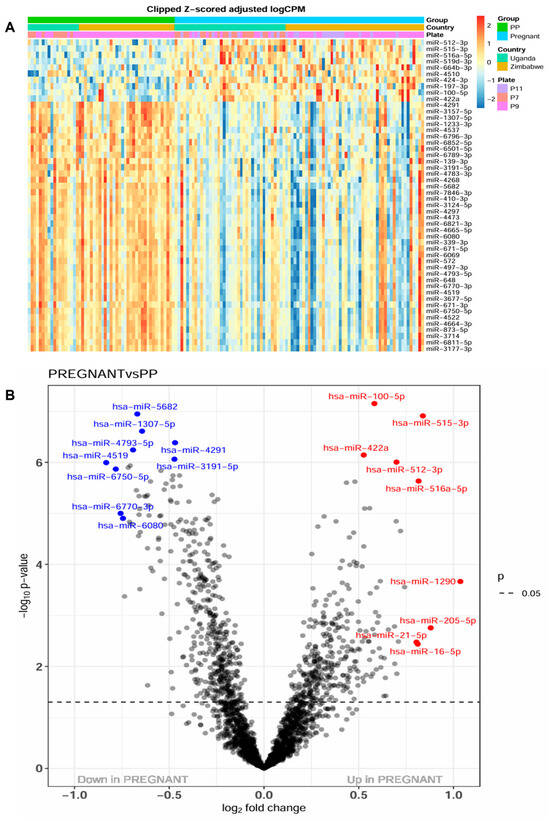
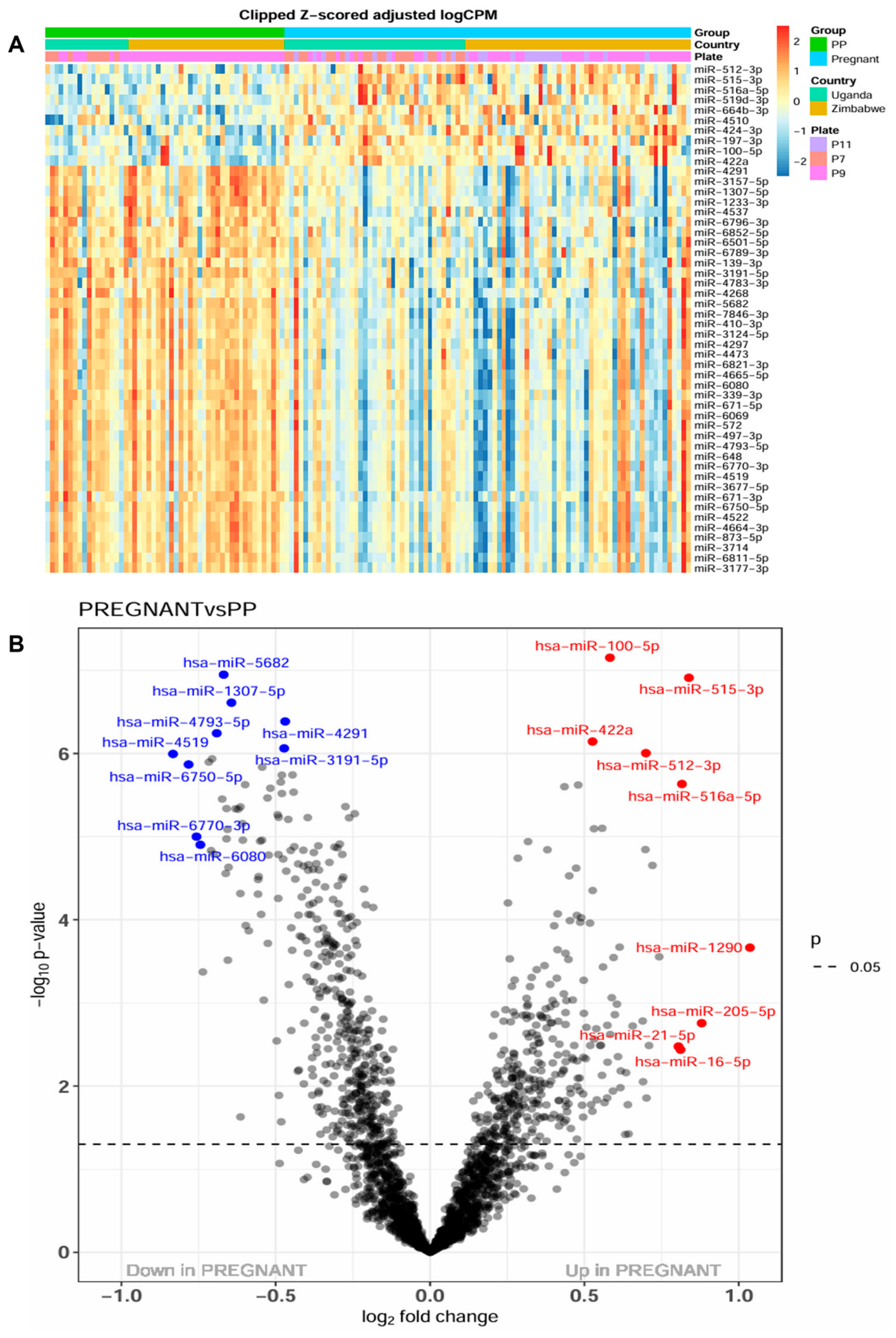
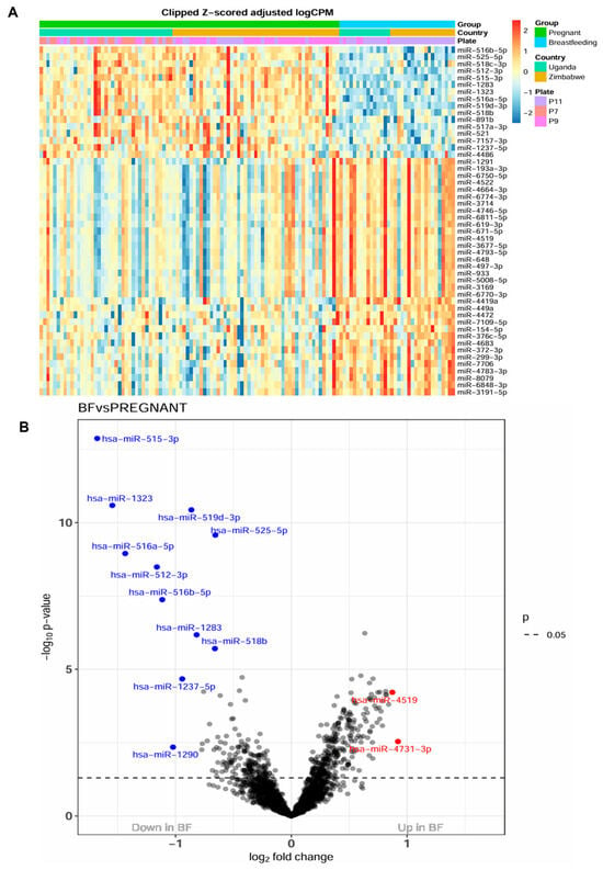
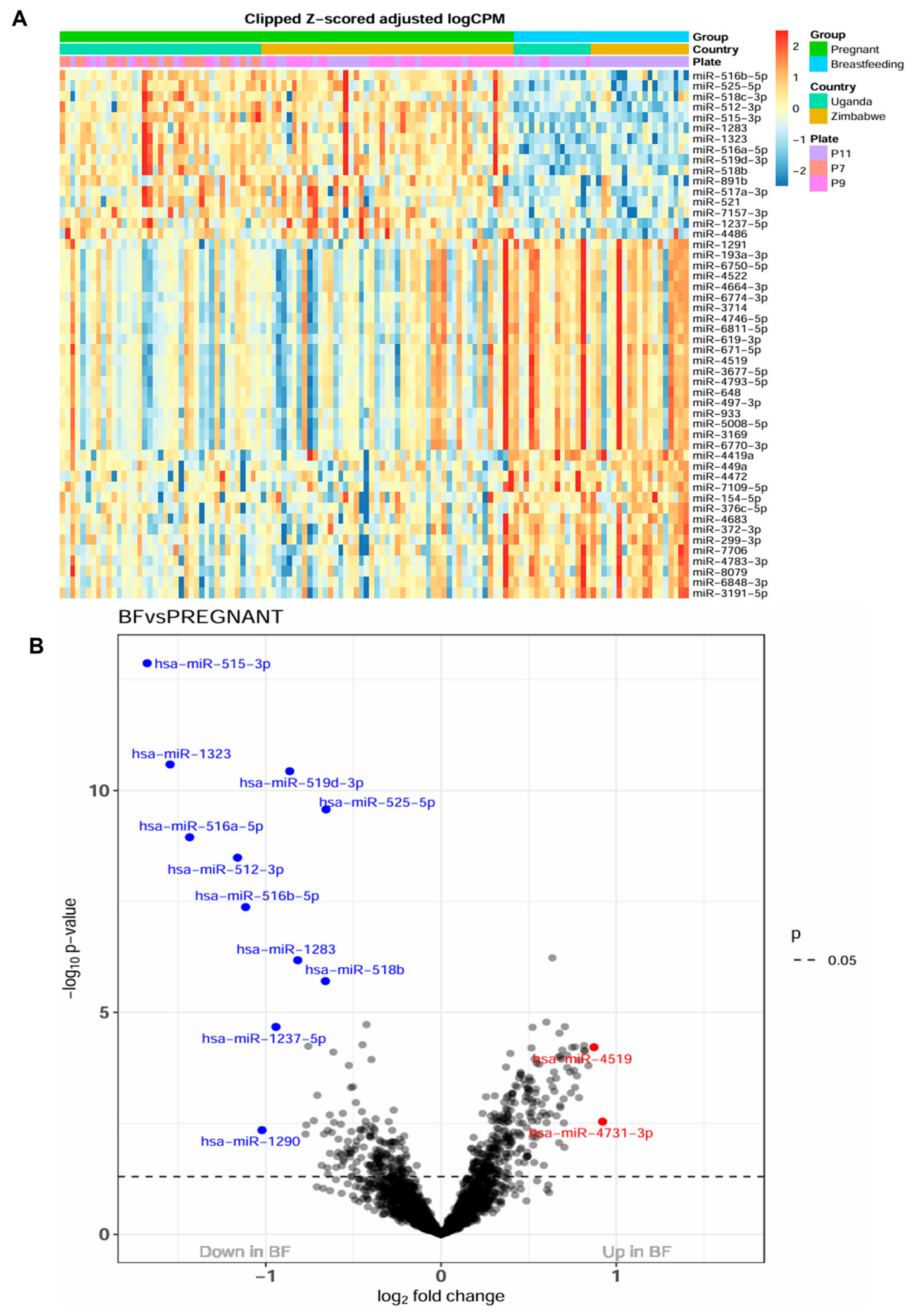
The PCA plots comparing sample variance showed that the samples from pre-pregnant (Supplemental Figure S1A) and breastfeeding (Supplemental Figure S1B) women displayed modest separation from those of pregnant women, while the total variance explained by PC1 (20%) and PC2 (12%) was low, suggesting lack of systematic bias between the comparison groups and warranting searching for a defined set of differentially expressed genes as biomarkers of reproductive condition. Pregnancy vs. Pre-pregnant comparison revealed 684 DE miRNAs and Pregnancy vs. Breastfeeding revealed 240 DE miRNAs with FDR < 0.1) (Table S3), corresponding to 32.8% and 11.5% of the miRWT whole transcriptome, respectively. Heatmaps illustrate the top 50 DE miRNAs across samples for the Pregnancy vs. Pre-pregnancy (Figure 1A) and Breastfeeding vs. Pregnant (Figure 2A) comparisons. Volcano plots annotate the miRNAs with the lowest FDR and the largest fold change in both comparisons (Figure 1B and Figure 2B).
Figure 1.
Serum miRNA expression by female reproductive status comparing pregnancy (P) versus pre-pregnancy (PP). (A) A heatmap displays the top 50 DE miRNAs (rows) across individual samples (column) for the comparison P vs. pre-pregnancy PP with clustering by country and batch effect (plate). Expression levels are represented by color intensity: red for upregulation and blue for downregulation. (B) A volcano plot shows differentially expressed (DE) miRNAs for P vs. PP. The most significantly downregulated (blue) and upregulated (red) miRNAs based on the smallest p-values or largest log fold changes (log FC) are annotated.
Figure 2.
Serum miRNA expression by female reproductive status comparing breastfeeding (BF) versus pregnancy (P). (A) A heatmap displays the top 50 DE miRNAs (rows) across individual samples (column) for the comparison of BF vs. pregnancy P with clustering by country and batch effect (Plate). Expression levels are represented by color intensity: red for upregulation and blue for downregulation. (B) A volcano plot shows differentially expressed (DE) miRNAs for BF vs. P. The most significantly downregulated (blue) and upregulated (red) miRNAs based on smallest p-values or largest log fold changes (log FC) are annotated.
Pregnancy-driven serum miRNAs identified by the overlap between the Pregnant vs. Pre-pregnant and Pregnant vs. Breastfeeding comparisons included 160 DE miRNAs (FDR < 0.1), of which 29 were upregulated and 131 downregulated (Table S4). Among those DE miRNAs, 19 belonged to the placenta-expressed clusters C19MC (upregulated hsa-miR-517a-3p, -516a-5p, -516b-5p, -525-5p, -519d-3p, -518c-3p, -512-3p, -522-3p, -1283, -521, -515-3p and -1321), C14MC (downregulated hsa-miR-410-3p, -376c-5p, -377-3p, -299-3p, -299-5p, and -1247-5p) and hsa-miR-371-3 cluster (downregulated hsa-miR-372-3p).
3.2. Pregnancy-Driven Serum miRNAs Target Genes Shared with the HIV Interactome
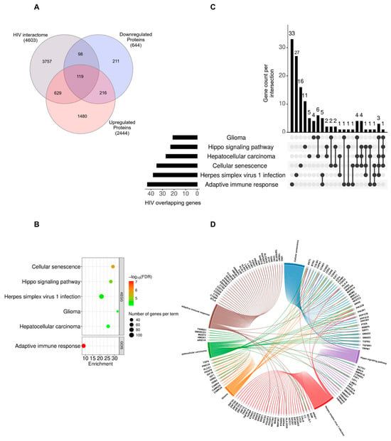
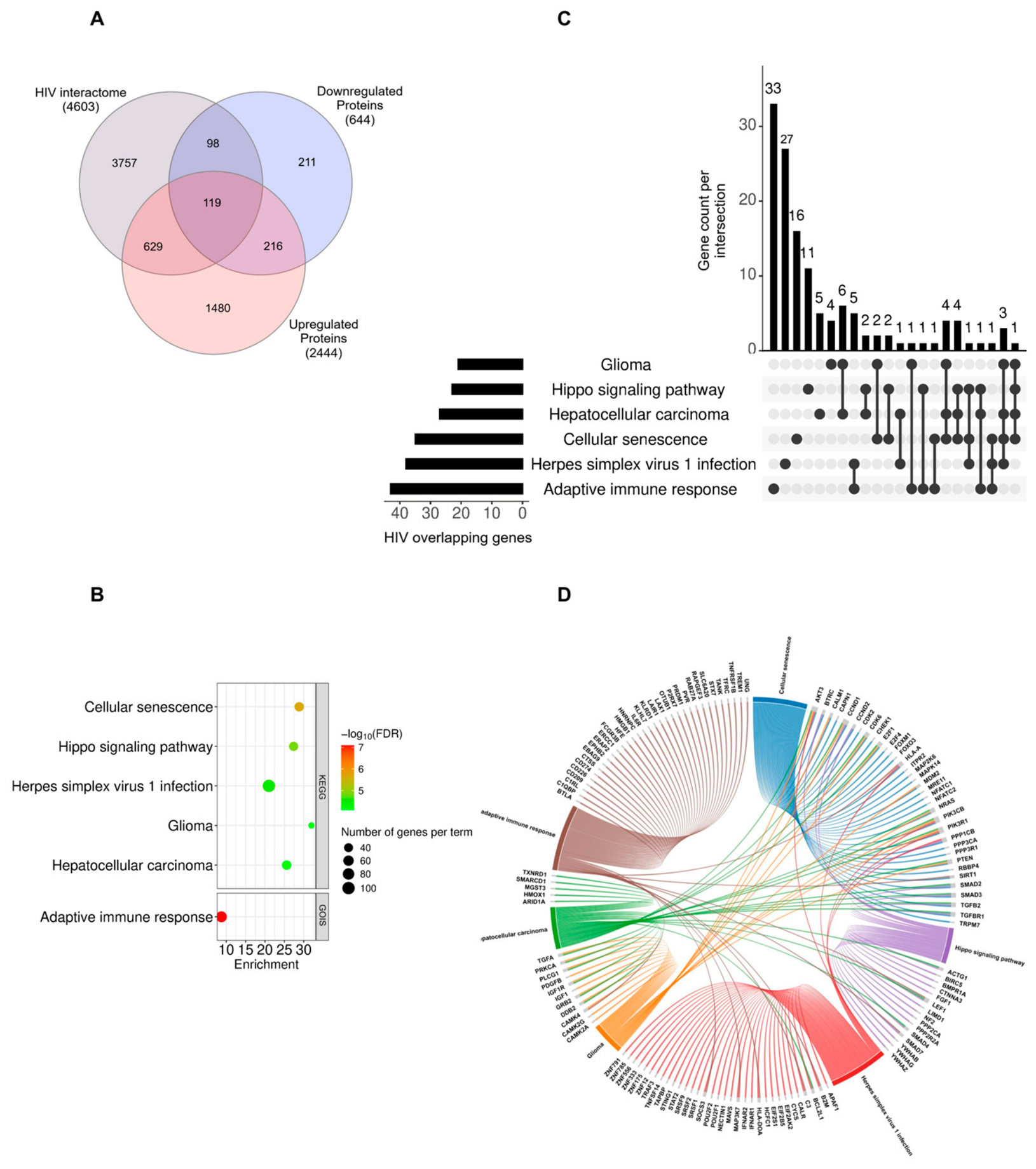
Pregnancy-driven DE miRNAs were predicted to downregulate 644 genes and upregulate 2444 genes, based on miRNA-mRNA regulatory relationships (Table S5). Among them, 98 (15.2%) of downregulated genes and 629 (25.7%) of upregulated genes overlapped with the HIV interactome dataset (Figure 3A), while 119 proteins fell into the intersection of the three groups and 216 were predicted to be bidirectionally expressed (both up- and downregulated) (Figure 3A, Table S6).
Figure 3.
Modular enrichment analysis (MEA) of validated targets of serum miRNAs altered during pregnancy and enriched in the overlap with the HIV interactome. (A) A Venn diagram shows the overlap (intersections) and unique elements (differences) between the HIV interactome and the validated targets of pregnancy-driven serum miRNAs. (B) A bubble plot presents MEA of all validated targets (2444 upregulated and 644 downregulated) of pregnancy-driven serum miRNAs. The Y-axis annotates the names of enriched pathways (KEGG, upper plot) and terms (GOIS, lower plot) and the X-axis shows enrichment as % of genes per pathway or term. Bubble color represents the significance level of each pathway/term (-log10 FDR), while size indicates the total number of genes enriched in each pathway/term. (C) An UpSet plot and (D) a cord plot highlight the HIV interactome gene overlap contributing to the enrichment of the biological pathways shown in (B). The Y-axis of upper graph on the UpSet plot shows the count of HIV-overlapping genes that are either unique to or shared among specific pathway combinations, as indicated by the connected dots below the X-axis. Horizontal bars on the lower left of the Upset plot represent the total number of HIV-overlapping genes associated with each individual enriched pathway. The chord plot illustrates all genes within enriched biological terms that overlap with the HIV interactome and their distribution across pathways. Ribbons and colors in the cord plot distinguish the 6 enriched biological groups/pathways. The percentages below the labels indicate the proportion of genes in each term that are shared with the HIV interactome.
3.3. The Genes Targeted by Pregnancy-Driven Serum miRNAs Are Linked to Pathways of Adaptive Immune Response, Hippo Signaling, Cancer, Cellular Senescence and Viral Infection Shared with the HIV Interactome
All confirmed targets of pregnancy-modulated serum miRNAs were employed to identify associated biological pathways. Targets were found to be functionally associated with “adaptive immune response” (GO:0002250), “Cellular Senescence” (KEGG:04218), “Hippo Signaling Pathway” (KEGG:04390), “Herpes Simplex Virus 1 (HSV1) Infection” (KEGG:05168), “Glioma” (KEGG:05214) and “Hepatocellular Carcinoma” (HCC) (KEGG:05225) with an FDR < 0.05 (Figure 3B). The GOIS term “adaptive immune response” was the most significant (corrected p = 0.000015) while “Cellular Senescence” showed the greatest enrichment score (28.85% of genes per pathway) (Figure 3B, Table S7). Over 71% of the genes associated with those six biological functions are predicted to be upregulated as a result of downregulated pregnancy-driven DE miRNAs (Table S7). Among the genes contributing to enriched biological pathways, 132 were found to overlap with the HIV interactome (Figure 3C, Table S8), highlighting shared molecular mechanisms. Of these, 36 genes are shared across the six key pathways identified (Figure 3C,D, Table S8).
The differential miRNA-targeted HIV interactome genes were unevenly distributed between the six pregnancy-enriched pathways (Figure 3C and Table S8). Viral infection and immune response contained a higher number of HIV-interactome genes not functionally shared with the other terms (27 in “HSV-1 infection” pathway and 33 in “adaptive immune response”). Functional connections between the DE miRNA target genes within the enriched biological pathways (Figure 3D) showed that cancer-related pathways and “Cellular Senescence” functionally share the largest number of HIV-interactome genes. The percentage of genes shared with the HIV interactome within each pathway ranged from 37% to 88% (Tables S8 and S9).
3.4. Protein–Protein Interaction (PPI) Network Mapping of the Pregnancy-Enriched HIV Interactome Reveals Hub Gene Interactions While miRNA–hub Gene Networks Highlight Pregnancy-Driven miRNA Control of These Hub Genes
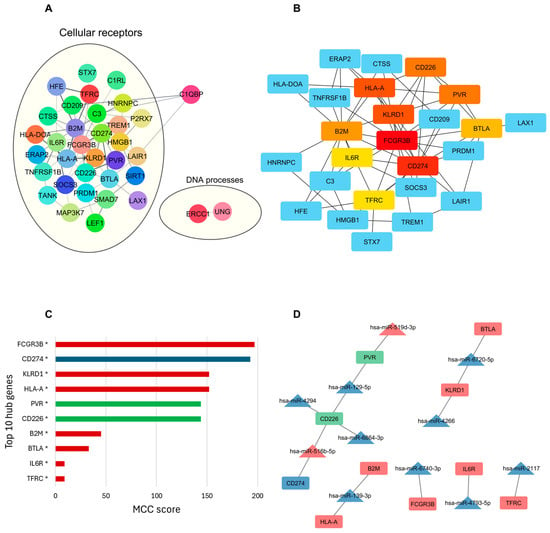
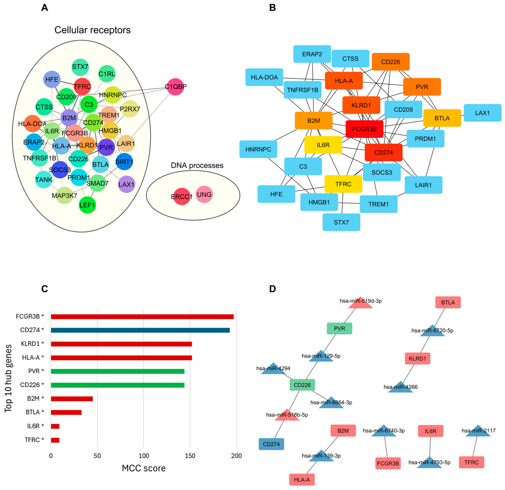
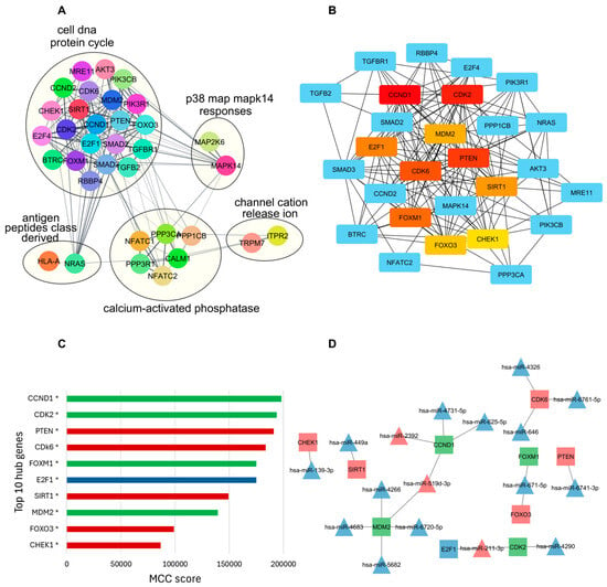
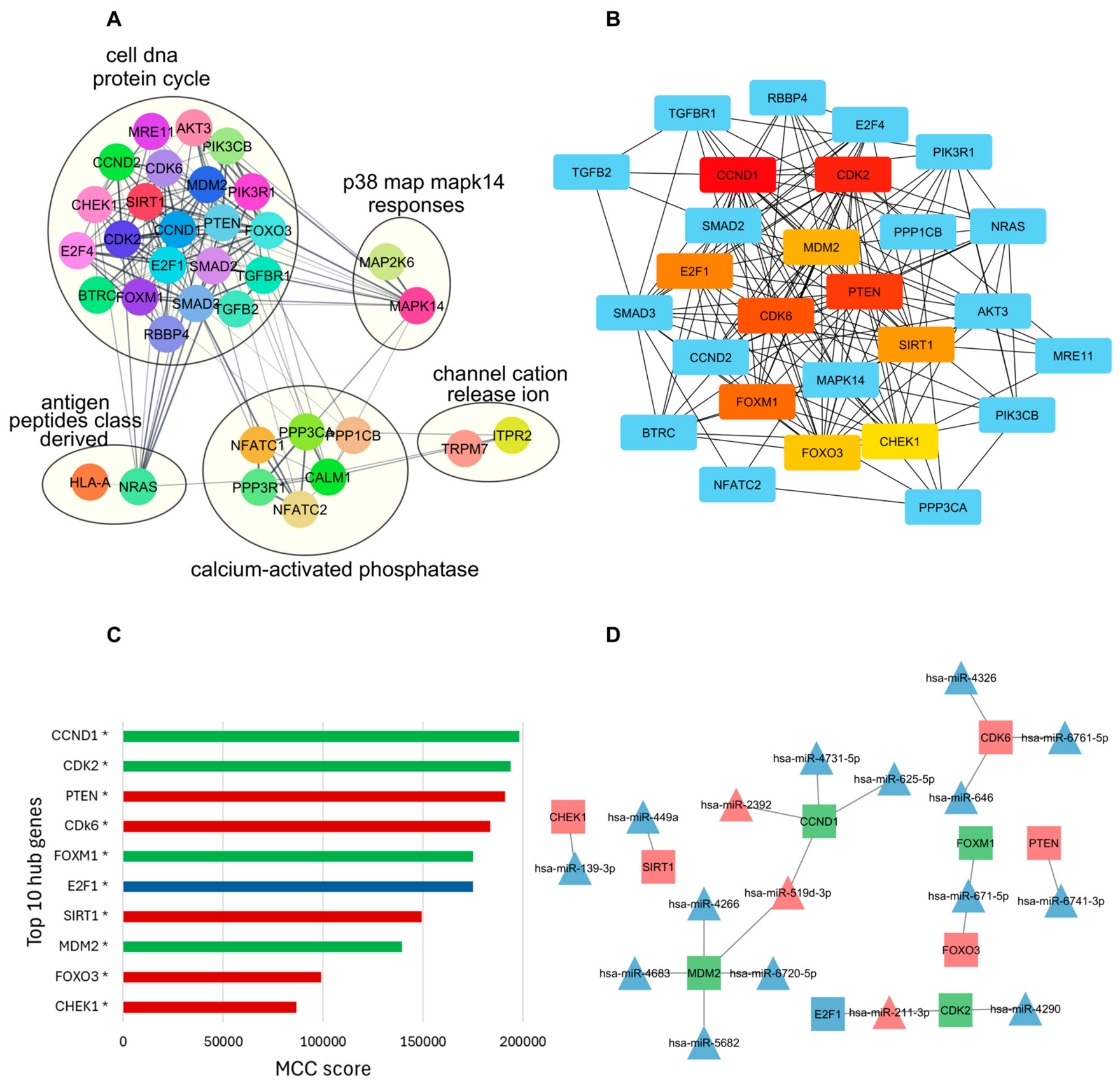
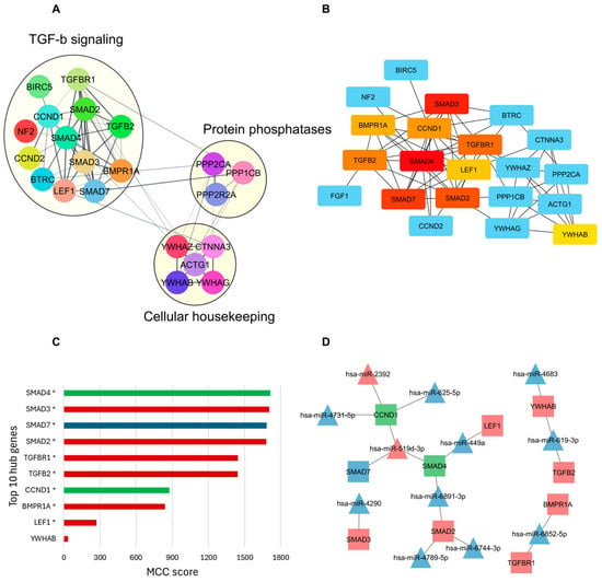
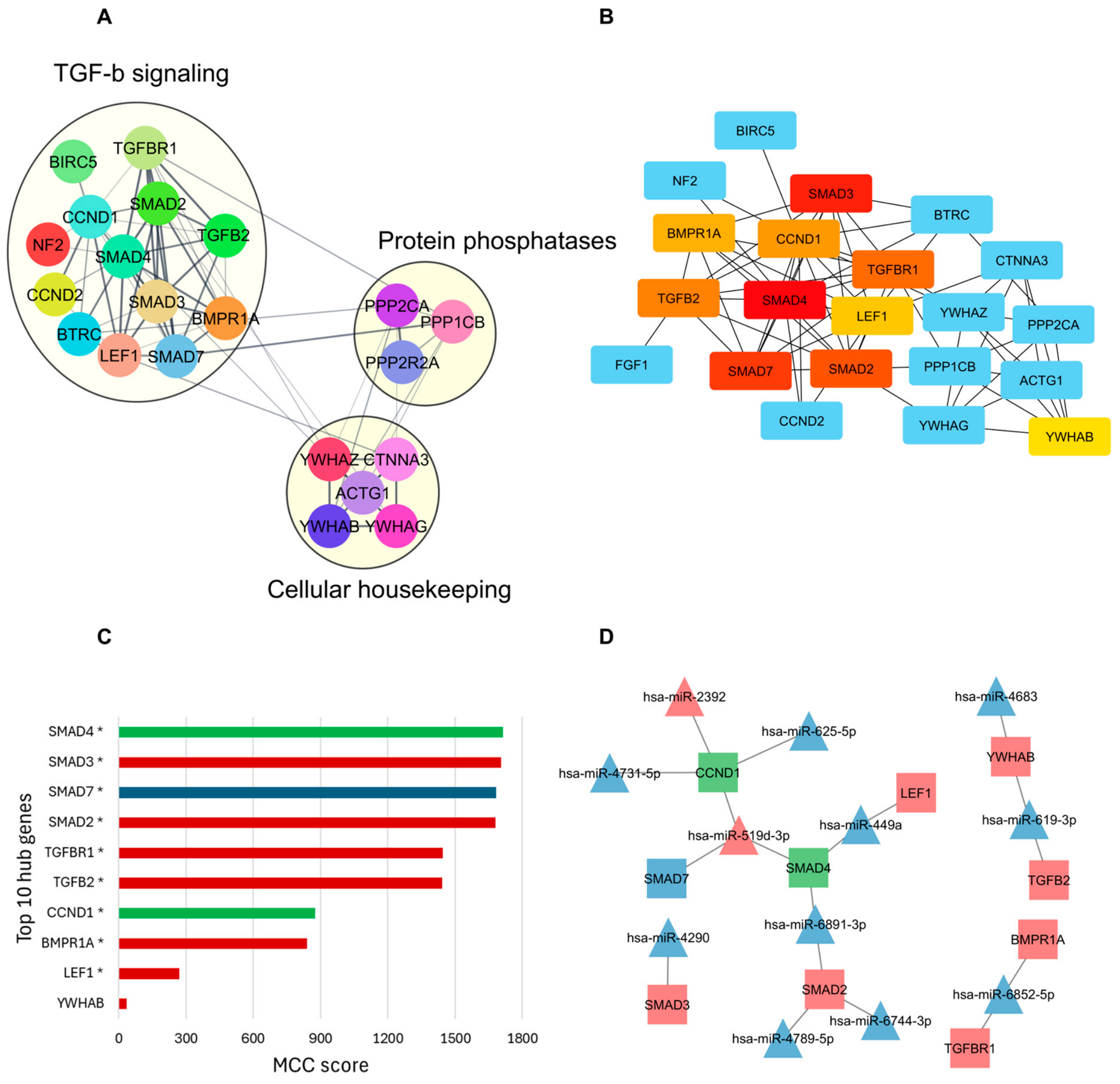
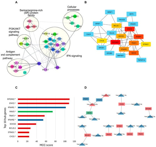
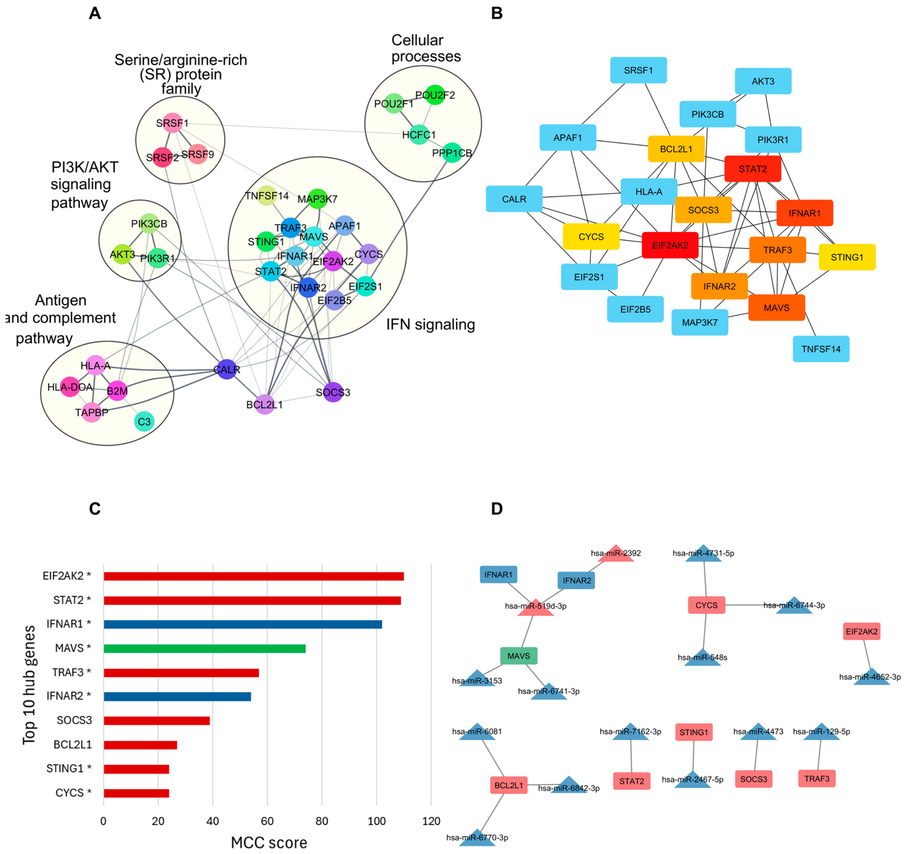
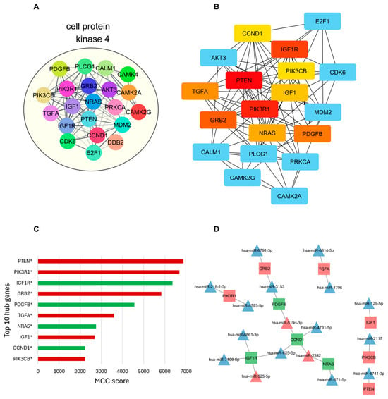
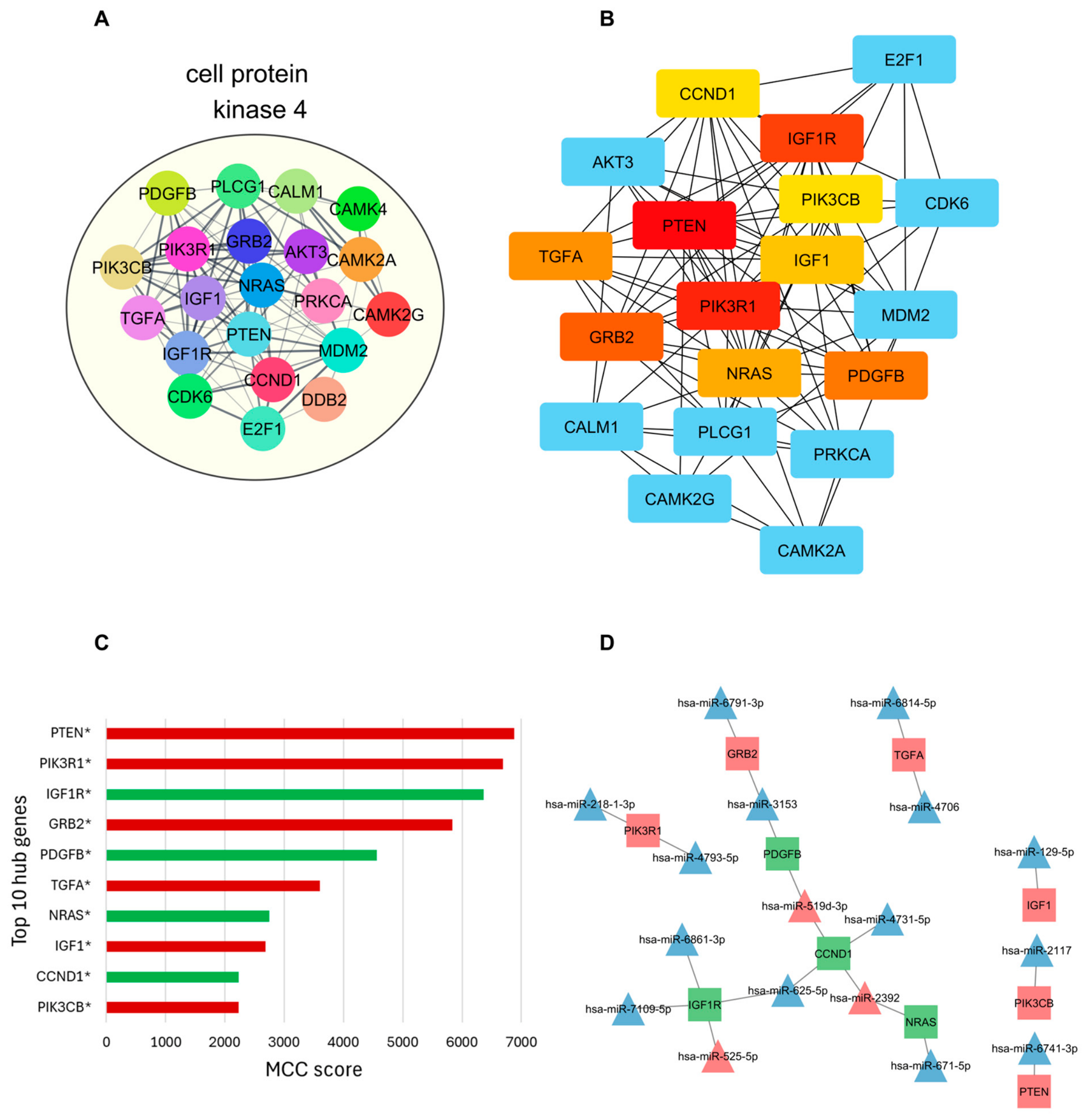
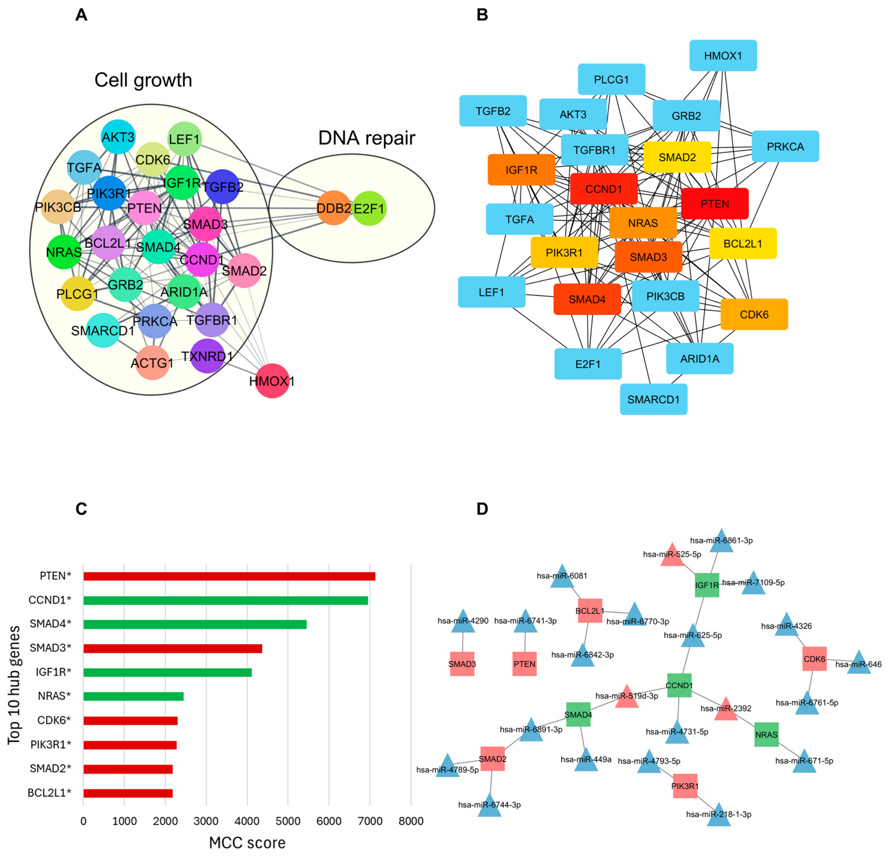
PPI networks built for genes targeted by pregnancy-driven miRNA overlapping with the HIV interactome contributed to six enriched and functionally mapped biological pathways. The “adaptive immune response” PPI network (Figure 4A) included 43 nodes and 80 edges mapped into two functional groups (cellular receptors and DNA processes), while “Cellular Senescence” comprised 35 nodes and 180 edges assigned to five functional groups (cell cycle, calcium-activated phosphatase, ion channel, MAPK signaling and plasma membrane proteins) (Figure 5A). “Hippo Signaling Pathway” included 23 nodes and 69 edges distributed into three functional groups (TGF-β signaling, cellular housekeeping and protein phosphatases) (Figure 6A). The “HSV-1 Infection” PPI network (Figure 7A) included 38 nodes and 77 edges mapped into five functional groups (IFN signaling, antigen presentation and complement pathway, cellular processes, PI3K/AKT signaling pathway and serine/arginine-rich (SR) protein family). The cancer-related pathways, “Glioma” and “HCC” included 21 nodes and 102 edges associated with cell protein kinase (Figure 8A), and 27 nodes and 131 edges linked to Cell Growth and DNA repair, respectively (Figure 9A). In summary, network mapping revealed essential functional groups that were likely altered during pregnancy.
Figure 4.
Protein–protein interaction (PPI) and hub gene networks of HIV-interactome genes within the adaptive immune response GO term, including predicted miRNA–target interactions. (A) Protein–protein interaction (PPI) networks display HIV-interactome genes contributing to the adaptive immune response term enrichment. Functional annotations (cellular responses and DNA processes) highlight commonalities in protein roles. (B) PPI network of the top 10 hub genes within the term, identified using cytoHubba based on Maximum Clique Centrality (MCC) analysis. Node colors indicate MCC scores, from high (red) to low (yellow). (C) Bar plots of the top 10 hub genes by MCC score. Asterisks indicate genes annotated under cellular receptor-related functions, as shown in Figure 4A. Bar colors represent the predicted regulation of each gene in pregnancy based on differential miRNA expression: blue for downregulated, red for upregulated, and green for mixed regulation (both up- and downregulated). (D) Networks of miRNAs and their hub gene targets show 21 nodes (triangles) and 15 edges (lines). Triangles represent miRNAs and rectangles represent hub genes. Node colors reflect inferred regulation in pregnancy: blue (downregulated), red (upregulated), and green (mixed).
Figure 5.
PPI and hub gene networks of HIV-interactome genes within the Cellular Senescence KEGG pathway, including predicted miRNA–target interactions. (A) Protein–protein interaction (PPI) networks display HIV-interactome genes contributing to the cellular senescence pathway enrichment. Functional annotations highlight commonalities in protein roles. (B) PPI network of the top 10 hub genes within the term, identified using cytoHubba based on Maximal Clique Centrality (MCC) analysis. Node colors indicate MCC scores, from high (red) to low (yellow). (C) Bar plots of the top 10 hub genes by MCC score. Asterisks indicate genes annotated under a cell cycle, as shown in Figure 5A. Bar colors represent the predicted regulation of each gene in pregnancy based on differential miRNA expression: blue for downregulated, red for upregulated, and green for mixed regulation (both up- and downregulated). (D) miRNA–hub gene target networks show 27 nodes (triangles and rectangles) and 20 edges (connecting lines). Triangles represent miRNAs and rectangles represent hub genes. Node colors reflect inferred regulation in pregnancy: blue (downregulated), red (upregulated), and green (mixed).
Figure 6.
PPI and hub gene networks of HIV-interactome genes within the Hippo Signaling KEGG pathway, including predicted miRNA–target interactions. (A) Protein–protein interaction (PPI) networks display HIV-interactome genes contributing to the Hippo Signaling pathway enrichment. Functional annotations highlight commonalities in protein roles. (B) PPI network of the top 10 hub genes within the term, identified using cytoHubba based on Maximal Clique Centrality (MCC) analysis. Node colors indicate MCC scores, from high (red) to low (yellow). (C) Bar plots of the top 10 hub genes by MCC score. Asterisks indicate genes annotated under TGFβ signaling, as shown in Figure 6A. Bar colors represent the predicted regulation of each gene in pregnancy based on differential miRNA expression: blue for downregulated, red for upregulated, and green for mixed regulation (both up- and downregulated). (D) miRNA–hub gene target networks show 22 nodes (triangles and quadrangles) and 18 edges (connecting lines). Triangles represent miRNAs and rectangles represent hub genes. Node colors reflect inferred regulation in pregnancy: blue (downregulated), red (upregulated), and green (mixed).
Figure 7.
PPI and hub gene networks of HIV-interactome genes within the herpes simplex virus 1 Infection KEGG pathway, including predicted miRNA–target interactions. (A) Protein–protein interaction (PPI) networks display HIV-interactome genes contributing to the herpes simples virus 1 (HSV1) infection pathway enrichment. Functional annotations highlight commonalities in protein roles. (B) PPI network of the top 10 hub genes within the term, identified using cytoHubba based on Maximal Clique Centrality (MCC) analysis. Node colors indicate MCC scores, from high (red) to low (yellow). (C) Bar plots of the top 10 hub genes by MCC score. Asterisks indicate genes annotated under IFN signaling, as shown in Figure 7A. Bar colors represent the predicted regulation of each gene in pregnancy based on differential miRNA expression: blue for downregulated, red for upregulated, and green for mixed regulation (both up- and downregulated). (D) miRNA–hub gene target networks show 25 nodes and 17 edges. Triangles represent miRNAs and rectangles represent hub genes. Node colors reflect inferred regulation in pregnancy: blue (downregulated), red (upregulated), and green (mixed).
Figure 8.
PPI and hub gene networks of HIV interactome genes within the glioma KEGG pathway, including predicted miRNA–target interactions. (A) Protein–protein interaction (PPI) networks display HIV interactome genes contributing to the glioma pathway enrichment. Functional annotations highlight commonalities in protein roles. (B) PPI network of the top 10 hub genes within the term, identified using cytoHubba based on Maximal Clique Centrality (MCC) analysis. Node colors indicate MCC scores from high (red) to low (yellow). (C) Bar plots of the top 10 hub genes by MCC score. Asterisks indicate genes annotated under cell protein kinase, as shown in Figure 8A. Bar colors represent the predicted regulation of each gene in pregnancy based on differential miRNA expression: blue for downregulated, red for upregulated, and green for mixed regulation (both up- and downregulated). (D) miRNA–hub gene target networks show 27 nodes and 21 edges. Triangles represent miRNAs and rectangles represent hub genes. Node colors reflect inferred regulation in pregnancy: blue (downregulated), red (upregulated), and green (mixed).
Figure 9.
PPI and hub gene networks of HIV interactome genes within the Hepatocellular Carcinoma (HCC) KEGG pathway, including predicted miRNA–target interactions. (A) Protein–protein interaction (PPI) networks display HIV interactome genes contributing to the HCC pathway enrichment. Functional annotations highlight commonalities in protein roles. (B) PPI network of the top 10 hub genes within the term, identified using cytoHubba based on Maximal Clique Centrality (MCC) analysis. Node colors indicate MCC scores from high (red) to low (yellow). (C) Bar plots of the top 10 hub genes by MCC score. Asterisks indicate genes annotated under cell growth, as shown in Figure 9A. Bar colors represent the predicted regulation of each gene in pregnancy based on differential miRNA expression: blue for downregulated, red for upregulated, and green for mixed regulation (both up- and downregulated). (D) miRNA–hub gene target networks show 32 nodes and 26 edges. Triangles represent miRNAs and rectangles represent hub genes. Node colors reflect inferred regulation in pregnancy: blue (downregulated), red (upregulated), and green (mixed).
Hub genes are highly connected within a PPI network. They are prime candidates for drug targeting, as their central roles within the network suggest they are likely essential for disease initiation and progression. To further explore this, we identified the top 10 hub genes that support and maintain the structural integrity of all PPI networks (Figure 4B, Figure 5B, Figure 6B, Figure 7B, Figure 8B and Figure 9B), presenting them according to their MCC scores and predicted direction of regulation, which is either upregulated, downregulated, or bidirectional (Figure 4C, Figure 5C, Figure 6C, Figure 7C, Figure 8C and Figure 9C). We identified pregnancy-driven miRNAs that regulate the hub genes and constructed their corresponding regulatory networks, highlighting the predicted miRNA–hub gene interactions within the following pathways: “adaptive immune response” (Figure 4D), “Cellular Senescence” (Figure 5D), “Hippo Signaling Pathway” (Figure 6D), “HSV1 Infection” (Figure 7D), “Glioma” (Figure 8D), and “HCC” (Figure 9D). Our data show a total of 47 unique hub genes across all the enriched pathways (Table 2) likely under post-transcriptional “one-to-many” and “many-to-one” regulation by 45 pregnancy-driven miRNAs, of which 40 are downregulated and five are upregulated. Moreover, C19MC-derived DE miRNAs target 12 top-ranked hub genes, representing 25.5% of all hub genes.
Table 2.
Hub genes with top MCC scores featuring pregnancy-enriched pathways overlapping with the HIV interactome.
Figure 10.
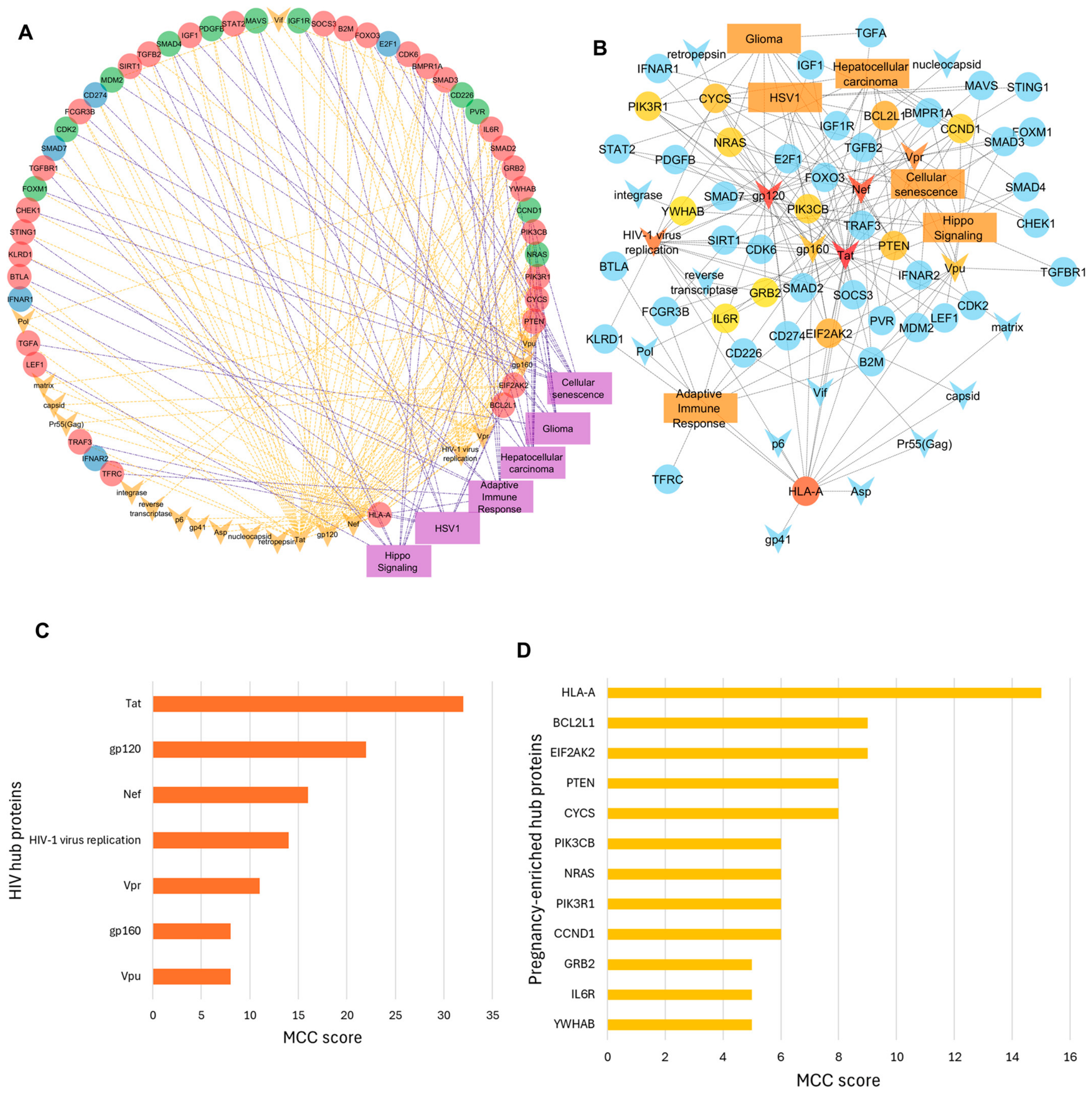
Circular network of hub genes, enriched pathways, and HIV-interacting modulators in pregnancy. (A) Ellipses represent hub genes with colors indicating their predicted regulation during pregnancy: downregulated (blue), upregulated (red), and mixed regulation (green). Enriched pathways are shown as purple rectangles, and HIV-interacting proteins are depicted as orange V-shapes. Orange dashed lines represent interactions between hub genes and HIV proteins, while purple dash-dot lines represent interactions between hub genes and enriched pathways. The network contains 72 nodes and 189 edges. (B) This subnetwork emphasizes highly connected HIV proteins (V-shapes), miRNA targets (circles), and enriched pathways (squares) extracted from panel A. The color gradient from intense orange to light yellow represents increasing Maximum Clique Centrality (MCC). All pathways in pregnancy showed an MCC = 10. (C) MCC score for HIV-1 proteins. (D) MCC for pregnancy-regulated host hub genes.
3.5. Host Hub Proteins Are Co-Targeted by Pregnancy-Driven miRNAs and Viral Proteins
Interactions between hub genes and HIV-1 proteins were sourced from the HIV-NCBI database [31]. These data were leveraged to construct a network incorporating the pathways associated with each hub gene (Figure 10A) and compiled into tables (Table 3, Table 4, Table 5, Table 6 and Table 7). We found that the hub genes were reported to interact with 19 HIV modulators, including an HIV biological process (HIV replication) and 18 HIV proteins (Capsid, gp120, gp160—precursor of gp120 and gp41, gp41, Integrase, Matrix, Nef, Nucleocapsid, p6, Pol, Pr55(gag)—(gag polyprotein precursor, Retropepsin—a protease, Reverse transcriptase, Tat, Vif, Vpr and Vpu). A subnetwork focuses on highly connected proteins (Figure 10B). Among these HIV-1 proteins, Tat and gp120 showed the highest connectivity within the network, followed by Nef, HIV-1 virus replication, Vpr, gp160, and Vpu (Figure 10C). HLA-A stood out as the most highly connected host protein, with BCL2L1 ranking second, followed by EIF2AK2, PTEN, and CYCS, completing the top five host proteins (Figure 10D).
Table 3.
Interactions between HIV-1 and the top 10 adaptive immune response hub genes ranked by Maximum Clique Centrality Score (MCC).
Table 4.
Interactions between HIV-1 and cellular senescence hub genes ranked by Maximum Clique Centrality Score (MCC).
Table 5.
Interactions between HIV-1 and Hippo Signaling Pathway hub genes ranked by Maximum Clique Centrality Score (MCC).
Table 6.
Interactions between HSV1 and hub genes ranked by Maximum Clique Centrality Score (MCC).
Table 7.
Interactions between HIV-1 and glioma hub genes ranked by Maximum Clique Centrality Score (MCC).
4. Discussion
Our study leverages a robust cohort of women at higher risk of acquiring HIV throughout their reproductive cycles, providing a unique opportunity to investigate systemic molecular changes during pregnancy. Only a small number of prior longitudinal studies have reported peripheral blood miRNAs in pregnancy and primarily focus on preterm birth [36] and maternal blood pressure as underlying risk of adverse birth outcomes [37] and pre-eclampsia [38]. Our work specifically examines cell-free serum miRNAs and provides new insights into molecular mechanisms, potentially shaping HIV susceptibility and pathogenesis. By following women before and after pregnancy, we capture miRNAs that are likely critical during this period, revealing their systemic roles in pregnancy.
One of the several physiological adaptations that occur during pregnancy involves the suppression of immune functions, irrespective of the woman’s HIV status. These changes include early pregnancy events such as reduction in complement events and cell-mediated immunity [39]. In this sense, scientists and clinicians have yet to reach a consensus on whether pregnancy itself constitutes a risk factor for HIV acquisition [17,40,41,42]. However, a recent study analyzing data from two large clinical trials concluded that the HIV risk increases during pregnancy and continues through the first six months postpartum, with the highest risk occurring in the later stages of pregnancy [1]. While association studies have laid the groundwork in exploring the link between pregnancy and HIV risk, too little is known about this topic, and limited empirical molecular evidence has been proposed.
MiRNAs are well-known immune modulators that regulate gene expression and, consequently, contribute to immune-related processes [43]. In this study, we determined changes in serum miRNAs across pre-pregnancy, pregnancy, and breastfeeding reproductive states, providing evidence that molecular mechanisms governed by systemic miRNAs also undergo physiological adaptation during those transitions. We tested the hypothesis that those individual miRNAs regulate pivotal genes and pathways related to the HIV interaction, influencing susceptibility and progression during pregnancy.
It has been proposed that in pregnancy some miRNAs are expressed mainly from the placenta and trophoblast cells, in a coordinated manner from specific chromosomal clusters rather than individually [44,45]. The human placenta expresses two key imprinted microRNA clusters: the paternally expressed chromosome 19 microRNA cluster (C19MC) and the maternally expressed chromosome 14 cluster (C14MC). C19MC is highly placenta-specific, enriched in trophoblast-derived vesicles such as exosomes, and its members are detectable in maternal serum [46,47]. C14MC is known as the largest miRNA cluster and is primarily maternally expressed, showing dynamic regulation across gestation [44]. We identified 133 HIV interactome genes regulated by the DE placental cluster miRNAs circulating in the maternal serum, constituting a large proportion (~25%) of the hub genes with high connectivity to HIV proteins, all targeted by upregulated maternal-imprinted C19MC.
C19MC miRNAs have been shown to modulate maternal immune responses, promoting tolerance at the maternal–fetal interface [48]. In vitro studies have shown that while C19CM is not modulated by exposure to certain viruses, such as Vesicular Stomatitis Virus (VSV) and Cytomegalovirus (CMV) [49], it can be protective against ZIKV infection [50], vaccinia virus, VSV, HSV-1, but not CMV [51]. Whether and which specific miRNA members of C14MC and C19MC affect maternal HIV susceptibility and vertical transmission remain to be investigated.
Here, we reported the enrichment of six pathways in pregnancy. Among those, the Hippo Signaling pathway stands out as physiologically relevant during pregnancy due to its proteins’ role in early cell fate decisions between placental and embryonic lineages, as well as in trophoblast invasion [52,53], supporting the validity and biological relevance of our analysis. The Hippo Signaling pathway is functionally linked to the Cellular senescence pathway [54], suggesting coordinated regulation. Cellular senescence is also physiologically linked to cancer-associated pathways, such as HCC and glioma, as dysregulated cell cycle control is a hallmark of tumorigenesis [55,56]. The additional two pathways reflect over-representation of the adaptive immune response and viral infection processes, particularly those related to HSV-1. Curiously, a parallel has been proposed between maternal immune tolerance to the fetus during pregnancy and immune evasion mechanisms in cancer [57,58]. The idea is that the same immune system adaptations that help a fetus grow safely might also help cancer cells avoid being attacked. This might explain the cancer-related pathways found in this study and their linked genes. For instance, PDL-1—a known cancer suppressor, predicted to be downregulated in our dataset—is one of the top hub genes found in the “adaptive immune response” category. Blood levels of PDL-1 were similar in pregnant women without cancer and non-pregnant cancer patients when compared to healthy non-pregnant women [58]. Genes overlapping the HIV interactome are present across all enriched pathways. Therefore, our data highlights a gene network targeted by miRNAs DE in pregnancy that also shares similarities with cancer pathways and potentially interacts with HIV-1.
The pregnancy-enriched pathway analysis revealed protein–protein interaction networks converging on 47 hub genes shared with the HIV interactome. The miRNA–hub gene networks revealed “one-to-many” and “many-to-one” relationships, shedding light on a complex regulatory landscape in which individual miRNAs can influence multiple hub genes, while key hub genes may be co-regulated by several miRNAs. This reflects coordinated regulatory control over critical biological processes.
The hub genes identified in the “adaptive immune response” term converge on immune regulation with implications for tumor immunity, infection, and tolerance. They include immune checkpoints (PD-L1/CD274, BTLA, and PVR/CD155, the latter interacting with the co-stimulatory receptor CD226 [59]), a major histocompatibility complex (MHC) class I antigen processing and presentation machinery (HLA-A, B2M, TFRC), and additional inflammatory mediators such as IL6R and FCGR3B. Complementing these findings, hub genes linked to the “HSV1 infection” pathway fall into three categories: components of the type I interferon response (EIF2AK2, MAVS, TRAF3, STING1, STAT2, IFNAR1/2), negative feedback regulators (SOCS3), and apoptotic mediators (BCL2L1, CYCS), which together influence whether T cells, NK cells, and other immune populations undergo survival or programmed cell death, underscoring the integration of antiviral sensing with cytokine regulation and immune cell fate decisions.
Hub genes in the cellular senescence and Hippo Signaling pathways highlight coordinated regulation of cell fate. Senescence-associated genes balance cell cycle progression (CCND1, also in the Hippo Signaling pathway, CDK2/6, E2F1, FOXM1, CHEK1) with growth arrest and stress responses (PTEN, FOXO3, SIRT1, MDM2), while Hippo pathway genes are largely composed of TGF-β signaling components (SMAD2/3/4/7, TGFBR1, TGFB2, BMPR1A). Downstream effectors (LEF1 and YWHAB) integrate transcriptional programs controlling proliferation, differentiation, and tissue homeostasis. Together, they seem to reflect an interconnected network orchestrating proliferation versus arrest and survival decisions. The remaining three hub genes in both Glioma and HCC pathways act in growth factor signaling, likely with IGF1R upstream of NRAS and PIK3R1 driving proliferation and survival. Together, these hub genes may be key determinants of HIV risk during pregnancy and highlight three major themes—immune regulation, cell cycle control, and growth/homeostasis—targeted or dysregulated by HIV-1 as part of its immune evasion strategy.
The global miRNA transcriptome during pregnancy identified specific interactions between hub gene targets, host proteins, and HIV proteins in the HIV interactome. Among the host proteins, HLA-A, an MHC class I transmembrane protein, showed the highest number of interactions, while Tat emerged as the central HIV modulator, alongside gp120 and Nef. HLA-A, predicted to be upregulated, belongs to a gene complex that accounts for much of the observed variation in immune responses. It is highly polymorphic, and genetic variations have been demonstrated to be relevant in the vertical transmission of HIV [60,61]. A large study of ~9800 HIV-infected individuals found that elevated HLA-A expression was associated with significantly higher viral load and lower CD4+ T-cell counts, indicating poorer virologic control [62]. HIV Nef exhibits strong interaction with HLA-A and is known to downregulate its expression [63].
BCL2L1 (also known as BCL-2) was identified as the second most highly connected host protein within the HIV hub-gene network. HIV-infected cells are enriched in BCL-2, an anti-apoptotic factor that enhances their survival [64]. By sustaining the reservoir and reinforcing latency, BCL2L1 contributes to the long-term persistence of infection [65]. Pharmacologic inhibition of BCL-2 with venetoclax has been shown to promote the clearance of actively replicating HIV-infected cells in both in vitro and ex vivo models [66], and this approach is currently being evaluated in clinical trials (NCT05668026). In this context, miRNAs that regulate these pathways may offer additional opportunities for therapeutic intervention.
EIF2AK2, which is predicted to be upregulated, shows increased expression at late gestation in both circulating macrophages and placental tissues [67]. Activation of EIF2AK2 (PKR) by viral or RNA signals induces phosphorylation of eIF2α, a central antiviral pathway that normally limits protein synthesis. However, HIV can exploit this mechanism to enhance splicing of its rev/tat mRNA and promote productive viral gene expression [68]. In addition, the HIV Tat protein can compete with eIF2α, increasing transcription from the HIV-1 long terminal repeat (LTR), a regulatory DNA sequence [69]. EIF2AK2 also interacts with mitochondrial antiviral signaling proteins on the outer mitochondrial membrane, linking viral RNA sensing to mitochondrial-mediated immune responses. Notably, PKR has been implicated in placental immune regulation, suggesting a role in maintaining pregnancy homeostasis and responding to cellular stress. PTEN, also predicted to be upregulated in our dataset, appears to have a dual role in HIV infection. It can act as a barrier to viral replication by silencing unintegrated HIV-1 DNA [70], yet HIV integration into the PTEN gene may disrupt its function [71], potentially affecting immune responses. Investigating PTEN’s role during HIV exposure in pregnancy could provide valuable insights into maternal immune regulation and inform potential therapeutic strategies.
CYCS, an apoptotic protein predicted to be upregulated in our dataset, encodes cytochrome c, a mitochondrial factor central to placental function and pregnancy outcomes [72,73]. Mitochondrial dysfunction, marked by impaired cytochrome c oxidase activity and excessive cytochrome c-mediated apoptosis, has been linked to pre-eclampsia and related complications [74]. Elevated circulating cytochrome c has also been observed in HIV patients with antiretroviral therapy-related toxicity [75]. Cytochrome c is also central to HIV-1-induced apoptosis, in which viral proteins such as Env and Vpr directly or indirectly promote mitochondrial membrane permeabilization [76]. Thus, cytochrome c is likely a convergence point between pregnancy disorders and HIV pathogenesis.
Our results reveal a complex interplay in which pregnancy-associated miRNAs and HIV-1 proteins converge on shared hub genes, suggesting two-way co-targeting. This convergence points to a molecular tug-of-war between host regulatory mechanisms and viral interference during pregnancy. This overlap may reveal potential points of susceptibility in pregnant women, where physiological adaptations to pregnancy intersect with viral strategies to hijack host pathways. A number of experimental studies have provided evidence for direct interaction of circulating cell-free RNAs with HIV–host cells providing the basis of direct enhancement of HIV infection [77,78]. Future studies should validate key pregnancy-associated miRNA interactions with HIV in relevant models and explore their timing throughout gestation. Integrating additional non-coding RNAs and transcriptomic data may uncover broader regulatory networks, opening up new possibilities in informed targeted HIV prevention strategies tailored to reproductive stages.
5. Conclusions
Our longitudinal study reveals that systemic miRNAs undergo regulation during gestation, likely supporting maternal–fetal immune tolerance. We identified 47 hub genes targeted by these pregnancy-driven miRNAs (with 25% of those expressed by placental cluster C19MC) that overlap with the HIV interactome. Those genes include immune checkpoints (PD-L1, BTLA), apoptotic mediators (BCL2L1, CYCS), and antiviral signaling components (EIF2AK2, PTEN). Pregnancy-regulated miRNA converged on pathways of immune regulation, cell cycle control, and viral sensing, highlighting mechanisms by which HIV-1 may exploit pregnancy-associated molecular adaptations, or alternatively could be counteracted by host protective responses. Some of the differentially regulated direction of miRNAs mechanistically supported an increased risk of HIV-1 during pregnancy by targeting genes promoting host cell permissiveness (e.g., ↑ CD16) or viral replication (e.g., ↓ E2F1, ↑ FOXO3, ↑ CHEK-1). Other effects were bidirectional, holding wider potential to protect depending on the environmental context and exposures that have not been studied here. Future work should validate these interactions, establish the predictive value of miRNA signature for HIV-1 acquisition in longitudinal cohorts, and examine gestational timing to inform stage-specific HIV prevention or treatment strategies.
Supplementary Materials
The following supporting information can be downloaded at: https://www.mdpi.com/article/10.3390/v18070753/s1, Table S1: List of miRNAs gene symbols used in the HTG assay; Table S2: List of genes pertaining to the HIV interactome dataset; Table S3: Serum miRNAs differentially expressed in Pregnancy versus Pre-pregnancy at FDR < 0.1; Table S4: Differentially expressed serum miRNAs in pregnancy (FDR < 0.1) identified consistently across P vs. PP and P vs. BF comparisons; Table S5: Validated targets of serum miRNAs upregulated in Pregnancy; Table S6: Venn diagram analysis of overlap between the HIV interactome and proteins predicted to be downregulated or upregulated in pregnancy; Table S7: Functional enrichment analysis of pregnancy-altered genes based on KEGG and Gene Ontology terms; Table S8: Genes in pregnancy-enriched pathways targeted by DE-miRNAs and shared with the HIV interactome; Table S9: Proportion of genes overlapping the HIV interactome per pregnancy-enriched pathway. Supplemental Figure S1: Principal Component Analysis (PCA) for comparisons of Pregnant vs. Pre-pregnant Breastfeeding vs. Pregnant groups, including batch effect.
Author Contributions
R.N.F. conceived and designed the study, acquired funding, administered the project, supervised the laboratory data acquisition and provided writing and critical editing of the manuscript. P.F.T.C.-d.-M. performed the functional analysis and prepared the original draft. H.Y. contributed to the miRNA data acquisition, R.L.B., X.G., H.P., C.M., G.F.D., contributed to study design, data curation, and methodology. J.M.D. and P.-L.C. performed the statistical analysis in R. P.F.T.C.-d.-M., J.M.D., H.P. and R.N.F. contributed to visualization. All authors contributed to the review and editing. All authors have read and agreed to the published version of the manuscript.
Funding
Research reported in this publication was supported by the Eunice Kennedy Shriver National Institute of Child Health & Human Development of the National Institutes of Health under Award Number R01HD099091. The content is solely the responsibility of the authors and does not necessarily represent the official views of the National Institutes of Health.
Institutional Review Board Statement
The study was conducted in accordance with the Declaration of Helsinki and approved by the Institutional Review Board of Brigham and Women’s Hospital (protocol 2019P000273 approved on 21 March 2019). The transfer of biospecimens to the United States was approved by institutional authorities in Uganda and Zimbabwe.
Informed Consent Statement
Informed consent was obtained from all subjects involved in the study.
Data Availability Statement
All data are provided in Supplemental Tables.
Acknowledgments
The authors like to acknowledge the contribution of the study participants and all investigators and study staff of the HC-HIV study who made this nested study possible.
Conflicts of Interest
The authors declare no conflicts of interest. The funders had no role in the design of the study; in the collection, analyses, or interpretation of data; in the writing of the manuscript; or in the decision to publish the results.
Abbreviations
The following abbreviations are used in this manuscript:
| BF | Breastfeeding |
| BV | Bacterial vaginosis |
| CMV | Cytomegalovirus |
| CPM | Counts per million |
| DE | Differentially expressed |
| FC | Fold change |
| FDR | False Discovery Rate |
| GO | Gene Ontology |
| GOIS | Gene Ontology of Immune System |
| HCC | Hepatocellular carcinoma |
| HIV | Human immunodeficiency virus |
| HSV | Herpes simplex virus |
| KEGG | Kyoto Encyclopedia of Genes and Genomes |
| MC | miRNA cluster |
| MCC | Maximum Clique Centrality |
| MEA | Modular Enrichment Analysis |
| miRWT | miRNA whole transcriptome |
| NGS | Next-generation sequencing |
| P | Pregnant |
| PCA | Principal component analysis |
| PP | Pre-pregnant |
| PPI | Protein–protein interaction |
| STIs | Sexually transmitted infections |
| VSV | Vesicular stomatitis virus |
References
- Thomson, K.A.; Hughes, J.; Baeten, J.M.; John-Stewart, G.; Celum, C.; Cohen, C.R.; Ngure, K.; Kiarie, J.; Mugo, N.; Heffron, R.; et al. Increased Risk of HIV Acquisition Among Women Throughout Pregnancy and During the Postpartum Period: A Prospective Per-Coital-Act Analysis Among Women with HIV-Infected Partners. J. Infect. Dis. 2018, 218, 16–25. [Google Scholar] [CrossRef] [Scilit] [PubMed]
- Calvert, C.; Ronsmans, C. Pregnancy and HIV disease progression: A systematic review and meta-analysis. Trop. Med. Int. Health 2015, 20, 122–145. [Google Scholar] [CrossRef] [Scilit] [PubMed]
- Tai, J.H.; Udoji, M.A.; Barkanic, G.; Byrne, D.W.; Rebeiro, P.F.; Byram, B.R.; Kheshti, A.; Carter, J.D.; Graves, C.R.; Raffanti, S.P.; et al. Pregnancy and HIV disease progression during the era of highly active antiretroviral therapy. J. Infect. Dis. 2007, 196, 1044–1052. [Google Scholar] [CrossRef] [Scilit] [PubMed]
- Abu-Raya, B.; Michalski, C.; Sadarangani, M.; Lavoie, P.M. Maternal Immunological Adaptation During Normal Pregnancy. Front. Immunol. 2020, 11, 575197. [Google Scholar] [CrossRef] [Scilit] [PubMed]
- Liu, X.; Zhu, L.; Huang, Z.; Li, Z.; Duan, R.; Li, H.; Xie, L.; Chen, X.; Ding, W.; Chen, B.; et al. A dynamic peripheral immune landscape during human pregnancy. Fundam. Res. 2025, 5, 391–406. [Google Scholar] [CrossRef] [Scilit] [PubMed]
- Li, L.P.; Kang, J.L.; Xia, W. Effect of first, second, and third trimester placental factors on CD4, CCR5, and CXCR4 expression in human peripheral blood lymphocytes. J. Cent. South Univ. Med. Sci. 2008, 33, 461–467. [Google Scholar]
- Mor, G.; Cardenas, I. The immune system in pregnancy: A unique complexity. Am. J. Reprod. Immunol. 2010, 63, 425–433. [Google Scholar] [CrossRef] [Scilit] [PubMed]
- Ren, J.; Jin, H.; Zhu, Y. The Role of Placental Non-Coding RNAs in Adverse Pregnancy Outcomes. Int. J. Mol. Sci. 2023, 24, 5030. [Google Scholar] [CrossRef] [Scilit] [PubMed]
- Kamity, R.; Sharma, S.; Hanna, N. MicroRNA-Mediated Control of Inflammation and Tolerance in Pregnancy. Front. Immunol. 2019, 10, 718. [Google Scholar] [CrossRef] [Scilit] [PubMed]
- Bartel, D.P. MicroRNAs: Genomics, biogenesis, mechanism, and function. Cell 2004, 116, 281–297. [Google Scholar] [CrossRef] [Scilit] [PubMed]
- Nail, H.M.; Chiu, C.-C.; Leung, C.-H.; Ahmed, M.M.M.; Wang, H.-M.D. Exosomal miRNA-mediated intercellular communications and immunomodulatory effects in tumor microenvironments. J. Biomed. Sci. 2023, 30, 69. [Google Scholar] [CrossRef] [Scilit] [PubMed]
- Tribolet, L.; Kerr, E.; Cowled, C.; Bean, A.G.D.; Stewart, C.R.; Dearnley, M.; Farr, R.J. MicroRNA Biomarkers for Infectious Diseases: From Basic Research to Biosensing. Front. Microbiol. 2020, 11, 1197. [Google Scholar] [CrossRef] [Scilit] [PubMed]
- de Candia, P.; De Rosa, V.; Casiraghi, M.; Matarese, G. Extracellular RNAs: A Secret Arm of Immune System Regulation. J. Biol. Chem. 2016, 291, 7221–7228. [Google Scholar] [CrossRef] [Scilit] [PubMed]
- Turchinovich, A.; Samatov, T.R.; Tonevitsky, A.G.; Burwinkel, B. Circulating miRNAs: Cell-cell communication function? Front. Genet. 2013, 4, 119. [Google Scholar] [CrossRef] [Scilit] [PubMed]
- Abel, T.; Moodley, J.; Naicker, T. The Involvement of MicroRNAs in SARS-CoV-2 Infection Comorbid with HIV-Associated Preeclampsia. Curr. Hypertens. Rep. 2021, 23, 20. [Google Scholar] [CrossRef] [Scilit] [PubMed]
- Fichorova, R.; Dreyfuss, J.; Pan, H.; Nartey, S.; Yamamoto, H.; Chen, P.; Gao, X.; Doncel, G.F.; Barbieri, R. MicroRNAs as Regulators of Drug Metabolism and Transport in Pregnant and Lactating Women. bioRxiv 2025. [Google Scholar] [CrossRef] [Scilit] [PubMed]
- Morrison, C.S.; Wang, J.; Van Der Pol, B.; Padian, N.; Salata, R.A.; Richardson, B.A. Pregnancy and the risk of HIV-1 acquisition among women in Uganda and Zimbabwe. Aids 2007, 21, 1027–1034. [Google Scholar] [CrossRef] [Scilit] [PubMed]
- Morrison, C.S.; Richardson, B.A.; Mmiro, F.; Chipato, T.; Celentano, D.D.; Luoto, J.; Mugerwa, R.; Padian, N.; Rugpao, S.; Brown, J.M.; et al. Hormonal contraception and the risk of HIV acquisition. Aids 2007, 21, 85–95. [Google Scholar] [CrossRef] [Scilit] [PubMed]
- Fichorova, R.N.; Chen, P.L.; Morrison, C.S.; Doncel, G.F.; Mendonca, K.; Kwok, C.; Chipato, T.; Salata, R.; Mauck, C. The Contribution of Cervicovaginal Infections to the Immunomodulatory Effects of Hormonal Contraception. mBio 2015, 6, e00221-15. [Google Scholar] [CrossRef] [Scilit] [PubMed]
- Nowak, R.G.; Gravitt, P.E.; Morrison, C.S.; Gange, S.J.; Kwok, C.; Oliver, A.E.; Howard, R.; Van der Pol, B.; Salata, R.A.; Padian, N.S.; et al. Increases in human papillomavirus detection during early HIV infection among women in Zimbabwe. J. Infect. Dis. 2011, 203, 1182–1191. [Google Scholar] [CrossRef] [Scilit] [PubMed]
- Cezar-de-Mello, P.F.T.; Ryan, S.; Fichorova, R.N. The microRNA Cargo of Human Vaginal Extracellular Vesicles Differentiates Parasitic and Pathobiont Infections from Colonization by Homeostatic Bacteria. Microorganisms 2023, 11, 551. [Google Scholar] [CrossRef] [Scilit] [PubMed]
- Law, C.W.; Chen, Y.; Shi, W.; Smyth, G.K. voom: Precision weights unlock linear model analysis tools for RNA-seq read counts. Genome Biol. 2014, 15, R29. [Google Scholar] [CrossRef] [Scilit] [PubMed]
- Bolstad, B.M.; Irizarry, R.A.; Astrand, M.; Speed, T.P. A comparison of normalization methods for high density oligonucleotide array data based on variance and bias. Bioinformatics 2003, 19, 185–193. [Google Scholar] [CrossRef] [Scilit] [PubMed]
- Hoffman, G.E.; Roussos, P. Dream: Powerful differential expression analysis for repeated measures designs. Bioinformatics 2020, 37, 192–201. [Google Scholar] [CrossRef] [Scilit] [PubMed]
- Sticht, C.; De La Torre, C.; Parveen, A.; Gretz, N. miRWalk: An online resource for prediction of microRNA binding sites. PLoS ONE 2018, 13, e0206239. [Google Scholar] [CrossRef] [Scilit] [PubMed]
- Koopmans, F.; van Nierop, P.; Andres-Alonso, M.; Byrnes, A.; Cijsouw, T.; Coba, M.P.; Cornelisse, L.N.; Farrell, R.J.; Goldschmidt, H.L.; Howrigan, D.P.; et al. SynGO: An Evidence-Based, Expert-Curated Knowledge Base for the Synapse. Neuron 2019, 103, 217–234.4. [Google Scholar] [CrossRef] [Scilit] [PubMed]
- Heberle, H.; Meirelles, G.V.; da Silva, F.R.; Telles, G.P.; Minghim, R. InteractiVenn: A web-based tool for the analysis of sets through Venn diagrams. BMC Bioinform. 2015, 16, 169. [Google Scholar] [CrossRef] [Scilit]
- Lex, A.; Gehlenborg, N.; Strobelt, H.; Vuillemot, R.; Pfister, H. UpSet: Visualization of Intersecting Sets. IEEE Trans. Vis. Comput. Graph. 2014, 20, 1983–1992. [Google Scholar] [CrossRef] [Scilit]
- Conway, J.R.; Lex, A.; Gehlenborg, N. UpSetR: An R package for the visualization of intersecting sets and their properties. Bioinformatics 2017, 33, 2938–2940. [Google Scholar] [CrossRef] [Scilit]
- Luo, Y.; Jacobs, E.Y.; Greco, T.M.; Mohammed, K.D.; Tong, T.; Keegan, S.; Binley, J.M.; Cristea, I.M.; Fenyö, D.; Rout, M.P.; et al. HIV–host interactome revealed directly from infected cells. Nat. Microbiol. 2016, 1, 16068. [Google Scholar] [CrossRef] [Scilit] [PubMed]
- Ako-Adjei, D.; Fu, W.; Wallin, C.; Katz, K.S.; Song, G.; Darji, D.; Brister, J.R.; Ptak, R.G.; Pruitt, K.D. HIV-1, human interaction database: Current status and new features. Nucleic Acids Res. 2015, 43, D566–D570. [Google Scholar] [CrossRef] [Scilit] [PubMed]
- Bindea, G.; Mlecnik, B.; Hackl, H.; Charoentong, P.; Tosolini, M.; Kirilovsky, A.; Fridman, W.H.; Pagès, F.; Trajanoski, Z.; Galon, J. ClueGO: A Cytoscape plug-in to decipher functionally grouped gene ontology and pathway annotation networks. Bioinformatics 2009, 25, 1091–1093. [Google Scholar] [CrossRef] [Scilit] [PubMed]
- Huang, D.W.; Sherman, B.T.; Lempicki, R.A. Bioinformatics enrichment tools: Paths toward the comprehensive functional analysis of large gene lists. Nucleic Acids Res. 2009, 37, 1–13. [Google Scholar] [CrossRef] [Scilit] [PubMed]
- Kucera, M.; Isserlin, R.; Arkhangorodsky, A.; Bader, G.D. AutoAnnotate: A Cytoscape app for summarizing networks with semantic annotations. F1000Research 2016, 5, 1717. [Google Scholar] [CrossRef] [Scilit] [PubMed]
- Chin, C.H.; Chen, S.H.; Wu, H.H.; Ho, C.W.; Ko, M.T.; Lin, C.Y. cytoHubba: Identifying hub objects and sub-networks from complex interactome. BMC Syst. Biol. 2014, 8, S11. [Google Scholar] [CrossRef] [Scilit] [PubMed]
- Menon, R.; Debnath, C.; Lai, A.; Guanzon, D.; Bhatnagar, S.; Kshetrapal, P.K.; Sheller-Miller, S.; Salomon, C.; The Garbhini Study Team. Circulating Exosomal miRNA Profile During Term and Preterm Birth Pregnancies: A Longitudinal Study. Endocrinology 2018, 160, 249–275. [Google Scholar] [CrossRef] [Scilit] [PubMed]
- Anderson, E.C.; Muse, M.E.; Niu, Z.; Foley, H.B.; Levy, J.J.; Romano, M.E.; Gui, J.; Bentz, J.L.; Farzan, S.F.; Bastain, T.M.; et al. Blood Pressure and Late Pregnancy Circulating miRNAs in the MADRES Study. J. Am. Heart Assoc. 2025, 14, e040416. [Google Scholar] [CrossRef] [Scilit] [PubMed]
- Subramanian, A.; Weiss, D.; Nyhan, K.; Dewan, A.; Jukic, A.M.Z. Circulating miRNAs in the first trimester and pregnancy complications: A systematic review. Epigenetics 2023, 18, 2152615. [Google Scholar] [CrossRef] [Scilit] [PubMed]
- WHO/RHT/9. HIV in Pregnancy: A Review; World Health Organization: Geneva, Switzerland, 1999. [Google Scholar]
- Marston, M.; Newell, M.L.; Crampin, A.; Lutalo, T.; Musoke, R.; Gregson, S.; Nyamukapa, C.; Nakiyingi-Miiro, J.; Urassa, M.; Isingo, R.; et al. Is the risk of HIV acquisition increased during and immediately after pregnancy? A secondary analysis of pooled HIV community-based studies from the ALPHA network. PLoS ONE 2013, 8, e82219. [Google Scholar] [CrossRef] [Scilit] [PubMed]
- Mugo, N.R.; Heffron, R.; Donnell, D.; Wald, A.; Were, E.O.; Rees, H.; Celum, C.; Kiarie, J.N.; Cohen, C.R.; Kayintekore, K.; et al. Increased risk of HIV-1 transmission in pregnancy: A prospective study among African HIV-1-serodiscordant couples. Aids 2011, 25, 1887–1895. [Google Scholar] [CrossRef] [Scilit] [PubMed]
- Gray, R.H.; Li, X.; Kigozi, G.; Serwadda, D.; Brahmbhatt, H.; Wabwire-Mangen, F.; Nalugoda, F.; Kiddugavu, M.; Sewankambo, N.; Quinn, T.C.; et al. Increased risk of incident HIV during pregnancy in Rakai, Uganda: A prospective study. Lancet 2005, 366, 1182–1188. [Google Scholar] [CrossRef] [Scilit] [PubMed]
- Mehta, A.; Baltimore, D. MicroRNAs as regulatory elements in immune system logic. Nat. Rev. Immunol. 2016, 16, 279–294. [Google Scholar] [CrossRef] [Scilit] [PubMed]
- Morales-Prieto, D.M.; Ospina-Prieto, S.; Chaiwangyen, W.; Schoenleben, M.; Markert, U.R. Pregnancy-associated miRNA-clusters. J. Reprod. Immunol. 2013, 97, 51–61. [Google Scholar] [CrossRef] [Scilit] [PubMed]
- Wommack, J.C.; Trzeciakowski, J.P.; Miranda, R.C.; Stowe, R.P.; Ruiz, R.J. Micro RNA clusters in maternal plasma are associated with preterm birth and infant outcomes. PLoS ONE 2018, 13, e0199029. [Google Scholar] [CrossRef] [Scilit] [PubMed]
- Ouyang, Y.; Mouillet, J.F.; Coyne, C.B.; Sadovsky, Y. Review: Placenta-specific microRNAs in exosomes—Good things come in nano-packages. Placenta 2014, 35, S69–S73. [Google Scholar] [CrossRef] [Scilit] [PubMed]
- Doridot, L.; Miralles, F.; Barbaux, S.; Vaiman, D. Trophoblasts, invasion, and microRNA. Front. Genet. 2013, 4, 248. [Google Scholar] [CrossRef] [Scilit] [PubMed]
- Zaga-Clavellina, V.; Diaz, L.; Olmos-Ortiz, A.; Godínez-Rubí, M.; Rojas-Mayorquín, A.E.; Ortuño-Sahagún, D. Central role of the placenta during viral infection: Immuno-competences and miRNA defensive responses. Biochim. Biophys. Acta (BBA)-Mol. Basis Dis. 2021, 1867, 166182. [Google Scholar] [CrossRef] [Scilit] [PubMed]
- Dumont, T.M.F.; Mouillet, J.F.; Bayer, A.; Gardner, C.L.; Klimstra, W.B.; Wolf, D.G.; Yagel, S.; Balmir, F.; Binstock, A.; Sanfilippo, J.S.; et al. The expression level of C19MC miRNAs in early pregnancy and in response to viral infection. Placenta 2017, 53, 23–29. [Google Scholar] [CrossRef] [Scilit] [PubMed]
- Bayer, A.; Lennemann, N.J.; Ouyang, Y.; Sadovsky, E.; Sheridan, M.A.; Roberts, R.M.; Coyne, C.B.; Sadovsky, Y. Chromosome 19 microRNAs exert antiviral activity independent from type III interferon signaling. Placenta 2018, 61, 33–38. [Google Scholar] [CrossRef] [Scilit] [PubMed]
- Delorme-Axford, E.; Donker, R.B.; Mouillet, J.-F.; Chu, T.; Bayer, A.; Ouyang, Y.; Wang, T.; Stolz, D.B.; Sarkar, S.N.; Morelli, A.E.; et al. Human placental trophoblasts confer viral resistance to recipient cells. Proc. Natl. Acad. Sci. USA 2013, 110, 12048–12053. [Google Scholar] [CrossRef] [Scilit] [PubMed]
- Kruger, R.E.; Aziz, F.; Ralston, A. Hippo signaling in mammalian reproduction. Reproduction 2025, 169, e250016. [Google Scholar] [CrossRef] [Scilit] [PubMed]
- Soncin, F.; Parast, M.M. Role of Hippo signaling pathway in early placental development. Proc. Natl. Acad. Sci. USA 2020, 117, 20354–20356. [Google Scholar] [CrossRef] [Scilit] [PubMed]
- Miyajima, C.; Nagasaka, M.; Aoki, H.; Toriuchi, K.; Yamanaka, S.; Hashiguchi, S.; Morishita, D.; Aoyama, M.; Hayashi, H.; Inoue, Y. The Hippo Signaling Pathway Manipulates Cellular Senescence. Cells 2024, 14, 13. [Google Scholar] [CrossRef] [Scilit] [PubMed]
- Lu, X.; Luo, Y.; Huang, Y.; Zhu, Z.; Yin, H.; Xu, S. Cellular Senescence in Hepatocellular Carcinoma: Immune Microenvironment Insights via Machine Learning and In Vitro Experiments. Int. J. Mol. Sci. 2025, 26, 773. [Google Scholar] [CrossRef] [Scilit] [PubMed]
- Chojak, R.; Fares, J.; Petrosyan, E.; Lesniak, M.S. Cellular senescence in glioma. J. Neuro-Oncol. 2023, 164, 11–29. [Google Scholar] [CrossRef] [Scilit] [PubMed]
- Wilczyński, J.R. Cancer and pregnancy share similar mechanisms of immunological escape. Chemotherapy 2006, 52, 107–110. [Google Scholar] [CrossRef] [Scilit] [PubMed]
- Holtan, S.G.; Creedon, D.J.; Haluska, P.; Markovic, S.N. Cancer and pregnancy: Parallels in growth, invasion, and immune modulation and implications for cancer therapeutic agents. Mayo Clin. Proc. 2009, 84, 985–1000. [Google Scholar] [CrossRef] [Scilit]
- Kučan Brlić, P.; Lenac Roviš, T.; Cinamon, G.; Tsukerman, P.; Mandelboim, O.; Jonjić, S. Targeting PVR (CD155) and its receptors in anti-tumor therapy. Cell. Mol. Immunol. 2019, 16, 40–52. [Google Scholar] [CrossRef] [Scilit] [PubMed]
- Mackelprang, R.D.; John-Stewart, G.; Carrington, M.; Richardson, B.; Rowland-Jones, S.; Gao, X.; Mbori-Ngacha, D.; Mabuka, J.; Lohman-Payne, B.; Farquhar, C. Maternal HLA homozygosity and mother-child HLA concordance increase the risk of vertical transmission of HIV-1. J. Infect. Dis. 2008, 197, 1156–1161. [Google Scholar] [CrossRef] [Scilit] [PubMed]
- Mekue, L.M.; Nkenfou, C.N.; Ndukong, E.; Yatchou, L.; Dambaya, B.; Ngoufack, M.N.; Kameni, J.K.; Kuiaté, J.R.; Ndjolo, A. HLA A*32 is associated to HIV acquisition while B*44 and B*53 are associated with protection against HIV acquisition in perinatally exposed infants. BMC Pediatr. 2019, 19, 249. [Google Scholar] [CrossRef] [Scilit] [PubMed]
- Ramsuran, V.; Naranbhai, V.; Horowitz, A.; Qi, Y.; Martin, M.P.; Yuki, Y.; Gao, X.; Walker-Sperling, V.; Del Prete, G.Q.; Schneider, D.K.; et al. Elevated HLA-A expression impairs HIV control through inhibition of NKG2A-expressing cells. Science 2018, 359, 86–90. [Google Scholar] [CrossRef] [Scilit] [PubMed]
- Rajapaksa, U.S.; Li, D.; Peng, Y.C.; McMichael, A.J.; Dong, T.; Xu, X.N. HLA-B may be more protective against HIV-1 than HLA-A because it resists negative regulatory factor (Nef) mediated down-regulation. Proc. Natl. Acad. Sci. USA 2012, 109, 13353–13358. [Google Scholar] [CrossRef] [Scilit] [PubMed]
- Chandrasekar, A.P.; Badley, A.D. Prime, shock and kill: BCL-2 inhibition for HIV cure. Front. Immunol. 2022, 13, 1033609. [Google Scholar] [CrossRef] [Scilit] [PubMed]
- Chandrasekar, A.P.; Cummins, N.W.; Badley, A.D. The Role of the BCL-2 Family of Proteins in HIV-1 Pathogenesis and Persistence. Clin. Microbiol. Rev. 2019, 33, 10–1128. [Google Scholar] [CrossRef] [Scilit] [PubMed]
- Chandrasekar, A.P.; Cummins, N.W.; Natesampillai, S.; Misra, A.; Alto, A.; Laird, G.; Badley, A.D. The BCL-2 Inhibitor Venetoclax Augments Immune Effector Function Mediated by Fas Ligand, TRAIL, and Perforin/Granzyme B, Resulting in Reduced Plasma Viremia and Decreased HIV Reservoir Size during Acute HIV Infection in a Humanized Mouse Model. J. Virol. 2022, 96, e01730-22. [Google Scholar] [CrossRef] [Scilit] [PubMed]
- Wang, Z.; Ou, Q.; Gao, L. The increased cfRNA of TNFSF4 in peripheral blood at late gestation and preterm labor: Its implication as a noninvasive biomarker for premature delivery. Front. Immunol. 2023, 14, 1154025. [Google Scholar] [CrossRef] [Scilit] [PubMed]
- Namer, L.S.; Harwig, A.; Heynen, S.P.; Das, A.T.; Berkhout, B.; Kaempfer, R. HIV co-opts a cellular antiviral mechanism, activation of stress kinase PKR by its RNA, to enable splicing of rev/tat mRNA. Cell Biosci. 2023, 13, 28. [Google Scholar] [CrossRef] [Scilit] [PubMed]
- Endo-Munoz, L.; Warby, T.; Harrich, D.; McMillan, N.A. Phosphorylation of HIV Tat by PKR increases interaction with TAR RNA and enhances transcription. Virol. J. 2005, 2, 17. [Google Scholar] [CrossRef] [Scilit] [PubMed]
- Phan, A.T.; Zhu, Y. PTEN Mediates the Silencing of Unintegrated HIV-1 DNA. Viruses 2024, 16, 291. [Google Scholar] [CrossRef] [Scilit] [PubMed]
- Smith, D.M.; Rowland, E.F.; Gianella, S.; Patel, S.P.; Solso, S.; Dullano, C.; Deiss, R.; Wells, D.; Ignacio, C.; Caballero, G.; et al. HIV Integration into the PTEN Gene and Its Tumor Microenvironment Implications for Lung Cancer. Curr. Oncol. 2025, 32, 389. [Google Scholar] [CrossRef] [Scilit] [PubMed]
- Toledano, J.M.; Puche-Juarez, M.; Galvez-Navas, J.M.; Moreno-Fernandez, J.; Diaz-Castro, J.; Ochoa, J.J. Pregnancy Disorders: A Potential Role for Mitochondrial Altered Homeostasis. Antioxidants 2024, 13, 979. [Google Scholar] [CrossRef] [Scilit] [PubMed]
- Hu, X.Q.; Zhang, L. Mitochondrial Dysfunction in the Pathogenesis of Preeclampsia. Curr. Hypertens. Rep. 2022, 24, 157–172. [Google Scholar] [CrossRef] [Scilit] [PubMed]
- Matsubara, S.; Minakami, H.; Sato, I.; Saito, T. Decrease in cytochrome c oxidase activity detected cytochemically in the placental trophoblast of patients with pre-eclampsia. Placenta 1997, 18, 255–259. [Google Scholar] [CrossRef] [Scilit] [PubMed]
- Langs-Barlow, A.; Selvaraj, S.; Ogbuagu, O.; Shabanova, V.; Shapiro, E.D.; Paintsil, E. Association of circulating cytochrome c with clinical manifestations of antiretroviral-induced toxicity. Mitochondrion 2015, 20, 71–74. [Google Scholar] [CrossRef] [Scilit] [PubMed]
- Castedo, M.; Perfettini, J.L.; Andreau, K.; Roumier, T.; Piacentini, M.; Kroemer, G. Mitochondrial apoptosis induced by the HIV-1 envelope. Ann. N. Y. Acad. Sci. 2003, 1010, 19–28. [Google Scholar] [CrossRef] [Scilit] [PubMed]
- Kimura, K.; Hohjoh, H.; Fukuoka, M.; Sato, W.; Oki, S.; Tomi, C.; Yamaguchi, H.; Kondo, T.; Takahashi, R.; Yamamura, T. Circulating exosomes suppress the induction of regulatory T cells via let-7i in multiple sclerosis. Nat. Commun. 2018, 9, 17. [Google Scholar] [CrossRef] [Scilit] [PubMed]
- Okoye, I.; Xu, L.; Oyegbami, O.; Shahbaz, S.; Pink, D.; Gao, P.; Sun, X.; Elahi, S. Plasma Extracellular Vesicles Enhance HIV-1 Infection of Activated CD4(+) T Cells and Promote the Activation of Latently Infected J-Lat10.6 Cells via miR-139-5p Transfer. Front. Immunol. 2021, 12, 697604. [Google Scholar] [CrossRef] [Scilit] [PubMed]

Disclaimer/Publisher’s Note: The statements, opinions and data contained in all publications are solely those of the individual author(s) and contributor(s) and not of MDPI and/or the editor(s). MDPI and/or the editor(s) disclaim responsibility for any injury to people or property resulting from any ideas, methods, instructions or products referred to in the content. |
© 2026 by the authors. Licensee MDPI, Basel, Switzerland. This article is an open access article distributed under the terms and conditions of the Creative Commons Attribution (CC BY) license.