Super-Resolution Imaging of Nuclear Pore Responses to Mechanical Stress and Energy Depletion


Abstract
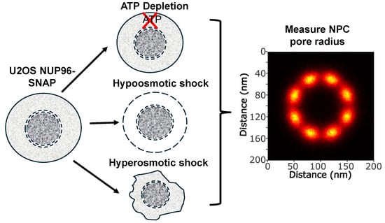
1. Introduction
2. Methods
2.1. Cells and Reagents
2.2. Production and Characterization of HIV-1 Pseudoviruses
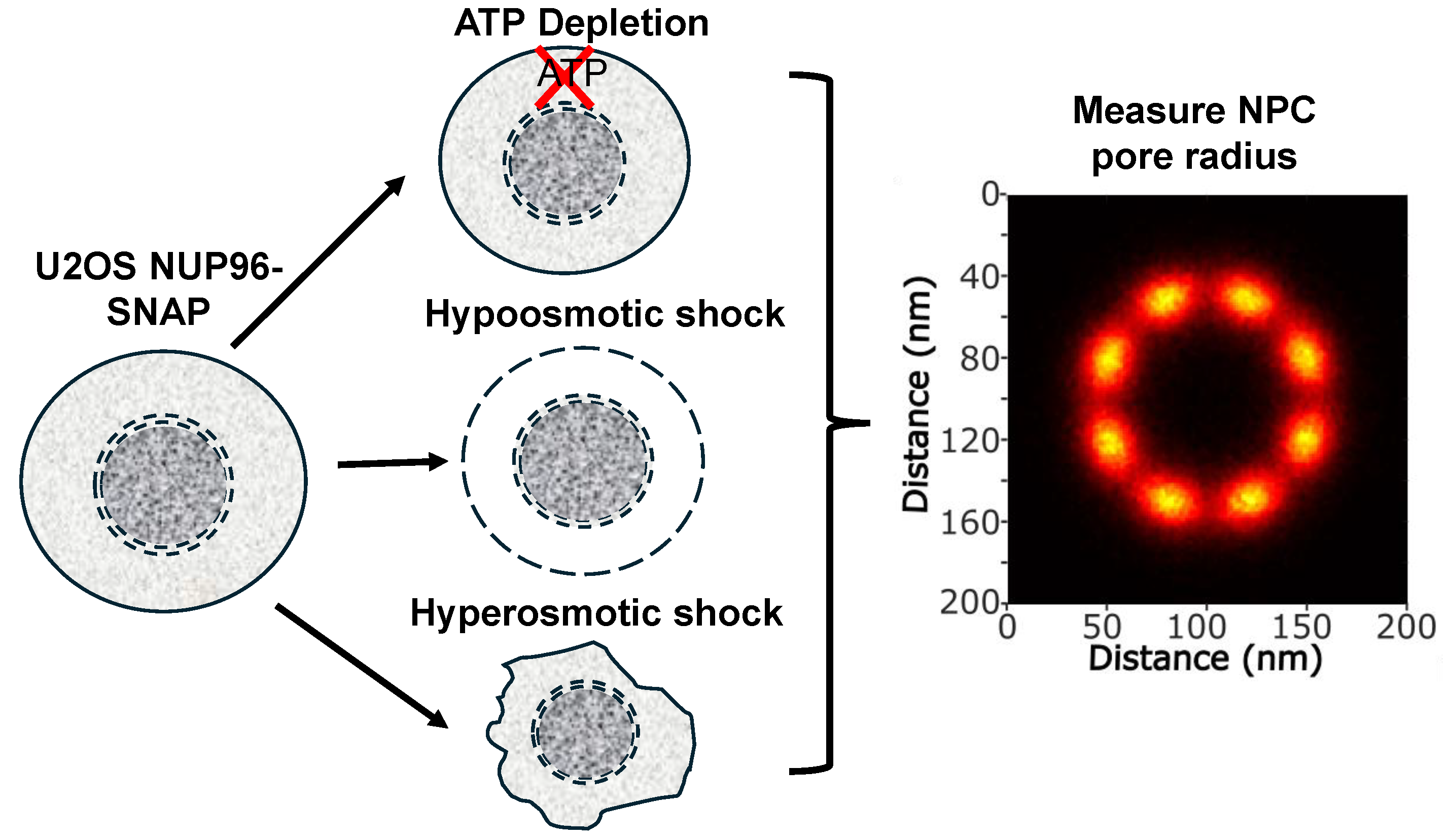
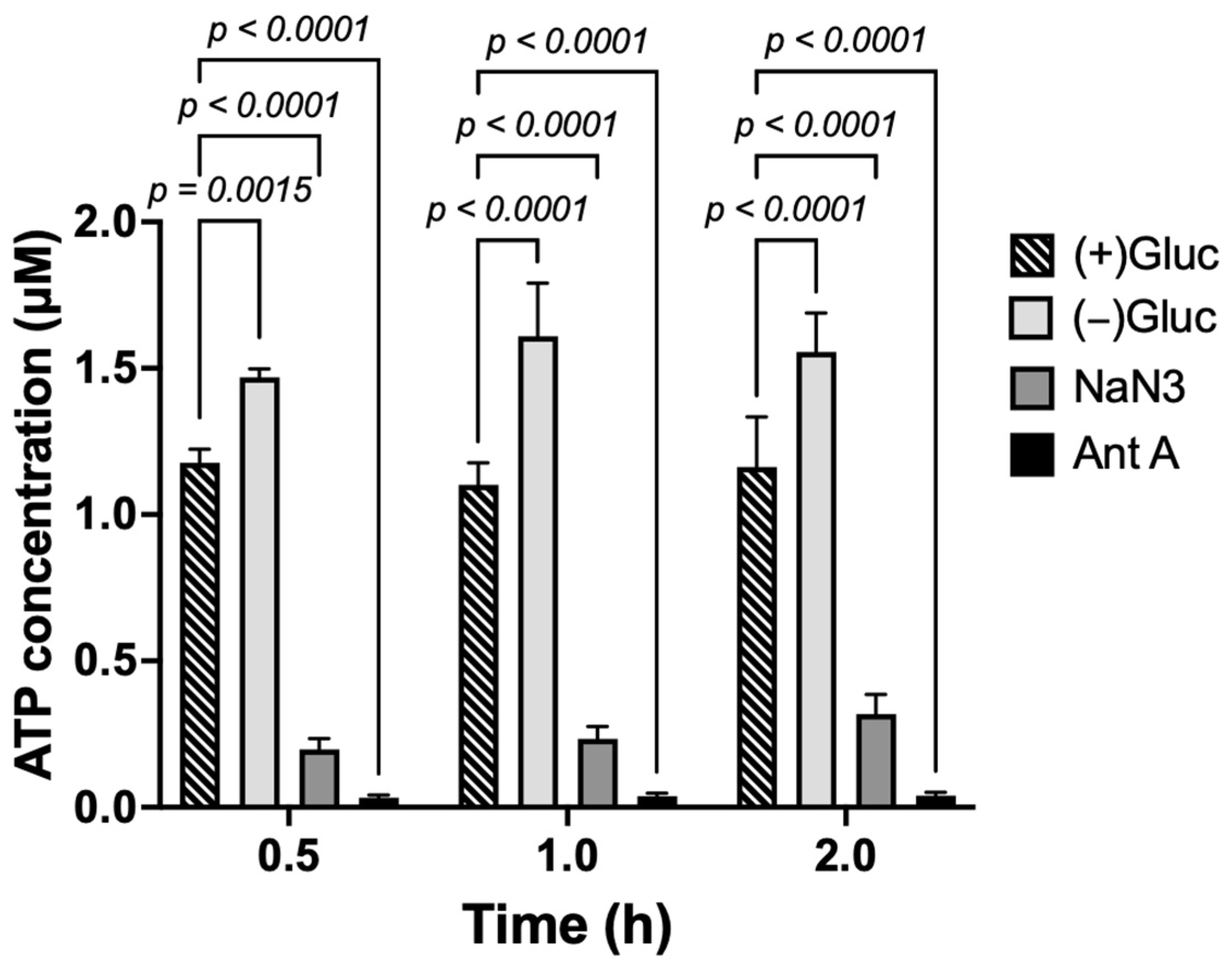
2.3. ATP Depletion
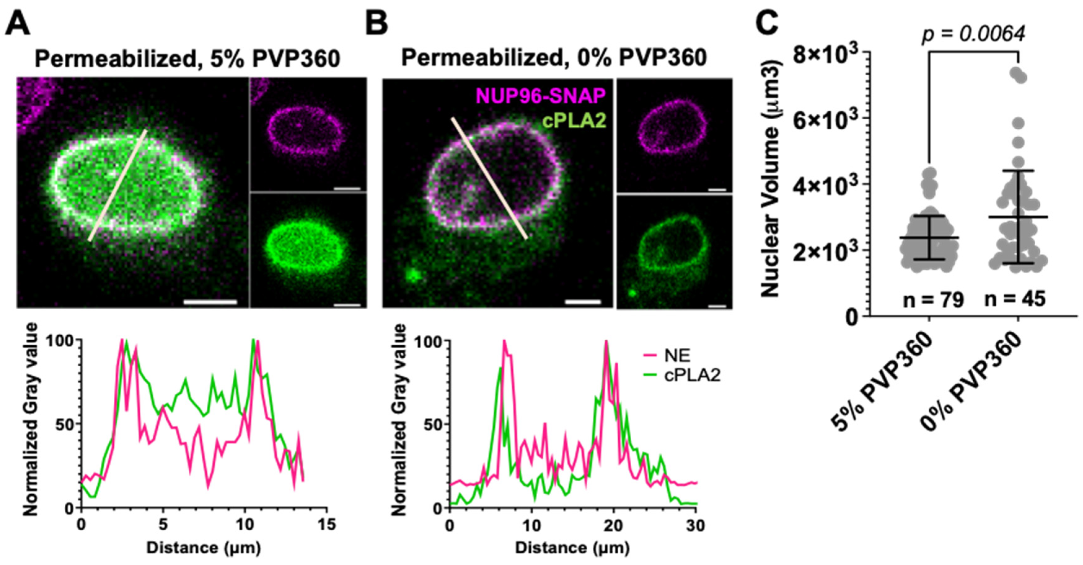
2.4. Osmotic Stretching and Shrinkage of Nuclear Envelope
2.5. Preparation of Samples for Imaging
2.6. Confocal and STORM Imaging
2.7. Image Analysis and Statistics
3. Results
3.1. NUP96-SNAP Tag Does Not Affect the HIV-1 Nuclear Import Kinetics
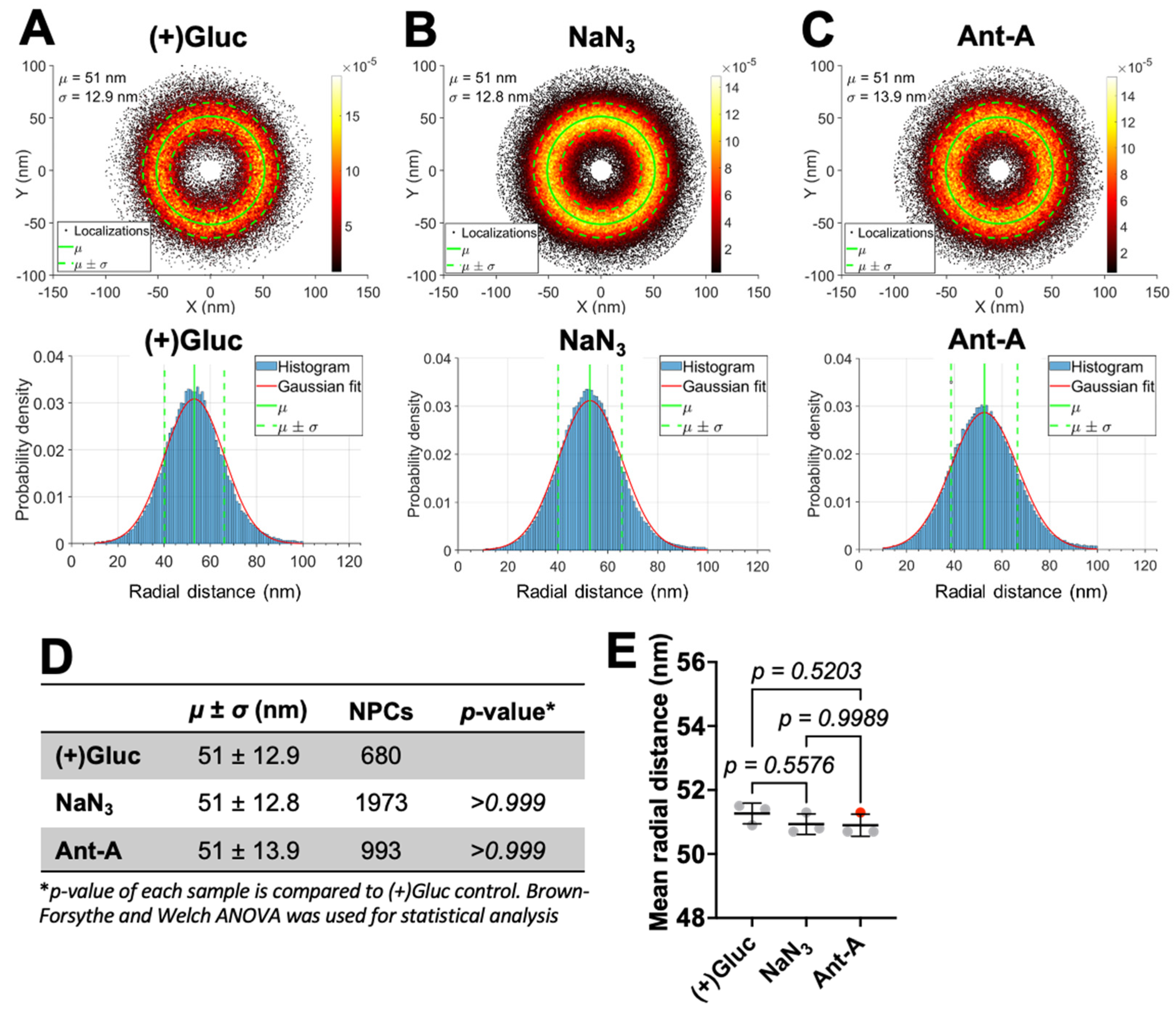
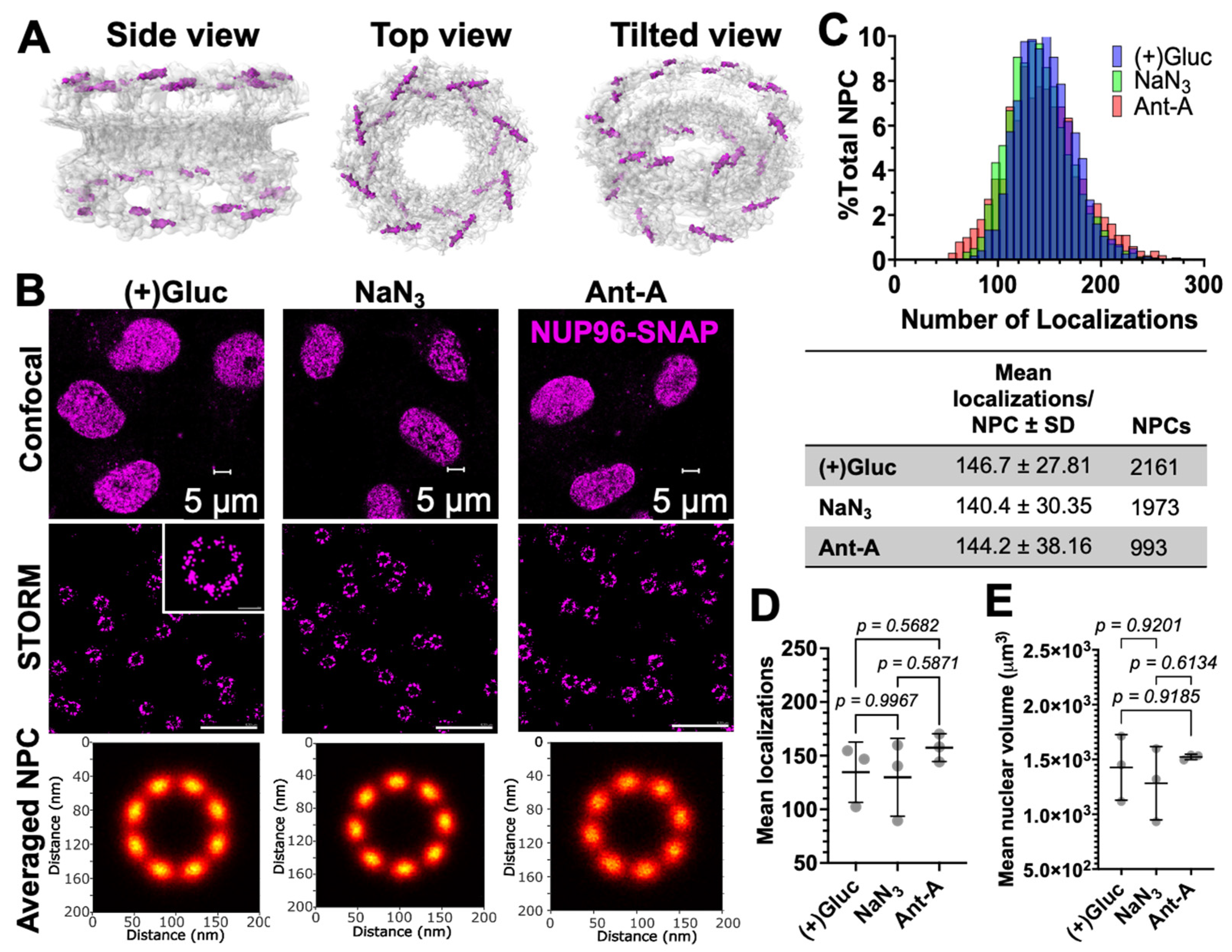
3.2. ATP Depletion Does Not Cause Detectable Changes in NPC Size
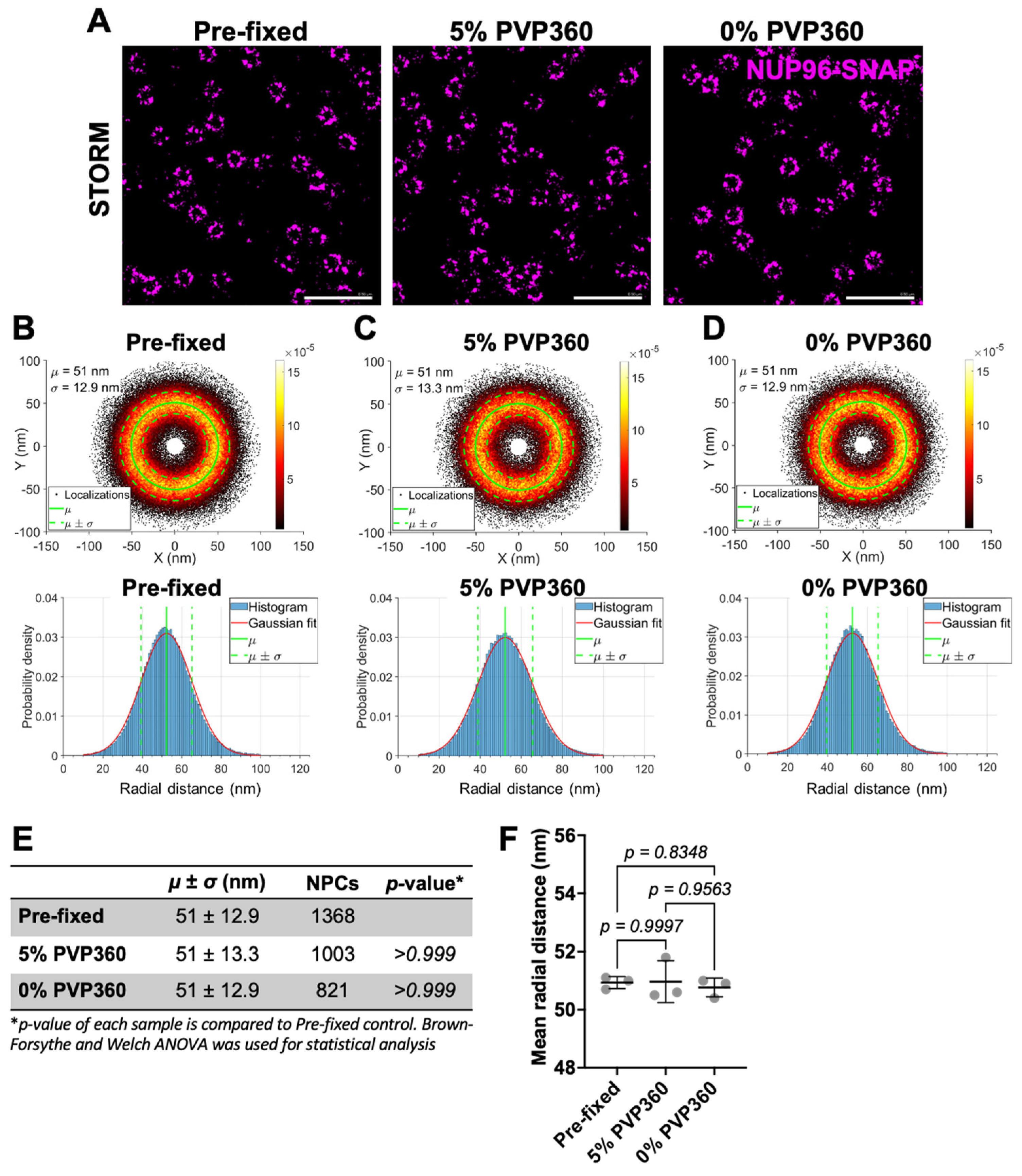
3.3. NPC Size Is Resistant to Stretching and Shrinking of the Nuclear Envelope
4. Discussion
Supplementary Materials
Author Contributions
Funding
Data Availability Statement
Acknowledgments
Conflicts of Interest
References
- Beck, M.; Hurt, E. The nuclear pore complex: Understanding its function through structural insight. Nat. Rev. Mol. Cell Biol. 2016, 18, 73–89. [Google Scholar] [CrossRef]
- Knockenhauer, K.E.; Schwartz, T.U. The Nuclear Pore Complex as a Flexible and Dynamic Gate. Cell 2016, 164, 1162–1171. [Google Scholar] [CrossRef] [PubMed]
- Lin, D.H.; Hoelz, A. The Structure of the Nuclear Pore Complex (An Update). Annu. Rev. Biochem. 2019, 88, 725–783. [Google Scholar] [CrossRef] [PubMed]
- Paci, G.; Caria, J.; Lemke, E.A. Cargo transport through the nuclear pore complex at a glance. J. Cell Sci. 2021, 134. [Google Scholar] [CrossRef]
- Bui, K.H.; von Appen, A.; DiGuilio, A.L.; Ori, A.; Sparks, L.; Mackmull, M.-T.; Bock, T.; Hagen, W.; Andrés-Pons, A.; Glavy, J.S.; et al. Integrated Structural Analysis of the Human Nuclear Pore Complex Scaffold. Cell 2013, 155, 1233–1243. [Google Scholar] [CrossRef] [PubMed]
- Celetti, G.; Paci, G.; Caria, J.; VanDelinder, V.; Bachand, G.; Lemke, E.A. The liquid state of FG-nucleoporins mimics permeability barrier properties of nuclear pore complexes. J. Cell Biol. 2019, 219, e201907157. [Google Scholar] [CrossRef]
- Frey, S.; Richter, R.P.; Görlich, D. FG-Rich Repeats of Nuclear Pore Proteins Form a Three-Dimensional Meshwork with Hydrogel-Like Properties. Science 2006, 314, 815–817. [Google Scholar] [CrossRef]
- Panté, N.; Kann, M. Nuclear Pore Complex Is Able to Transport Macromolecules with Diameters of ∼39 nm. Mol. Biol. Cell 2002, 13, 425–434. [Google Scholar] [CrossRef]
- Paci, G.; Zheng, T.; Caria, J.; Zilman, A.; A Lemke, E. Molecular determinants of large cargo transport into the nucleus. eLife 2020, 9, e55963. [Google Scholar] [CrossRef]
- Zila, V.; Margiotta, E.; Turoňová, B.; Müller, T.G.; Zimmerli, C.E.; Mattei, S.; Allegretti, M.; Börner, K.; Rada, J.; Müller, B.; et al. Cone-shaped HIV-1 capsids are transported through intact nuclear pores. Cell 2021, 184, 1032–1046.e18. [Google Scholar] [CrossRef]
- Schuller, A.P.; Wojtynek, M.; Mankus, D.; Tatli, M.; Kronenberg-Tenga, R.; Regmi, S.G.; Dip, P.V.; Lytton-Jean, A.K.R.; Brignole, E.J.; Dasso, M.; et al. The cellular environment shapes the nuclear pore complex architecture. Nature 2021, 598, 667–671. [Google Scholar] [CrossRef]
- Mahamid, J.; Pfeffer, S.; Schaffer, M.; Villa, E.; Danev, R.; Cuellar, L.K.; Förster, F.; Hyman, A.A.; Plitzko, J.M.; Baumeister, W. Visualizing the molecular sociology at the HeLa cell nuclear periphery. Science 2016, 351, 969–972. [Google Scholar] [CrossRef]
- Zimmerli, C.E.; Allegretti, M.; Rantos, V.; Goetz, S.K.; Obarska-Kosinska, A.; Zagoriy, I.; Halavatyi, A.; Hummer, G.; Mahamid, J.; Kosinski, J.; et al. Nuclear pores dilate and constrict in cellulo. Science 2021, 374, eabd9776. [Google Scholar] [CrossRef]
- Morgan, K.J.; Carley, E.; Coyne, A.N.; Rothstein, J.D.; Lusk, C.P.; King, M.C. Visualizing nuclear pore complex plasticity with pan-expansion microscopy. J. Cell Biol. 2025, 224, e202409120. [Google Scholar] [CrossRef]
- Taniguchi, R.; Orniacki, C.; Kreysing, J.P.; Zila, V.; Zimmerli, C.E.; Böhm, S.; Turoňová, B.; Kräusslich, H.-G.; Doye, V.; Beck, M. Nuclear pores safeguard the integrity of the nuclear envelope. Nat. Cell Biol. 2025, 27, 762–775. [Google Scholar] [CrossRef] [PubMed]
- Sellés, J.; Penrad-Mobayed, M.; Guillaume, C.; Fuger, A.; Auvray, L.; Faklaris, O.; Montel, F. Nuclear pore complex plasticity during developmental process as revealed by super-resolution microscopy. Sci. Rep. 2017, 7, 14732. [Google Scholar] [CrossRef]
- Löschberger, A.; van de Linde, S.; Dabauvalle, M.-C.; Rieger, B.; Heilemann, M.; Krohne, G.; Sauer, M. Super-resolution imaging visualizes the eightfold symmetry of gp210 proteins around the nuclear pore complex and resolves the central channel with nanometer resolution. J. Cell Sci. 2012, 125, 570–575. [Google Scholar] [CrossRef] [PubMed]
- Hou, Z.; Shen, Y.; Fronik, S.; Shen, J.; Shi, J.; Xu, J.; Chen, L.; Hardenbrook, N.; Engelman, A.N.; Aiken, C.; et al. HIV-1 nuclear import is selective and depends on both capsid elasticity and nuclear pore adaptability. Nat. Microbiol. 2025, 10, 1868–1885. [Google Scholar] [CrossRef]
- Kreysing, J.P.; Heidari, M.; Zila, V.; Cruz-León, S.; Obarska-Kosinska, A.; Laketa, V.; Rohleder, L.; Welsch, S.; Köfinger, J.; Turoňová, B.; et al. Passage of the HIV capsid cracks the nuclear pore. Cell 2025, 188, 930–943.e21. [Google Scholar] [CrossRef]
- Hou, Z.; Fronik, S.; Shen, Y.; Chen, L.; Thompson, C.; Neumann, S.; Zhang, P. Direct visualization of HIV-1 core nuclear import and its interplay with the nuclear pore. EMBO Rep. 2025, 26, 5133–5153. [Google Scholar] [CrossRef]
- Müller, T.G.; Zila, V.; Peters, K.; Schifferdecker, S.; Stanic, M.; Lucic, B.; Laketa, V.; Lusic, M.; Müller, B.; Kräusslich, H.-G. HIV-1 uncoating by release of viral cDNA from capsid-like structures in the nucleus of infected cells. eLife 2021, 10, e64776. [Google Scholar] [CrossRef] [PubMed]
- Ay, S.; Burlaud-Gaillard, J.; Gazi, A.; Tatirovsky, Y.; Cuche, C.; Diana, J.-S.; Scoca, V.; Di Santo, J.P.; Roingeard, P.; Mammano, F.; et al. In vivo HIV-1 nuclear condensates safeguard against cGAS and license reverse transcription. EMBO J. 2024, 44, 166–199. [Google Scholar] [CrossRef]
- Müller, T.G.; Klaus, S.; Zila, V.; Lucic, B.; Penzo, C.; Nopper, S.L.; Golani, G.; Anders-Össwein, M.; Sonntag-Buck, V.; Heuser, A.-M.; et al. Lenacapavir-induced capsid damage uncovers HIV-1 genomes emanating from nuclear speckles. EMBO J. 2025, 45, 449–470. [Google Scholar] [CrossRef]
- Hou, Z.; Shen, Y.; Fronik, S.; Shen, J.; Shi, J.; Xu, J.; Chen, L.; Hardenbrook, N.; Thompson, C.; Neumann, S.; et al. Correlative In Situ Cryo-ET Reveals Cellular and Viral Remodeling Associated with Selective HIV-1 Core Nuclear Import. bioRxiv 2025. [Google Scholar] [CrossRef]
- Sabinina, V.J.; Hossain, M.J.; Hériché, J.-K.; Hoess, P.; Nijmeijer, B.; Mosalaganti, S.; Kueblbeck, M.; Callegari, A.; Szymborska, A.; Beck, M.; et al. Three-dimensional superresolution fluorescence microscopy maps the variable molecular architecture of the nuclear pore complex. Mol. Biol. Cell 2021, 32, 1523–1533. [Google Scholar] [CrossRef]
- Ye, X.; Guan, M.; Guo, Y.; Liu, X.; Wang, K.; Chen, T.; Zhao, S.; Chen, L. Live-cell super-resolution imaging unconventional dynamics and assemblies of nuclear pore complexes. Biophys. Rep. 2023, 9, 206–214. [Google Scholar] [CrossRef] [PubMed]
- Thevathasan, J.V.; Kahnwald, M.; Cieśliński, K.; Hoess, P.; Peneti, S.K.; Reitberger, M.; Heid, D.; Kasuba, K.C.; Hoerner, S.J.; Li, Y.; et al. Nuclear pores as versatile reference standards for quantitative superresolution microscopy. Nat. Methods 2019, 16, 1045–1053. [Google Scholar] [CrossRef]
- Pizzato, M.; Erlwein, O.; Bonsall, D.; Kaye, S.; Muir, D.; McClure, M.O. A one-step SYBR Green I-based product-enhanced reverse transcriptase assay for the quantitation of retroviruses in cell culture supernatants. J. Virol. Methods 2009, 156, 1–7. [Google Scholar] [CrossRef]
- Shen, Z.; Belcheva, K.T.; Jelcic, M.; Hui, K.L.; Katikaneni, A.; Niethammer, P. A synergy between mechanosensitive calcium- and membrane-binding mediates tension-sensing by C2-like domains. Proc. Natl. Acad. Sci. USA 2021, 119, e2112390119. [Google Scholar] [CrossRef] [PubMed]
- Enyedi, B.; Jelcic, M.; Niethammer, P. The Cell Nucleus Serves as a Mechanotransducer of Tissue Damage-Induced Inflammation. Cell 2016, 165, 1160–1170. [Google Scholar] [CrossRef]
- Super Resolution Microscope N-STORM Protocol; N-STORM PROTOCOL 2MJ-SCIH-4; Nikon Corporation: Tokyo, Japan, 2013.
- Haghparast, S.; Stallinga, S.; Rieger, B. Detecting continuous structural heterogeneity in single-molecule localization microscopy data. Sci. Rep. 2023, 13, 19800. [Google Scholar] [CrossRef]
- Wang, W.; Heydarian, H.; Huijben, T.A.P.M.; Stallinga, S.; Rieger, B. Joint registration of multiple point clouds for fast particle fusion in localization microscopy. Bioinformatics 2022, 38, 3281–3287. [Google Scholar] [CrossRef]
- Nieuwenhuizen, R.P.J.; A Lidke, K.; Bates, M.; Puig, D.L.; Grünwald, D.; Stallinga, S.; Rieger, B. Measuring image resolution in optical nanoscopy. Nat. Methods 2013, 10, 557–562. [Google Scholar] [CrossRef]
- van Vliet, L.J.; Verbeek, P.W. Edge localization by MoG filters: Multiple-of-Gaussians. Pattern Recognit. Lett. 1994, 15, 485–496. [Google Scholar] [CrossRef]
- von Appen, A.; Kosinski, J.; Sparks, L.; Ori, A.; DiGuilio, A.L.; Vollmer, B.; Mackmull, M.-T.; Banterle, N.; Parca, L.; Kastritis, P.; et al. In situ structural analysis of the human nuclear pore complex. Nature 2015, 526, 140–143. [Google Scholar] [CrossRef] [PubMed]
- Schlichthaerle, T.; Strauss, M.T.; Schueder, F.; Auer, A.; Nijmeijer, B.; Kueblbeck, M.; Sabinina, V.J.; Thevathasan, J.V.; Ries, J.; Ellenberg, J.; et al. Direct Visualization of Single Nuclear Pore Complex Proteins Using Genetically-Encoded Probes for DNA-PAINT. Angew. Chem. Int. Ed. Engl. 2019, 58, 13004–13008. [Google Scholar] [CrossRef] [PubMed]
- Rankovic, S.; Ramalho, R.; Aiken, C.; Rousso, I. PF74 Reinforces the HIV-1 Capsid To Impair Reverse Transcription-Induced Uncoating. J. Virol. 2018, 92, e00845-18. [Google Scholar] [CrossRef]
- Bhattacharya, A.; Alam, S.L.; Fricke, T.; Zadrozny, K.; Sedzicki, J.; Taylor, A.B.; Demeler, B.; Pornillos, O.; Ganser-Pornillos, B.K.; Diaz-Griffero, F.; et al. Structural basis of HIV-1 capsid recognition by PF74 and CPSF6. Proc. Natl. Acad. Sci. USA 2014, 111, 18625–18630. [Google Scholar] [CrossRef] [PubMed]
- Bönisch, I.Z.; Dirix, L.; Lemmens, V.; Borrenberghs, D.; De Wit, F.; Vernaillen, F.; Rocha, S.; Christ, F.; Hendrix, J.; Hofkens, J.; et al. Capsid-Labelled HIV To Investigate the Role of Capsid during Nuclear Import and Integration. J. Virol. 2020, 94, e01024-19. [Google Scholar] [CrossRef]
- Hulme, A.E.; Kelley, Z.; Foley, D.; Hope, T.J. Complementary Assays Reveal a Low Level of CA Associated with Viral Complexes in the Nuclei of HIV-1-Infected Cells. J. Virol. 2015, 89, 5350–5361. [Google Scholar] [CrossRef]
- Balasubramaniam, M.; Zhou, J.; Addai, A.; Martinez, P.; Pandhare, J.; Aiken, C.; Dash, C. PF74 Inhibits HIV-1 Integration by Altering the Composition of the Preintegration Complex. J. Virol. 2019, 93, e01741-18. [Google Scholar] [CrossRef]
- Robinson, G.L.; Dinsdale, D.; MacFarlane, M.; Cain, K. Switching from aerobic glycolysis to oxidative phosphorylation modulates the sensitivity of mantle cell lymphoma cells to TRAIL. Oncogene 2012, 31, 4996–5006. [Google Scholar] [CrossRef]
- Song, S.B.; Hwang, E.S. A Rise in ATP, ROS, and Mitochondrial Content upon Glucose Withdrawal Correlates with a Dysregulated Mitochondria Turnover Mediated by the Activation of the Protein Deacetylase SIRT1. Cells 2018, 8, 11. [Google Scholar] [CrossRef]
- Zhang, C.-S.; Hardie, D.G.; Lin, S.-C. Glucose Starvation Blocks Translation at Multiple Levels. Cell Metab. 2020, 31, 217–218. [Google Scholar] [CrossRef]
- Wu, Y.-L.; Tschanz, A.; Krupnik, L.; Ries, J. Quantitative Data Analysis in Single-Molecule Localization Microscopy. Trends Cell Biol. 2020, 30, 837–851. [Google Scholar] [CrossRef]
- Bates, M.; Jones, S.A.; Zhuang, X. Stochastic Optical Reconstruction Microscopy (STORM): A Method for Superresolution Fluorescence Imaging. Cold Spring Harb. Protoc. 2013, 2013, 498–520. [Google Scholar] [CrossRef]
- Holzer, G.; Antonin, W. Breaking the Y. PLoS Genet. 2019, 15, e1008109. [Google Scholar] [CrossRef] [PubMed]
- Goldberg, M.W. Nuclear pore complex tethers to the cytoskeleton. Semin. Cell Dev. Biol. 2017, 68, 52–58. [Google Scholar] [CrossRef]
- Szymborska, A.; de Marco, A.; Daigle, N.; Cordes, V.C.; Briggs, J.A.G.; Ellenberg, J. Nuclear Pore Scaffold Structure Analyzed by Super-Resolution Microscopy and Particle Averaging. Science 2013, 341, 655–658. [Google Scholar] [CrossRef] [PubMed]
- Rajoo, S.; Vallotton, P.; Onischenko, E.; Weis, K. Stoichiometry and compositional plasticity of the yeast nuclear pore complex revealed by quantitative fluorescence microscopy. Proc. Natl. Acad. Sci. USA 2018, 115, E3969–E3977. [Google Scholar] [CrossRef]
- Allegretti, M.; Zimmerli, C.E.; Rantos, V.; Wilfling, F.; Ronchi, P.; Fung, H.K.H.; Lee, C.-W.; Hagen, W.; Turoňová, B.; Karius, K.; et al. In-cell architecture of the nuclear pore and snapshots of its turnover. Nature 2020, 586, 796–800. [Google Scholar] [CrossRef] [PubMed]
- Elosegui-Artola, A.; Andreu, I.; Beedle, A.E.M.; Lezamiz, A.; Uroz, M.; Kosmalska, A.J.; Oria, R.; Kechagia, J.Z.; Rico-Lastres, P.; Le Roux, A.-L.; et al. Force Triggers YAP Nuclear Entry by Regulating Transport across Nuclear Pores. Cell 2017, 171, 1397–1410.e14. [Google Scholar] [CrossRef] [PubMed]
- Meng, X.; Zhu, Y.; Tan, H.; Daraqel, B.; Ming, Y.; Li, X.; Yang, G.; He, X.; Song, J.; Zheng, L. The cytoskeleton dynamics-dependent LINC complex in periodontal ligament stem cells transmits mechanical stress to the nuclear envelope and promotes YAP nuclear translocation. Stem Cell Res. Ther. 2024, 15, 284. [Google Scholar] [CrossRef] [PubMed]



Disclaimer/Publisher’s Note: The statements, opinions and data contained in all publications are solely those of the individual author(s) and contributor(s) and not of MDPI and/or the editor(s). MDPI and/or the editor(s) disclaim responsibility for any injury to people or property resulting from any ideas, methods, instructions or products referred to in the content. |
© 2026 by the authors. Licensee MDPI, Basel, Switzerland. This article is an open access article distributed under the terms and conditions of the Creative Commons Attribution (CC BY) license.
Share and Cite
Torres-Rivera, D.; Haghparast, S.; Rieger, B.; Melikyan, G.B. Super-Resolution Imaging of Nuclear Pore Responses to Mechanical Stress and Energy Depletion. Viruses 2026, 18, 167. https://doi.org/10.3390/v18020167
Torres-Rivera D, Haghparast S, Rieger B, Melikyan GB. Super-Resolution Imaging of Nuclear Pore Responses to Mechanical Stress and Energy Depletion. Viruses. 2026; 18(2):167. https://doi.org/10.3390/v18020167
Chicago/Turabian StyleTorres-Rivera, Dariana, Sobhan Haghparast, Bernd Rieger, and Gregory B. Melikyan. 2026. "Super-Resolution Imaging of Nuclear Pore Responses to Mechanical Stress and Energy Depletion" Viruses 18, no. 2: 167. https://doi.org/10.3390/v18020167
APA StyleTorres-Rivera, D., Haghparast, S., Rieger, B., & Melikyan, G. B. (2026). Super-Resolution Imaging of Nuclear Pore Responses to Mechanical Stress and Energy Depletion. Viruses, 18(2), 167. https://doi.org/10.3390/v18020167