


Abstract
Staphylococcus aureus is one of the major pathogenic species affiliated to ESKAPE (Enterococcus faecium, Staphylococcus aureus, Klebsiella pneumoniae, Acinetobacter baumannii, Pseudomonas aeruginosa, and Enterobacter spp.) group, frequently encountered in the lower respiratory tract infections (LRTI). Here, we investigated the applicability of Staphylococcus phage RuSa1 as a bioreporter for S. aureus infections in human respiratory tract and assessed the prophage landscape of clinical strains of S. aureus with reference to P. aeruginosa for safe phage therapeutics. While BOX-A1R-based repetitive extragenic palindromic-polymerase chain reaction (BOX-PCR) displayed discrete signatures corresponding to Staphylococcus in sputum samples, RuSa1 specifically identified S. aureus in LRTI. Pangenome analysis revealed the prevalence of holin and major capsid proteins as prophages in clinical strains of S. aureus (n = 50). Peeveelvirus, Biseptimavirus, Dubowvirus and Triavirus are the major lysogenic taxa in S. aureus, whereas genetic signatures corresponding to Kayvirus-like prophages were absent. The abundance and evenness of prophages were high in S. aureus, whereas the diversity and richness peaked in P. aeruginosa. Interestingly, holins exhibited high diversity, abundance, richness and evenness specifically in S. aureus. In contrast, tail proteins showed high diversity, abundance, and richness in P. aeruginosa, while their distribution was more even in S. aureus. We identified a tail fibre protein in RuSa1, which exhibited strong binding with wall teichoic acids of S. aureus (binding energy −8.1 kcal/mol), followed by S. aureus ST395 (binding energy −7.2 kcal/mol). This study demonstrated the potential ability of RuSa1 to sense and lyse respiratory S. aureus, and decoded taxonomic landscape of prophages, which encourages safe applicability of Kayvirus in diagnostics and therapy targeted to S. aureus in LRTI.
1. Introduction
Staphylococcus aureus is a Gram-positive bacterium extensively studied for its flexible and diverse nature, and its pathogenicity is influenced by prophages [1,2]. Strains of S. aureus are both commensal and opportunistic pathogens, with 20–30% of the human population being colonised in nose, ear and skin, present in different ecological niches. While colonisation is often harmless, it increases the risk of developing serious infections such as bacteremia and infective endocarditis, contributing to increased morbidity and mortality globally [1,3,4]. S. aureus is also known to cause skin and wound infections, resulting in a serious public threat in hospital settings due to increasing antimicrobial resistance (AMR), which contributes to high mortality [5,6]. S. aureus poses serious clinical threat, predominantly due to their antibiotic resistance and virulence features [7]. Since they are able to form biofilm and cause infection in the body, conventional antibiotic therapy is becoming ineffective and needs a new alternative approach, such as phage therapy [8,9]. Most phages known to infect S. aureus belong to class Caudoviricetes, which includes tailed phages and replaced the former order Caudovirales in the International Committee on Taxonomy of Viruses (ICTV) [10,11]. Virulent phages used for the treatment of pathogenic infections have several advantages in comparison with traditional antibiotics due to high specificity and minimal effect on beneficial microbiota [10,12].
Prophages are an important part of S. aureus genome, making up to 10% of their genetic material, with 90% of the isolates containing at least one prophage in their genome [13]. Temperate phages contribute to the persistence and pathogenic potential of S. aureus by serving as a reservoir of virulence, AMR, and phage resistance genes. Importantly, prophages increase bacterial evolution by introducing new genes through lysogenic conversion. As temperate phages that integrate into host genome, boosting genetic diversity and adaptation between phages and their hosts, prophages are key to the success of the S. aureus pathogens; they modulate their survival fitness and help them persist in different niches [13,14,15]. Thus, prophages provide insight into their role in fitness and pathogenicity of S. aureus and may aid safety assurance of lytic phages for therapeutic applications.
Earlier studies have analysed prophages using Rapid annotations using subsystems technology (RAST), Phage search tool-enhanced release (PHASTER), PhiSpy, Pathosystems resource integration center (PATRIC), VGAS and Prokaryotic annotation (PROKKA), emphasising the variation in prophages as per bacterial genomes analysed as well as different bioinformatic tools utilised [16,17,18]. Previous studies have shown that prophages play an important role in the adaptation of S. aureus, carrying structural and functional genes that encode holin, major capsid proteins (MCP) and tail proteins [17]. Prophage-encoded accessory proteins influence phage-host interaction, blocking the lytic activity of phages, and highlighting the functional role in host survival and adaptation [19]. Along with analysing prophage proteins, studies have shown S. aureus prophages belong exclusively to the class Caudoviricetes, with different genera, along with clustering based on geographical locations [20,21]. Furthermore, studies have identified wall teichoic acid as the primary receptor for bacteriophage binding in S. aureus. This interaction includes tail fibre protein and wall teichoic acid playing an important role. Structural variations have reported to influence the binding affinity, highlighting the importance of wall teichoic acid in phage host interactions [22,23].
Lytic phages play a key role in phage therapy due to their ability to infect multiple clinical strains of pathogenic bacteria [24,25]. For comparative study and functional analysis, we employed lytic bacteriophage RuSa1, affiliated to the genus Kayvirus. The experimental validation of lytic attributes was deduced through biofilm removal assay, lack of lysogenic genes, and negative results obtained on lysogenic test [24,25]. However, possible incorporation of Kayvirus genetic material into S. aureus remained elusive.
Here, we investigated the potential ability of Staphylococcus phage RuSa1, affiliated to the family Herelliviridae and phylum Uroviricota, to sense and lyse S. aureus in lower respiratory tract infections (LRTI). In addition, we probed the taxonomic landscape of prophages in clinical strains of S. aureus, predominantly based on holin, MCP and tail proteins, followed by their comparative analysis of diversity indices with clinical stains of P. aeruginosa. We further identified a putative tail fibre protein in RuSa1 that could interact with S. aureus wall teichoic acid (WTA) and may confer specificity.
2. Materials and Methodology
2.1. Bacterial Strains, Bacteriophage and Culture/Propagation Conditions
S. aureus strain ATCC BAA-44 was revived from −80 °C. Clinical strains, designated L1, L2, L5 and L7, affiliated to species S. haemolyticus; SA5, SA3A and SA3B affiliated to the species S. epidermidis; NAS1, NAS2, NAS3, NAS4 and NAS5 affiliated to species S. aureus were isolated from the sputum samples originating from human lower respiratory tract infections (LRTI) (Protocol number YEC-1/2025/174). All these cultures were maintained on Mueller–Hinton agar or broth (HiMedia Laboratories, Mumbai, Maharashtra, India). The bacteriophage RuSa1 was previously isolated from a water sample collected at Sambar deer (Rusa unicolor) enclosure, Pilikula Biological Park, Mangalore [24]. RuSa1 was propagated using S. aureus ATCC BAA-44 as a host and stored at −80 °C in lysate form.
2.2. Genotyping of Clinical Stains of Staphylococcus
Genomic DNA was extracted from Staphylococcus isolates using a DNA extraction kit manufacturer’s protocol (Qiagen, Germany). BOX Polymerase chain reaction (PCR) was performed using the primer (5′-CTACGGCAAGGCGACGC TGACG-3′) and conditions defined earlier [24]. The BOX PCR band patterns were converted into binary matrix, where 1 represents presence of band and 0 represents absence of band at corresponding position. The relatedness of bacterial isolates were analysed through cladogram (https://cladogrammaker.com/).
2.3. Biochemical Characterisation
Gram-positive identification card, Vitek 2 GP ID, bioMérieux (Marcy-l’Étoile, France) was used for biochemical characterisation of all the considered species of Staphylococcus as per the manufacturer’s protocol. Single colonies of each of the species were inoculated into saline, and optical density (OD) was adjusted to McFarland No. 0.50 to 0.63. The cassette consisting of the culture tubes and the identification card were incubated at 37 °C and results were noted within a period of 10 h [26].
2.4. Assessment of Lytic Activity of RuSa1 Using Spot Assay, Plaque Assay and Adsorption Assay
Spot assay was performed according to the workflow described elsewhere [24]. To assess the lytic activity of RuSa1, S. aureus was evenly spread on the Muller–Hinton agar plate. To this plate, 10 µL of RuSa1 phage lysate was drop coated. The plates were incubated at 37 °C, overnight. Following the overnight incubation, the plates were checked for the presence of spots to confirm the lytic activity. Plaque assay was performed using RuSa1 lysate on S. aureus ATCC BAA-44 using double layer agar, in accordance with the previously described workflow [24]. Serial dilution of phage lysate (10−1 to 10−10) was performed using phage buffer and 100 µL of each dilution was mixed with 100 µL of freshly prepared S. aureus ATCC BAA-44. Semi solid agar was added, mixed well and poured on Mueller-Hinton agar plate, allowed to solidify, then incubated at 37 °C, overnight. Plaque-forming units (PFU) were counted, and phage titre was determined. Phage adsorption assay was performed according to the workflow described elsewhere, with slight modification [27]. To two ml of late log phase bacterial culture, 10 µL of phage lysate was added and incubated at 37 °C with shaking for required time intervals (5, 10, 15 and 20 min). The mixture was centrifuged and filtered with a 0.2 µm filter. The number of phages present in the supernatant was determined using plaque assay.
2.5. Light and Transmission Electron Microscopy
Clinical sputum samples and purified strains were subjected Gram staining [28], followed by light microscopy. For transmission electron microscopy, phage lysate (109 PFU/mL) was centrifuged (12,000× g, 5 min, 4 °C) and supernatant was placed onto carbon-coated copper grids, followed by critical point drying. The sample placed on grid was stained with 2% (w/v) phosphotungstic acid and observed under a transmission electron microscope (JEM-2100 plus, JEOL Japan, Tokyo, Japan).
2.6. Comparative Taxonomic Assessment of Prophages in Clinical S. aureus
Genome sequences of clinical S. aureus (n = 50) and P. aeruginosa (n = 50) strains were retrieved from the National Centre for Biotechnology Information (NCBI) database (https://www.ncbi.nlm.nih.gov/) (accessed from 3 August 2025 to 30 August 2025) [29] and annotated using the RAST (Rapid Annotations using Subsystems Technology) (https://rast.nmpdr.org/) (accessed from 3 August 2025 to 30 August 2025) [30] for prophage identification. Genome-sequenced S. aureus of clinical origins, such as respiratory tract, wound and skin, and other defined clinical settings, were included (Table S1). Non-clinical S. aureus were excluded from analysis. Prophages were screened manually for genetic signatures of RuSa1, a lytic Kayvirus species infecting multiple clinical strains of methicillin-resistant S. aureus (MRSA) [24]. Similarly, prophages were screened for genetic signatures of PseuPha1, a lytic Pakpunavirus phage infecting multiple multi-drug resistant (MDR) strains of P. aeruginosa [31]. The taxonomic distributions of holins, major capsid proteins (MCP) and tail proteins in S. aureus and P. aeruginosa (n = 50) were assessed in RAST (BLAST tool) using reference sequences obtained from the UniProt (https://www.uniprot.org/) (accessed from 3 August 2025 to 30 August 2025) [32]. The top five hits were considered with the highest similarity scores for taxonomic resolution. Comparative BLAST analysis against RuSa1 phage was performed and the first hit of phage RuSa1 obtained from RAST was noted down. The distribution of prophages was visualised through a heatmap generated using the Heatmapper (http://heatmapper.ca/) (accessed on 4 September to 10 September 2025) [33].
2.7. Comparative Analysis of Prophage Diversity
A comparative prophage analysis was performed between S. aureus and P. aeruginosa based on occurrence of prophage proteins, total prophage counts and genus-level taxonomical distribution. Alpha diversity indices at the genus-level and protein-level were evaluated; diversity was measured through Shannon–Wiener and Simpson’s indices, richness was quantified through the Margalef index, and evenness was scored through Pielou’s index [34].
2.8. Identification, Annotation and Structural Prediction of Tail Fiber Proteins of Staphylococcus Phage RuSa1
Putative tail fibre proteins in RuSa1 were identified according to the workflow described elsewhere with slight modifications [35]. The genome sequence of phage RuSa1 was first annotated using the RAST server and screened for tail fibre proteins using a reference sequence retrieved from NCBI. Open reading frames exhibiting high sequence similarities and query coverage, and low E-value were considered. The 3D structures of RuSa1 phage tail fibre proteins NTF1 (WEU67506.1, VHWIEOXL_CDS138), NTF2 (WEU67489.1, VHWIEOXL_CDS134) and NTF3 (WEU67490.1, VHWIEOXL_CDS127) were modelled using ab initio protein structure prediction methods. The NTF1 and NTF3 were modelled using protenix-server [36], and the NTF2 was modelled using Alphafold3 [37]. All the modelling was performed in five replicates, with each replicate having five modelled structures. For each protein, the structure with highest pLDDT score was selected as the candidate structure and visualised using UCSF-ChimeraX (https://www.rbvi.ucsf.edu/chimerax/) (accessed from 3 August 2025 to 30 August 2025) [38].
2.9. Phylogenetic Analysis
A maximum likelihood phylogenetic tree was constructed for tail proteins and tail fibre proteins of RuSa1 using closely related reference sequences of Staphylococcus phages [24]. Sequences were retrieved from UniProt and multiple sequence alignment was carried out using ClustalX (https://clustalx.software.informer.com/2.1/) (accessed from 3 August 2025 to 30 August 2025) [39]. The phylogenetic tree was constructed using MEGA software (http://www.megasoftware.net) [40] after 1000 bootstrap replications.
2.10. Identification of Receptor for RuSa1 Tail Fiber in S. aureus
Preliminary indication for the possible receptor in S. aureus for Kayvirus tail fibre was gained through previously published studies [23,41]. Chemical structures of two WTA repeating units were obtained from the literature [22]. The three-dimensional structure of the RuSa1 tail fibre protein (NTF1) trimer was predicted using AlphaFold3 [37]. The SMILES IDs for WTA structures were retrieved using decimer.ai [42], and the molecular structures were modelled using the ChimeraX server [38]. Protein-ligand interaction was performed using docking to understand the interaction between NTF1 and WTA using the HADDOCK 2.4 (High Ambiguity Driven Biomolecular DOCKing) docking platform [43]. The polar region of the tail fibre protein was selected as a putative binding region, and docking was performed only in that region. Multiple docking analyses were generated, and they were ranked based on docking scores and binding energies. The top-ranked complex was visualised, and its interaction was evaluated using ChimeraX.
3. Results
3.1. RuSa1 Senses and Lyses Multiple S. aureus Originating from Lower Respiratory Tract Infections (LRTI)
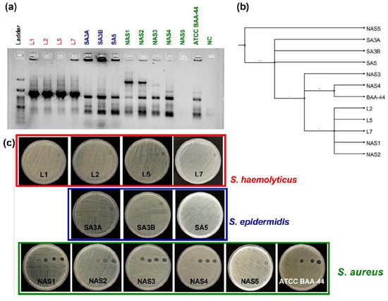
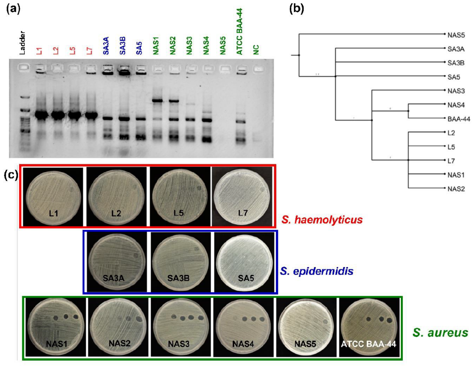
The BOX-PCR fingerprint generated for the clinical Staphylococcus isolated from sputum sample and the associated cladogram are shown in Figure 1a,b. The antibiotic and phage sensitivity of isolates are summarised in Table 1. We observed mild heterogeneity in terms of DNA banding pattern of S. haemolyticus, suggesting clonal origins of possible Staphylococcus as they grew on Mueller-Hinton agar. The spot assays performed for phage RuSa1 against Staphylococcus are shown in Figure 1b. RuSa1 produced clear zones of lysis only against S. aureus, substantiating lytic activity on multiple clinical strains of S. aureus. No such distinct zones occurred despite the load of high-titre phage particles, suggesting its poor sensitivity to S. haemolyticus and S. epidermidis. Plaque assay confirmed phage activity by producing distinct plaques on double-layer agar, with a decrease in the plaque number with increasing dilution, showing countable plaques for phage titre determination. The typical lytic response of Staphylococcus phage RuSa1 on the lawn of S. aureus indicates the sensitivity and specificity of RuSa1 towards S. aureus. Thus, lytic RuSa1, acting as a reporter for S. aureus in LRTI, provided evidence for its possible diagnostic application.
Figure 1.
Lytic/sensing response of RuSa1 on Staphylococcus isolated from Lower Respiratory Tract Infection (LRTI). BOX-PCR genotyping of staphylococcal colonies showing distinct DNA fingerprinting patterns generated for respiratory Staphylococcus (a). Cladogram generated for the BOX-PCR DNA fingerprint is shown (b). Spot assay images showing the lytic activity of RuSa1 on strains of S. haemolyticus (L1, L2, L5 and L7), S. epidermidis (SA3A, SA3B and SA5), and S. aureus (NAS1, NAS2, NAS3, NAS4, NAS5 and ATCC BAA-44) (c). 1 × 10⁸ CFU/mL vs. 1.8 × 10⁹ PFU/mL (left) to 1.8 × 10¹² PFU/mL (right).
Table 1.
Antibiotic and RuSa1 sensitivity pattern of Staphylococcus originating from lower respiratory tract infection (LRTI). Antibiotic sensitivity was determined using Vitek. Phage sensitivity was determined using 1 × 10⁸ CFU/mL vs. 1.8 × 10⁹ PFU/mL to 1.8 × 10¹² PFU/mL.
3.2. Contrasting Occurrence of Prophages in Clinical Strains of S. aureus
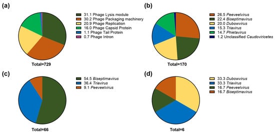
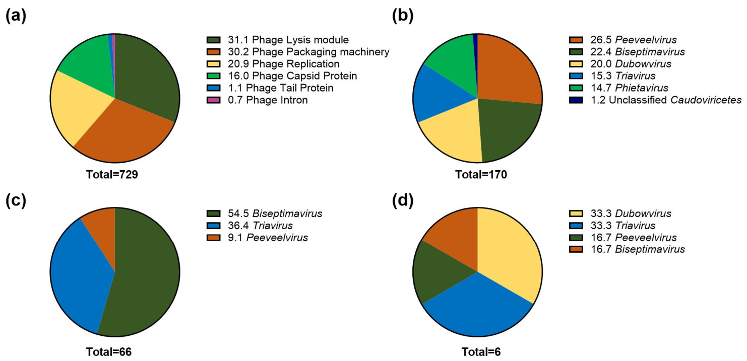
S. aureus and P. aeruginosa strains analysed in this study were from diverse geographical locations and predominantly originated from clinical sources (Tables S1 and S2). We identified 729 prophages spread across 48 genomes (ranging 2.77–3.0 Mb in size each) in S. aureus. Among these strains, S. aureus CUVET18-370 and CUVET20-2256 lacked prophages, whereas prophage count peaked in S. aureus Gv69 (n = 37) (Table S1). The phage lysis module was the most predominant (n = 227) across S. aureus genomes, accounting for 31.1% of prophages, indicating their active integration into the host genome. Phage packaging machinery represented the second highest (n = 220), which amounts to 30.2% of total phages, followed by phage replication (n = 152) and phage capsid proteins (n = 117) amounts to 20.9% and 16% of total phages, respectively. The phage tail protein and phage introns were present in the lowest numbers (n = 8 and n = 5, respectively), amounting to 1.1% and 0.7% of prophages (Figure 2a). Thus, genome-wide occurrence of prophages was found to differ in clinical strains of S. aureus.
Figure 2.
Genome-wide distribution and taxonomic assignments of prophages in clinical strains of Staphylococcus aureus. Relative distribution of prophage proteins (a). Taxonomic assignments based on holins (b), major capsid proteins (c), and phage tail proteins (d). Values are in percentages.
3.3. Structural and Functional Attributes of Prophages in Clinical S. aureus
Prophages were investigated to gain detailed insights into their structural and functional attributes in S. aureus. We identified 84 holins across S. aureus genomes, with S. aureus Gv69 and NMR08 harbouring the maximum (n = 4 each) (Table S1). Similarly, we found 66 MCP, which exhibited dominance in S. aureus Gv69 and 103744 (n = 3 each) (Table S1). The group of tail proteins, including phage tail protein, phage major tail protein, and phage tail tube protein, known for host specificity, were absent in almost all the genomes except for S. aureus subsp. aureus 11819-97 and S. aureus Gv88. Only eight tail proteins were identified across S. aureus genomes (Table S1). Thus, holin and MCP, a unit of the phage lysis module and a building block for the protective envelope surrounding viral genetic material, respectively, dominated S. aureus prophages.
3.4. Taxonomic Landscape of Prophages in Clinical S. aureus
S. aureus genomes were screened for the presence of Kayvirus-like prophages, and holins and MCP were exploited to deduce the genome-wide association and cross-strain dominance of prophages. The prophages were exclusively affiliated with class Caudoviricetes. Among 84 identified holins, the majority belonged to the genus Peeveelvirus (26.5%), followed by Biseptimavirus (22.4%), Dubowvirus (20.0%), Triavirus (15.3%) and Phietavirus (14.7%) and unclassified Caudoviricetes (1.2%) (Figure 2b). Among 66 identified MCP, Biseptimavirus dominated (54.5%), followed by Triavirus (36.4%) and Peeveelvirus at (9.1%) (Figure 2c). Analysis of tail proteins (n = 8) revealed the prevalence of Triavirus and Dubowvirus (33.3% each), followed by Biseptimavirus and Peeveelvirus (16.7% each), respectively (Figure 2d). None of the prophages showed significant hits to holins, MCP and/or tail proteins of Kayvirus phage RuSa1 (Tables S3–S5).
3.5. Clustered Heatmap Analysis of Prophages in Clinical Strains of S. aureus
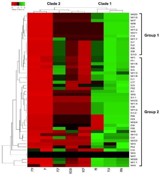
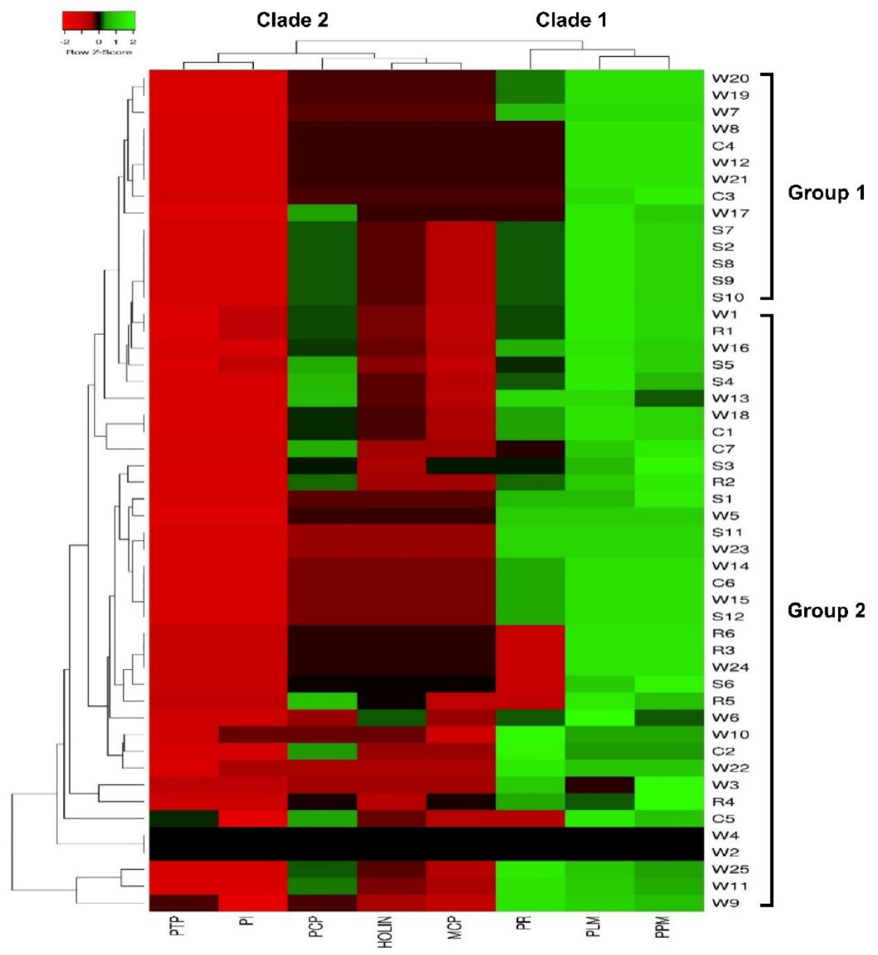
Prophages were subjected to clustered heatmap to trace their numerical abundance and geographical segregation. The clustered heatmap showed two distinct prophage clades designated as clade 1 and clade 2. While clade 1 accommodated dominant phage lysis module (PLM), phage packaging machinery (PPM) and phage replication (PR), clade 2 harboured prophages of moderate-to-minor dominance. The strain-wise clustering into group 1 and group 2 revealed geographical segregation of prophages (Figure 3). Clinical samples (skin, wound, etc.) collected from the European continent (64.28%) predominated in group 1, followed by North America (28.57%) and Africa (7.14%). In contrast, Group 2 mainly accommodated samples (wound, skin and/or respiratory) collected from the Asian continent (47.22%), followed by North America (22.22%), Europe (19.44%), South America (5.55%) and Africa (2.77%), respectively. Prophage-wise and strain-wise clustering reflected their differential prevalence in clinical S. aureus (Table S6).
Figure 3.
Relative genome-wide occurrence of prophages in clinical strains of Staphylococcus aureus. Clustered heatmap showing the row-wise distribution of phage lysis module (PLM), phage packaging machinery (PPM), phage replication (PR), phage capsid protein (PCP), phage tail protein (PTP), phage intron (PI), holin (HOLIN) and major capsid protein (MCP) in clinical strains of S. aureus. C, defined clinical; R, respiratory; W, wound; S, skin swab or wound.
3.6. Taxonomic Identity of Major Prophages in Clinical Strains of S. aureus
Taxonomic affiliation and dominance of phages infecting S. aureus were probed through holins, MCP and tail proteins. Holins were found to be predominantly affiliated to Biseptimavirus and Peeveelvirus (25% each), followed by Dubowvirus (21.43%), Phietavirus (14.29%), Triavirus (12.50%), and unclassified Caudoviricetes (1.79%) in group 1 (Europe) (Figure S1a). In group 2 (Asia), holins were predominantly affiliated to Peeveelvirus (27.52%), followed by Biseptimavirus (22.02%), Dubowvirus (19.27%), Phietavirus (14.68%), Triavirus (15.6%), and unclassified Caudoviricetes (0.92%) (Figure S1b). Taxonomic analysis of MCP revealed the dominance of Biseptimavirus in groups 1 and 2 (56.52% and 53.66%, respectively), followed by Triavirus (39.13% and 34.15%, respectively) and Peeveelvirus (4.35% and 12.20%, respectively) (Figure S1c,d). Tail proteins, which were found exclusively in group 2, were affiliated with Triavirus and Dubowvirus (33.33% each), followed by Biseptimavirus and Peeveelvirus (16.67% each) (Figure S1e). Thus, the present study indicated heterogeneous occurrence of Biseptimavirus, Peeveelvirus, Triavirus and Dubowvirus in S. aureus across Europe and Asia.
3.7. Comparative Analysis of S. aureus and P. aeruginosa Prophages
Out of the 12 prophage proteins, eight (66.7%) were common in S. aureus and P. aeruginosa and four (33.3%) remained unique to P. aeruginosa. No prophage protein exclusive to S. aureus was found. Lysis module, packaging machinery, replication, capsid protein, tail protein, introns, holin and MCP of phages were common in S. aureus and P. aeruginosa. In contrast, IbrA and IbrB: co-activators of prophage gene expression, phage DNA synthesis, baseplate protein and tail fibre protein were unique to P. aeruginosa. The shared prophage proteins constituted structural and functional modules (Table S7).
Out of 22 genera identified, 16 (72.7%) belonged to P. aeruginosa and 5 (22.7%) belonged to S. aureus, whereas unclassified Caudoviricetes (4.5%) were common in both hosts. Biseptimavirus, Triavirus, Peeveelvirus, Dubowvirus and Phietavirus were found to be unique to S. aureus. In contrast, Detrevirus, Haihevirus, Bugaksanvirus, Torontovirus, Casadabanvirus, Citexvirus, Nankolanvirus, Primolicivirus, Pakpunavirus, Baldwinvirus, Torinovirus, Elvirus, Otagovirus, Miecznikowavirus and Tunggulvirus were found to be unique to P. aeruginosa. Among these, Pakpunavirus, Baldwinvirus, Torinovirus and Otagovirus belonged to the family Vandenendeviridae, whereas Citexvirus, Primolicivirus, Elvirus and Tunggulvirus belonged to the families Peduoviridae, Inoviridae, Chimalliviridae and Autoscriptoviridae, respectively (Table S7).
3.8. Diversity Indices of Prophages in S. aureus vs. P. aeruginosa
Analysis of prophages revealed high Shannon–Wiener/Simpson diversity and Margalef’s richness in P. aeruginosa, whereas the abundance and Pielou’s evenness were found to be high in S. aureus. The taxonomic distribution of MCP displayed a similar trend by showing high diversity and richness in P. aeruginosa, while high abundance and evenness were recorded in S. aureus. Interestingly, the diversity, abundance, richness and evenness of holins were found to be high in S. aureus. In contrast, the diversity, abundance and richness of tail proteins were high in P. aeruginosa, whereas evenness was found to be high in S. aureus (Table S8).
3.9. Identification of a Tail Fiber Protein in RuSa1 Interacting with S. aureus
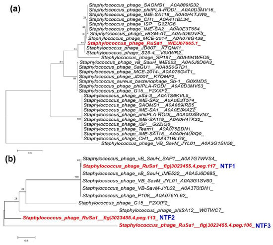
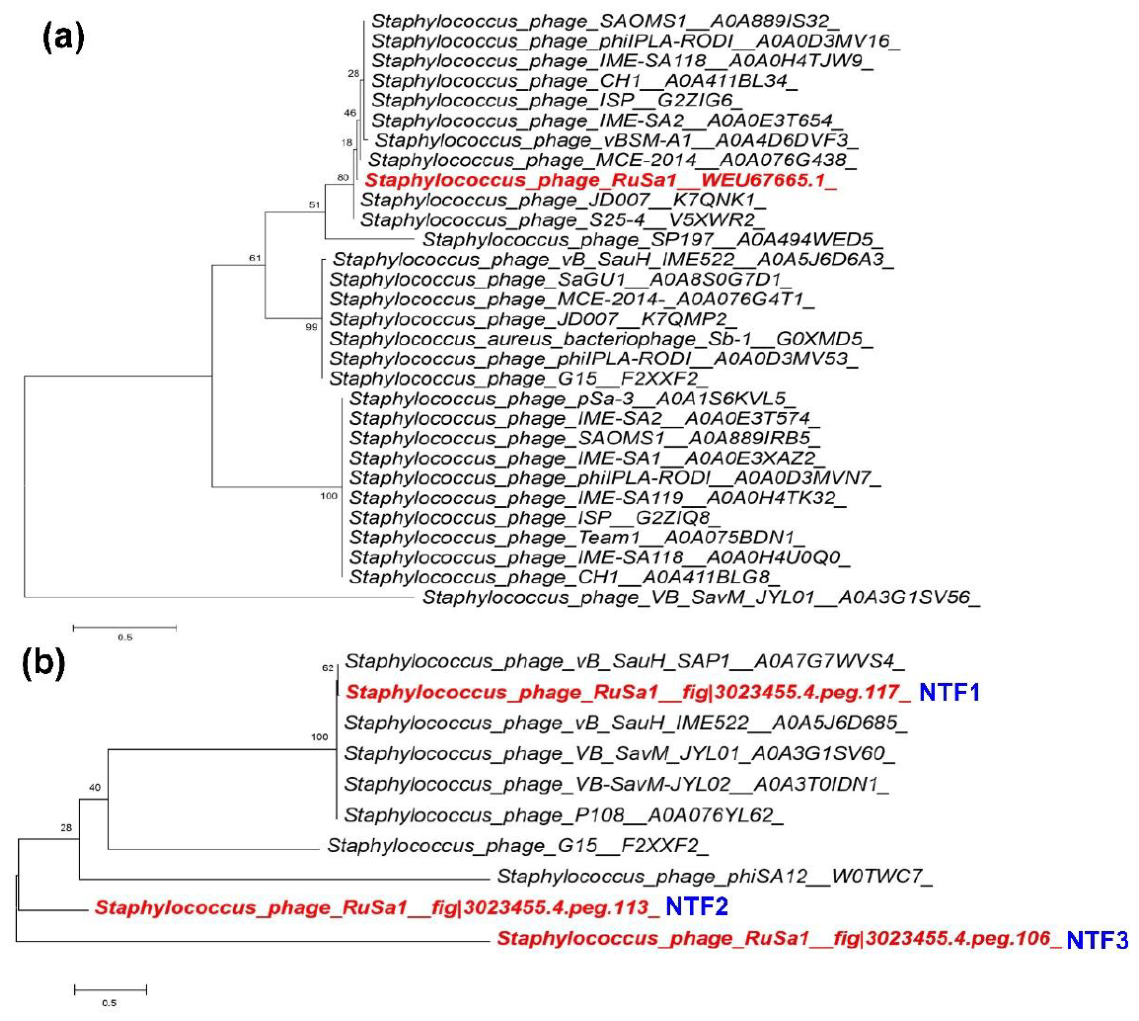
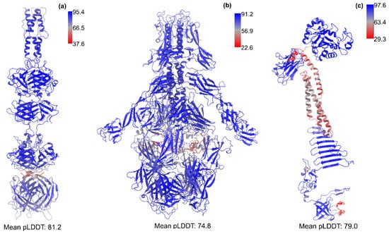
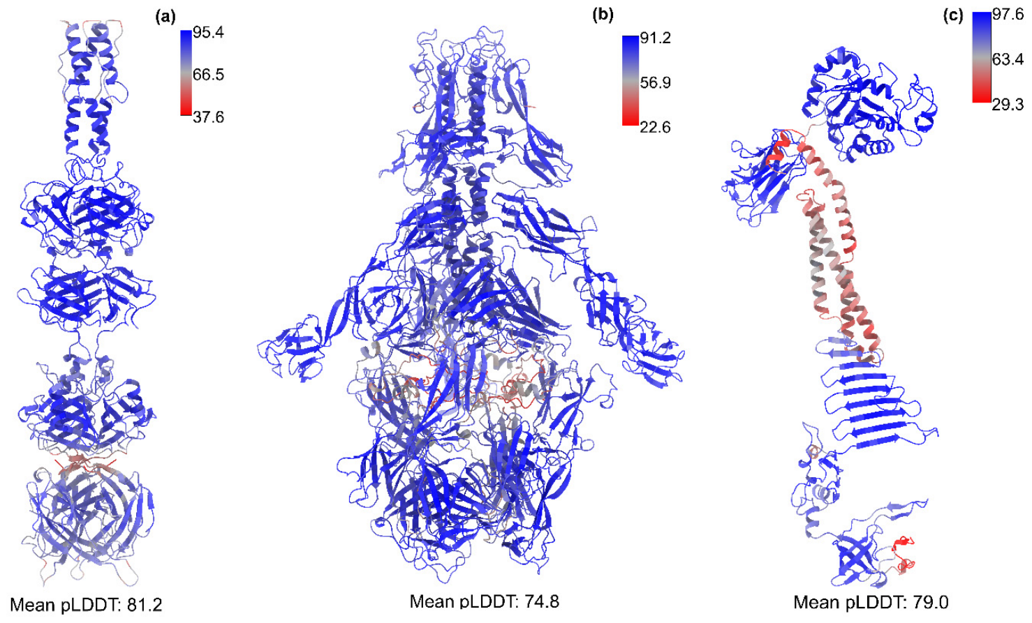
We identified three potential tail fibre proteins designated as NTF1, NTF2 and NTF3 in RAST through BLAST analysis. RuSa1 tail fibre protein, designated as NTF1, was clustered within known Staphylococcus phages during maximum likelihood phylogenetic analysis, suggesting their possible structural similarities. On the other hand, the other two tail fibre proteins of RuSa1, designated as NTF2 and NTF3, formed distinct lineages distantly associated with the clade that accommodated NTF1 (Figure 4). A secondary tree was constructed using all closely related sequences, indicating Phage RuSa1 to have distinct tail-associated protein architectures (Figure S2). We next modelled the tail fibre proteins NTF1, 2 and 3 identified through the phylogenetic analysis (Figure 5a–c). To ascertain the oligomeric states of these proteins, we examined the structures of tail fibre proteins of S. aureus phage phi812, which is evolutionarily related to the phage RuSa1. Since NTF1, 2 and 3 have very high sequence identities with Receptor Binding Protein, DUF4815 domain-containing protein and GP-PDE domain-containing proteins of phi812 phage (sequence identities of 91.7%, 99.7% and 98.7% respectively), we inferred that oligomeric orientations of NTF proteins would be the same as the tail fibre proteins of phi812 phage. Thus, we modelled NTF1 and 2 as homotrimers and NTF3 as a monomer. All the models were found to have high pLDDT scores (>70). In NTF1, though most of the regions of the protein were modelled with high pLDDT scores, certain regions, particularly the loops around the apex of the trimer, had relatively low pLDDT values (Figure 5a). Recent studies have examined thousands of modelled alphafold3 structures and compared these models with molecular dynamics simulations as well as with experimental flexibility descriptors such as B-factors [44]. This study showed that regions with low pLDDT scores can predict conformational flexibility to a certain extent. Based on this, we inferred that these loops may participate in binding with the biomolecules that are part of host cell surface.
Figure 4.
Maximum likelihood phylogeny of tail protein and tail fibre protein of RuSa1 and other related Staphylococcus phages. Maximum likelihood tree showing the phylogenetic position of tail protein (a) and tail fibre protein (b) of Staphylococcus phage RuSa1 and other related S. aureus-infecting phages. Tail protein and tail fibre protein sequences were retrieved from UniProt and publications, and the tail protein and tail fibre protein of Phage RuSa1. The bootstrap values are indicated at branching points, with the scale bar having 0.5 substitutions per site. Sequences highlighted in red denotes the tail fiber protein sequences of Staphylococcus phage RuSa1.
Figure 5.
Structural analysis of tail fibre proteins detected in Staphylococcus Phage RuSa1. Modelled 3D structures of RuSa1 phage NTF1 (a), NTF2 (b) and NTF3 (c) proteins. The modelled structures are represented as cartoons and coloured according to the pLDDT scores.
3.10. Molecular Docking of Tail Fibre Protein of RuSa1 Versus WTA
NTF1 exists as a trimer; this is because the 3D structure of NTF1 from bacteriophage phi812, evolutionarily related to RuSa1, is characterised as a trimer from cryo-electron microscopy studies [PMID:42350675; PDB:9EUI; EMDB:19971] [45]. The NTF1 proteins of phi812 and RuSa1 have very high sequence identity of 92% (Figure S3), which indicates that the trimer interface observed in phi812 NTF would also be conserved in RuSa1. Based on these observations, we inferred that the tail fiber protein of RuSa1NTF1 would exist as a trimer and hence modelled it as a trimer for the docking calculations.
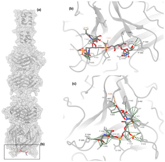
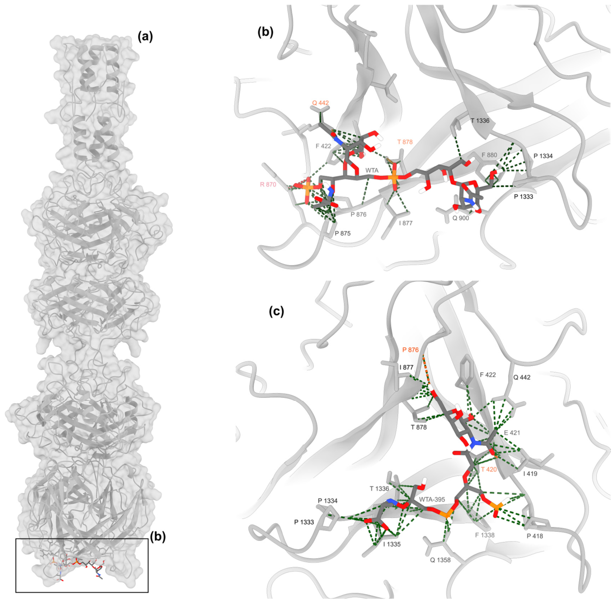
WTA, a potential receptor for phages in S. aureus, typically consists of repeating units of RboP along with GlcNAc and D-alanine, in which specific GlcNAc is required for phage adsorption. TarM and TarS are two glycosyltransferases essential for the addition of either α-GlcNAc or β-GlcNAc, respectively [41,46]. Docking analysis of the NTF1 trimer was performed with a single WTA repeating unit from S. aureus and S. aureus ST395 (Figure 6). The analysis showed WTA from S. aureus had stronger binding than the WTA from S. aureus ST395 (Table 2). This was evident from the lower binding free energy of NTF1 with S. aureus WTA (−8.1 kcal/mol) than the ST395 WTA complex (−7.2 kcal/mol). In addition to this, S. aureus WTA showed a higher interface area (893.3 Å2 vs. 735.8 Å2, respectively) and a high number of hydrogen bonds (3 vs. 2), indicating high stability, and also formed a salt bridge interaction between the phosphate moiety (1 vs. 0), and the R412 residue of the NTF1 protein, which was not observed in the S. aureus ST395 WTA complex. Apart from high hydrogen bonds, the presence of additional electrostatic interaction rendered more specificity to the interaction between WTA and NTF1.
Figure 6.
Molecular docking of tail fibre protein of RuSa1 (NTF1) with wall teichoic acid of S. aureus. Top-ranked docked complex of RuSa1 NTF1 trimer with S. aureus WTA. The NTF1 trimer is represented as light grey cartoon and surfaces; WTA is represented as dark grey sticks (a). Binding cavity of the NTF1-WTA complex showing intermolecular contacts between the molecules (b). Binding cavity of NTF1-WTA395 complex showing the intermolecular contacts (c). Van der Waals’ interactions are represented as green dashed lines; hydrogen bonds are represented as dashed orange lines and salt bridges are represented as red lines. The residues forming atomic contacts with WTA are represented as red dashed lines. The NTF1 residues forming contacts are represented as sticks and labelled according to the nature of interactions (VdW contacts: Black label, Hydrogen bonds: Orange label, Salt bridges: Red label). WTA atoms are coloured according to their chemistry (Dark grey: Carbon, Red: Oxygen, White: Hydrogen, Blue: Nitrogen & Orange: Phosphorus).
Table 2.
Interaction metrics of top-ranked complexes of WTA molecules with RuSa1 NTF1 trimer.
4. Discussion
The lytic ability of Staphylococcus phage RuSa1 on multiple clinical strains of methicillin-resistant S. aureus (MRSA) originating from diabetic foot infection was shown earlier [24]. However, the potential applicability of RuSa1 to sense and lyse S. aureus, which infect respiratory tract of humans remained uncertain. The present study is significant as it extrapolated lytic attributes of RuSa1 to probe S. aureus infecting the lower respiratory tract. The drug resistance patterns and the virulence of the respiratory S. aureus need further detailed investigations in vitro.
Prophage plays an important role in bacterial adaptation, fitness, genetic diversity, virulence and pathogenicity [18]. Thus, we assessed the taxonomic landscape of prophages in clinical strains of S. aureus to identify genetic signatures of phages for safe therapeutics. We identified 729 prophages in S. aureus through RAST, which were further compared with the prophages of clinical P. aeruginosa. We found considerable variations in terms of occurrence, diversity, abundance, richness and evenness of prophages in S. aureus and P. aeruginosa, two key species of pathogens affiliated with the ESKAPE group. One study identified 87 intact prophages from 73 isolates of S. aureus in Egypt using PHASTER [16]. Another study identified 1968 prophages from 500 S. aureus through PATRIC [17]. Similarly, one study identified 196,727 prophages in 10,011 S. aureus genomes using combined application of PhiSpy, VGAS and PROKKA [18].
We found predominance of holins and MCP in clinical strains of S. aureus, whereas tail fibre protein was exclusive to few genomes. A previous study placed holin into 10 different groups based on 92% to 100% sequence similarity, with the majority of the Staphylococcus phages clustered in one of the five groups [47]. Between the lytic phages and prophages, 834 orthogroups were shared, of which 8 orthogroups were holin, 5 orthogroups capsid protein and 21 orthogroups tail protein [17]. Although proteins were shared, holin was observed only in 23.6% (35/148) of intact prophages and 4.4% (23/522) of incomplete prophages, compared with 93.8% (15/16) in lytic phages, suggesting frequent loss of prophage proteins during evolution. Similarly, analysis of S. aureus Newman prophage showed that though holin and endolysin were retained, prophage (φNM1–4) only produced defective DNA elements, demonstrating that structural and lytic activity degrade gradually over time [19]. Some S. aureus prophages encode accessory protein PdpSau, which blocks the lytic activity of Kayvirus, highlighting their functional role [48]. We found no evidence for the integration of the genetic element of Kayvirus into the S. aureus genome. Our findings substantiate the conserved nature of holin, MCP, along with tail protein in prophage structural modules in S. aureus.
We identified the genera Peeveelvirus, Biseptimavirus, Dubowvirus, Triavirus and Phietavirus, affiliated to class Caudoviricetes in S. aureus. While Biseptimavirus and Peeveelvirus were affiliated to the subfamily Bronfenbrennervirinae, Dubowvirus and Phietavirus were classified under subfamily Azeredovirinae. The taxonomic lineage of Triavirus affiliated to class Caudoviricetes at family- or subfamily-level remains currently unknown. The presence of Biseptimavirus, Phietavirus and Triavirus, typical tailed phages previously placed under the family Siphoviridae, have been reported earlier in S. aureus [16]. A recent study also reported the occurrence of these genera in S. aureus [20]. While Biseptimavirus and Peeveelvirus predominate among S. aureus lineages, Triavirus, Dubowvirus and Phietavirus contribute to prophage diversity and carry accessory genes for bacterial adaptation. Similar to our work, a study reported intact prophages of S. aureus belonging to Biseptimavirus, Dubowvirus, Peeveelvirus, Phietavirus and Triavirus across S. aureus isolates, demonstrating their conserved taxonomic distribution across S. aureus [49].
Heatmap clustering was performed to trace the predominant prophages in S. aureus and their distribution as per geographical location. Clustering revealed two clades, with one clade including phage lysis module, phage packaging machinery, and phage replication proteins, while another clade includes moderately and less abundant prophage proteins. Strain-wise clustering suggested partial segregation of prophage protein with the European continent clustering separately from the Asian continent. Taxonomic distribution further demonstrated the occurrence of prophage belonging to the class Caudoviricetes with genera Peeveelvirus, Biseptimavirus, Dubowvirus, Triavirus and Phietavirus. Biseptimavirus and Peeveelvirus consistently represented holin, whereas Biseptimavirus dominated in the MCP. Tail protein was detected only in the Asian continent with the genus Triavirus and Dubowvirus, suggesting variation in prophages. In a previous study, prophages formed different clades where the geographical distribution was observed [21]. In another study, the isolation of the bacterial genomes was from Egypt, belonging to the genus Biseptimavirus, Phietavirus and Triavirus [16]. The recombination events were analysed for intact prophages, incomplete prophages and lytic phages, and their geographic regions were analysed based on the nodes, which included North America, Asia, South America, Europe, Africa, Oceania, and somewhere unknown, representing genetic relationships [17]. This suggests the diversification of prophage proteins across different regions and how the bacterium and prophage have evolved and adapted to different geographical regions.
The comparative study showed considerable difference in prophage composition and diversity in S. aureus and P. aeruginosa. Although both bacterial populations had several common prophage proteins, proteins such as IbrA and IbrB co-activators, DNA synthesis, and Baseplate protein were only present in P. aeruginosa, suggesting host-specific adaptations. The numerical dominance of unclassified Caudoviricetes in P. aeruginosa suggests the presence of novel prophages within this Gram-negative species. The comparative analysis based on alpha diversity indices showed higher prophage diversity and richness in P. aeruginosa, whereas their abundance and evenness were higher in S. aureus. A similar trend was observed in MCP and tail protein, while holin exhibited higher diversity, abundance, richness, and evenness in S. aureus, suggesting differences in prophage functional modules highlighting specific diversity and adaptation in P. aeruginosa and S. aureus.
The binding specificity of phage towards S. aureus is mainly dependent on WTA, particularly the GlcNAc moieties required for adsorption and subsequent infection [50]. A study identified 15 receptor-binding proteins and their binding towards different WTA types, highlighting the role of WTA as the receptor [51]. The studies showed that many Rosenblumviruses as well as some Twortvirinae were unable to bind to Tar M-modified WTA, indicating how a small variation can affect phage host interaction. TarM is the first enzyme responsible for α-GlcNAc modifications whose effects have been confirmed by in vitro studies and identified that WTA glycosylation is species-specific [52]. Modification in TarM affects the binding of Podoviridae, as these require TarS for binding to S. aureus, which in this case protects S. aureus from only Podoviridae, hence proving that this effect is limited to a specific phage type and not by other phages, representing a phage-specific resistance mechanism [50]. We found that the interaction involving WTA with NTF1 is more stable and more specific compared to WTA from the ST395 strain with NTF1. Though there are a greater number of hydrophobic contacts in ST395, these could be non-specific and promiscuous interactions, which may not ultimately result in conformational changes leading to integration of the viral genome into the host. There are some limitations to this approach, which includes only one repeating unit used for docking, despite the possibility of more than one repeating unit forming a complex. However, this is a distant possibility, as the interface is not large enough to accommodate multiple repeating units. This discussion is based on cryo-EM structure of phi812 phage, whose NTF1 protein has high sequence identity (92%) with RuSa1 NTF1 (Figure 1). In phi812 phage, the tail apparatus cryoEM structure was resolved in both pre-contraction state [EMDB:19968; PDB:9EUF] and the post-contraction state [EMD:19971; PDB:9EUI]. While in the pre-contraction state, the NTF1 is present perpendicular to the baseplate core (parallel to the host membrane), once the phage tail transitions to the post-contraction state, the conformation of the baseplate apparatus transitions so that NTF1 is present parallel to the baseplate core (perpendicular to the host membrane) [45]. We defined the potential binding site as the polar region of NTF1 during docking; this is because, since NTF1 is arranged longitudinally in the tail apparatus in the cryo-EM structure, the pole region could be the first region that comes in contact with WTA and might be involved in host recognition.
The present prophage analysis is limited as it is based on a tiny fraction of S. aureus. We recommend the inclusion of more S. aureus strains from the respiratory tract for in silico and in vitro analyses to check the lytic attributes of phages towards MRSA. The present study considered phage-associated proteins for taxonomic delineation, although they represent fundamentally different biological entities. Since phage genomes are highly modular and frequently undergo recombination, future taxonomic segregation efforts targeted to prophages should therefore be based on the overall sequence of complete prophage regions. RuSa1 appears to be optimally suited for diagnostics, and future studies are warranted to rule out the risk of lytic phage-driven horizontal gene transfer when applied in therapeutics.
5. Conclusions
In summary, this study demonstrated the potential applicability of Kayvirus RuSa1 to sense and lyse S. aureus of lower respiratory tract infections (LRTI) origins. Our study further revealed a distinct structural, functional and taxonomic landscape of prophages in S. aureus, which potentiates the applicability of RuSa1 in diagnostics and phage therapy. Peeveelvirus, Biseptimavirus, Dubowvirus and Triavirus are major lysogenic taxa invading S. aureus. The abundance and evenness of prophages were high in S. aureus, whereas their diversity and richness peaked in P. aeruginosa. Interestingly, holins exhibited high diversity, abundance, richness and evenness in S. aureus, indicating its crucial role in invading thick cell walls of Gram-positive species. The high binding affinity of RuSa1 tail fibre with WTA of S. aureus and the lack of Kayvirus-like prophages with clinical S. aureus genomes provide new dimensions to the potential applicability of the present phage in diagnostics of LRTI.
Supplementary Materials
The following supporting information can be downloaded at: https://www.mdpi.com/article/10.3390/v18111207/s1, Figure S1: Taxonomic distribution of prophages in clinical strains of S. aureus (n = 50) in clade I and clade II. (a) Holin-group 1; (b) Holin-group 2; (c) Major capsid proteins-group 1; (d) Major capsid proteins-group 2; (e) Tail proteins-group 2; Figure S2: Phylogeny of tail proteins (a) and tail fiber proteins (b) of different Staphylococcus phages and RuSa1. The analysis includes different tail fiber protein and tail protein sequences retrieved from UniProt versus Phage RuSa1. The bootstrap values are indicated as branching points, with the scale bar having 0.005 (a) and 0.0005 (b) substitutions per site; Figure S3: Pairwise alignment of NTF1 sequences of phages RuSa1 and phi812. Amino acid residues are coloured according to their chemistry (White: Hydrophobic, Green: Uncharged Hydrophilic, Blue: Basic, Red: Acidic); Table S1: Heatmap showing the characteristics and distribution of prophages associated with Staphylococcus aureus (n = 50); Table S2: Strain name and GeneBank accession number for reference prophages of P. aeruginosa used for estimating diversity indices; Table S3: Distribution and taxonomical dissection of holins in Staphylococcus phages. Staphylococcus phages showing low homology with phage RuSa1; Table S4: Distribution of taxonomical classification of major capsid protein along with homology with Staphylococcus phages. Staphylococcus Phages showing low homology with phage RuSa1; Table S5: Distribution of taxonomical classification of phage tail protein along with homology with Staphylococcus phages. Staphylococcus phages showing low homology with phage RuSa1; Table S6: Heatmap showing the distribution of prophages in geographical Group 1 and 2 of S. aureus; Table S7: Heatmap showing the comparison between P. aeruginosa and S. aureus isolates based on composition of prophage proteins, geographical distribution and genus level distribution of prophage proteins; Table S8: Heatmap summary of alpha diversity indices of genus-level distribution of prophage in S. aureus and P. aeruginosa.
Author Contributions
R.S.N.: Writing—Original draft, Methodology, Investigation, Data Curation, Formal Analysis, Visualisation and Validation. V.C.S.: Investigation, Data Curation, Formal Analysis, Writing—Review and Editing. R.P.S.: Methodology, Formal Analysis, Software, Resources, Writing—Review and Editing. R.M.R.: Investigation, Data curation, Formal Analysis, Visualisation, Writing—Review and Editing. A.B.M.: Resources, Formal Analysis, Writing—Review and Editing. K.A.: Methodology, Formal Analysis, Writing—Review and Editing. A.H.: Supervision, Conceptualisation, Methodology, Project Administration, Funding Acquisition, Writing—Review and Editing. All authors have read and agreed to the published version of the manuscript.
Funding
This work is funded by a grant from ICMR, New Delhi (EMDR/SG/10/2025-02816).
Institutional Review Board Statement
All human participants were recruited from Yenepoya Medical College Hospital, Mangalore, and the study was approved by the Institutional Ethics Committee, Yenepoya Ethics Committee-1, Yenepoya (Deemed to be University) (Protocol number YEC-1/2025/174, Approval Date: 5 February 2026).
Informed Consent Statement
Informed consent was obtained from all subjects involved in the study.
Data Availability Statement
The data produced in this study are available upon reasonable request to the corresponding author.
Acknowledgments
Authors R.S.N. and V.C.S. acknowledges the junior research fellowship from Yenepoya (Deemed to be University). All the individuals included in this section have consented to the acknowledgement.
Conflicts of Interest
The authors declare no conflicts of interest.
Abbreviations
| Abbreviations | Definition |
| BLAST | Basic Local Alignment Search Tool |
| ExPASy | Expert Protein Analysis System |
| HADDOCK 2.4 | High Ambiguity Driven Biomolecular DOCKing |
| ICTV | International Committee on Taxonomy of Viruses |
| LRTI | Lower Respiratory Tract Infections |
| MCP | Major Capsid Proteins |
| MDR | Multidrug-resistant |
| MEGA | Molecular Evolutionary Genetics Analysis |
| MRSA | Methicillin-Resistant Staphylococcus aureus |
| NCBI | National Centre for Biotechnology Information |
| NTF | Tail fibre protein |
| PATRIC | Pathosystems Resource Integration Centre |
| PCP | Phage Capsid Protein |
| PCR | Polymerase Chain Reactions |
| PFU | Plaque Forming Units |
| PHASTER | PHAge Search Tool—Enhanced Release |
| PI | Phage Intron |
| PLM | Phage Lysis Module |
| PPM | Phage Packaging Machinery |
| PR | Phage Replication |
| PROKKA | PROKaryotic Annotation |
| PTP | Phage Tail Protein |
| RAST | Rapid Annotations using Subsystems Technology |
| SMILES | Simplified Molecular Input Line Entry System |
| VGA | Viral Genome Assembler |
| WTA | Wall Teichoic Acid |
References
- Wang, Z.; Hulpusch, C.; Traidl-Hoffmann, C.; Reiger, M.; Schloter, M. Understanding the role of Staphylococcus aureus in atopic dermatitis: Strain diversity, microevolution, and prophage influences. Front. Med. 2024, 11, 1480257. [Google Scholar] [CrossRef] [Scilit]
- Kałuski, Ł.; Stefańczyk, E.; Głowacka-Rutkowska, A.; Gawor, J.; Empel, J.; Orczykowska-Kotyna, M.; Szczypkowska, A.; Żuchniewicz, K.; Gromadka, R.; Łobocka, M. Characterization of a novel Phietavirus genus bacteriophage and its potential for efficient transfer of modified shuttle plasmids to Staphylococcus aureus strains of different clonal complexes. Microbiol. Spectr. 2025, 13, e0333224. [Google Scholar] [CrossRef] [Scilit]
- Howden, B.P.; Giulieri, S.G.; Wong Fok Lung, T.; Baines, S.L.; Sharkey, L.K.; Lee, J.Y.H.; Hachani, A.; Monk, I.R.; Stinear, T.P. Staphylococcus aureus host interactions and adaptation. Nat. Rev. Microbiol. 2023, 21, 380–395. [Google Scholar] [CrossRef] [Scilit]
- Tong, S.Y.; Davis, J.S.; Eichenberger, E.; Holland, T.L.; Fowler, V.G., Jr. Staphylococcus aureus infections: Epidemiology, pathophysiology, clinical manifestations, and management. Clin. Microbiol. Rev. 2015, 28, 603–661. [Google Scholar] [CrossRef] [Scilit]
- Morguette, A.E.B.; Bartolomeu-Gonçalves, G.; Andriani, G.M.; Bertoncini, G.E.S.; Castro, I.M.d.; Spoladori, L.F.d.A.; Bertão, A.M.S.; Tavares, E.R.; Yamauchi, L.M.; Yamada-Ogatta, S.F. The Antibacterial and Wound Healing Properties of Natural Products: A Review on Plant Species with Therapeutic Potential against Staphylococcus aureus Wound Infections. Plants 2023, 12, 2147. [Google Scholar] [CrossRef] [Scilit]
- Lakhundi, S.; Zhang, K. Methicillin-Resistant Staphylococcus aureus: Molecular Characterization, Evolution, and Epidemiology. Clin. Microbiol. Rev. 2018, 31, e00020-18. [Google Scholar] [CrossRef] [Scilit]
- Mobarezi, Z.; Esfandiari, A.H.; Abolbashari, S.; Meshkat, Z. Efficacy of phage therapy in controlling staphylococcal biofilms: A systematic review. Eur. J. Med. Res. 2025, 30, 605. [Google Scholar] [CrossRef] [Scilit]
- Ponce Benavente, L.; Wagemans, J.; Hinkel, D.; Aguerri Lajusticia, A.; Lavigne, R.; Trampuz, A.; Gonzalez Moreno, M. Targeted enhancement of bacteriophage activity against antibiotic-resistant Staphylococcus aureus biofilms through an evolutionary assay. Front. Microbiol. 2024, 15, 1372325. [Google Scholar] [CrossRef] [Scilit]
- Tran, M.; Hernandez Viera, A.J.; Tran, P.Q.; Nilsen, E.D.; Tran, L.; Mo, C.Y. Bacteriophage infection drives loss of β-lactam resistance in methicillin-resistant Staphylococcus aureus. eLife 2025, 13, R102743. [Google Scholar] [CrossRef] [Scilit]
- Liu, K.; Wang, C.; Zhou, X.; Guo, X.; Yang, Y.; Liu, W.; Zhao, R.; Song, H. Bacteriophage therapy for drug-resistant Staphylococcus aureus infections. Front. Cell. Infect. Microbiol. 2024, 14, 1336821. [Google Scholar] [CrossRef] [Scilit]
- Xia, G.; Wolz, C. Phages of Staphylococcus aureus and their impact on host evolution. Infect. Genet. Evol. 2014, 21, 593–601. [Google Scholar] [CrossRef] [Scilit]
- Plumet, L.; Magnan, C.; Costechareyre, D.; Sotto, A.; Lavigne, J.P.; Molle, V. Phage therapy: A promising approach for Staphylococcus aureus diabetic foot infections. J. Virol. 2025, 99, e0045825. [Google Scholar] [CrossRef] [Scilit]
- Li, Y.; Berti, A.D.; Abdelhady, W.; Xiong, Y.Q. Prophage ϕSA169 Enhances Vancomycin Persistence in Methicillin-Resistant Staphylococcus aureus (MRSA). Antibiotics 2025, 14, 191. [Google Scholar] [CrossRef] [Scilit]
- Chaguza, C.; Smith, J.T.; Bruce, S.A.; Gibson, R.; Martin, I.W.; Andam, C.P. Prophage-encoded immune evasion factors are critical for Staphylococcus aureus host infection, switching, and adaptation. Cell Genom. 2022, 2, 100194. [Google Scholar] [CrossRef] [Scilit]
- Zhou, W.; Li, Y.; Wu, Y.; Hu, W.; Li, W.; Deng, A.; Han, Y.; Zhu, G.; Yang, Z. Temperate bacteriophage SapYZUs7 alters Staphylococcus aureus fitness balance by regulating expression of phage resistance, virulence and antimicrobial resistance gene. Microbiol. Res. 2025, 292, 128040. [Google Scholar] [CrossRef] [Scilit]
- Ene, A.; Miller-Ensminger, T.; Mores, C.R.; Giannattasio-Ferraz, S.; Wolfe, A.J.; Abouelfetouh, A.; Putonti, C. Examination of Staphylococcus aureus Prophages Circulating in Egypt. Viruses 2021, 13, 337. [Google Scholar] [CrossRef] [Scilit]
- Zhou, W.; Li, Y.; Xu, X.; Rao, S.; Wen, H.; Han, Y.; Deng, A.; Zhang, Z.; Yang, Z.; Zhu, G. Whole-genome analysis showed the promotion of genetic diversity and coevolution in Staphylococcus aureus lytic bacteriophages and their hosts mediated by prophages via worldwide recombination events. Front. Microbiol. 2023, 14, 1088125. [Google Scholar] [CrossRef] [Scilit]
- Sweet, T., Jr.; Sindi, S.; Sistrom, M. Going through phages: A computational approach to revealing the role of prophage in Staphylococcus aureus. Access Microbiol. 2023, 5, acmi000424. [Google Scholar] [CrossRef] [Scilit]
- Bae, T.; Baba, T.; Hiramatsu, K.; Schneewind, O. Prophages of Staphylococcus aureus Newman and their contribution to virulence. Mol. Microbiol. 2006, 62, 1035–1047. [Google Scholar] [CrossRef] [Scilit]
- Aguayo-González, A.; Martínez-Flores, I.; Bustos, P.; Santamaría, R.I.; Cabrera-Contreras, R.; Martínez-Gamboa, A.; Ibarra-Chávez, R.; González, V. Predicted and inducible prophages display contrasting virulence gene profiles within the prophage–SaPI mobilome of Staphylococcus aureus. mSphere 2026, 11, e00103-26. [Google Scholar] [CrossRef] [Scilit]
- Naorem, R.S.; Goswami, G.; Gyorgy, S.; Fekete, C. Comparative analysis of prophages carried by human and animal-associated Staphylococcus aureus strains spreading across the European regions. Sci. Rep. 2021, 11, 18994. [Google Scholar] [CrossRef] [Scilit]
- Van Dalen, R.; Peschel, A.; van Sorge, N.M.J.T.I.M. Wall teichoic acid in Staphylococcus aureus host interaction. Trends Microbiol. 2020, 28, 985–998. [Google Scholar] [CrossRef] [Scilit]
- Jurado, A.; Fernández, L.; Rodríguez, A.; García, P. Understanding the Mechanisms That Drive Phage Resistance in Staphylococci to Prevent Phage Therapy Failure. Viruses 2022, 14, 1061. [Google Scholar] [CrossRef] [Scilit]
- Suchithra, K.V.; Hameed, A.; Rekha, P.D.; Stothard, P.; Arun, A.B. A novel Kayvirus species phage RuSa1 removes biofilm and lyses multiple clinical strains of methicillin resistant Staphylococcus aureus. Sci. Rep. 2025, 15, 7358. [Google Scholar] [CrossRef] [Scilit]
- Suchithra, K.V.; Hameed, A.; Surya, S.; Mahammad, S.; Arun, A.B. Dual phage-incorporated electrospun polyvinyl alcohol-eudragit nanofiber matrix for rapid healing of diabetic wound infected by Pseudomonas aeruginosa and Staphylococcus aureus. Drug Deliv. Transl. Res. 2025, 15, 1092–1108. [Google Scholar] [CrossRef] [Scilit]
- Simpson, A.C.; Sengupta, P.; Zhang, F.; Hameed, A.; Parker, C.W.; Singh, N.K.; Miliotis, G.; Rekha, P.D.; Raman, K.; Mason, C.E.; et al. Phylogenomics, phenotypic, and functional traits of five novel (Earth-derived) bacterial species isolated from the International Space Station and their prevalence in metagenomes. Sci. Rep. 2023, 13, 19207. [Google Scholar] [CrossRef] [Scilit]
- Le, S.; He, X.; Tan, Y.; Huang, G.; Zhang, L.; Lux, R.; Shi, W.; Hu, F. Mapping the tail fiber as the receptor binding protein responsible for differential host specificity of Pseudomonas aeruginosa bacteriophages PaP1 and JG004. PLoS ONE 2013, 8, e68562. [Google Scholar] [CrossRef] [Scilit]
- Murray, R.G.; Doetsch, R.N.; Robinow, C.F. Determinative and cytological light microscopy. In Methods for General and Molecular Bacteriology; American Society for Microbiology: Washington, DC, USA, 1994. [Google Scholar]
- Benson, D.A.; Boguski, M.S.; Lipman, D.J.; Ostell, J.; Ouellette, B.F.F. GenBank. Nucleic Acids Res. 1998, 26, 1–7. [Google Scholar] [CrossRef] [Scilit]
- Aziz, R.K.; Bartels, D.; Best, A.A.; DeJongh, M.; Disz, T.; Edwards, R.A.; Formsma, K.; Gerdes, S.; Glass, E.M.; Kubal, M.; et al. The RAST Server: Rapid annotations using subsystems technology. BMC Genom. 2008, 9, 75. [Google Scholar] [CrossRef] [Scilit]
- Suchithra, K.V.; Hameed, A.; Rekha, P.D.; Arun, A.B. Description and host-range determination of phage PseuPha1, a new species of Pakpunavirus infecting multidrug-resistant clinical strains of Pseudomonas aeruginosa. Virology 2023, 585, 222–231. [Google Scholar] [CrossRef] [Scilit]
- Apweiler, R.; Bairoch, A.; Wu, C.H.; Barker, W.C.; Boeckmann, B.; Ferro, S.; Gasteiger, E.; Huang, H.; Lopez, R.; Magrane, M.; et al. UniProt: The Universal Protein knowledgebase. Nucleic Acids Res. 2004, 32, D115–D119. [Google Scholar] [CrossRef] [Scilit]
- Kernick, K.; Woudstra, R.; Berjanskii, M.; MacKay, S.; Wishart, D.S. Heatmapper2: Web-enabled heat mapping made easy. Nucleic Acids Res. 2025, 53, W316–W323. [Google Scholar] [CrossRef] [Scilit]
- Kumar, P.; Dobriyal, M.; Kale, A.; Pandey, A.K.; Tomar, R.S.; Thounaojam, E. Calculating forest species diversity with information-theory based indices using sentinel-2A sensor’s of Mahavir Swami Wildlife Sanctuary. PLoS ONE 2022, 17, e0268018. [Google Scholar] [CrossRef] [Scilit]
- Chang, K.-C.; Lin, N.-T.; Hu, A.; Lin, Y.-S.; Chen, L.-K.; Lai, M.-J. Genomic analysis of bacteriophage ϕAB1, a ϕKMV-like virus infecting multidrug-resistant Acinetobacter baumannii. Genomics 2011, 97, 249–255. [Google Scholar] [CrossRef] [Scilit]
- Zhang, Y.; Gong, C.; Sun, J.; Guan, J.; Ren, M.; Xue, S.; Zhang, H.; Ma, W.; Liu, Z.; Chen, X.; et al. Protenix-v2: Broadening the Reach of Structure Prediction and Biomolecular Design. bioRxiv 2026. [Google Scholar] [CrossRef] [Scilit]
- Abramson, J.; Adler, J.; Dunger, J.; Evans, R.; Green, T.; Pritzel, A.; Ronneberger, O.; Willmore, L.; Ballard, A.J.; Bambrick, J.; et al. Accurate structure prediction of biomolecular interactions with AlphaFold 3. Nature 2024, 630, 493–500. [Google Scholar] [CrossRef] [Scilit]
- Pettersen, E.F.; Goddard, T.D.; Huang, C.C.; Meng, E.C.; Couch, G.S.; Croll, T.I.; Morris, J.H.; Ferrin, T.E. UCSF ChimeraX: Structure visualization for researchers, educators, and developers. Protein sci. 2021, 30, 70–82. [Google Scholar] [CrossRef] [Scilit]
- Thompson, J.D.; Gibson, T.J.; Plewniak, F.; Jeanmougin, F.; Higgins, D.G. The CLUSTAL_X Windows Interface: Flexible Strategies for Multiple Sequence Alignment Aided by Quality Analysis Tools. Nucleic Acids Res. 1997, 25, 4876–4882. [Google Scholar] [CrossRef] [Scilit]
- Tamura, K.; Peterson, D.; Peterson, N.; Stecher, G.; Nei, M.; Kumar, S. MEGA5: Molecular Evolutionary Genetics Analysis Using Maximum Likelihood, Evolutionary Distance, and Maximum Parsimony Methods. Mol. Biol. Evol. 2011, 28, 2731–2739. [Google Scholar] [CrossRef] [Scilit]
- Leprince, A.; Mahillon, J. Phage Adsorption to Gram-Positive Bacteria. Viruses 2023, 15, 196. [Google Scholar] [CrossRef] [Scilit]
- Rajan, K.; Brinkhaus, H.O.; Agea, M.I.; Zielesny, A.; Steinbeck, C. DECIMER.ai: An open platform for automated optical chemical structure identification, segmentation and recognition in scientific publications. Nat. Commun. 2023, 14, 5045. [Google Scholar] [CrossRef] [Scilit]
- Honorato, R.V.; Trellet, M.E.; Jiménez-García, B.; Schaarschmidt, J.J.; Giulini, M.; Reys, V.; Koukos, P.I.; Rodrigues, J.P.G.L.M.; Karaca, E.; van Zundert, G.C.P.; et al. The HADDOCK2.4 web server for integrative modeling of biomolecular complexes. Nat. Protoc. 2024, 19, 3219–3241. [Google Scholar] [CrossRef] [Scilit]
- Vander Meersche, Y.; Diharce, J.; Gelly, J.C.; Galochkina, T. Flexibility or uncertainty? A critical assessment of AlphaFold 2 pLDDT. Structure 2025, 33, 2157–2163.e2. [Google Scholar] [CrossRef] [Scilit]
- Bíňovský, J.; Šiborová, M.; Zlatohurska, M.; Nováček, J.; Bárdy, P.; Baška, R.; Škubník, K.; Botka, T.; Benešík, M.; Pantůček, R.; et al. Conformational changes of the baseplate regulating tail contraction of Staphylococcus phage 812. Embo J. 2026, 45, 5514–5536. [Google Scholar] [CrossRef] [Scilit]
- Koç, C.; Xia, G.; Kühner, P.; Spinelli, S.; Roussel, A.; Cambillau, C.; Stehle, T. Structure of the host-recognition device of Staphylococcus aureus phage ϕ11. Sci. Rep. 2016, 6, 27581. [Google Scholar] [CrossRef] [Scilit]
- Goerke, C.; Pantucek, R.; Holtfreter, S.; Schulte, B.; Zink, M.; Grumann, D.; Bröker, B.M.; Doskar, J.; Wolz, C. Diversity of prophages in dominant Staphylococcus aureus clonal lineages. J. Bacteriol. 2009, 191, 3462–3468. [Google Scholar] [CrossRef] [Scilit]
- Kuntová, L.; Mašlaňová, I.; Obořilová, R.; Šimečková, H.; Finstrlová, A.; Bárdy, P.; Šiborová, M.; Troianovska, L.; Botka, T.; Gintar, P.; et al. Staphylococcus aureus Prophage-Encoded Protein Causes Abortive Infection and Provides Population Immunity against Kayviruses. mBio 2023, 14, e0249022. [Google Scholar] [CrossRef] [Scilit]
- Ndhlovu, G.O.N.; Javkar, K.G.; Matuvhunye, T.; Ngondoh, F.; Jamrozy, D.; Bentley, S.; Shittu, A.O.; Dube, F.S. Investigating genomic diversity of Staphylococcus aureus associated with pediatric atopic dermatitis in South Africa. Front. Microbiol. 2024, 15, 1422902. [Google Scholar] [CrossRef] [Scilit]
- Li, X.; Gerlach, D.; Du, X.; Larsen, J.; Stegger, M.; Kühner, P.; Peschel, A.; Xia, G.; Winstel, V. An accessory wall teichoic acid glycosyltransferase protects Staphylococcus aureus from the lytic activity of Podoviridae. Sci. Rep. 2015, 5, 17219. [Google Scholar] [CrossRef] [Scilit]
- Krusche, J.; Beck, C.; Lehmann, E.; Gerlach, D.; Daiber, E.; Mayer, C.; Müller, J.; Onallah, H.; Würstle, S.; Wolz, C.; et al. Characterization and host range prediction of Staphylococcus aureus phages through receptor-binding protein analysis. Cell Rep. 2025, 44, 115369. [Google Scholar] [CrossRef] [Scilit]
- Xia, G.; Maier, L.; Sanchez-Carballo, P.; Li, M.; Otto, M.; Holst, O.; Peschel, A. Glycosylation of Wall Teichoic Acid in Staphylococcus aureus by TarM. J. Biol. Chem. 2010, 285, 13405–13415. [Google Scholar] [CrossRef] [Scilit]

Disclaimer/Publisher’s Note: The statements, opinions and data contained in all publications are solely those of the individual author(s) and contributor(s) and not of MDPI and/or the editor(s). MDPI and/or the editor(s) disclaim responsibility for any injury to people or property resulting from any ideas, methods, instructions or products referred to in the content. |
© 2026 by the authors. Licensee MDPI, Basel, Switzerland. This article is an open access article distributed under the terms and conditions of the Creative Commons Attribution (CC BY) license.