


Abstract
Mung bean (Vigna radiata L. Wilczek) is an important short-duration grain legume; however, the development of reliable molecular approaches for studying Mungbean Yellow Mosaic Virus (MYMV) is limited for its recalcitrance to in vitro regeneration and inefficient transformation protocols. The present study aimed to optimize an Agrobacterium tumefaciens-mediated transformation and regeneration system for MYMV resistance and molecular assessment of viral transmission in mung bean cv. VBN 4. A hypocotyl-assisted wounding approach combined with pre-induced A. tumefaciens strain LBA4404 carrying MYMV infectious clones was optimized using 100 µM acetosyringone for 2 h. The optimized protocol resulted in 96.0% post-inoculation survival. Following 48 h of dark co-cultivation, MS medium supplemented with 4 mg L−1 BAP, 250 mg L−1 kanamycin and 200 mg L−1 cefotaxime resulted in rapid shoot regeneration, with the success rate of 95.2%. Root induction with 1 mg L−1 IBA resulted in 87.5% rooting, while primary and secondary acclimatization achieved 80.0% and 90.0% survival, respectively. Molecular analysis of regenerated plants confirmed MYMV infectious clones through amplification of coat protein (CP) and movement protein (MP) sequences in 88.88% of plants, while the nptII selectable marker was detected in 77.77% of regenerants. T1 progeny showed 100% seed germination, and MYMV-specific PCR amplification was detected in 72.2% of seedlings, indicating persistence of viral sequences across generations. Overall, the optimized hypocotyl-wounding and Agrobacterium-mediated system provides a reproducible platform for controlled MYMV transformation, plant regeneration, molecular screening and investigation of viral persistence and transmission in mung bean.
1. Introduction
Mung bean (Vigna radiata L. Wilczek) is an important pulse crop which is mainly grown in tropical and sub-tropical regions particularly in South and Southeast Asia [1,2,3]. It is a good source of digestible proteins, essential amino acids, and minerals, and is low in anti-nutritional factors and rich in pro-vitamin A; thus, it is a key protein source [1,3,4,5]. Beyond its nutritional value, mung bean promotes sustainable agriculture through nitrogen fixation, crop rotation, and improved soil fertility. Its short growth cycle and low water requirement make it a climate-resilient crop [2,3,6].
Despite its importance, mung bean productivity remains low (0.50–0.75 tonnes/ha), well below its yield potential [2,7], largely due to various biotic and abiotic stresses. Abiotic stresses, including water scarcity, extreme temperatures, salinity, and waterlogging, further affect seedling establishment and reproductive growth [2,4,5,7]. Yellow Mosaic Disease (YMD) of mung bean (Vigna radiata) is the most destructive viral disease in South and Southeast Asian countries and causes 85% to 100% loss of yield, primarily associated with members of the genus Begomovirus caused by Mungbean Yellow Mosaic Virus (MYMV) and Mungbean Yellow Mosaic India Virus (MYMIV) [8,9,10,11]. The conventional field screening using natural whitefly (Bemisia tabaci), however, is often limited by irregularity of the vector, seasonal variations, intensive labor requirement and disease escape [11,12]. Dimeric or partial tandem repeat infectious clones can be used as a disease evaluation platform that is robust and vector independent, to create a uniform and reproducible disease evaluation framework [8,11].
Conventional breeding is constrained by limited compatibility with wild relatives, linkage of undesirable traits, and a narrow genetic base [3,7,9]. Biotechnology, particularly genetic transformation and gene editing, offers promising approaches to overcome these limitations. However, inefficient tissue culture and plant regeneration continue to restrict the application of genetic engineering in mung bean [3,7,13]. Advances in tissue culture using diverse explants and regeneration media have enabled shoot organogenesis, providing opportunities for developing improved mung bean cultivars [1,3,7,14,15].
Transgenic resistance to MYMV is mediated by a sequence-specific gene-silencing mechanism called RNA interference (RNAi). When the expression of MYMV-derived sequences is triggered, double-stranded RNA (dsRNA) is produced which is subsequently cut by Dicer-like enzymes into small interfering RNAs (siRNAs). They direct the RNA-induced silencing complex (RISC) to viral transcripts which are complementary to the siRNAs, and subsequent degradation of the viral transcripts occurs, resulting in down-regulation of viral gene expression, accumulation and spread in the host plants [16,17,18].
Agrobacterium-mediated genetic transformation is an effective approach for achieving stable, low-copy integration of transgenes in mung bean. In this context, the main genetic engineering efforts have been aimed at creating resistance to MYMV/MYMIV as well as increased drought and salinity tolerance through the incorporation of genes for viral resistance and stress responses [1,3,7,14,15]. Shoot tips, cotyledonary nodes and embryonic axes are considered to be meristematic tissues, which show greater transformation and regeneration potential and less soma-clonal variation. Biolistic transformation is reported as efficient (around 14.22%) and has the drawback of incurring risks of multiple insertions and silencing of the introduced gene. On the other hand, Agrobacterium transformation has been reported to be associated with low efficiency (0.9–1.5%). Poor rooting remains a major bottleneck in Agrobacterium-mediated transformation of mung bean, limiting the recovery of healthy transformed plants [1,3,7,14,15].
Although progress has been made in mung bean biotechnology, a reproducible Agrobacterium-mediated protocol for introducing MYMV infectious clones remains important. Conventional whitefly-mediated inoculation often causes variable virus exposure due to differences in vector activity and environmental conditions. Therefore, this study aimed to develop a reproducible Agrobacterium-mediated method for delivering an MYMV infectious clone into mung bean, detect MYMV-derived sequences in inoculated plants, and examine their presence in the T1 progeny. The novelty of the study lies in establishing a controlled infectious-clone delivery system and assessing the persistence of MYMV-derived sequences across generations. This approach provides standardized virus exposure compared with whitefly-mediated inoculation and forms a basis for developing RNA-silencing-based resistance targeting the essential MYMV CP and MP genes. Such a strategy can be applied to elite but susceptible cultivars without relying on natural resistance alleles and may complement marker-assisted selection and CRISPR-Cas-based genome editing.
2. Materials and Methods
2.1. Plant Material
Genetically pure seeds of mung bean (Vigna radiata (L.) R. Wilczek) were used in this study. The cultivars VBN 4 were obtained from the Department of Pulses, Tamil Nadu Agricultural University (TNAU), Coimbatore, Tamil Nadu, India. Healthy, uniform, fully mature, and disease-free seeds were carefully selected by hand to reduce variation during the experiment. These seeds were used for Agrobacterium-mediated transformation, plant regeneration, molecular testing for viral infection, and checking whether the virus was passed to the T1 generation.
2.2. Mobilization of MYMV Infectious Clones
The previously reported MYMV infectious clone constructs containing partial tandem repeats of DNA-A (GenBank accession no. MK317961) and DNA-B (GenBank accession no. MK317962) in the binary vector pCAMBIA2300 were used in the present study [8]. The constructs, designated pCamA0.8 mer and pCamB0.4 mer, were transferred from Escherichia coli XL1-Blue to Agrobacterium tumefaciens strain LBA4404 using the triparental mating method described by [19] (Figure 1).
Figure 1.
Schematic representation of the T-DNA region of the pCAMBIA2300-based MYMV–AtDREB1A construct, showing the left and right borders (LB and RB), the nptII selectable marker, CaMV 35S promoter, MYMV A- and B-DNA components, AtDREB1A gene, Prd29A promoter, NOS terminator, restriction enzyme sites, fragment sizes, and the probe region used for molecular analysis.
The successful transfer of the MYMV constructs into Agrobacterium was confirmed by colony PCR using primers specific to the respective DNA-A and DNA-B components. The confirmed Agrobacterium cultures were subsequently used to introduce the MYMV infectious clone into mung bean plants.
2.3. Preparation of Agrobacterium Culture Carrying MYMV Infectious Clones
The Agrobacterium cultures carrying the previously published MYMV infectious clones were prepared following the method described by [8], with slight modifications based on [7]. A single confirmed colony was grown overnight in 5 mL of LB broth containing rifampicin (20 mg/L), spectinomycin (100 mg/L), and streptomycin (300 mg/L) at 28 °C with shaking.
A 300 µL aliquot of the overnight culture was transferred to 30 mL of fresh LB broth containing the same antibiotics and incubated at 28 °C with shaking until an OD600 of 0.8–1.0 was reached. The cells were collected by centrifugation at 5000× g for 10 min at 4 °C and resuspended in 30 mL MS broth. Acetosyringone was added to a final concentration of 100 µM, and the suspension was incubated with shaking for 2 h at 28 °C to induce Agrobacterium virulence genes. The prepared bacterial suspension was then used for plant inoculation.
2.4. Optimization of Agrobacterium-Mediated Transformation in Mung Bean
Mung bean (VBN 4) seeds were washed with sterile distilled water and surface sterilized by immersion in 70% ethanol for 1 min and then washed with sterile distilled water. The seeds were then treated with 0.1% mercuric chloride (HgCl2) for 10 min and washed three times with sterile distilled water. The seeds were placed in sterile Petri dishes with sterile cotton and germinated overnight under sterile conditions. Aseptic germinated seedlings were gently pricked with a sterile needle just below the hypocotyl region. Then, the pricked seedlings were placed in the pre-induced Agrobacterium suspension with 100 µM acetosyringone and incubated in an incubator for 2 h. The hypocotyl wounding depth, acetosyringone concentration (100 µM) and infection time (two hours) were the conditions used for Agrobacterium-mediated delivery of the MYMV infectious clone into mung bean seedlings.
2.5. In Vitro Regeneration of Genetically Transformed Seedlings
Following 2 h incubation, the inoculated seedlings were removed from the bacterial suspension, washed with sterile distilled water and blotted dry on sterile filter paper. The seedlings were transferred to co-cultivation medium consisting of full-strength solid MS medium supplemented with 6-benzylaminopurine (BAP, 4 mL/L of a 1 mg mL−1 stock solution, equivalent to 4 mg L−1) and incubated under dark conditions for 2 days. After co-cultivation, the explants were washed two to three times with sterile distilled water and treated with MS broth containing cefotaxime for 20 min to eliminate excess Agrobacterium. The explants were then transferred to selection medium composed of full-strength MS medium supplemented with BAP (4 mL/L of a 1 mg mL−1 stock solution, equivalent to 4 mg L−1), kanamycin (250 mg/L) and cefotaxime (200 mg/L) and maintained in culture bottles for 15 days under controlled tissue culture conditions at 25 ± 2 °C under a 16 h light/8 h dark photoperiod with a light intensity of 35 µmol m−2 s−1 PPFD provided by cool white fluorescent lamps. Healthy, non-necrotic shoots regenerating on selection medium were excised and transferred to rooting medium consisting of full-strength MS medium supplemented with indole-3-butyric acid (IBA, 1 mL/L of a 1 mg mL−1 stock solution, equivalent to 1 mg L−1) until well-developed root systems were induced.
2.6. Hardening and Acclimatization of Regenerated Plants
Rooted plantlets were carefully removed from the culture bottles and residual media was gently washed from the roots using sterile water. The plantlets were initially transferred to autoclaved vermiculite in plastic cups for primary hardening under controlled growth chamber conditions. Following primary establishment, acclimatized plants were transferred to pots filled with a mixture of soil, sand and farmyard manure (1:1:1) for secondary hardening under natural conditions, where they were maintained until maturity and seed set.
2.7. Molecular Confirmation of MYMV Infectious Clones and Transgene-Specific Primers
Total genomic DNA was isolated from young foliar tissues of transformed plants following our previously published modified CTAB extraction procedure [20]. Molecular screening of putatively transformed plantlets was performed by PCR amplification using nptII-specific primers to detect the presence of the neomycin phosphotransferase II (nptII) gene sequences. This selection transformation process was conducted in parallel with the molecular transformation analysis for systemic Mungbean Yellow Mosaic Virus (MYMV) infection. PCR analysis was performed using virG-specific primers to check for Agrobacterium contamination in putative transgenic plants, alongside MYMV detection using CP- and MP-specific primers. The coat protein (CP) gene and the movement protein (MP) gene were chosen as targets of RNA silencing-mediated resistance because they have a complementary and essential role in the infection of MYMV. CP silencing could affect encapsidation and systemic movement of viruses while MP silencing could affect cell-to-cell movement of viruses through plasmodesmata. Replicase (AC1) and (AC4) are alternative targets; however, the relative effectiveness of these genes will depend on the degree of sequence conservation, siRNA accumulation and the expression of the transgenes. Thus, it is rational to develop MYMV resistance towards CP and MP. PCR reactions were assembled in a total volume of 14 µL, comprising: 7.0 µL of 2× PCR Master Mix, 0.5 µL each of forward and reverse primers, 2.0 µL of genomic DNA template and Nuclease-free water is 4 µL.
Amplification products CP, MP and virG gene were resolved by agarose gel electrophoresis on a 1.5% (w/v) agarose gel prepared and run in 0.5× TBE (Tris-Borate-EDTA) buffer. In contrast, the amplification products for the neomycin phosphotransferase II (nptII) selectable marker gene were resolved on a high-density 3.0% (w/v) agarose gel to ensure maximum band sharpness and optimal separation. PCR amplification was performed using a Bio-Rad T100™ Thermal Cycler (Bio-Rad Laboratories, Hercules, CA, USA) under the specified PCR conditions (Table 1). Gels were stained with ethidium bromide, visualized under UV transillumination and documented using a gel documentation system to identify PCR-positive infected plants.
Table 1.
PCR primers, amplification conditions, and expected amplicon sizes of the selected genes used in this study.
2.8. Molecular Analysis of Transformed Progenies
PCR-confirmed transformed plants were grown to reproductive maturity under secondary hardening conditions. Fully dried pods were harvested on an individual plant basis and T1 progeny seeds were manually extracted, dried and stored under temperature and humidity-controlled conditions for downstream inheritance analysis.
Harvested T1 seeds from PCR-positive transformed plants were sown in sterilized soil and germinated under controlled growth chamber conditions to raise the progeny. Total genomic DNA was isolated from young foliar tissues of seedling progeny using the modified CTAB extraction method [20]. To verify transgenerational viral persistence and inheritance, PCR assays were performed using MYMV-specific primers and selectable gene markers under optimized thermal cycling conditions. Amplification products were resolved by 1.5% (w/v) agarose gel electrophoresis in 0.5× TBE buffer and visualized under UV transillumination to confirm transgenerational viral inheritance in the T1 generation.
3. Results
3.1. Optimization of Key Factors Influencing Transformation Efficiency
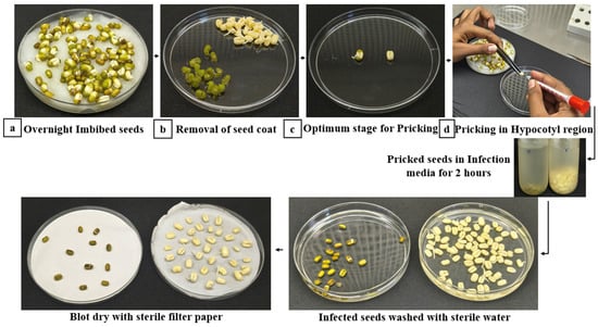
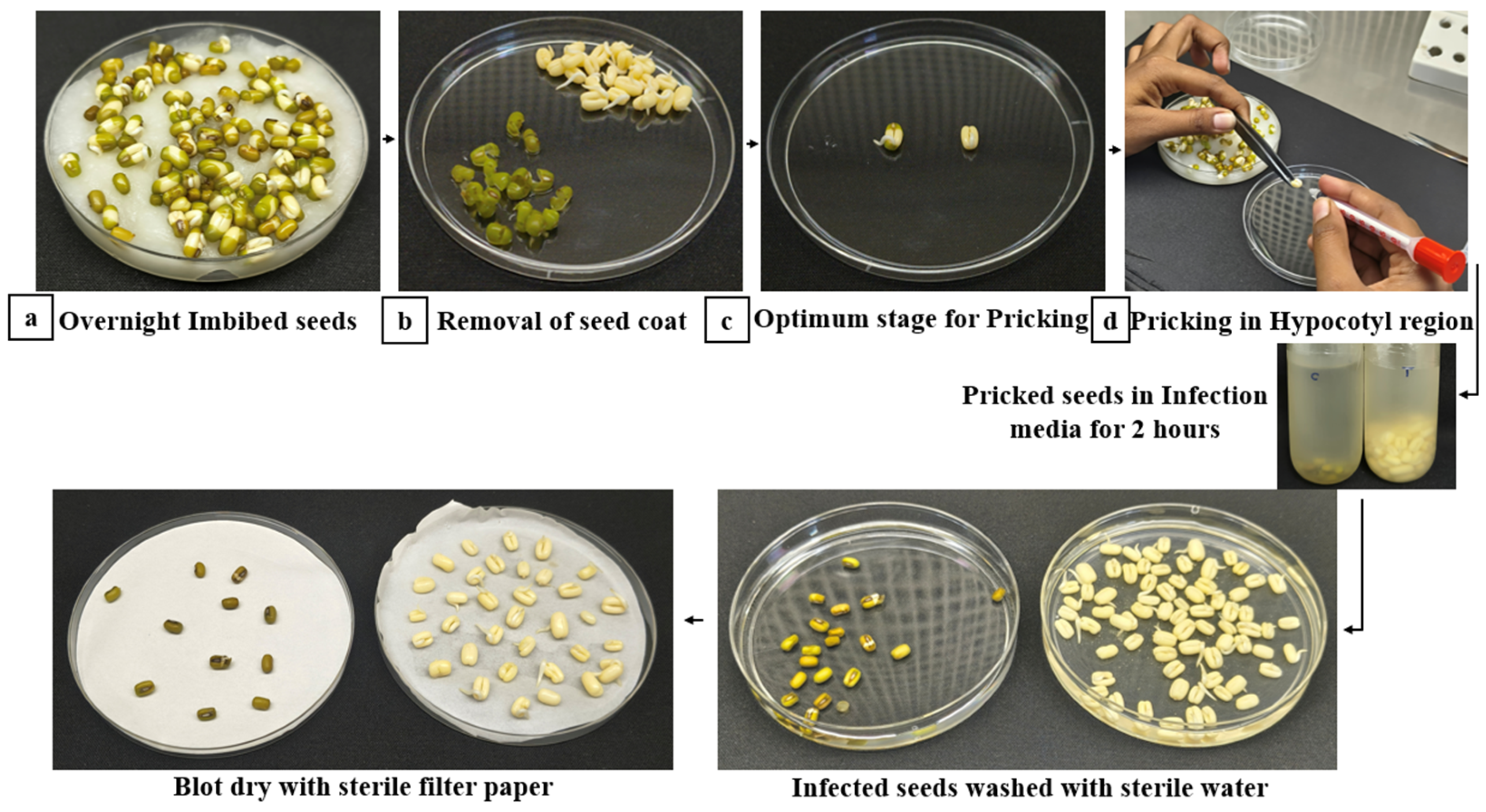
Genetic transformation of mung bean (Vigna radiata) seedlings via hypocotyl wounding resulted in efficient delivery of the pCAMBIA2300-based MYMV infectious clone construct. Surface-sterilized seeds exhibited uniform overnight germination on sterile cotton beds. Following mechanical wounding at the hypocotyl region and a 2 h incubation in MS broth supplemented with 15 microliters of 100 µM acetosyringone, inoculated seedlings demonstrated high post-inoculation survival (Figure 2).
Figure 2.
Micro-dissection of explants, seed de-coating, and hypocotyl wounding parameters. (a) Overnight imbibed sprouted seeds at the early germination stage; (b) aseptic removal of the seed coat utilizing a sterile scalpel and forceps; (c) identification of the optimal physiological stage for wounding, characterized by early radicle emergence and greening cotyledons; and (d) hypocotyl wounding of de-coated seeds, followed by 2 h Agrobacterium infection, sterile washing, and blot-drying before co-cultivation.
Data represent three independent transformation replicates, with n = 50 explants per replicate (N = 150 explants in total for the transformation treatments), conducted under optimized co-cultivation conditions (100 µM acetosyringone and 2 h incubation). Kanamycin selection was performed at 250 mg L−1. Non-inoculated control explants were cultured on MS medium without kanamycin selection. Mock-inoculated control explants were subjected to micro-wounding and cultured on MS medium supplemented with 250 mg L−1 kanamycin; all explants exhibited complete tissue necrosis within 7–10 days. Values in parentheses indicate percentage frequencies relative to the corresponding surviving or evaluated explants. Data are expressed as mean ± standard error (SE). Across three independent experimental replicates (N = 150 total explants; n = 50 per replicate), a total of 144 explants survived following hypocotyl micro-wounding and co-cultivation, achieving a mean post-inoculation explant survival rate of 96.0 ± 0.6% (48.0 ± 0.6 explants per replicate) under optimized co-cultivation conditions (Table 2 and Table 3).
Table 2.
Experimental design, replication, and explant responses in Agrobacterium-mediated transformation of mung bean (Vigna radiata cv. VBN 4).
Table 3.
Effects of acetosyringone concentration and co-cultivation duration on explant survival, selection-resistant shoot regeneration, and transformation efficiency in mung bean (Vigna radiata cv. VBN 4).
3.2. In Vitro Shoot Regeneration and Selection Efficiency
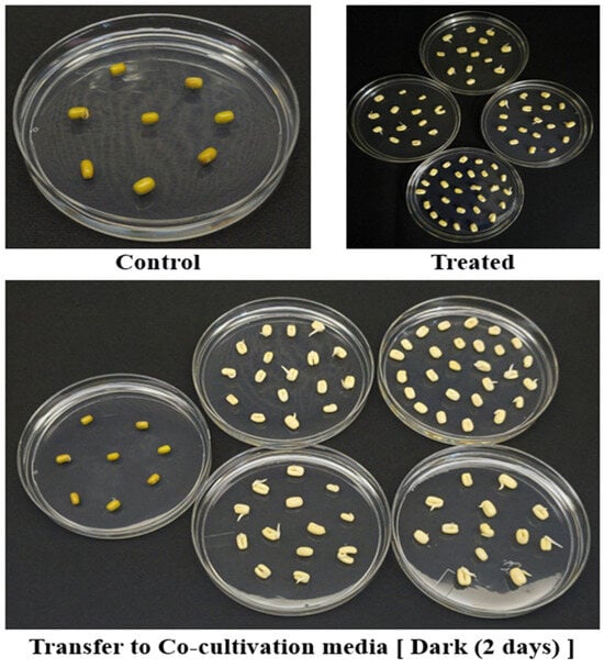
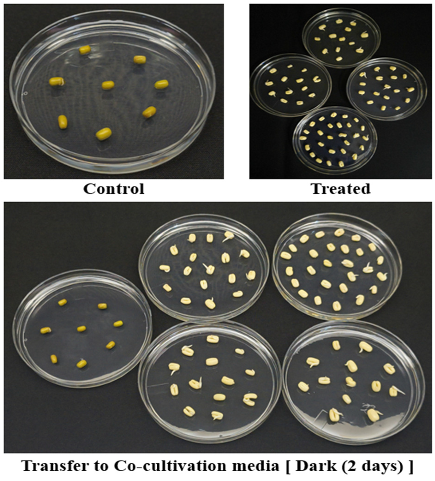
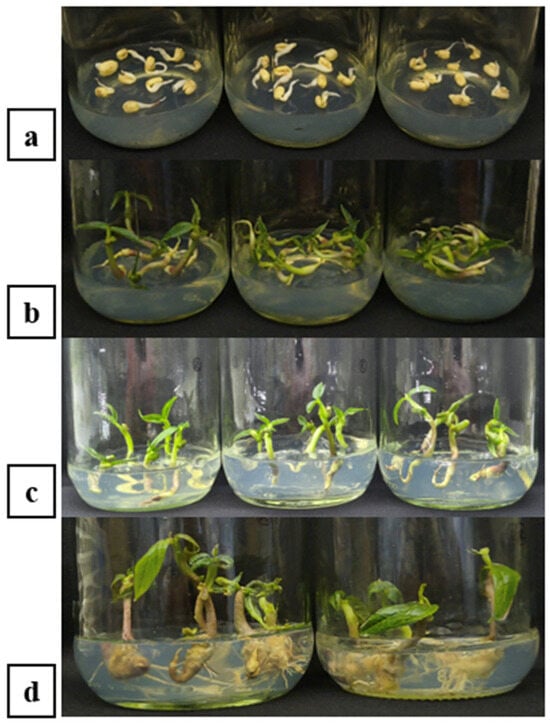
Following 48 h co-cultivation in total darkness on solid MS medium fortified with BAP (4 mL/L of a 1 mg mL−1 stock solution, equivalent to 4 mg L−1), explants were treated with cefotaxime to effectively eliminate residual Agrobacterium without compromising plant tissue viability (Figure 3). Transfer of explants to antibiotic selection medium containing kanamycin (250 mg/L), cefotaxime (200 mg/L) and BAP (4 mL/L of a 1 mg mL−1 stock solution, equivalent to 4 mg L−1) allowed for stringently distinguishing transformed tissues from non-transformed escapes (Figure 4a).
Figure 3.
Growth response of control and treated samples during co-cultivation. Transfer of infected explants to plates, showing treated seeds compared to mock-inoculated control seeds.
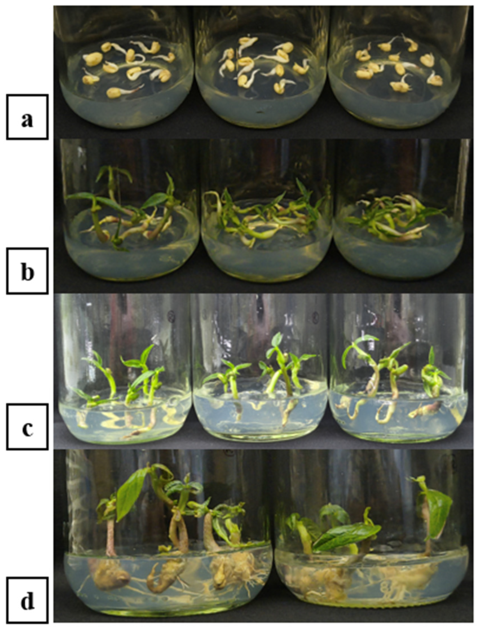
Figure 4.
In vitro shoot regeneration, elongation, and root development. (a) Two-days after dark co-cultivation transferred to MS selection medium; (b) emergence of healthy green shoots after 5 days on MS medium containing 250 mg/L kanamycin and 4 mL/L BAP; (c) transfer of selected shoots to rooting medium; and (d) development of adventitious roots with lateral branching on MS medium supplemented with IBA.
Due to the optimized concentration of BAP (4 mL/L of a 1 mg mL−1 stock solution, equivalent to 4 mg L−1), rapid shoot organogenesis and enhanced bud initiation were recorded as early as within 5 days of culture (Figure 4b). During the 15-day selection phase in culture vessels, resurgent, non-necrotic adventitious shoots actively emerged from the hypocotyl-cotyledonary junction. The frequency of shoot organogenesis on selection medium reached 95.2% explants producing healthy green shoots. In contrast, non-transformed control explants subjected to kanamycin selection exhibited severe bleaching, tissue necrosis, and complete mortality within 5–7 days.
3.3. Adventitious Root Induction and Greenhouse Acclimatization
Excised green shoots transferred to full-strength MS rooting medium supplemented with IBA (1 mL/L of a 1 mg mL−1 stock solution, equivalent to 1 mg L−1) initiated adventitious root growth within 7–10 days (Figure 4c). Rooting efficiency reached 87.5%, with rooted plantlets developing a well-branched primary and lateral root architecture suitable for ex vitro establishment (Figure 4d and Table 4).
Table 4.
In vitro root induction, primary hardening, and secondary hardening outcomes across three independent transformation replicates in mung bean (Vigna radiata cv. VBN 4).
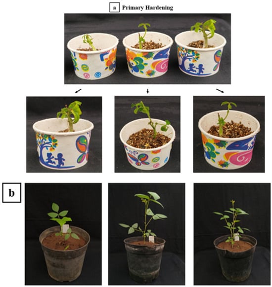
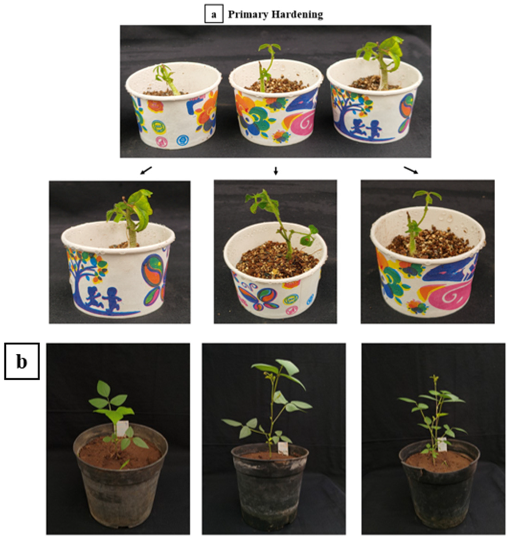
During primary hardening in autoclaved vermiculite under high relative humidity, plantlets achieved a survival frequency of 80.0% (Figure 5a). Following transfer to larger pots containing the farm soil, sand and farmyard manure mixture (1:1:1) for secondary hardening, acclimatized plants developed normally under natural greenhouse conditions, showing full flowering, pod development and seed setting (Figure 5b).
Figure 5.
Ex vitro acclimatization and establishment of regenerated plants. (a) Transfer of rooted plantlets to vermiculite for primary hardening; and (b) transfer to soil: sand: compost substrate for secondary hardening and greenhouse establishment.
3.4. Molecular Validation of the MYMV Construct and nptII Selectable Marker in T0 Transgenic Plants
To confirm systemic viral infection and nptII selectable marker gene at the molecular level, total genomic DNA extracted from young foliar tissues of hardened T0 plants was subjected to PCR diagnostic assays using MYMV coat protein (CP)-specific primers, MYMV movement protein (MP)-specific primers and selectable marker (nptII) gene.
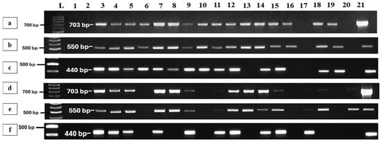
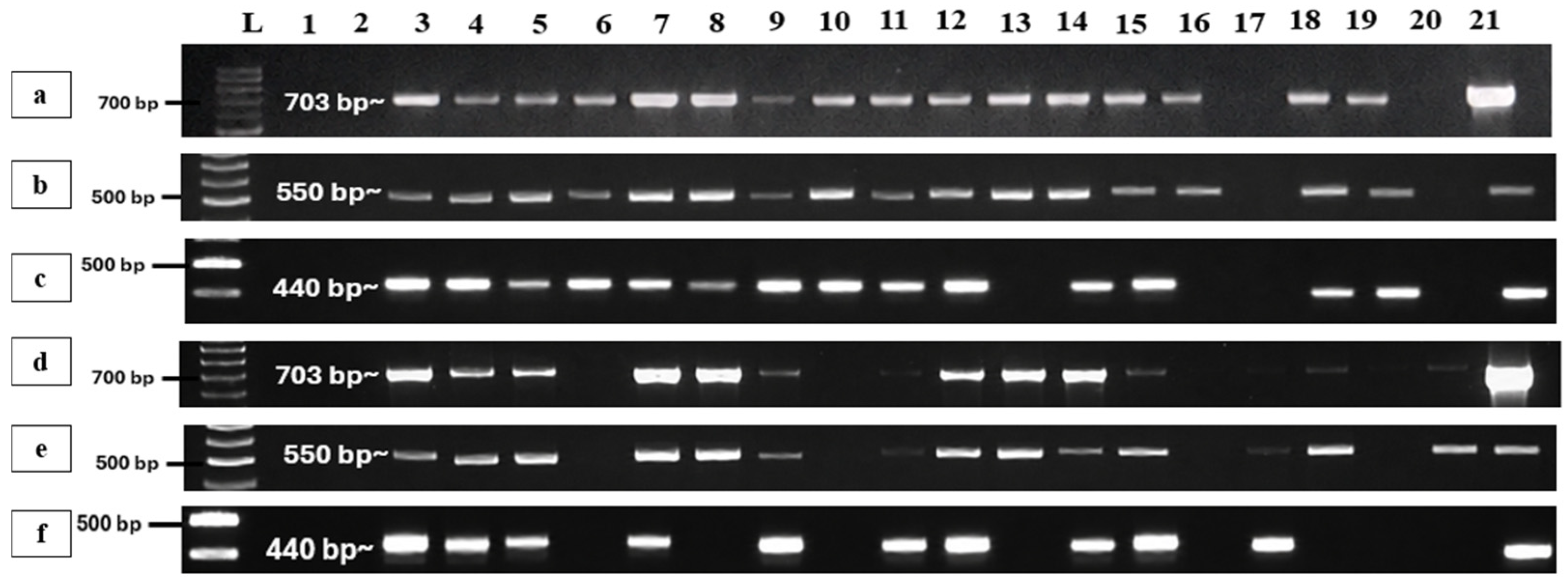
Electrophoretic separation on 1.5% agarose gels in 0.5× TBE buffer revealed the expected 703 bp diagnostic amplicons for CP and 550 bp diagnostic amplicons for MP in symptomatic plants. Out of 18 regenerated plants screened, 16 tested positive CP and 16 tested positive MP for MYMV infection, representing a molecular confirmation rate of 88.88% for both primers (Figure 6). No PCR amplification was observed in non-inoculated or control plant samples. Electrophoretic separation on 3.0% agarose gels in 0.5× TBE buffer revealed the expected 440 bp diagnostic amplicons for nptII. Out of 18 regenerated plants screened, 14 tested positive for the kanamycin resistance gene, representing a molecular confirmation rate of 77.77% (Figure 6 and Table 5).
Figure 6.
PCR confirmation of CP, MP, and nptII positive plants and their harvested seeds. (a–c) PCR detection in primary T0 transformants for CP (703 bp, 1.5% agarose), MP (550 bp, 1.5% agarose), and nptII (440 bp, 3.0% agarose). (d–f) PCR detection in T1 progeny seedlings for CP (703 bp, 1.5% agarose), MP (550 bp, 1.5% agarose), and nptII (440 bp, 3.0% agarose). Lane assignments (all panels): Lane L: 100 bp DNA ladder; Lane 1: No-template control (water blank); Lane 2: Non-transformed control plant (Vigna radiata cv. VBN 4); Lanes 3–20: Genomic DNA from T0 Plants 1–18 (panels (a–c)) or T1 Seedlings 1–18 (panels (d–f)); Lane 21: Positive control (pCAMBIA2300 plasmid/A. tumefaciens LBA4404 culture).
Table 5.
PCR-based molecular validation of T-DNA transformation (nptII, CP, and MP) in hardened T0 mung bean (Vigna radiata cv. VBN 4) regenerants across three independent replicates.
3.5. Transgenerational Viral Persistence in T1 Progeny
PCR-confirmed MYMV-transformed T0 plants were grown to reproductive maturity and produced normal, fully developed pods. Pods harvested individually at full maturity yielded viable T1 progeny seeds. Mature pods harvested from 16 PCR-positive plants yielded viable progeny seeds. Upon germination in sterilized soil under controlled environmental conditions, a 100% germination frequency (18/18 seeds) was recorded. Foliar genomic DNA extracted from 10-day-old seedlings was screened via PCR using virus-specific primers for both MYMV CP (703 bp) and MP (550 bp) gene regions [8,21]. Molecular diagnostic analysis confirmed the persistent transmission of both viral targets in 13 of the 18 seedlings, demonstrating a 72.2% T1 progeny PCR-positivity frequency in the progeny group. Out of 18 regenerated plants screened, 10 tested positive for the kanamycin resistance gene, representing a molecular confirmation rate of 55.56%.
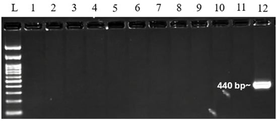
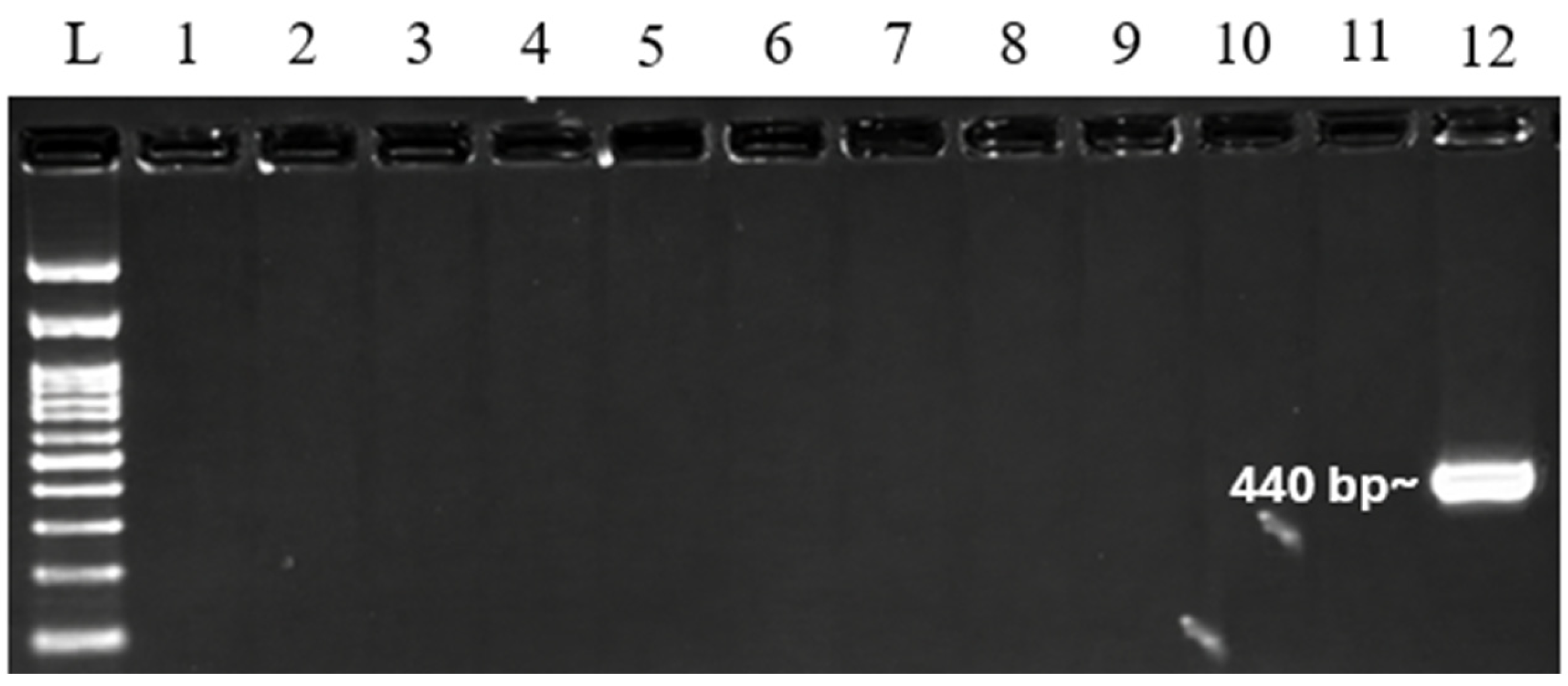
Interestingly, the diagnostic band profiles exhibited varying levels of PCR product abundance on the gel. Specifically, eight of the positive seedlings displayed highly prominent, intense (strong) bands for both the 703 bp CP, 550 bp MP amplicons and 440 bp diagnostic amplicons for nptII (Figure 6). In contrast, the remaining five positive seedlings exhibited distinct but noticeably fainter (light) bands [8,22]. This variation in band intensity likely reflects differences in template DNA concentration, systemic viral replication kinetics, or varying accumulation levels of the replicating viral genome among individual segregating progeny lines. To assess residual Agrobacterium contamination, DNA extracted from putative T0 plants was analyzed by PCR using virG-specific primers. No detectable amplification was observed in the plant samples (lanes 1–11), whereas the Agrobacterium positive control (lane 12) produced the expected approximately 440 bp amplicons (Figure 7 and Table 6). These results support the absence of detectable residual Agrobacterium DNA in the tested plant samples under the assay conditions.
Figure 7.
PCR amplification of the virG gene from genomic DNA of putative transgenic (T0) plants. Lane L: 100 bp DNA ladder; Lane 1: no-template control (water blank); Lane 2: genomic DNA from a non-transformed plant (negative control); Lanes 3–11: genomic DNA samples from putative transgenic plants; Lane 12: Agrobacterium culture cloned with MYMV construct.
Table 6.
Analysis of T1 progeny seedling germination, nptII marker segregation, and target gene inheritance across three independent transformation replicates in mung bean (Vigna radiata cv. VBN 4).
3.6. Evaluation of Overall Transformation Efficiency
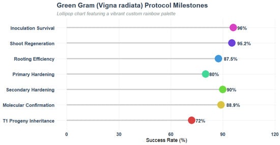
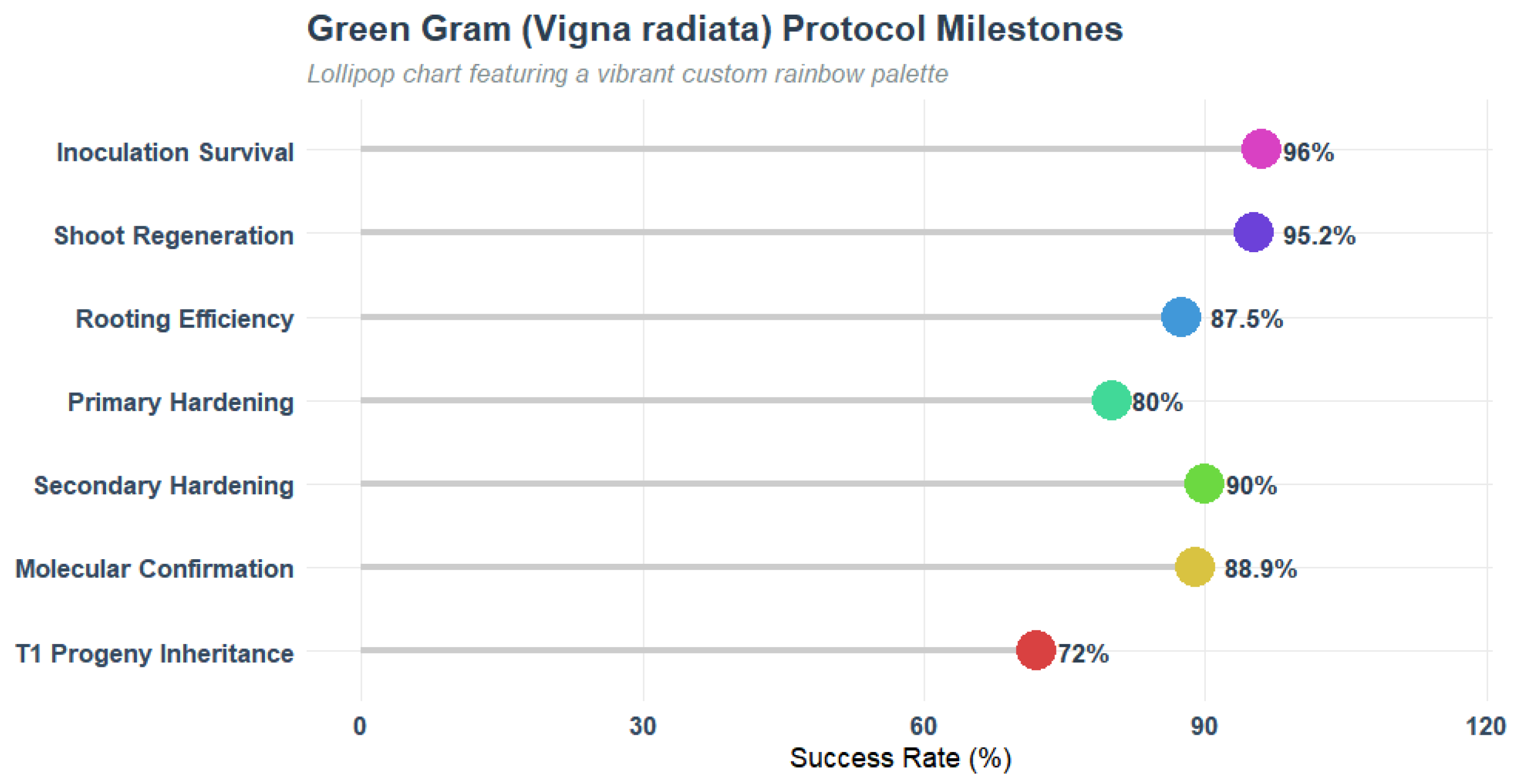
To assess overall pipeline efficiency and locate developmental bottlenecks, we mapped success rates across seven critical milestones (Figure 8). Unlike traditional legume tissue culture systems that suffer compounding exponential losses across developmental phases, this optimized protocol maintained remarkably high and stable survival throughout.
Figure 8.
Chronological milestone map illustrating phase-specific success rates.
Initial biophysical barriers to gene transfer were successfully bypassed, yielding 96.0% survival post-inoculation and micro-wounding, followed by 95.2% shoot regeneration under selection. Downstream transitions proceeded with high efficiency: 87.5% rooting, 80.0% primary hardening under high humidity, and a resurgent 90.0% secondary hardening in greenhouse soil. At the molecular level, 88.9% of regenerants displayed target gene integration, with stable vertical transmission established in 72.0% of the tested progeny.
3.7. Systemic Physiological Response Rates Across Experiments
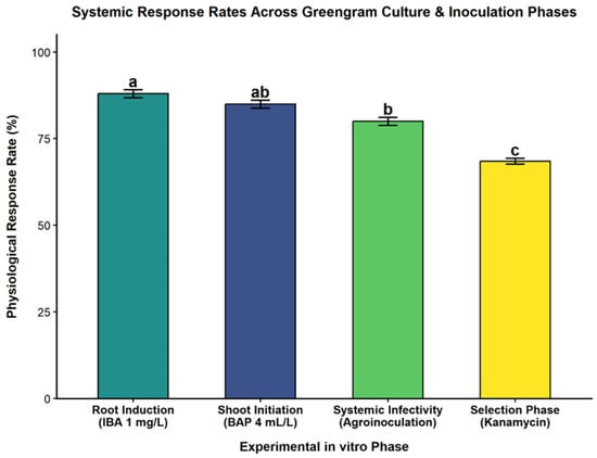
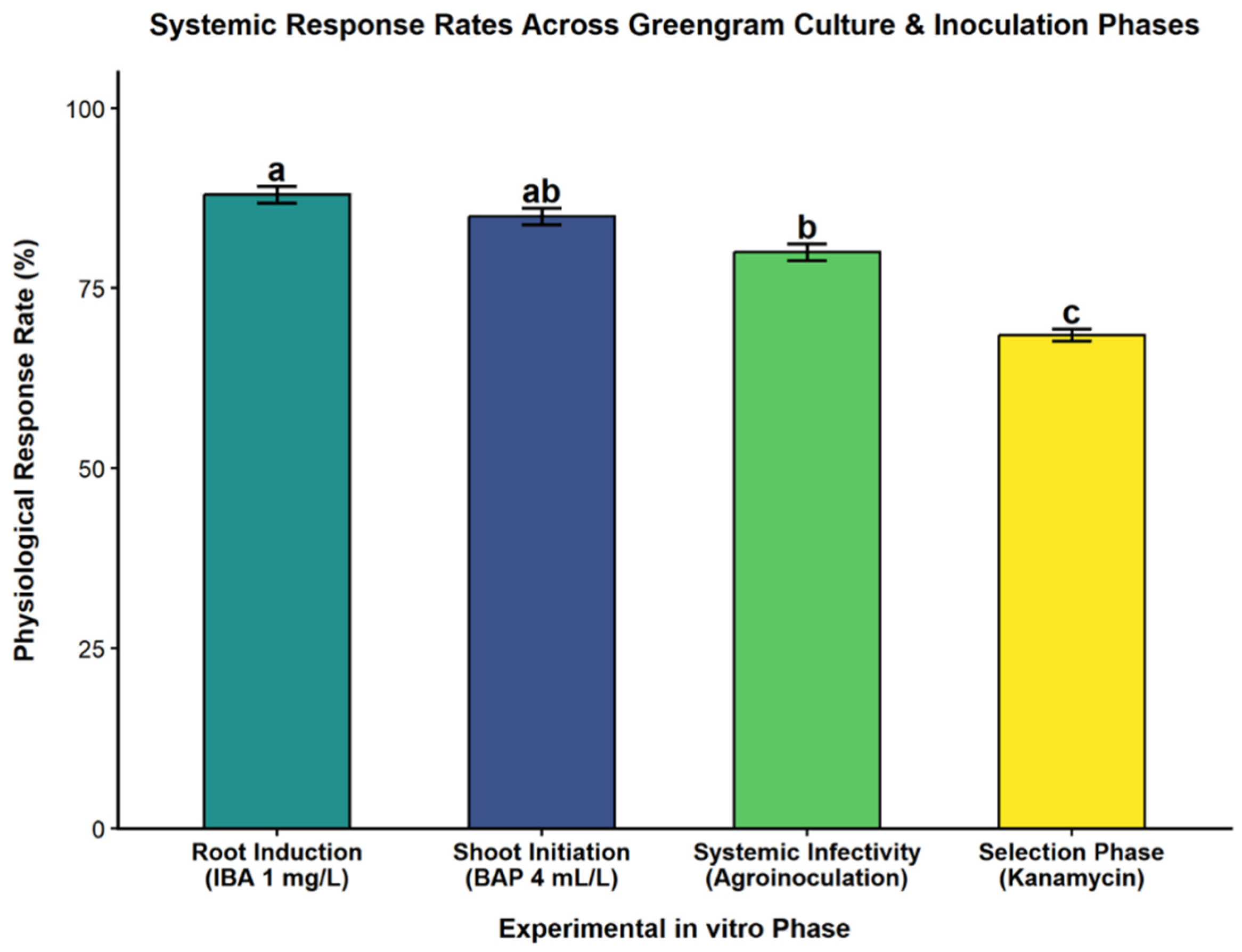
The one-way Analysis of Variance (ANOVA) applied only to the various values of a given parameter showed significant differences between the different parameter gradients (p < 0.001). Post-inoculation explant survival was observed to be at its highest level, 96.0 ± 1.2% (144/150 explants from three independent replicates) with optimized co-cultivation conditions (100 µM acetosyringone for 2 h) as a statistically superior response group (‘a’) than non-induced or over-exposed treatments. Shoots were regenerated from the surviving explants and were found to have high tissue tolerance and to produce shoots which were resistant to the selection medium’s kanamycin. 95.2 ± 1.1% (40/42) of the explants were found to regenerate shoots that were resistant to the selection medium’s kanamycin, whereas the non-transformed control explants were all found to have necrotized within 5–7 days. The root organogenesis on MS with 1.0 mg L−1 IBA produced an 87.5 ± 3.6% rooting efficiency (n = 28/32 elongated shoots) in subsequent treatments. The primary hardening survival rate in sterile substrate was 80.0 ± 2.6% (20/25 plantlets) and secondary hardening survival rate in soil pots in the greenhouse was 90.0 ± 4.8% (18/20 plantlets) for 25 selected rooted plantlets, following ex vitro acclimatization. The overall transformation protocol efficiency was 36.0% (18/50) based on the number of fully hardened, PCR confirmed T0 transformants obtained per batch of 50 inoculated explants (Figure 9).
Figure 9.
Systemic physiological response rates (%) of mung bean (Vigna radiata L. Wilczek cv. VBN 4) across critical in vitro culture, selection, and inoculation phases. Different letters (a, b, c) indicate statistically significant differences among means, while means sharing the same letter are not significantly different (p < 0.05), according to the post hoc multiple-comparison test.
4. Discussion
4.1. Optimized Agrobacterium-Mediated Transformation Protocol for Mung Bean
The post-inoculation explant survival rate was 96.0% as of three replicates (50) when mechanical wounding was done at the hypocotyl region with a 2 h incubation period in MS broth with 100 µM acetosyringone. This high infection rate is quite higher than the historical efficiencies of genetic transformations in mung bean through pin pricking and radicle submersion, which ranged between 15% and 50% [10,12]. The significant improvement in systemic infection can be explained by the fact that the hypocotyl-cotyledonary junction is the site where actively growing cells that supply optimal phloem-associated and meristematic tissues needed for the propagation of begomovirus and systemic infection are most abundant [11,23]. In addition, by adding 100 µM acetosyringone to the 2 h pre-inoculation incubation, the Agrobacterium vir gene cascade was effectively induced to facilitate the transfer of the T-DNA into plant tissues before post-inoculation recovery [24,25]. The results are consistent with those of [8,11] who underscored the importance of using p liquid co-incubation to remove delivery barriers and achieve comparable disease pressure among the plants.
4.2. Cytokinin-Mediated Rapid Shoot Regeneration in Mung Bean
Grain legumes are famous for being highly sensitive to in vitro culture, rapid polyphenolic oxidation and limited shoot bud differentiation when cultured in an in vitro tissue culture and organogenesis system [3,15]. Explant selection and plant growth regulator (PGR) formulation are pivotal factors in overcoming recalcitrance to in vitro organogenesis in grain legumes. While cotyledonary node and hypocotyl explants derived from germinated seedlings possess pre-existing meristematic tissues that are highly responsive to cytokinin-induced shoot organogenesis, this regeneration pathway carries an inherent risk of generating chimeric or mosaic T0 regenerants. To prevent non-transformed tissue escapes from pre-existing meristems, explants were subjected to multi-stage selection under high antibiotic pressure (250 mg L−1 kanamycin). Potential chimerism was subsequently ruled out by confirming uniform T-DNA integration across foliage tissues and demonstrating Mendelian nptII transgene segregation in T1 progeny seedlings [3].
The addition of 4 mL/L BAP (6-benzylaminopurine) in the MS regeneration medium resulted in quick induction of adventitious shoot organogenesis and bud emergence within 5 days of culture initiation in this investigation. In general, the organogenesis of mung bean takes 10 to 15 days on media supplemented with lower concentration of BAP (1.0–2.0 mg/L) [15]. This is an accelerated shoot proliferation, which occurred after 5 days, and shows that increased exogenous BAP supplementation is an effective way to overcome the apical dominance and to initiate synchronized mitotically dividing cells in the peripheral layers of the cotyledonary nodal zone. BAP has been considered as the most effective cytokinin to induce direct organogenesis in Vigna species. The optimal pre-conditioning or exposure of BAP in seedling explants allowed multiple bud primordia differentiation without intervening callus generation, thus maintaining genetic fidelity and speeding up shoot extension as documented by [14,15].
4.3. Shoot Selection, Root Formation, and Plant Acclimatization
In vitro organogenesis requires the use of an appropriate antibiotic to prevent the presence of escapes which are not infected or transformed. A high level of selection pressure was applied using the explants that had been inoculated into the medium with kanamycin at 250 mg/L + cefotaxime at 200 mg/L. Severe foliar bleaching, necrosis and total mortality occurred in non-transformed control explants in 5–7 days, while revived shoots that survived exhibited dark green leaves and active organogenesis. In the present case, the residual growth of Agrobacterium tumefaciens was successfully arrested with less phytotoxicity and without affecting the shoot elongation of the plantlets with the use of cefotaxime at 200 mg/L [7,14].
In legume micropropagation, rhizogenesis and vascular discontinuity are common problems in the regenerated shoots, and subsequent induction of roots is a significant bottleneck [26]. Excised shoots were transferred to full-strength MS medium supplemented with 1 mg/L IBA (indole-3-butyric acid) to obtain high rooting efficiency of 87.5% formation of well-branched primary and lateral root system in 7–10 days. In recalcitrant legumes, auxins, especially IBA, have been well established for encouraging lateral root initiation and adventitious root elongation [3,14]. Acclimatized plantlets showed high ex vitro survival with 100% of surviving plantlets reaching full maturity and producing viable pods after primary hardening in sterile vermiculite and secondary establishment in a potted soil mixture (1:1:1 soil: sand: FYM).
4.4. Transgenerational Inheritance and Detection of MYMV in Progenys
To validate that the optimized Agrobacterium-mediated transformation protocol successfully delivered the MYMV Infectious clone and enabled the systemic virus movement between generations, molecular diagnostic assays were performed on the Coat Protein (CP, 703 bp amplicon) and Movement Protein (MP, 550 bp amplicon) gene regions [21,27,28]. Systemic MYMV infection was confirmed in 88.88% of the transformed plants and showed that the MYMV viral DNA was transferred from the inoculation site through the vascular tissues to the newly developed trifoliate leaves, thereby validating the successful transfer of viral DNA from the inoculation site to the leaf [8].
All the seeds harvested from transformed T0 progeny germinated successfully under controlled growth chamber conditions. A total of 13/18 progeny seedlings had both 703 bp CP and 550 bp MP amplicons when analyzed by diagnostic PCR of 10-day-old seedlings, resulting in a 72.2% transgenerational transmission rate. The discovery has confirmed the molecular presence of MYMV-derived DNA in the progeny. Seed borne transmission of MYMV is a newly recognized route for the survival and long-distance spread of the virus in pulse crops, although begomoviruses are mainly seed-transmissible via whiteflies in a circulative persistent manner [21]. The detection of viral DNA in seedling leaves suggests that the virus was maintained during pod development and colonized emerging tissues without requiring agro-inoculation. These findings indicate the presence of MYMV-derived DNA in a proportion of T1 progeny, suggesting vertical transmission of the infectious clone or viral genome. Further analyses, such as infectivity assays or quantitative PCR, would further clarify the degree of seed transmission.
5. Conclusions
The present study successfully established a robust, reproducible, and efficient Agrobacterium-mediated genetic transformation and regeneration system for mung bean (Vigna radiata L.) cv. VBN 4 using targeted hypocotyl wounding. Optimization of transformation and tissue-culture conditions enhanced explant response, regeneration, rooting, and transformation efficiency. The established protocol provides a reliable and reproducible approach for the genetic manipulation of VBN 4, an agronomically important mung bean cultivar. The efficient regeneration and transformation system overcomes a major bottleneck in mung bean genetic improvement. This protocol provides a reliable platform for functional genomics, gene editing, and genetic improvement of mung bean, particularly for developing resistance to biotic stresses and tolerance to abiotic stresses.
Author Contributions
Conceptualization, S.M. and R.M.; investigation, H.C.G.; writing—original draft preparation, H.C.G.; writing—review and editing, S.A.R. and V.R.R.; supervision, S.M., S.A.R. and V.R.R.; funding acquisition, R.M. All authors have read and agreed to the published version of the manuscript.
Funding
The research was funded by the Department of Biotechnology (DBT), Ministry of Science and Technology, India for providing financial assistance under the Grant No. BT/INF/22/SP45584/2022.
Institutional Review Board Statement
Ethical review and approval were not applicable for studies not involving humans or animals.
Data Availability Statement
The data generated during the current study are available from the corresponding author upon reasonable request.
Conflicts of Interest
The authors declare no conflicts of interest.
References
- Bhajan, S.K.; Begum, S.; Islam, M.N.; Hoque, M.I.; Sarker, R.H. In Vitro Regeneration and Agrobacterium-Mediated Genetic Transformation of Local Varieties of Mungbean (Vigna radiata (L). Wilczek). Plant Tissue Cult. Biotechnol. 2019, 29, 81–97. [Google Scholar] [CrossRef] [Scilit]
- Dikr, W. Mung Bean (Vigna radiata L.) Production Status and Challenges in Ethiopia. Glob. Acad. J. Agric. Biosci. 2023, 5, 13–22. [Google Scholar] [CrossRef] [Scilit]
- Jaiwal, P.K.; Kumari, R.; Ignacimuthu, S.; Potrykus, I.; Sautter, C. Agrobacterium Tumefaciens-Mediated Genetic Transformation of Mungbean (Vigna radiata L. Wilczek)—A Recalcitrant Grain Legume. Plant Sci. 2001, 161, 239–247. [Google Scholar] [CrossRef] [Scilit] [PubMed]
- Li, M.; Yu, Q.; Wei, J.; Li, X.; Hao, A.; Liu, F.; Xing, B. Genome-Wide Characterization and Expression Analysis of the MYB Transcription Factor Family in Mung Bean (Vigna radiata). BMC Plant Biol. 2026, 26, 766. [Google Scholar] [CrossRef] [Scilit] [PubMed]
- Li, S.; Cong, Y.; Liu, Y.; Wang, T.; Shuai, Q.; Chen, N.; Gai, J.; Li, Y. Optimization of Agrobacterium-Mediated Transformation in Soybean. Front. Plant Sci. 2017, 8, 246. [Google Scholar] [CrossRef] [Scilit] [PubMed]
- Fernandez-Gutierrez, A.; Curtin, S.J.; Gutierrez-Gonzalez, J.J. Gene Editing and Transformation Strategies for Engineering Drought-Tolerant Legumes. Legume Sci. 2026, 8, e70080. [Google Scholar] [CrossRef] [Scilit]
- Tripathi, A.; Rathore, M.; Shukla, S.; Das, A.; Debnath, S.C. Agrobacterium and Biolistic Mediated Genetic Transformation of Mungbean Cultivar Samrat Using Embryogenic Explant. Plant Cell Tissue Organ Cult. PCTOC 2024, 157, 72. [Google Scholar] [CrossRef] [Scilit]
- Balasubramaniam, M.; Thangavel, T.; Aiyanathan, K.E.A.; Rathnasamy, S.A.; Rajagopalan, V.R.; Subbarayalu, M.; Natesan, S.; Kanagarajan, S.; Muthurajan, R.; Manickam, S. Unveiling Mungbean Yellow Mosaic Virus: Molecular Insights and Infectivity Validation in Mung Bean (Vigna radiata) via Infectious Clones. Front. Plant Sci. 2024, 15, 1401526. [Google Scholar] [CrossRef] [Scilit] [PubMed]
- Ramachandra, A.; Roshni, M.; Anitha, P.; Bhat, S.G.; Raviteja, C. Isolation, cloning and agrobacterium mediated transformation of MYMV replicase (Rep) gene in Vigna radiata (L.) Wilczek for MYMV resistance. Asian J. Microbiol. Biotechnol. Environ. Sci. 2022, 24, 102–107. [Google Scholar] [CrossRef] [Scilit]
- Karthikeyan, A.; Shobhana, V.G.; Sudha, M.; Raveendran, M.; Senthil, N.; Pandiyan, M.; Nagarajan, P. Mungbean Yellow Mosaic Virus (MYMV): A Threat to Green Gram (Vigna radiata) Production in Asia. Int. J. Pest Manag. 2014, 60, 314–324. [Google Scholar] [CrossRef] [Scilit]
- Sivalingam, P.N.; Dokka, N.; Mahajan, M.M.; Sahu, B.; Marathe, A.; Kaushal, P.; Ghosh, P.K. Achieving Maximum Efficiency of Mungbean Yellow Mosaic India Virus Infection in Mungbean by Agroinoculation. 3 Biotech 2022, 12, 29. [Google Scholar] [CrossRef] [Scilit] [PubMed]
- Sudha, M.; Karthikeyan, A.; Anusuya, P.; Ganesh, N.M.; Pandiyan, M.; Senthil, N.; Raveendran, M.; Nagarajan, P.; Angappan, K. Inheritance of Resistance to Mungbean Yellow Mosaic Virus (MYMV) in Inter and Intra Specific Crosses of Mungbean (Vigna radiata). Am. J. Plant Sci. 2013, 4, 1924–1927. [Google Scholar] [CrossRef]
- Aasim, M.; Barpete, S.; Usta, A.; Sevinc, C. An insight into Agrobacterium tumafaciens-mediated genetic transformation studies in Mungbean (Vigna radiata L. Wilczek). J. Glob. Innov. Agric. Soc. Sci. 2019, 7, 47–51. [Google Scholar] [CrossRef] [Scilit]
- Mekala, G.K.; Juturu, V.N.; Mallikarjuna, G.; Kirti, P.B.; Yadav, S.K. Optimization of Agrobacterium-Mediated Genetic Transformation of Shoot Tip Explants of Green Gram (Vigna radiata (L.) Wilczek). Plant Cell Tissue Organ Cult. PCTOC 2016, 127, 651–663. [Google Scholar] [CrossRef] [Scilit]
- Yadav, S.K.; Katikala, S.; Yellisetty, V.; Kannepalle, A.; Narayana, J.L.; Maddi, V.; Mandapaka, M.; Shanker, A.K.; Bandi, V.; Bharadwaja, K.P. Optimization of Agrobacterium Mediated Genetic Transformation of Cotyledonary Node Explants of Vigna radiata. SpringerPlus 2012, 1, 59. [Google Scholar] [CrossRef] [Scilit] [PubMed]
- Taliansky, M.; Samarskaya, V.; Zavriev, S.K.; Fesenko, I.; Kalinina, N.O.; Love, A.J. RNA-Based Technologies for Engineering Plant Virus Resistance. Plants 2021, 10, 82. [Google Scholar] [CrossRef] [Scilit] [PubMed]
- Zhao, Y.; Yang, X.; Zhou, G.; Zhang, T. Engineering Plant Virus Resistance: From RNA Silencing to Genome Editing Strategies. Plant Biotechnol. J. 2020, 18, 328–336. [Google Scholar] [CrossRef] [Scilit] [PubMed]
- Pooggin, M.M. RNAi-Mediated Resistance to Viruses: A Critical Assessment of Methodologies. Curr. Opin. Virol. 2017, 26, 28–35. [Google Scholar] [CrossRef] [Scilit] [PubMed]
- Ditta, G.; Stanfield, S.; Corbin, D.; Helinski, D.R. Broad Host Range DNA Cloning System for Gram-Negative Bacteria: Construction of a Gene Bank of Rhizobium Meliloti. Proc. Natl. Acad. Sci. USA 1980, 77, 7347–7351. [Google Scholar] [CrossRef] [Scilit] [PubMed]
- Manickam, S. DNA Isolation Protocol for Vigna radiata with Free of Phenolics. Protoc. Exch. 2009. [Google Scholar] [CrossRef] [Scilit]
- Talakayala, A.; Dokka, N.; Baloji, G.; Ankanagari, S.; Garladinne, M. Efficient Agrobacterium Mediated Genetic Transformation of CRISPR/Cas9 Construct Using Cotyledonary Node Explants of Greengram. J. Agric. Appl. Biol. 2023, 4, 107–115. [Google Scholar] [CrossRef] [Scilit]
- Talakayala, A.; Prathyusha, V.B.; Divya, D.; Ankanagari, S.; Garladinne, M. Molecular Cloning of MYMV Genome and Infectivity of Yellow Mosaic Virus in Green Gram Using Different Viral Transmission Tools. Biosci. Biotechnol. Res. Asia 2021, 18, 467–478. [Google Scholar] [CrossRef] [Scilit]
- Vuorinen, A.L.; Kelloniemi, J.; Valkonen, J.P.T. Why Do Viruses Need Phloem for Systemic Invasion of Plants? Plant Sci. 2011, 181, 355–363. [Google Scholar] [CrossRef] [Scilit] [PubMed]
- Sheikholeslam, S.N.; Weeks, D.P. Acetosyringone Promotes High Efficiency Transformation of Arabidopsis Thaliana Explants by Agrobacterium Tumefaciens. Plant Mol. Biol. 1987, 8, 291–298. [Google Scholar] [CrossRef] [Scilit] [PubMed]
- Stachel, S.E.; Messens, E.; Van Montagu, M.; Zambryski, P. Identification of the Signal Molecules Produced by Wounded Plant Cells That Activate T-DNA Transfer in Agrobacterium Tumefaciens. Nature 1985, 318, 624–629. [Google Scholar] [CrossRef] [Scilit]
- Eapen, S. Advances in Development of Transgenic Pulse Crops. Biotechnol. Adv. 2008, 26, 162–168. [Google Scholar] [CrossRef] [Scilit] [PubMed]
- Maheshwari, R.; Panigrahi, G.; Angappan, K. Molecular Characterization of Distinct YMV (Yellow Mosaic Virus) Isolates Affecting Pulses in India with the Aid of Coat Protein Gene as a Marker for Identification. Mol. Biol. Rep. 2014, 41, 2635–2644. [Google Scholar] [CrossRef] [Scilit] [PubMed]
- Talakayala, A.; Mekala, G.K.; Reddy, M.K.; Ankanagari, S.; Garladinne, M. Manipulating Resistance to Mungbean Yellow Mosaic Virus in Greengram (Vigna radiata L): Through CRISPR/Cas9 Mediated Editing of the Viral Genome. Front. Sustain. Food Syst. 2022, 6, 911574. [Google Scholar] [CrossRef] [Scilit]



Disclaimer/Publisher’s Note: The statements, opinions and data contained in all publications are solely those of the individual author(s) and contributor(s) and not of MDPI and/or the editor(s). MDPI and/or the editor(s) disclaim responsibility for any injury to people or property resulting from any ideas, methods, instructions or products referred to in the content. |
© 2026 by the authors. Licensee MDPI, Basel, Switzerland. This article is an open access article distributed under the terms and conditions of the Creative Commons Attribution (CC BY) license.