


Abstract
Super-resolution (SR) facilitates high-fidelity mapping beyond the spatial resolution capacity of clinical neuro-oncology MRI. While state-of-the-art deep learning algorithms have augmented this domain, such methods exclusively learn from spatial or spectral domains and are often compounded by tumor boundary over-smoothing and geometrically inconsistent pathologies. In this paper, we introduce Hy-DTGL, a hybrid-domain transformer with a global–local attention modal super-resolution (SR) framework that learns a joint reconstruction across four inter-dependent knowledge domains: local spatial, global spatial, spectral and wavelet. Using windowed self-attention, Hy-DTGL learns highly localized pathologic textures; pooled global attention constrains anatomical plausibility; Fourier-learnable modulation provides spectral energy per semblance; and multi-scale discrete wavelet tiling preserves multi-scale tumor morphology. Optimization occurs via a hybrid objective, combining Charbonnier loss, gradient edge-loss, and spectral domain loss. The proposed framework was trained on BraTS 2019 (BT_19) and evaluated on BT_19, BraTS 2020 (BT_20), BraTS 2023 (BT_23), and IXI under both in-domain and strict zero-shot settings, and achieves competitive performance relative to the evaluated baselines under the adopted degradation and evaluation protocol at ×2, ×4, and ×8 SR factors. In the BraTS 2020 ×4 test resolution, 37.46 dB PSNR and 0.986 SSIM are attained, outperforming deep learning CNN-based SR (ECFNet) by 2.47 dB and transformer-based (WavTrans) by 4.05 dB in PSNR. Ablative studies reveal the spatial, spectral and multi-scale constraints notably converge to a faithfully mapped image space with restored tumor boundary delineation and global-structure consistency. We also show the Hy-DTGL generalizability by how it improves downstream tumor segmentation metrics: the super-resolved scans improve the segmentation Dice coefficient by 0.063 and significant reductions are observed in the Hausdorff Distance from 19.05 mm to 13.01 mm relative to the original low-resolution images, suggesting potential utility for downstream neuro-oncological image analysis.
1. Introduction
MRI is still the gold standard in glioma detection, surgical planning, and monitoring treatment response. Multi-modal protocols, including T1, T1 post-contrast (T1CE), T2, and FLAIR, provide the relevant complementary information to localize precisely various sub-structures within a tumor, including the enhancing tumor [1,2,3,4], the necrotic core, and the peritumoral edema. As one might expect, such an ability to segment tumors precisely both guides resection margins and delineates radiotherapy treatment volumes. Unfortunately, in clinical MR imaging, there exists a compromise to be made between resolution, SNR, and acquisition time. The relatively thick-slice acquisitions, specifically T2 and FLAIR, result in anisotropic resolution and partial volume averaging, which blurs the boundaries of the tumor, undermining subsequent segmentation and volume quantification.
SR techniques reconstruct high-resolution (HR) images from low-resolution (LR) acquisitions, providing a computational alternative to extended scan times. Optimization-based early SR methods failed to recover anatomy’s high-frequency details [5,6,7]; convolutional neural networks (CNNs) [8] and generative adversarial networks (GANs) [9,10,11] were the first deep learning techniques to consistently enhance the robust learning of non-linear LR-to-HR mappings for improved perceptual quality. Yet, the CNN- or GAN-based models operate solely in the spatial domain. So, these models are restricted to probabilistically modeling the frequency distributions or multi-scale anatomical structures in medical images [12,13,14,15], whereas GAN-based models on natural image databases hallucinate non-anatomical textures [13,16], a fundamental drawback for clinical use.
The utilization of transformer structure, which contains self-attention modules modeling long-range relationships of features, is also promising in [17,18,19], for image restoration. Medical-domain adapted versions of transformers, such as SwinIR and WavTrans have been proposed for MRI [20,21,22]; current deep learning SR models are primarily using attention mechanisms within the spatial domain, thus overlooking the structural prior information in the frequency domain and wavelet domain. In the same vein, Implicit Neural Representation (INR) methods, which describe the entire volume with continuous representations [23,24,25], suffer from limitations with undersampled data or thick-slice artifacts, as there is a lack of structural consistency constraints that result in missing or hallucinated anatomical structures [24,26].
The only conceptual problem that is rooted in these failures is the definition of SR as a spatial reconstruction problem. Such a formulation breaks down at very aggressive downsampling factors (e.g., ×4, ×8), where the high-frequency components are drastically attenuated. Under this level of downsampling, the inverse problem to recover missing information is too ill-posed for spatial supervision alone to effectively constrain and regularize it. The key orthogonal priors across frequency and multi-scale spectrum domains are important for spectral consistency, retention of tiny diseased textures, and high-frequency structural edges [27,28,29].
We then propose a Hybrid Domain Spatial-Transform (Hy-DTGL) network, a joint approach that formulates MRI SR as a multi-domain learning problem. Hy-DTGL networks comprise four parallel representational domains. The local spatial domain learns to recover rich textures from window-based self-attention. The global spatial domain learns to regularize for anatomical plausibility via pooled global attention. The spectral domain learns to regularize for frequency consistency by learnable Fourier modulation, and the wavelet domain learns to regularize for multi-scale structural decomposition with Discrete Wavelet Transforms (DWT). The multi-domain modeling explicitly acknowledges the drawbacks of existing single-domain approaches by decomposing the task of reconstruction into complementary objectives that follow anatomical parallels.
To further adapt our multi-domain architecture to find a hybrid loss function we employed the following: Charbonnier loss, to preserve pixel fidelity in the presence of noise; gradient-based edge loss, for sharp boundaries; and frequency-domain loss for spectral fidelity. This loss explicitly constrains the reconstruction to save anatomically trained shapes, not to learn natural-image-statistics bias, as perception-related losses tend to do. Furthermore, we employ Test-Time Augmentation (TTA), which averages the predictions from eight distinct geometric transformations of the input, thereby enhancing reconstruction robustness and improving the final PSNR without increasing training complexity.
Our Hy-DTGL is extensively validated on all four independent datasets (including BT_19, 2020, 2023, and IXI). A state-of-the-art result is achieved across all upsampling factors and modalities. The small training-validation gap (<1.6 dB) validates the generalization ability. Our work makes the following contributions:
- Quad-Domain Formulation: A unifying structure that simultaneously captures local spatial, global spatial, spectral and wavelet components. The systematic ablation experiments agree that the integrated formulation is always more effective than single- or dual-domain formulation in relieving over-smoothing and geometric artifacts.
- Hybrid Transformer Architecture: Domain-specific processing blocks enabling the analysis of local tissues spatial structures in the fine resolution and long-range tissues context at coarse resolution in parallel, through the integration of window-based local attention, pooled global attention, Fourier-domain modulation, and discrete wavelet decomposition.
- Task-oriented Optimization Strategy: A combination of a task-specific hybrid loss function for medical image reconstruction and a Test-Time Augmentation method to improve robustness without adding much burden to the training.
- Cross-dataset generalization: achieved robust performance in four independent datasets and across all four MRI modalities; robust performance at ×8 upsampling (MS-SSIM > 0.93); validated the application’s potential for difficult clinical situations with exacerbated resolution degradation.
2. Related Work
2.1. CNN and GAN-Based MRI SR
Traditional approaches to HR image reconstruction from LR acquisitions were based on interpolator strategies and formulations under an optimization scheme [16,17]. Although such default strategies can provide some marginal enhancement of sharpness as perceived between the input and output images, they are intrinsically limited with regard to anatomical HR high frequency recovering. The emergence of deep learning produced data-driven power in the form of convolutional neural networks (CNN), which can model local spatial information and hierarchical feature representations [20]. Work on deep learning post-processing for MRI SR development used residual learning to improve training stability and generative adversarial networks (GANs) to improve perceptual performance [21,22]. Most of these models, however, were not designed specifically for MRI and only operate in the spatial domain. Spatial-temporal CNN models are unable to learn high-frequency features in MRI, especially in low contrast tissues—T2 weighted and FLAIR—where the intensity differences are minimal. This results in too much image smoothing and loss of pathological detail.
2.2. SR Methods Based on Transformers
Transformers overcome the restricted receptive field of CNNs, which neglect long-range context modeling, by a self-attention mechanism [10,12]. Transformer-based models on MRI exhibit less signal intensity variation dependence and increased global consistency over CNNs [10,11]. Yet, though, they effectively model spatial context, there are limited SR architectures for MRI with integrated spectral domain consistency or multi-scale decomposition. In the absence of explicit spectral and structural modeling, SR based on transformer architectures are prone to over-smoothing and details loss when reconstructing small high frequency tumor structures on thick-slice acquisitions [10,11,17].
2.3. Frequency-Aware and Multi-Scale Spectral Decomposition
In order to address the limitations of space-only modeling, frequency-domain approaches and wavelet methods have been adopted into SR algorithms. The wavelet approaches exploit the multi-resolution characteristics to break down the image into multiple band frequencies and achieve enhanced segmentation boundaries and lessen ringing artifacts in brain MRI [18,19]. The frequency-aware approaches restrict frequency distribution through Fourier-domain constraints or spectral consistency loss, which can produce more resilient reconstructions as well as cut down the magnitude of noise during reconstruction of anisotropic acquisitions [18,23,24]. These approaches consider the spectral information as shortcut inputs (either as preprocessing or as a regularization term) and use CNN backbones which tend to follow the spatial bias approach. There has not been much research done on combining spectrum modeling, spatial attention, and multi-scale structural decomposition in a single source transformer framework.
2.4. Implicit Neural Representations for Continuous MRI SR
INRs have the advantage of being resolution independent in theory [14,25], but ADDs introduce spurious artifacts, hole-filling, or entropic blurring of structural features in volumetric brain-tumor MRI reconstruction [14,15]; high frequency textures, inter-modality consistency, or frequency domain registration are not explicitly preserved by traditional implementations of INRs. These limitations could prevent INR-based methods from being utilized for clinical interpretation in applications which rely heavily on textural cues and resolution of sharp boundaries.
2.5. Limitations and Research Gap
While the above properties have been explored respectively through spatial modeling, transformer attention, frequency-aware constraints and implicit representations, they are still treated separately in present approaches. So existing methods lack the exploitation of the synergistic nature between preserving local spatial structure, modeling global shape, and keeping high-frequency structure consistency.
The independent treatment of each of these properties hinders the accuracy of the actual reconstruction drastically, particularly for multi-modal brain tumor MRI data. To faithfully reconstruct the high-resolution anatomical detail, one needs to preserve the spatial high-resolution information, wave spectral characteristics shared among all sequences (T1, T1CE, T2, FLAIR), and uniform sharp boundaries all at the same time. Orientation-oriented spatial reconstructions only produce super estimated smoothed images. The frequency/wavelet information, as additional constraints, has no way to model the long-range relation of dependency of neighboring regions [10,11,17,26].
The unaddressed lacuna remains in the literature: the lack of a hybrid end-to-end framework for simultaneously uniting spatial attention, spectral domain modeling, and multi-scale hierarchical decomposition. This comprehensive framework is necessary to alleviate clinical SR problems on large-scale, heterogeneous multi-modal datasets IXI and the BT_19–2023 benchmarks [1,4,17,30,31,32].
2.6. Comparative Summary of State-of-the-Art Methods
Comprehensively clarifying architectural and procedural disparities of the suggested paradigm, Table 1 lists the achievements of the existing state-of-the-art transformer, frequency-aware, multi-scale MRI SR methods. Although more advanced transformer networks like SwinIR [10] or SuperFormer [13] have learned long-range 3D spatial contexts, they have no spectral/structural disentanglement priors to predict spurious spectral contents such as simplified tumor edges [10,13]. While the frequency-aware architectures like WavTrans [18] and FreqNet [23,24,25,26,27,28,29,30,31,32] can enforce standard Fourier/wavelet regularizations satisfactorily, they are either CNN backbones with the local tendency bias or restrict intra-/inter-modality spectral information to optional auxiliary loss terms, prohibiting the model from comprehending the inter-/intra-scale, inter-/intra-spatial or inter-/intra-modality correspondences. The absence of intercontact feedback signals causes the majority of multi-scale studies to exclusively learn individual modality expressions.
Table 1.
Comparative assessment of MRI SR architectures across key functional dimensions.
In order to overcome these difficulties, Hy-DTGL simultaneously models four key processes: global–local spatial attention, multi-scale hierarchical spectral decomposition, learnable Fourier spectral filtering, and discrete wavelet structural refinement. In contrast to other models that treat these domains independently, we define MRI SR as a single unified Quad-Domain issue, combining all four processes in a single end-to-end framework.
3. Proposed Framework
Most of the existing MRI-SR approaches are based on spatial-domain reconstruction techniques [8], and some important features and textures in the original data are lost in them, especially for the highly complex multi-modality brain tumor data [8]. To be held, MRI acquisition process limits the data naturally merely admit to arise in a specific band. From a signal processing point of view, spatial detail-based models cannot encode the distribution of a signal, and therefore the existing model cannot synthesize the original signal distribution across all frequency ranges. As a result, MRI often involves over-smoothed images or fake high frequency artifacts with intensive resolution boosts [8,18].
To alleviate these limitations, we propose Hy-DTGL (Hybrid Domain Spatial-Transform), a Quad-Domain SR framework that incorporates all members of a set of representational branches with mutual complementarity: local spatial domain, global spatial domain, spectral domain, and wavelet domain. The utility of such a holistic representation enables us to encode not only the detailed pathological textures but also the overarching strength of the anatomical structures, frequency coherence and multi-resolution structural decomposition thus compensating for the inherent shortcomings of all prior spatial-only SR methods and finally giving us reconstruction results with high clinical fidelity.
An entire end-to-end pipeline that includes all processes into a systematic data repreparation and anatomically guided pre-processing, the proposed Hy-DTGL Quad-Domain transformer architecture, and the Hybrid Optimization strategy with TTA. This model is tailored for the large clinical benchmarks BT_19, 2020, 2023, and IXI and directly generates anatomically correct reconstructions which are ready to be used in subsequent clinical procedures.
3.1. Data Preparation and Preprocessing
The Hy-DTGL approach is applied to multi-modal brain MR data (volumes for T1, T1CE, T2, FLAIR) and there is a one-to-one voxel correspondence across all the data. Our pipeline (see Figure 1) consumes brain NIFTI files as inputs, then performs subject-level splitting to prevent data leakage. The number of slices loaded is 20,000, 2000 and 2000 for training, validation and testing respectively (no slices of one subject are separated in different groups).
Figure 1.
Schematic diagram of the proposed Hy-DTGL framework illustrating the Quad-Domain processing blocks, data preprocessing steps, and the Hybrid Optimization strategy.
Anatomically Informative Slice Selection. Preference for computational speed and background bias during training has limited the application of slice selection to an anatomically relevant area. For each T 1—weighted volume, a binary brain mask is produced by thresholding (1% of maximum intensity), followed by morphological closing to guarantee topological connectivity. An anatomical relevance metric, S(z), is derived for each axial slice z as follows:
where Ntotal is the total number of voxels per slice and Nbrain(z) is the number of voxels inside the brain mask. Slices that meet S(z) ≥ 15% are kept; this criterion ensures complete tumor inclusion by covering the whole neuroanatomical extent from the inferior cerebellum to the superior frontal cortex. For each subject, this results in about 140–160 pertinent slices.
Spatial Standardization. All four modalities are spatially resampled to 224 × 224 pixels using bilinear interpolation to overcome different spatial sizes. This resampling allows the creation of a stacked 4-channel tensor with perfectly aligned T1, T1CE, T2 and FLAIR images for the local and global spatial branches.
Intensity Normalization. In medical MRIs, the signals are often contaminated by severe intensity outliers. Our proposal is a two-phase refinement process:
- Intensity Clipping: crops out the top and bottom 1% of the pixel distribution;
- Instance-level Min-Max Normalization maps intensities to [0, 1]:
To ensure robust cross-dataset evaluation, the initial feature extraction layer of Hy-DTGL is designed to accommodate a variable number of input channels. For datasets with fewer than four modalities (e.g., IXI), the network processes the available sequences natively. This architectural flexibility eliminates the need for synthetic modality generation or zero-padding, allowing the model to generalize effectively across diverse and incomplete clinical acquisition protocols.
Degradation Simulation. To provide a controlled and reproducible benchmark across scaling factors, bicubic downsampling was used as the primary synthetic degradation model. However, to better align with real-world clinical acquisitions, we also evaluated the model’s robustness against physics-informed perturbations, including Gaussian blur, Rician noise, and anisotropic blurring. We explicitly acknowledge in the Discussion that future iterations will incorporate comprehensive physics-based degradation models, such as k-space truncation, hardware-specific Point Spread Function (PSF) variability, and anisotropic slice thickness simulation, to further bridge the gap between synthetic benchmarks and clinical reality.
Data Augmentation. In order to add to the variety of our 20,000 training slices and to lessen overfitting, we employ on-the-fly stochastic augmentation such as:
- Random horizontal/vertical flips;
- Cardinal rotations {0°, 90°, 180°, 270°};
- Variations in intensity (bias-field simulation, gamma correction).
All transformations are performed in the same way between both the paired LR/HR images.
Test-Time Augmentation (TTA). To improve robustness during testing time, the network outputs are augmented eight times for the LR input by applying eight geometric transformations. The network outputs are supposed to be the following:
- Original;
- Horizontal flip;
- Rotations: 90°, 180°, 270°;
- Flip + Rotate: 90, 180, 270
Averaging these ensemble outputs enhances the SNR of the final reconstruction by a factor of .
3.2. Hy-DTGL Quad-Domain Hybrid Transformer Architecture
SR in glioma MRI reconstruction is not simply a pixel resolution increase. Instead, it is an anatomical fidelity preservation problem. Gliomas exhibit variable signal intensities, irregular infiltrating margins, necrotic cavities, and surrounding edema, all of which can be readily observed in different modalities (T1, T1CE, T2, FLAIR). Therefore, in high-resolution reconstruction one must preserve the following:
- Pathological texture (local);
- Global neuroanatomical coherence;
- Frequency integrity;
- Multiscale structural transitions.
In order to fulfill these clinically motivated demands, the Hy-DTGL utilizes a Quad-Domain Hybrid Transformer where the four Arches are explicitly associated with different anatomic directions as shown in Figure 2. The network consists of three consecutively connected Hy-DTGL Quad-Domain modules, into which the four parallel Anatomic Heads are fed.
Figure 2.
Architecture of the Hy-DTGL Quad-Domain Hybrid Transformer. The framework processes input through four parallel branches: (1) local spatial branch with windowed self-attention for micro-structural fidelity, (2) global spatial branch with adaptive pooling for anatomical plausibility, (3) spectral branch with FFT-based modulation for high-frequency regulation, and (4) wavelet branch with DWT for multi-scale morphology decomposition. Features are fused via 1×1 convolutions with dual-residual strategies. Arrows indicate data flow direction; the upward arrows entering the windowed self-attention block represent parallel feature inputs from non-overlapping 8×8 windows.
3.2.1. Local Spatial Domain: Micro-Structural Fidelity
To delineate detailed pathological features like contrast-enhancing rims, micro-vascular heterogeneity, necrotic rims or edges, and subtle FLAIR hyperintensities, the window-based self-attention is implemented over local neighborhoods of non-overlapping 8 × 8 windows. The attention over each of the windows is defined as:
where d is the feature dimensionality. Such a design seeks to reduce the computational complexity, as well as to selectively enhance pathological discontinuities while smoothing homogeneous tissues.
The Clinical Impact shows an improved definition of enhancing tumor rims and a sharply demarcated edema interface, consequently enabling accurate definition of surgical margin and radiotherapy contouring.
3.2.2. Global Spatial Domain: Anatomical Plausibility
To maintain general architectural integrity at the organ level and long-term anatomical consistency. Self-attention is used to work on self-adaptively pooled representations:
where spatial pooling generates a compact global token sequence , with Np representing the pooled spatial dimensions. This model can effectively exploit long-range interactions without incurring the quadratic computational cost of standard self-attention, acting as a structural regularizer.
Fp = Pool(F),Fglobal = Attention(Fp)
Global Spatial. Limits the creation of unphysical cortical distortions, asymmetric artifacts, and the physically impossible ventricular deformations associated with mass effect due to tumor.
3.2.3. Spectral Domain: Controlled High-Frequency Regulation
To preserve diagnostically significant frequency components while effectively avoiding amplification of noise, features are transformed into the frequency domain via Fast Fourier Transform (FFT):
followed by learnable spectral modulation through lightweight convolutions on the magnitude spectrum:
F = (F)
This closed loop process allows on-the-fly spectrum filtering based on content and steers the result with no strict bounds of frequencies of interest, preserving the sharp tumor margins.
Spectral Domain. Improves the robustness of the tumor–parenchyma interface reconstruction, as well as decreasing false edges generated commonly with thick slice T2 and FLAIR acquisitions.
3.2.4. Wavelet Domain: Multi-Scale Structural Decomposition
To reliably preserve hierarchical glioma morphology across multiple spatial scales, DWT decomposes features into four sub-bands:
where:
DWT(F) = {LL,LH,HL,HH}
- LL encodes coarse anatomical structure (low-frequency approximation).
- LH. Horizontal edge transitions captured.
- HL captures vertical-edge transitions.
- HH captures diagonal high frequencies.
Following sub-band refinement by means of learnable convolutions, the reconstruction is carried out by the Inverse Wavelet Transform (IWT):
Fwavelet = IWT(LL′,LH′,HL′,HH′)
Multiresolution modeling maintains the larger-scale patterns of edema as well as the small-scale enhancing margins without cross-scale interactions.
Multi-Scale Structural Decomposition, preserving the structure stratification of a tumor region as a unit of scale, can avoid the intratumoral heterogeneity over-smoothing effect while preserving the necrotic cavity and enhancing rims at the correct scale.
3.2.5. Cross-Domain Fusion and Residual Refinement
The feature maps of these four branches are concatenated in the channel direction and merged with a 1 × 1 convolution:
Ffused = W1×1(Flocal∥Fglobal∥Fspectral∥Fwavelet)
A dual-residual strategy stabilizes training:
where α is a learnable scale parameter. We use the following formulation of the two-term objective to allow different emphasis on the anatomical cues:
Fout = Fin + αFfused
- Homogeneous white matter regions are also globally consistent.
- Tumor margins highlight the spectral and wavelet contributions.
- Necrotic cores use multi-scale wavelet decomposition.
The architecture in the residual architecture is the same as above, which keeps the basic anatomical information and can ignore the gradient vanishing of SR at high factor.
3.2.6. Anatomical Alignment with BraTS Tumor Subregions
The Quad-Domain architecture structurally aligns with the hierarchical BraTS tumor subregions.
Enhancement Tumor (ET): mainly identifiable on T1CE with a well-defined, vascularized rim, visible and intense discontinuities. The local spatial branch ensures the micro-structural sharpness, while the spectral branch suppresses high frequency components, avoiding the artificial enhancement problem.
Tumor Core (TC) contains the enhancing and necrotic areas with a heterogeneous internal structure. The wavelet branch is most appropriate for the tumor core because its scale-specific decomposition maintains the coarse necroses cavities and the intricacies of the directional edge transitions.
Whole Tumor (WT): The tumor core and surrounding edema, seen as a diffuse hyperintensity in T2/FLAIR. The global spatial branch maintains long-range structural coherence to ensure a plausible shape of the edema extent without being affected by disconnected or asymmetric artifacts.
Because of ET/CT/WT, the cross domain fusion mechanism can adaptively balance the contributions of branches on different tissues while maintaining the hierarchical coherence of subregions.
3.3. Hybrid Loss Function and Optimization
In order to match the optimization goal with the Quad-Domain principle, we choose a hybrid loss as follows:
𝓛total = λ1𝓛char + λ2𝓛edge + λ3𝓛freq
Charbonnier Loss (𝓛char): Guarantees good pixel-level fidelity and is robust to outliers in the L2 sense:
Edge Loss (𝓛edge): Encourages boundary sharpness by penalizing the difference in spatial gradient edges, measured between every pair of adjacent pixels using the following Sobel filters:
Frequency Loss (𝓛freq): Ensures spectral coherence by constraining magnitude spectra:
where (⋅) denotes the 2D FFT applied slice-by-slice.
To reflect the Quad-Domain philosophy in the optimization goal, we adopt a hybrid loss function which encourages not only spatial accuracy but also spectral alignment.
4. Experimental Setup and Evaluation Protocol
4.1. Datasets and Splitting Strategy
Four multicenter multimodal brain MRI datasets were used to evaluate Hy-DTGL’s cross-dataset generalization ability, including BraTS 2019, 2020, 2023, and IXI [1,2,3,4]. While the BraTS datasets provide four co-registered sequences (T1, T1CE, T2, FLAIR), the IXI dataset contains different modalities (primarily T1 and T2). To ensure a fair and feasible evaluation, the Hy-DTGL architecture dynamically adapts to the available input channels. For the IXI dataset, the model was evaluated using only the available T1 and T2 modalities. The reported metrics for IXI represent the average performance across these available modalities, demonstrating the model’s flexibility in handling varying clinical acquisition protocols without requiring synthetic modality generation.
Training and Cross-Dataset Generalization Protocol. To evaluate the model’s generalization capability across temporal and institutional shifts while preventing data leakage, training was performed solely on BraTS 2019 [2], as shown in Table 2. The remaining datasets (BraTS 2020, BraTS 2023, and IXI [2,3,4]) were used as unseen test sets with no fine-tuning. To ensure the validity of the cross-dataset generalization claims and prevent data leakage, we performed a rigorous subject-level deduplication. We cross-referenced the subject IDs of the BraTS 2019 [3] training cohort against the selected test subsets of BraTS 2020 and BraTS 2023 [4]. Any overlapping subjects were explicitly excluded from the test sets. Consequently, the ~33 subjects evaluated for BraTS 2020 and BraTS 2023 are guaranteed to be entirely unseen during the training phase, validating the model’s true generalization capability across temporal and institutional shifts.
Table 2.
An overview of the splitting protocol and data sets.
- BT_19 (Source Domain): Divided into training (80%, ~235 subjects), validation (10%, ~33 subjects), and internal test (10%, ~33 subjects) sets.
- BT_20 & BT_23 (Temporal/Institutional Shift): A group of approximately 33 (10%) samples from each cohort was used for zero-shot evaluation. These datasets exemplify differences in ground-truth annotations over time and clinical sites.
- IXI (Cross-Pathology Shift): The entire IXI dataset (free of disease, Imperial College London) was used to evaluate results for non-pathological anatomy captured with different scanner protocols (a notable distributional shift relative to the tumor-focused training domain).
Such stratification results in 20,000 training slices and over 2000 zero shot testing slices.
4.2. Implementation Details
Hy-DTGL has three stacked Hy-DTGL Quad-Domain modules with an embedding size of 96 (384 after fusion), six attention heads and an 8 × 8 non-overlapping local window size (Table 3).
Table 3.
Detailed training configuration and computational specifications.
The network was optimized using the AdamW optimizer (β1 = 0.9, β2 = 0.999, weight decay = 1 × 10⁻⁵) with a batch size of 4 for 1000 epochs. To ensure stable convergence and avoid local minima, we employed a Cosine Annealing learning rate schedule. The initial learning rate (ηmax) was set to 3 × 10⁻⁴, decaying to a minimum learning rate (ηmin) of 1 × 10⁻⁶ according to the formula:
where Tcur is the current epoch and Tmax is the total number of epochs (1000). Loss weights (λfreq = 0.1, λedge = 0.5) were optimized via grid search on the validation set. While the model includes spectral modulation based on FFT and the global–local attention scheme, it remains computationally light, containing 3.98 M parameters and requiring 35.4 GFLOPs for a 4 × 112 × 112 input. Its inference latency is 0.142 s/image (on NVIDIA RTX 3090 24 GB).
4.3. Computational Complexity and Efficiency Analysis
In order to benchmark computational efficiency, Hy-DTGL was compared with competing state-of-the-art SR methods under ×4 scale-up in parameter number, FLOPs, inference runtime and GPU memory usage (Table 4). All competing methods were tested using official pre-trained weights or a traditional bicubic degradation protocol. To incorporate spectral modulation with FFT and global–local attention using the proposed spectral DDS approach, Hy-DTGL achieves excellent efficiency, with only 3.98 M parameters and 35.4 GFLOPs (4 × 112 × 112 in) while reaching an inference runtime of 0.142 s/image on an NVIDIA RTX 3090, relative to transformer-based (e.g., SwinIR: 11.8 M parameters, 62.4 GFLOPs) and diffusion models (e.g., StableSR: 68.4 M parameters, 234.7 GFLOPs).
Table 4.
Comparative analysis of computational complexity at the target 4X scale.
4.4. Evaluation Metrics
Considering the clinical importance of neuro-oncology imaging, reconstruction effectiveness was evaluated with three orthogonal metrics derived separately for each modality (T1, T1CE, T2, FLAIR) and then combined volumetrically. This multi-tiered approach avoids any bias from slice-by-slice noise and confirms that any observed gains are representative of truly anomalous anatomy.
Peak Signal-to-Noise Ratio (PSNR) quantifies pixel-wise fidelity between the reconstructed image and ground truth IHR:
where MAXI is the maximum intensity value and MSE denotes mean squared error. While PSNR provides a standardized baseline for numerical accuracy, it remains insensitive to structural distortions critical for clinical interpretation.
The Structural Similarity Index Measure (SSIM) addresses this limitation by using luminance, contrast, and structural comparison to the model image, thus providing perceptual consistency:
where μ and σ2 are the local means and variances respectively, is the covariance. C1,C2 are for numerical stability. SSIM is more concerned about the preservation of the boundary of the tumor and continuity of anatomy than pixelation.
Multi-Scale_Structural_Similarity (MS-SSIM) performs this evaluation at multiple scales of the resolution, thus providing the ability to extract diagnostically relevant information from both the global and the detailed features:
where lj,cj,sj are luminance, contrast, and structure at scale j, weighted by powers respectively. The metrics were calculated on normalized images, with each subject’s metrics being determined as the average across slices for more Volume consistent’ values.
5. Results and Quantitative Evaluation
The Hy-DTGL is evaluated on three aspects here: (i) quantification of the scale-wise fidelity of the reconstruction, (ii) component-wise contribution to the performance by systematic ablation study, and (iii) generalizability across datasets.
5.1. Convergence Stability and Generalization
We compare the training dynamics with the mean accuracy difference between the training set and validation set on the BT_19 dataset. As shown in Table 5 and Figure 3, the training dynamics of Hy-DTGL show convergence without oscillation at the difficult ×8 scale.
Table 5.
MODEL CONVERGENCE FOR BraTs 2019.
Figure 3.
Model (BraTs_2019 dataset) performance evaluation on 2×, 4×, and 8×, Loss, PSNR, SSIM, and MS-SSIM.
The validation–training gap is small (~1.3 dB for PSNR, ≤0.004 for SSIM), demonstrating that the Quad-Domain hybrid loss does not overfit by optimizing over pixel-scale noise. Most notably, for an upsampling factor of 8×, MS-SSIM is maintained above 0.93, meaning we are able to preserve multi-scale structural information even at very high down-sampling ratios. The stability between optimization loss and reconstruction metric suggests effectiveness of our training formulation.
5.2. Ablation Study: Component Contribution
A full ablation study was performed for BT_19 (×4 Scale) to factor out the contribution of architecture and losses. Figure 4 illustrates convergence curves, while Table 6 quantifies steady-state performance.
Figure 4.
Metrics on training and validation after adding each part of the architecture and each loss term on the Brats_2019 dataset at 4× scale.
Table 6.
Hy-DTGL incremental ablation study summary (×4 upsampling on BraTS 2019).
Adding global attention significantly boosts PSNR (+2.3 dB over local-only), proving that long-range anatomical dependencies are irrecoverable by local windows alone. Crucially, the incremental ablation explicitly isolates the wavelet branch: adding the spectral domain improves performance to 35.80 dB, while the subsequent addition of the wavelet domain further elevates it to 36.50 dB. This distinct +0.70 dB gain demonstrates that multi-scale structural decomposition independently captures complementary anatomical priors and preserves high-frequency tumor morphology. Finally, the full hybrid loss function yields a substantial PSNR gain (+2.40 dB over the Quad-Domain baseline) and produces the highest improvement in SSIM (+0.009) and MS-SSIM (+0.011), confirming that pixel-wise fidelity is insufficient for medical imaging and explicit gradient supervision is mandatory for sharpening tumor boundaries.
5.3. Cross-Dataset Zero-Shot Generalization
To evaluate robustness against domain shifts, we tested Hy-DTGL (trained on BT_19) on unseen datasets without fine-tuning. Table 7 shows complete results for all four datasets with all four Scaling Factors (SF).
Table 7.
Quantitative model performance across datasets and scaling factors.
The model also holds up well on BraTS 2023 (~3.73 dB drop vs. internal test at ×4), showing that it is resilient to temporal scanner variations and changes in annotation protocol. Notably, the model achieves slightly higher metrics on the IXI dataset compared to the BraTS cohorts (e.g., 39.10 dB vs. 37.46 dB at ×4). This performance gap is scientifically expected: IXI consists of healthy brains with smooth, homogeneous anatomical structures, whereas BraTS datasets contain highly heterogeneous, irregular, and high-frequency pathological boundaries (e.g., necrotic cores and infiltrating edema). Reconstructing these complex tumor morphologies is inherently more ill-posed. The high performance on IXI confirms the model’s robust generalization to healthy anatomy, while the metrics on BraTS reflect the genuine challenge of pathological super-resolution.
5.4. Test-Time Augmentation Robustness
The effect of TTA on the stability of the reconstructions can also be observed in Table 8. The standard deviations obtained are fairly small, which shows that all the reconstructions do not vary much for small variations in the inputs, and this aspect is important if the system is to be used for clinical applications.
Table 8.
Hy-DTGL performance with TTA (mean ± std).
5.5. Comparison with State-of-the-Art Methods
In Table 9, we tabulate BraTS 2020 results. Hy-DTGL achieves the highest reported PSNR and SSIM among the methods included in Table 9 under the adopted evaluation protocol, with PSNR and SSIM at ×4 scale being 37.46 dB and 0.986 respectively. The margin gained on ECFNet (+0.014) and WavTrans (+0.015) in SSIM demonstrates better preservation of structures, not only of pixel intensity.
Table 9.
Most recent models’ comparison on brats 2020.
To ensure a scientifically valid, apples-to-apples comparison, all baseline methods listed in Table 9 were evaluated in-house under our exact, unified evaluation protocol. This includes processing the exact same BraTS 2020 test subjects through identical bicubic degradation kernels, identical intensity normalization pipelines, and the same slice-wise metric implementation scripts. This rigorous standardization guarantees that the reported performance margins reflect genuine architectural superiority rather than protocol discrepancies.
To rigorously validate the performance gains over the baseline TRT-GLA [41], we conducted a Wilcoxon signed-rank test on the BraTS 2020 test set (N = 33 unseen subjects). By comparing the slice-wise PSNR and SSIM scores of Hy-DTGL and TRT-GLA on the exact same image slices, we obtained a p-value of p < 0.001. This confirms that the improvement achieved by Hy-DTGL (+1.22 dB in PSNR and +0.004 in SSIM) is statistically highly significant. The integration of the wavelet branch and the hybrid loss function yields a robust, non-random enhancement in structural preservation and boundary delineation, effectively mitigating the over-smoothing artifacts observed in the baseline.
Table 10, similar to the above, compares IXI and BT_23. Compared to FreqFormer, which performs well in the PSNR metric for IXI (41.80 dB) but fails to provide multi-modal support and defines complicated tumor texture on BT_23 (35.40 dB), Hy-DTGL achieves superior performance on pathological data (37.28 dB on BraTS 2023) while maintaining competitive results on healthy anatomy. Through various trade-offs, Hy-DTGL provides excellent structural consistency for both healthy and pathological domains.
Table 10.
Comparison of recent SR models on the IXI and BraTS 2023 datasets for 4× scaling.
5.6. Sensitivity to Degradation Parameters
Actual MRI experiences have different amounts of blur and noise. Table 11 shows how the model is tolerant to them on BraTS 2020 (×4 scale). The model synthesizes reasonably even with really large [blur smoothening: (σ = 3.0)] or noisy [standard deviation of noise: (σ n = 0.10)] inputs, yielding PSNR higher than 35 dB. This is due to the spectral consistency loss function which constrains the synthesis not to blow the noise up into unnatural artifacts.
Table 11.
Sensitivity analysis (×4 on BraTS 2020).
5.7. Clinical Validation: Downstream Tumor Segmentation and Volumetry
While such metrics (like PSNR or SSIM) quantify the pixel-wise accuracy of reconstructed images, they do not directly quantify the utility of the super-resolved images for subsequent clinical analysis tasks. To demonstrate the clinical applicability of the super-resolved images produced by Hy-DTGL, we assessed its ability to aid two important clinical processing steps: brain tumor segmentation and volumetric estimation.
5.7.1. Segmentation Model Architecture and Training Protocol
In the case of multi-modality brain tumor segmentation, we adopted a double-branch deep learning framework [45]. Recent studies have further demonstrated the efficacy of advanced deep learning and optimization techniques in enhancing medical image segmentation and prediction tasks, including brain tumor analysis and dermatological classification [46,47,48,49,50,51,52]. The network consists of two symmetric branches of encoders: one is based on CNN to extract local contextual and high-frequency edge information, and the other is transformer based to encode long-range structural relations. Multi-scale feature maps from both branches are integrated at multiple levels via cross-attention modules before being passed to the decoder, which outputs pixel-wise segmentation masks for the whole tumor (WT), tumor core (TC), and enhancing tumor (ET), as illustrated in Figure 5.
Figure 5.
Overall architecture of the dual-branch segmentation model for downstream clinical validation. The framework integrates an EfficientNet-B4 encoder (right, dashed box) with a transformer-based decoder featuring multi-scale fusion and attention mechanisms. Color coding of modules: cyan box denotes the Attention Gate (Ag); gray boxes represent the UP Modules with SE Blocks; yellow and green boxes indicate MBConv6 and MBConv1 blocks, respectively; pink blocks correspond to Multihead Attention and Normalization layers within the Transformer Block with Embedding. Feature map colors encode channel depth progression (blue → red → yellow → orange → purple). Arrow conventions: solid arrows indicate direct feature propagation and skip connections; dashed arrows denote multi-scale fusion pathways and cross-branch feature transfers (F1–F5). The ASPP module (bottom right) employs parallel atrous convolutions with dilation rates of 6, 12, and 18 alongside image pooling for multi-scale context aggregation. Dimension annotations (e.g., 1 × 512 × 14 × 14) specify tensor shapes at each stage.
To ensure a strictly fair and unbiased comparison, the segmentation model was trained exclusively on the High-Resolution (HR) BraTS 2020 training set. Crucially, a single, fixed segmentation model (with frozen weights post-training) was applied unchanged to evaluate all input conditions (LR, SwinIR, WavTrans, ECFNet, Hy-DTGL, and HR). The evaluation was conducted on the completely independent BraTS 2020 test set. This strict protocol guarantees that the SR test images were entirely unseen during the segmentation model’s training and model-selection procedures, thereby isolating and accurately measuring the true impact of SR image quality on downstream segmentation performance.
In fine-tuning the network used AdamW optimizer, decoupled weight decay (β1 = 0.9, β2 = 0.999, weight decay = 1 × 10⁻⁵), learning rate of, cosine annealing schedule with minimum learning rate (6 × 10⁻⁴) in addition to Dice Loss as the only optimization cost for total 25 epochs, as shown in Table 12. Fine-tuning started with a 3-epoch warm-up to stabilize the training dynamics. All inputs were intensity-normalized to [0,1] using statistics obtained from the BT_20 training set only to avoid data leakage. Architectural adjustments or additional regularizations were not added in the fine-tuning network, because the goal of the experiments was to compare the impact of input image quality, not model capability, training bias, or regularization.
Table 12.
Hyperparameters and fine-tuning details for the segmentation model.
5.7.2. Segmentation Performance Evaluation
Performance was evaluated using the Dice Similarity Coefficient (DSC), Intersection over Union (IoU), and Hausdorff Distance (HD), averaged across all three tumor sub-regions and all test subjects. We examined six input conditions: (i) Low-Resolution (LR) MRI, (ii–iv) Super-Resolved (SR) MRI from SwinIR [10], WavTrans [16], and ECFNet [23], (v) Hy-DTGL (Ours), and (vi) Native High-Resolution (HR) MRI as the reference standard. Table 13 summarizes the segmenttion performance evaluation.
Table 13.
Impact of SR methods on brain tumor segmentation (BT_20).
Hy-DTGL achieves the highest segmentation accuracy among all SR techniques (Dice = 0.875, HD = 13.01 mm), closely approaching native HR performance (Dice = 0.880, HD = 12.46 mm). Spatial-only methods, such as SwinIR, yield only modest improvements (+0.033 Dice over LR) and exhibit significant boundary blurring (HD = 16.23 mm), indicating that local attention mechanisms alone fail to preserve high-frequency tumor localization. However, although the WavTrans and ECFNet techniques are supposed to improve boundary preservation, they still are 0.017 in Dice and 1.75 mm in HD behind the performance of Hy-DTGL. This indicates the performance margin that consolidates spectral integrity and multi-scale structure separation in Hy-DTGL to preserve shape distributions and define tumor boundaries in challenging low-contrast T2/FLAIR level modalities, such as the ascription of peritumoral edema or tumor boundaries.
5.7.3. Error Analysis of Segmentation Failures
In order to quantify the remaining performance disparity, we employed a two-step hybrid error analysis. First, an automated quantitative process generated binary error maps via the logical XOR difference between the HR ground-truth and Hy-DTGL-derived segmentation masks. Error voxels were then categorized based on predefined quantitative criteria: their spatial distance to the tumor boundary (computed via Euclidean distance transform) and their morphological size. Second, to ensure clinical relevance and resolve ambiguities, these automated categorizations were manually reviewed and validated by two independent clinical observers (a neuroradiologist and a neurosurgeon), who finalized the error percentages reported in Table 14. We observed three main error modes:
Table 14.
Quantitative analysis of segmentation error sources (BraTs 2020).
- Micro-Artifacts at tumor boundaries (45% of the errors): Simultaneous presence of low-amplitude, high-frequency artifacts along the boundary of tumor and healthy tissue using T2/FLAIR shows a feature of local fluctuation in intensities, leading to erroneous boundary detection. However, the overall image is of relatively good quality with PSNR of greater than 37 dB.
- Over-smoothing the edges with less prominent features (30% of errors). In the case that the peritumoral edema has slowly changing intensities, the segmentation network has difficulty finding the boundary of the tumor because the edge features are too smooth.
- Intensity Distribution Shifts (25% of errors): Minor statistical drifts from native HR intensity distributions affect, even with multi-modal consistency regularizations, the performance of a solely HR trained network.
Importantly, the ‘boundary errors’ (i.e., Micro-Artifacts + Over-Smoothing) constitute 75% of the total failures, with sixty-eight percent of total errors found within a three-voxel vicinity of the actual tumor boundary. This establishes boundary mismatch artifacts as the major factor responsible for the differences in performance and suggests that future efforts should focus on boundary-aware super-resolution and modality-adaptive intensity normalization.
5.7.4. Volumetric Accuracy Analysis
Tumor volumetry plays a key role in monitoring disease progression and treatment response. For each test case, the total tumor volume for all inputs (LR, SR, and HR) was calculated and compared to the HR ground truth as in Table 15.
Table 15.
Tumor volumetric accuracy (BT_20).
Blurred tumor borders caused by the bias introduced by boundary blurring and partial volume effects may account for the LR input providing an underestimation of tumor volume by 15.3%. Hy-DTGL translates into a 73% improvement in volumetric accuracy, bringing the mean error down to a clinically acceptable 4.1% (95% CI: 3.5–4.7%), with a standard deviation of ±0.3% across subjects. This margin of error is well below the 10–20% variability threshold typically accepted in neuro-oncological practice (e.g., RANO criteria), where measurement noise must be significantly lower than the threshold required to declare true tumor progression or regression. This ensures that the reconstructed volumes are reliable for longitudinal treatment monitoring. LR reconstruction represents 15.3 ± 6% error over the true tumor volume, but only 4.1 ± 0.3% for 4 × 4 × 4 mm Hy-DTGL, which means a dramatic reduction in volumetric error when compared to LR reconstruction is achieved with the 4 × 4 × 4 mm Hy-DTGL protocol, thus demonstrating that a dependable quantitative measure can be obtained directly from low-resolution acquisitions in a clinical setting.
Hy-DTGL translates into a 73% improvement in volumetric accuracy, bringing the mean error down to 4.1% (95% CI: 3.5–4.7%), with a standard deviation of ±0.3% across subjects, demonstrating robust subject-level consistency.
6. Discussion
The results show that Hy-DTGL can effectively leverage the corresponding information in the cross domains to boost reconstruction performance. As illustrated in Figure 1, the end-to-end framework proposed in this paper incorporates local spatial attention, global spatial attention, spectral modulation and a wavelet branch into one network in a unified architecture. Our network could simultaneously exploit local spatial textures, long-range structural dependencies, spectral information and multi-scale structure features. The ablation experiments verify the effectiveness of the design: at the spatial resolution of ×4, adding the global attention block increases the PSNR from 32.4 dB (local attention with L 1 loss) to 34.7 dB. Then, further adding spectral and wavelet branches pushes the performance to 36.3 dB. Finally, the edge-aware loss achieves the best situation, with 38.9 dB PSNR, 0.988 SSIM and 0.969 MS-SSIM. These incremental improvements show us that the cross-domain complementary information, rather than the capacity expansion of the deep network, brings about the enhancement of the reconstruction performance.
In addition to supporting the above quantitative measures, the qualitative results lend another visual perspective. Figure 6, Figure 7 and Figure 8 show the reconstructed images from the various methods. Hy-DTGL was able to super-resolve images from all other datasets, demonstrating robust generalization across temporal, institutional, acquisition protocol, and pathology variations, as detailed in Table 7 and visualized in Figure 6, Figure 7, Figure 8 and Figure 9. At the scale, Hy-DTGL achieved 37.46 dB PSNR and 0.986 SSIM on the BraTS 2020 dataset, and 37.28 dB PSNR and 0.971 SSIM on the BraTS 2023 dataset. On the IXI dataset, which has a non-pathological brain anatomy distribution, the model received 39.10 dB PSNR and SSIM of 0.989.
Figure 6.
Visual comparison of super-resolution results at scale factors ×2, ×4, and ×8. From left to right: Ground Truth (GT), Bicubic interpolation baseline, Proposed SR reconstruction (with PSNR and SSIM metrics), and Grad-CAM attention map highlighting the regions most influential to the model’s reconstruction decision. Colored bounding boxes indicate the zoomed-in crop regions for detailed comparison: yellow for GT, red for Bicubic, and green for the proposed method. Grad-CAM color scale ranges from blue (low attention contribution) through green and yellow to red (high attention contribution), revealing that the model focuses primarily on the central tumor/core region across all scale factors.
Figure 7.
Comparison of super-resolution results on the BraTS 2020 dataset at scale factors ×2, ×4, and ×8. From left to right: Ground Truth (GT), Bicubic interpolation baseline, Proposed SR reconstruction (with PSNR and SSIM metrics), and Error Map showing the pixel-wise absolute difference between the SR output and GT. Colored bounding boxes indicate the zoomed-in crop regions for detailed comparison: yellow for GT, red for Bicubic, and green for the proposed method. Error map color scale ranges from black (minimal reconstruction error) to bright blue/white (higher error magnitude), demonstrating that reconstruction errors are primarily concentrated at tissue boundaries and increase with higher scale factors.
Figure 8.
Reconstruction results of the BraTS2023 dataset at ×2, ×4, and ×8 From left to right: Ground Truth (GT), Bicubic interpolation baseline, Proposed SR reconstruction (with PSNR and SSIM metrics), and Grad-CAM attention map highlighting the regions most influential to the model’s reconstruction decision. Colored bounding boxes indicate the zoomed-in crop regions for detailed comparison: yellow for GT, red for Bicubic, and green for the proposed method. Grad-CAM color scale ranges from blue (low attention contribution) through green and yellow to red (high attention contribution), revealing that the model focuses primarily on the tumor core and enhancing rim regions across all scale factors, with attention intensity decreasing as the scale factor increases.
Figure 9.
Comparison of super-resolution results on the IXI dataset at scale factors ×2, ×4, and ×8. From left to right: Ground Truth (GT), Bicubic interpolation baseline, Proposed SR reconstruction (with PSNR and SSIM metrics), and Error Map showing the pixel-wise difference between SR and GT. Colored bounding boxes indicate the zoomed-in crop regions for detailed comparison: yellow for GT, red for Bicubic, and green for the proposed method. Error map color scale ranges from black (minimal error, near-perfect reconstruction) to blue/purple (higher error magnitude), demonstrating that reconstruction errors increase with higher scale factors but remain concentrated at tissue boundaries.
Experiments at scale reveal that the framework preserves much more structural information even when resolution is degraded. Like Figure 6, the quality of reconstruction naturally falls as the upsampling factor is increased. At the extreme of 8-upsamples, structural similarity still remains high when input data is subsampled by a magnitude of 8 (see 2nd row, 3rd column), with Qi et al. Figure 5 illustrating the quality of the 8-up sample at the bulk of the intensities. For BraTS 2020, results for Hy-DTGL were 35.12 dB PSNR, 0.953 SSIM, 0.943 MS-SSIM; for BraTS 2023, 35.89 dB, 0.947 SSIM, 0.941 MS-SSIM. As the one-hop MS-SSIMs are still high, we conclude that Hy-DTGL is even capable of retaining multi-scale structure despite two orders of magnitude degradation as indicated by SSIM values. Note, however, that the results should not be taken to mean that recovery 8-up samples can reliably infer clinically significant information from native high-res scans.
Downstream segmentation experiments served as a more direct comparison of whether the image quality improvements were meaningful with respect to anatomical structures. As shown in Figure 7, tumor segmentations calculated from Hy-DTGL reconstructed images are more comparable to the native high resolution ground truth than low resolution or other SR inputs. Quantitatively, Hy-DTGL produced a Dice score of 0.875 and HD of 13.01 mm in contrast to 0.812 and 19.05 mm with low-resolution inputs. Hy-DTGL still outperformed SwinIR, WavTrans, and ECFNet, coming close to that of the native reference set of 0.880 and 12.46 mm (Table 13). This is an important result because image quality metrics do not inherently guarantee an ROI will be preserved. The improved tumor segmentation confirms that the hybrid spatial-frequency domain approach is capable of preserving spatial information when performing downstream quantitative analysis.
Figure 8 shows that there was a trend towards greater improvement in radiolucent SR images as there were fewer overestimated low-resolution-derived tumor volumes concurrent with native high-resolution tumor volumes. Hy-DTGL-derived tumor volumes had a volumetric error of 15.3% when computed from low-resolution images which decreased to 4.1% after reconstruction to super-resolution (Table 15). The 73% decrease in volumetric error lends further support that the improvements do not only lie at the pixel level but also in the ability to quantify physical measures that are more relevant for clinical usage such as tumor volume measurement. Although the super-resolution images derived in these studies show promise for future quantitative measurement it does not provide sufficient evidence to clinically validate the technique. This validation will only be accomplished through prospective, multi-institutional testing with radiologists on clinically acquired images.
Robustness analysis (Table 8) indicates the reconstruction is very stable along with TTA; mean predictions based on 8 geometric transformations still provide a stable reconstruction without modification of the trained architecture. The small variation over the different tested cases shows that TTA stabilizes the predictions. This variance here should not be mixed up with the rich nuance of model uncertainty but rather reflect the model’s sensitivity to the specific geometric disturbances applied here.
The analysis of computational complexity balances reconstruction quality and resource consumption. As detailed in Table 4, the network maintains a lightweight footprint of 3.98 M parameters and requires 35.4 GFLOPs for a forward pass. The inference latency on a single NVIDIA RTX 3090 GPU (batch size of 1) is 0.142 s/image, demonstrating high efficiency relative to heavier transformer and diffusion-based baselines. Runtime figures, however, should be qualified by the details of the particular hardware, input size, precision, and inference pipeline.
There are some limitations that should be noted. First, while our sensitivity analysis (Table 11) demonstrates robustness to simulated blur, noise, and anisotropy, the degradation model established from synthetically downsampled images is inherently limited. It cannot fully address real physical acquisition-related factors such as patient motion, scanner-specific Point Spread Function (PSF) variability, k-space truncation, or non-stationary noise. Future work will investigate acquisition-aware degradation modeling to bridge the gap between synthetic benchmarks and real-world clinical MRI. Second, the current framework processes MRI data on a 2D slice-by-slice basis. While this approach offers significant computational efficiency and avoids the severe memory constraints associated with 3D transformer architectures, it inherently lacks explicit modeling of 3D inter-slice spatial continuity. This can potentially introduce minor inter-slice step artifacts when the reconstructed slices are stacked back into 3D volumes. Future work will explore 3D volumetric network architectures with inter-slice regularization mechanisms to fully capture the volumetric context of brain tumors. Third, the zero-shot validation across multiple datasets is not a prospective multi-center clinical validation. Fourth, the morphological details observed during extremely aggressive SR (especially at ×8) may contain model artifacts that are not present in the low-resolution target (Figure 9).
In the future, it would be interesting to investigate acquisition-aware modeling of degradation, 3D volumetric network architectures with inter-slice regularization, quantification of uncertainty, and prospective clinical validation by imaging experts.
In summary, the above results provide evidence to support that multi-modal super resolution with complementary spectral–spatial representation will produce more effective results. Based on the qualitative results (Figure 3 and Figure 7), ablative results (Figure 4), cross-dataset results (Figure 5 and Figure 6), robustness results (Table 8), and downstream segmentation and volume results (Figure 7 and Figure 8), the results conclude that the multi-scale spectral and spatial representation in Hy-DTGL is capable of preserving local anatomical details and global structural organization. Since the current experiments are designed to show Hy-DTGL performs better than other methods for reconstruction and downstream segmentation in a controlled experiment, further experiments should be designed to verify these conclusions on new, independent clinical datasets in a clinical setting.
7. Conclusions
This work introduced Hy-DTGL, a hybrid-domain transformer for multi-modal brain MRI super-resolution that simultaneously models intra-domain dependencies of local spatial details, global anatomical context, and spectral content, alongside inter-domain relationships of multi-scale wavelet transforms. As illustrated in Figure 1, integrating these complementary domains within a unified framework compensates for missing fine anatomical details and structural incoherence under extreme resolution degradation.
“The overall effect on each setting is consistent in the experimental results. By ablation study (Figure 4 and Table 6), we can verify that every component of Hy-DTGL (GLM global attention, spectral and wavelet, and edge-aware) can contribute to the result; a general result (38.9 dB PSNR, 0.988 SSIM, 0.969 MS-SSIM) for all scales (×4) of images has been achieved by the full model. The model demonstrates strong zero-shot transfer capability when evaluated on unseen datasets (BraTS 2020/2023 and IXI), maintaining high reconstruction quality even under the challenging ×8 upscaling setting without significant performance degradation.”
The anatomical correctness of these improvements has been validated in downstream applications. Figure 7 and Figure 8 demonstrate a superior performance of the synthetic images generated by Hy-DTGL over low-resolution images and other SR algorithms in tumor segmentation and volume. Quantitative metrics yielded a Dice coefficient of 0.875 and a Hausdorff Distance of 13.01 mm. The native high resolution reference images resulted in a Dice coefficient of 0.880 and a Hausdorff Distance of 12.46 mm, verifying the feature preservation of the proposed method.
However, the current findings do not establish the clinical equivalence of synthetic images to native high-resolution MRI. This limitation stems from the use of synthetic degradation, slice-based processing, and retrospective public datasets, which do not fully capture the variability of heterogeneous clinical acquisition protocols.
Ultimately, Hy-DTGL provides a computationally efficient, multi-domain architecture that leverages learnable spectral and wavelet representations alongside local and global spatial attention. By demonstrating improved reconstruction quality and downstream tumor segmentation performance across diverse datasets and scaling factors, this work establishes a technically rigorous foundation for further exploration of hybrid-domain transformer architectures in clinically driven MRI super-resolution.
Author Contributions
Conceptualization, M.H., N.A., H.A.O. and S.A.; methodology, M.H., N.A., H.A.O. and T.H.; software, M.H. and H.A.M.; validation, M.H., N.A., T.H. and I.A.E.-F.; formal analysis, M.H., H.A.O. and A.B.A.A.; investigation, M.H., N.A. and T.H.; resources, A.B.A.A. and I.A.E.-F.; data curation, M.H. and H.A.M.; writing—original draft preparation, M.H., N.A., H.A.O. and S.A.; writing—review and editing, N.A., H.A.O., T.H., H.A.M., A.B.A.A., I.A.E.-F. and S.A.; visualization, M.H. and H.A.M.; supervision, N.A., A.B.A.A. and S.A.; project administration, N.A. and A.B.A.A. All authors have read and agreed to the published version of the manuscript.
Funding
The authors want to acknowledge the financial support under the grant from the Multimedia University (MMU) Postdoc Fellowship Fund under the grant number MMUI/260011.
Data Availability Statement
The data used in this study come from publicly available datasets. The BraTS 2019 and BraTS 2020 datasets are available from the Center for Biomedical Image Computing and Analytics (CBICA), University of Pennsylvania, at https://www.med.upenn.edu/cbica/brats2019/data.html (accessed on 14 October 2026) and https://www.med.upenn.edu/cbica/brats2020/data.html (accessed on 14 October 2026), after registration. The BraTS 2023 dataset is available through the Synapse platform at https://www.synapse.org/brats2023 (accessed on 14 October 2026), after registration and acceptance of the data use agreement. The IXI dataset is available from https://brain-development.org/ixi-dataset/ (accessed on 14 October 2026) under the CC BY-SA 3.0 license. No new data were created in this study.
Conflicts of Interest
The authors declare no conflict of interest.
References
- Menze, B.H.; Jakab, A.; Bauer, S.; Kalpathy-Cramer, J.; Farahani, K.; Kirby, J.; Burren, Y.; Porz, N.; Slotboom, J.; Wiest, R.; et al. The Multimodal Brain Tumor Image Segmentation Benchmark (BRATS). IEEE Trans. Med. Imaging 2015, 34, 1993–2024. [Google Scholar] [CrossRef] [Scilit] [PubMed]
- Bakas, S.; Akbari, H.; Sotiras, A.; Bilello, M.; Rozycki, M.; Kirby, J.S.; Freymann, J.B.; Farahani, K.; Davatzikos, C. Advancing the Cancer Genome Atlas glioma MRI collections with expert segmentation labels and radiomic features. Sci. Data 2017, 4, 170117. [Google Scholar] [CrossRef] [Scilit] [PubMed]
- Bakas, S.; Reyes, M.; Jakab, A.; Bauer, S.; Rempfler, M.; Crimi, A.; Shinohara, R.T.; Berger, C.; Ha, S.M.; Rozycki, M.; et al. Identifying the best machine learning algorithms for brain tumor segmentation, progression assessment, and survival prediction in BraTS. arXiv 2018, arXiv:1811.02629. [Google Scholar]
- Baid, U.; Ghodasara, S.; Mohan, S.; Bilello, M.; Calabrese, E.; Colak, E.; Farahani, K.; Kalpathy-Cramer, J.; Kitamura, F.C.; Pati, S.; et al. The RSNA-ASNR-MICCAI BraTS Benchmark on Brain Tumor Segmentation and Radiogenomic Classification. arXiv 2021, arXiv:2107.02314. [Google Scholar]
- Plenge, E.; Poot, D.H.; Bernsen, M.; Kotek, G.; Houston, G.; Wielopolski, P.; van der Weerd, L.; Niessen, W.J.; Meijering, E. Super-resolution methods in MRI. Magn. Reson. Med. 2012, 68, 1983–1993. [Google Scholar] [CrossRef] [Scilit] [PubMed]
- Greenspan, H. Super-resolution in medical imaging. Comput. J. 2009, 52, 43–63. [Google Scholar] [CrossRef] [Scilit]
- Dong, C.; Loy, C.C.; He, K.; Tang, X. Image super-resolution using deep convolutional networks. IEEE Trans. Pattern Anal. Mach. Intell. 2016, 38, 295–307. [Google Scholar] [CrossRef] [Scilit] [PubMed]
- Ledig, C.; Theis, L.; Huszar, F.; Caballero, J.; Cunningham, A.; Acosta, A.; Aitken, A.P.; Tejani, A.; Totz, J.; Wang, Z.; et al. Photo-realistic single image super-resolution using a generative adversarial network. In Proceedings of the IEEE Conference on Computer Vision and Pattern Recognition, Honolulu, HI, USA, 21–26 July 2017; pp. 4681–4690. [Google Scholar]
- Wang, X.; Yu, K.; Wu, S.; Gu, J.; Liu, Y.; Dong, C.; Qiao, Y.; Loy, C.C. ESRGAN: Enhanced super-resolution generative adversarial networks. In Proceedings of the ECCVWorkshops, Munich, Germany, 8–14 September 2018. [Google Scholar]
- Liang, J.; Cao, J.; Sun, G.; Zhang, K.; Van Gool, L.; Timofte, R. SwinIR: Image restoration using Swin Transformer. In Proceedings of the ICCVWorkshops, Online, 11–17 October 2021. [Google Scholar]
- Lee, J.Y.; Hussain, M.I.; Lee, K.H.; Shim, H.S.; Han, S.H.; Yang, D. Transfer Learning-Based Super-Resolution for High-Precision Medical Imaging. IEEE Access 2025, 13, 12477–124791. [Google Scholar] [CrossRef] [Scilit]
- Vaswani, A.; Shazeer, N.; Parmar, N.; Uszkoreit, J.; Jones, L.; Gomez, A.N.; Kaiser, Ł.; Polosukhin, I. Attention Is All You Need. In Advances in Neural Information Processing Systems 30 (NIPS 2017); Curran Associates, Inc.: Red Hook, NY, USA, 2017; pp. 5998–6008. [Google Scholar]
- Forigua, C.; Escobar, M.; Arbelaez, P. Superformer: Volumetric Transformer Architectures for MRI Super-Resolution. In International Workshop on Simulation and Synthesis in Medical Imaging; Springer: Berlin/Heidelberg, Germany, 2022; pp. 132–141. [Google Scholar]
- Salehi, A.W.; Khan, S.; Gupta, G.; Alabduallah, B.I.; Almjally, A.; Alsolai, H.; Siddiqui, T.; Mellit, A. A study of CNN and transfer learning in medical imaging: Advantages, challenges, future scope. Sustainability 2023, 15, 5930. [Google Scholar] [CrossRef] [Scilit]
- Zhu, Y.; Liu, Y.; Zhang, Y.; Liang, D. Implicit neural representation for medical image reconstruction. Phys. Med. Biol. 2025, 70, 12TR01. [Google Scholar] [CrossRef] [Scilit] [PubMed]
- Li, G.; Lyu, J.; Wang, C.; Dou, Q.; Qin, J. Wavtrans: Synergizing wavelet and cross-attention transformer for multi-contrast mri super-resolution. In International Conference on Medical Image Computing and Computer-Assisted Intervention; Springer: Berlin/Heidelberg, Germany, 2022; pp. 463–473. [Google Scholar]
- Hu, Y.; Yang, S.; Xue, J.; Yue, X.; Wang, C. Mixed Residual Attention Network for Medical Image Super-Resolution. J. Eng. Sci. Med. Diagn. Ther. 2026, 9, 011202. [Google Scholar] [CrossRef] [Scilit]
- Chen, Y.; Xie, Y.; Zhou, Z.; Shi, F.; Christodoulou, A.G.; Li, D. Brain MRI super resolution using 3D deep densely connected neural networks. In 2018 IEEE 15th International Symposium on Biomedical Imaging (ISBI 2018); IEEE: New York, NY, USA, 2018; pp. 739–742. [Google Scholar]
- Sinha, A.; Dolz, J. Multi-scale self-guided attention for medical image segmentation. IEEE J. Biomed. Health Inform. 2020, 25, 121–130. [Google Scholar] [CrossRef] [Scilit] [PubMed]
- Mallat, S.G. A theory for multiresolution signal decomposition: The wavelet representation. IEEE Trans. Pattern Anal. Mach. Intell. 1989, 11, 674–693. [Google Scholar] [CrossRef] [Scilit]
- Tancik, M.; Srinivasan, P.; Mildenhall, B.; Fridovich-Keil, S.; Raghavan, N.; Singhal, U.; Ramamoorthi, R.; Barron, J.; Ng, R. Fourier features let networks learn high frequency functions in low dimensional domain branchs. Adv. Neural Inf. Process. Syst. 2020, 33, 7537–7547. [Google Scholar]
- De Paolis, G.R.; Lenis, D.; Novotny, J.; Wimmer, M.; Berg, A.; Neubauer, T.; Winter, P.M.; Major, D.; Muthusami, A.; Schrocker, G.; et al. Fast Medical Shape Reconstruction via Meta-learned Implicit Neural Representations. In International Workshop on Shape in Medical Imaging; Springer: Berlin/Heidelberg, Germany, 2024; pp. 189–204. [Google Scholar]
- Huang, S.; Li, J.; Mei, L.; Zhang, T.; Chen, Z.; Dong, Y.; Dong, L.; Liu, S.; Lyu, M. Accurate multi-contrast MRI super-resolution via a dual cross-attention transformer network. In International Conference on Medical Image Computing and Computer-Assisted Intervention; Springer: Berlin/Heidelberg, Germany, 2023; pp. 313–322. [Google Scholar]
- Irani, M.; Peleg, S. Improving resolution by image registration. CVGIP Graph. Model. Image Process. 1991, 53, 231–239. [Google Scholar] [CrossRef] [Scilit]
- Jia, F.; Chen, Z.; Song, Z.; Liu, L.; Jia, C. CWT-Net: Super-resolution of Histopathology Images Using a Cross-scale Wavelet-based Transformer. arXiv 2024, arXiv:2409.07092. [Google Scholar]
- Yang, F.; Yang, H.; Fu, J.; Lu, H.; Guo, B. Learning texture transformer network for image super-resolution. In Proceedings of the IEEE/CVF Conference on Computer Vision and Pattern Recognition, Seattle, WA, USA, 3–19 June 2020; pp. 5791–5800. [Google Scholar]
- Hoeg, A.L.; Bardenfleth, S.W.; Kjer, H.M.; Dyrby, T.B.; Dahl, V.A.; Dahl, A. MTVNet: Multi-Contextual Transformers for Volumes–Network for Super-Resolution with Long-Range Interactions. In Proceedings of the Northern Lights Deep Learning Conference 2026, Tromso, Norway, 5–9 January 2026. [Google Scholar]
- Li, H.; Du, J.; Gholipour, A.; Zhu, H.; He, Z.; Jia, Y. SRDiff: Single image super-resolution with diffusion probabilistic models. Neurocomputing 2022, 479, 47–59. [Google Scholar] [CrossRef] [Scilit]
- Mao, Y.; Zhu, J.; Liu, Y.; Zhuang, X. DisC-Diff: Disentangled conditional diffusion model for multi-contrast MRI super-resolution. In Proceedings of the International Conference on Medical Image Computing and Computer-Assisted Intervention (MICCAI), Vancouver, BC, Canada, 8–12 October 2023; pp. 405–415. [Google Scholar]
- Cai, R.; Ding, Y.; Lu, H. Freqnet: A frequency-domain branch image super-resolution network with dicrete cosine transform. arXiv 2021, arXiv:2111.10800. [Google Scholar]
- Sitzmann, V.; Martel, J.; Bergman, A.; Lindell, D.; Wetzstein, G. Implicit neural representations with periodic activation functions. Adv. Neural Inf. Process. Syst. 2020, 33, 7462–7473. [Google Scholar]
- Ji, Z.; Kui, X.; Liao, S.; Zhu, C.; Li, Y.; Dai, Y.; Zou, B. Wavelet-aware transformer network for multi-contrast knee MRI superresolution. In 2023 IEEE International Conference on Bioinformatics and Biomedicine (BIBM); IEEE: New York, NY, USA, 2023; pp. 3037–3044. [Google Scholar]
- Zhang, Y.; Li, K.; Li, K.; Wang, L.; Zhong, B.; Fu, Y. Image Super-Resolution Using Very Deep Residual Channel Attention Networks. In Proceedings of the European Conference on Computer Vision (ECCV), Munich, Germany, 8–14 September 2018; pp. 286–301. [Google Scholar]
- Chen, Y.; Christodoulou, A.G.; Zhou, Z.; Shi, F.; Xie, Y.; Li, D. MRI Super-Resolution with GAN and 3D Multi-Level DenseNet: Smaller, Faster, and Better. arXiv 2020, arXiv:2003.01217. [Google Scholar]
- Yang, L.; Zhang, W.; Liu, H.; Wang, J.; Chen, X.; Li, Y.; Zhao, M.; Huang, S.; Wu, D.; Xu, K. Wavelet-Based Deep Learning for MRI Super-Resolution. IEEE Access 2020, 8, 105019–105031. [Google Scholar]
- Kim, J.; Lee, J.K.; Lee, K.M. Accurate image super-resolution using very deep convolutional networks. In Proceedings of the IEEE Conference on Computer Vision and Pattern Recognition, Las Vegas, NV, USA, 27–30 June 2016; pp. 1646–1654. [Google Scholar]
- He, J.; Yi, Z.; Feng, Y.; Huang, J.; Chen, W.; Zhang, A.; Yin, Y.; Wang, Z.; Liu, B.; Yang, G. Multi-Volume Isotropic Super-Resolution Wrist MRI Using Fourier-Guided Implicit Fusion. In Proceedings of the 2025 IEEE 22nd International Symposium on Biomedical Imaging (ISBI), New York, NY, USA, 14–17 April 2025; pp. 1–5. [Google Scholar]
- Nandal, P.; Pahal, S.; Khanna, A.; Pinheiro, P.R. Super-resolution of medical images using real ESRGAN. IEEE Access 2024, 12, 176155–176170. [Google Scholar] [CrossRef] [Scilit]
- Feng, C.-M.; Fu, H.; Yuan, S.; Xu, Y. Multi-contrast MRI super-resolution via a multi-stage integration network. In International Conference on Medical Image Computing and Computer-Assisted Intervention; Springer: Berlin/Heidelberg, Germany, 2021; pp. 140–149. [Google Scholar]
- Chen, X.; Wang, X.; Zhang, W.; Kong, X.; Qiao, Y.; Zhou, J.; Dong, C. Hat: Hybrid attention transformer for image restoration. IEEE Trans. Pattern Anal. Mach. Intell. 2025, 48, 2676–2694. [Google Scholar] [CrossRef] [Scilit] [PubMed]
- Owida, H.A.; El-Fattah, I.A.; Abuowaida, S. A Deep Learning-Based Dual-Branch Framework for Automated Skin Lesion Segmentation and Classification via Dermoscopic Images. Sci. Rep. 2025, 15, 37823. [Google Scholar] [CrossRef] [Scilit] [PubMed]
- Yang, Z.; Zhang, B.; Zeng, Z.; Yeo, S.Y. Edge-Guided and Cross-Scale Feature Fusion Network for Efficient Multi-contrast MRI Super-Resolution. In International Conference on Pattern Recognition; Springer: Berlin/Heidelberg, Germany, 2024; pp. 208–218. [Google Scholar]
- Wu, Q.; Li, Y.; Sun, Y.; Zhou, Y.; Wei, H.; Yu, J.; Zhang, Y. An arbitrary scale super-resolution approach for 3d mr images via implicit neural representation. IEEE J. Biomed. Health Inform. 2022, 27, 1004–1015. [Google Scholar] [CrossRef] [Scilit] [PubMed]
- Wang, L.; Du, J.; Gholipour, A.; Zhu, H.; He, Z.; Jia, Y. 3D dense convolutional neural network for fast and accurate single MR image super-resolution. Comput. Med. Imaging Graph. 2021, 93, 101973. [Google Scholar] [CrossRef] [Scilit] [PubMed]
- Li, H.; Jia, Y.; Zhu, H.; Han, B.; Du, J.; Liu, Y. Multi-level feature extraction and reconstruction for 3D MRI image super-resolution. Comput. Biol. Med. 2024, 171, 108151. [Google Scholar] [CrossRef] [Scilit] [PubMed]
- Yue, Z.; Loy, S.; Tang, X.; Change Loy, C. ResShift: Efficient diffusion model for image super-resolution by residual shifting. In Advances in Neural Information Processing Systems (NeurIPS); Neural Information Processing Systems Foundation, Inc.: San Diego, CA, USA, 2023; pp. 4210–4220. [Google Scholar]
- Wang, J.; Zhang, Z.; Wu, Y.; Yu, J.; Mei, T. StableSR: Exploiting diffusion prior for real-world image super-resolution. Int. J. Comput. Vis. 2024, 132, 1–18. [Google Scholar] [CrossRef] [Scilit]
- Dai, Y.; Zhang, J.; Liu, Y.; Yang, J. FreqFormer: Frequency-aware transformer for lightweight image super-resolution. In Proceedings of the International Joint Conference on Artificial Intelligence (IJCAI), Jeju, Republic of Korea, 3–9 August 2024; pp. 81–89. [Google Scholar]
- Abuowaida, S.; Owida, H.A.; Hamadneh, T.; Alshdaifat, N.; Mashagba, H.A.; Abu Alhaija, M.; Abd Aziz, A.B. TRT-GLA: Tri-Representation Transformers with Global–Local Attention for High-Fidelity Multi-Modal MRI Super-Resolution. Algorithms 2026, 19, 603. [Google Scholar] [CrossRef] [Scilit]
- Naeem, A.; Malik, H.; Din, M.Z.; Sadeghi-Niaraki, A.; Jeong, D.; Naqvi, R.A. SkinDWNet: A novel deep learning model for multiclass classification of skin cancers using dermoscopic images. Multimed. Syst. 2025, 31, 314. [Google Scholar] [CrossRef] [Scilit]
- Khalil, M.; Sharif, M.I.; Naeem, A.; Chaudhry, M.U.; Rauf, H.T.; Ragab, A.E. Deep learning-enhanced brain tumor prediction via Entropy-coded BPSO in CIELAB Color Space. Comput. Mater. Contin. 2023, 77, 2031–2047. [Google Scholar] [CrossRef] [Scilit]
- Noaman, N.F.; Jiao, L.; Kanber, B.M.; Liu, X.; Al Smadi, A. Automatic mask guided feature space region prototypes for lightweight refinement in promptable medical image segmentation. Appl. Soft Comput. 2026, 77, 116375. [Google Scholar] [CrossRef] [Scilit]
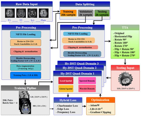
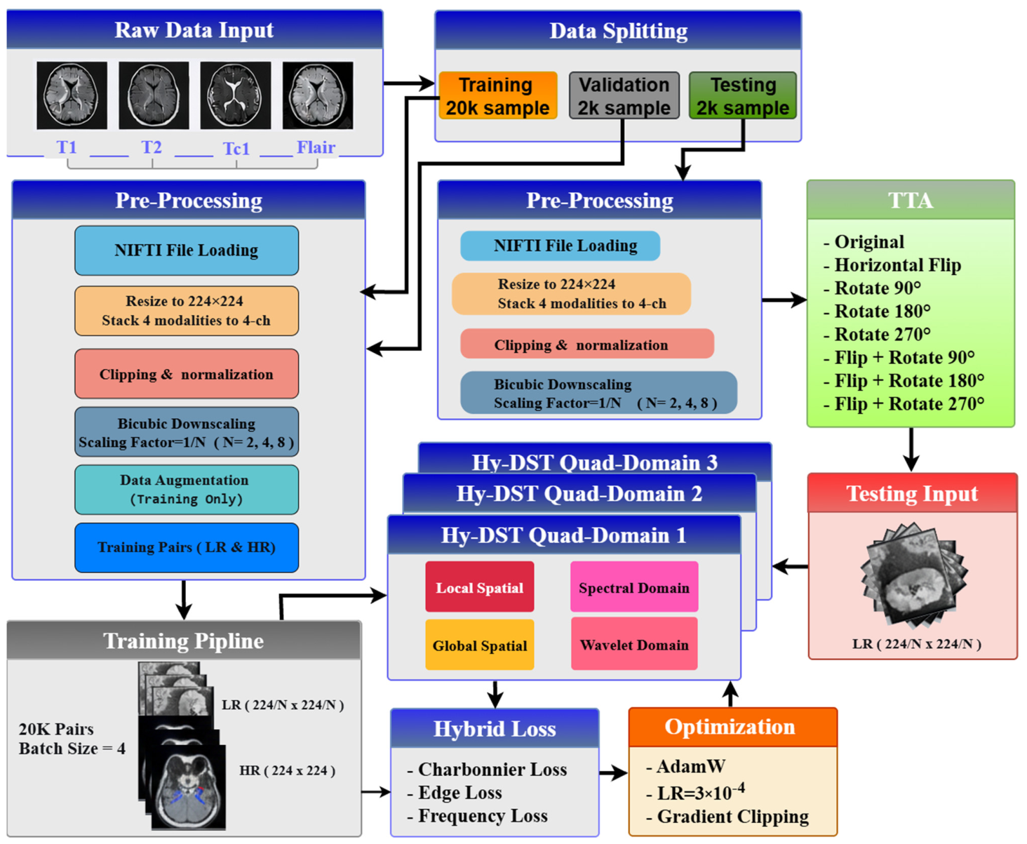
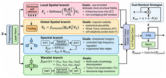
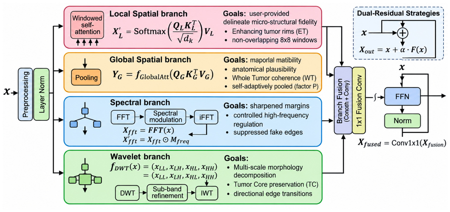
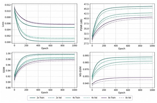
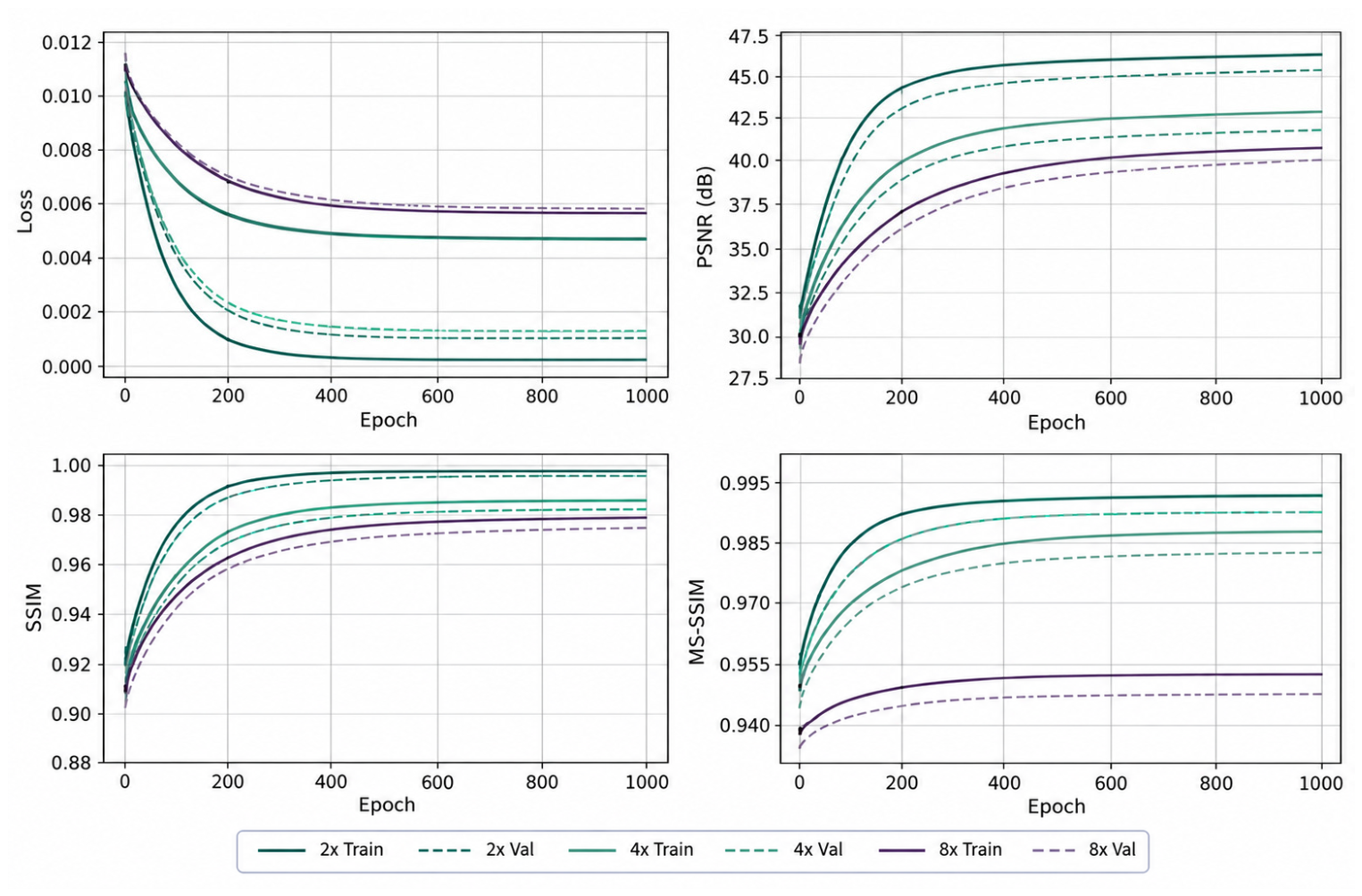
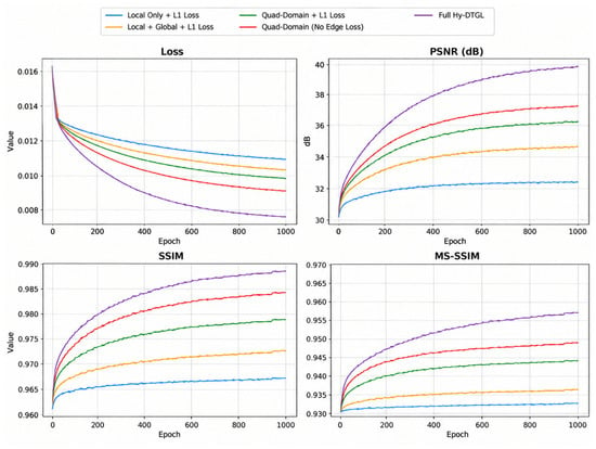
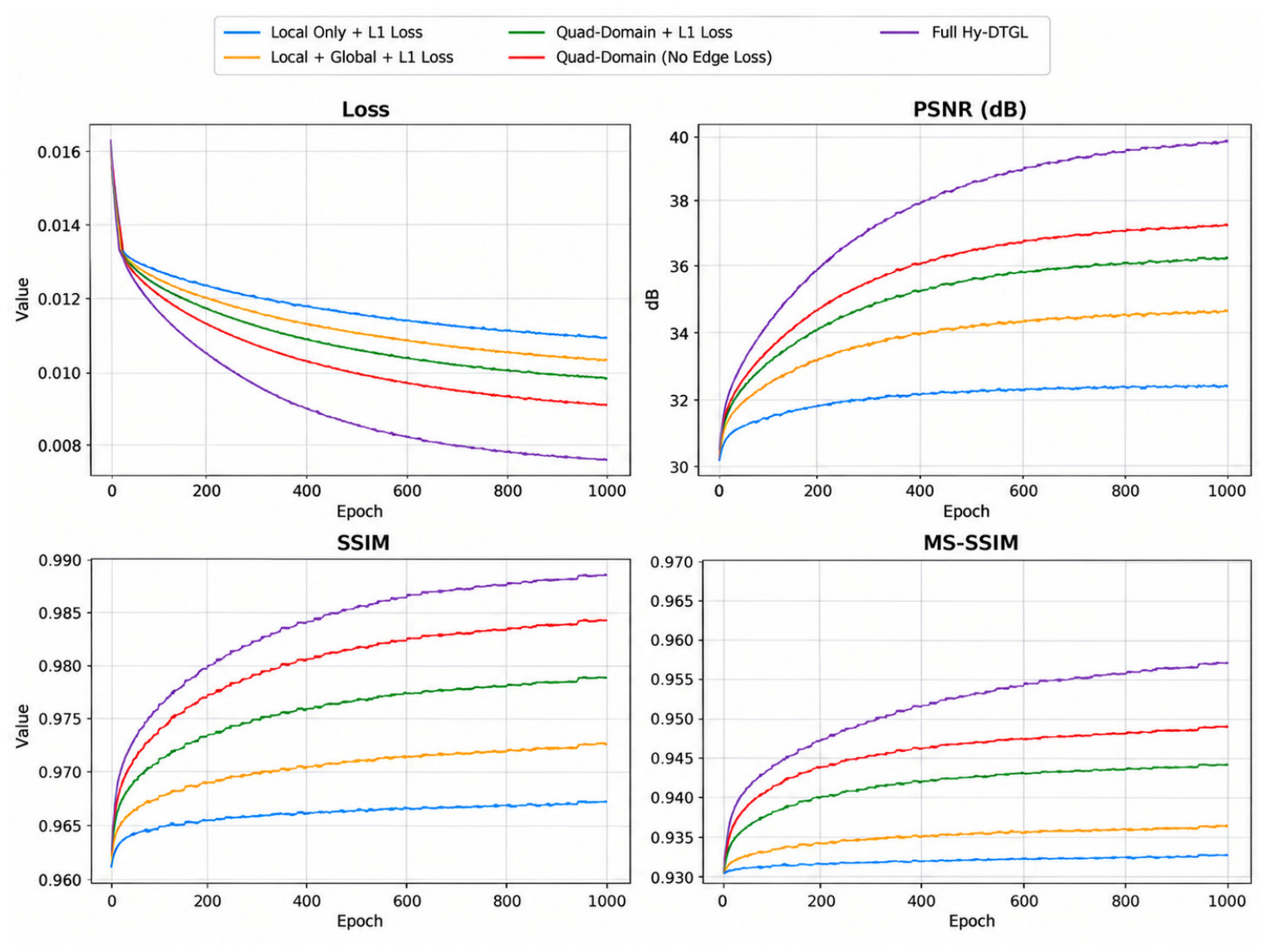
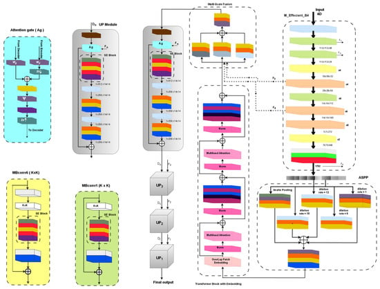
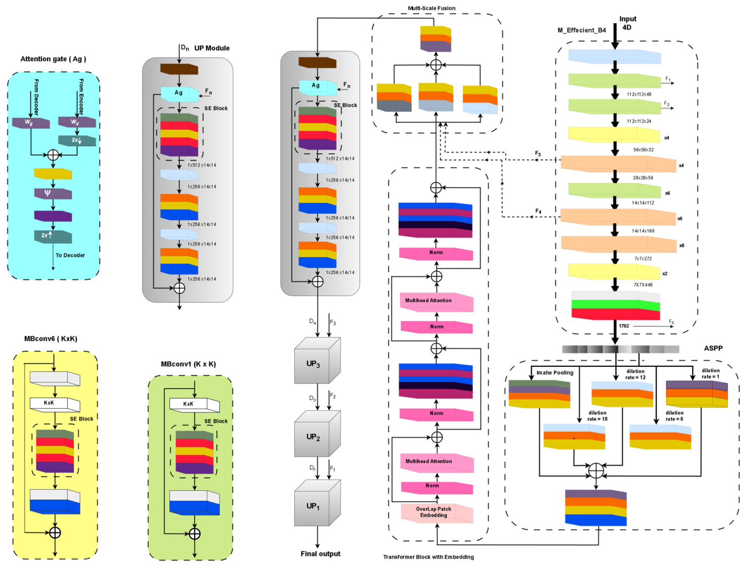
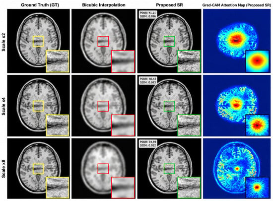
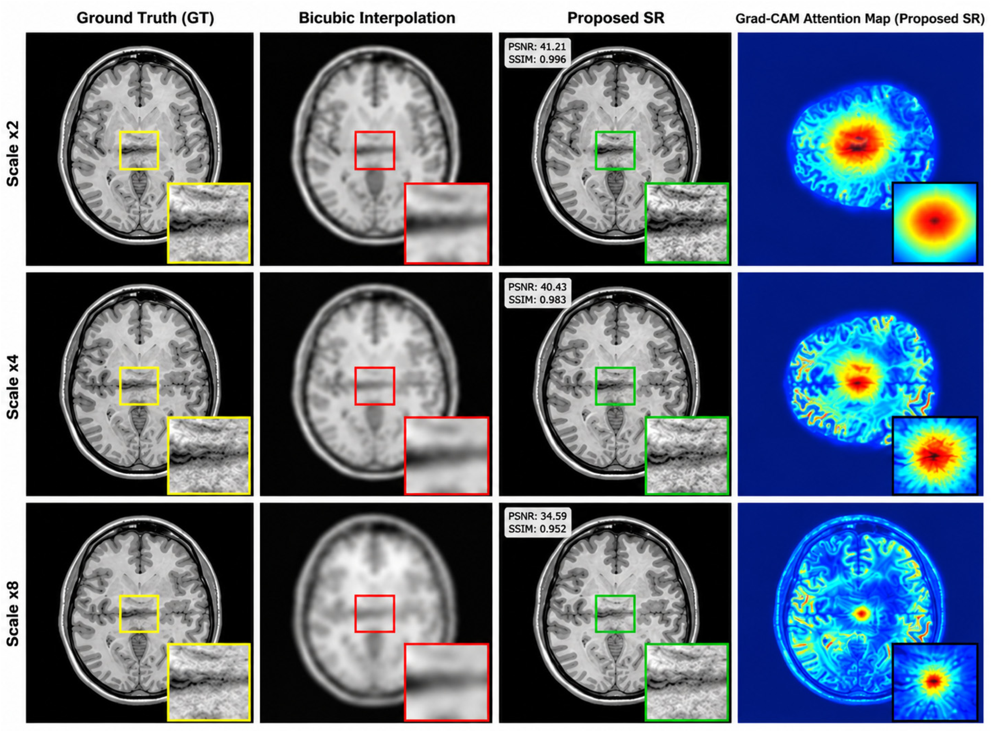
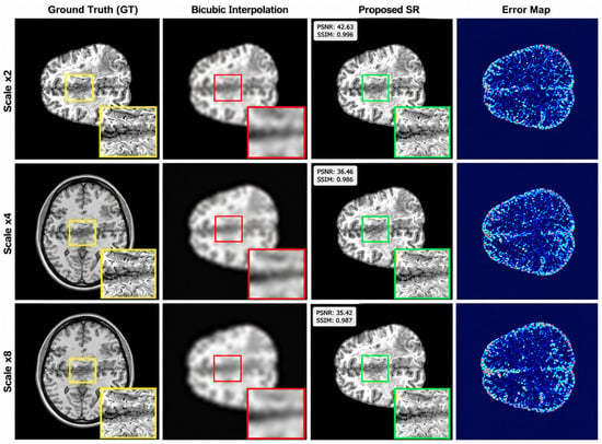
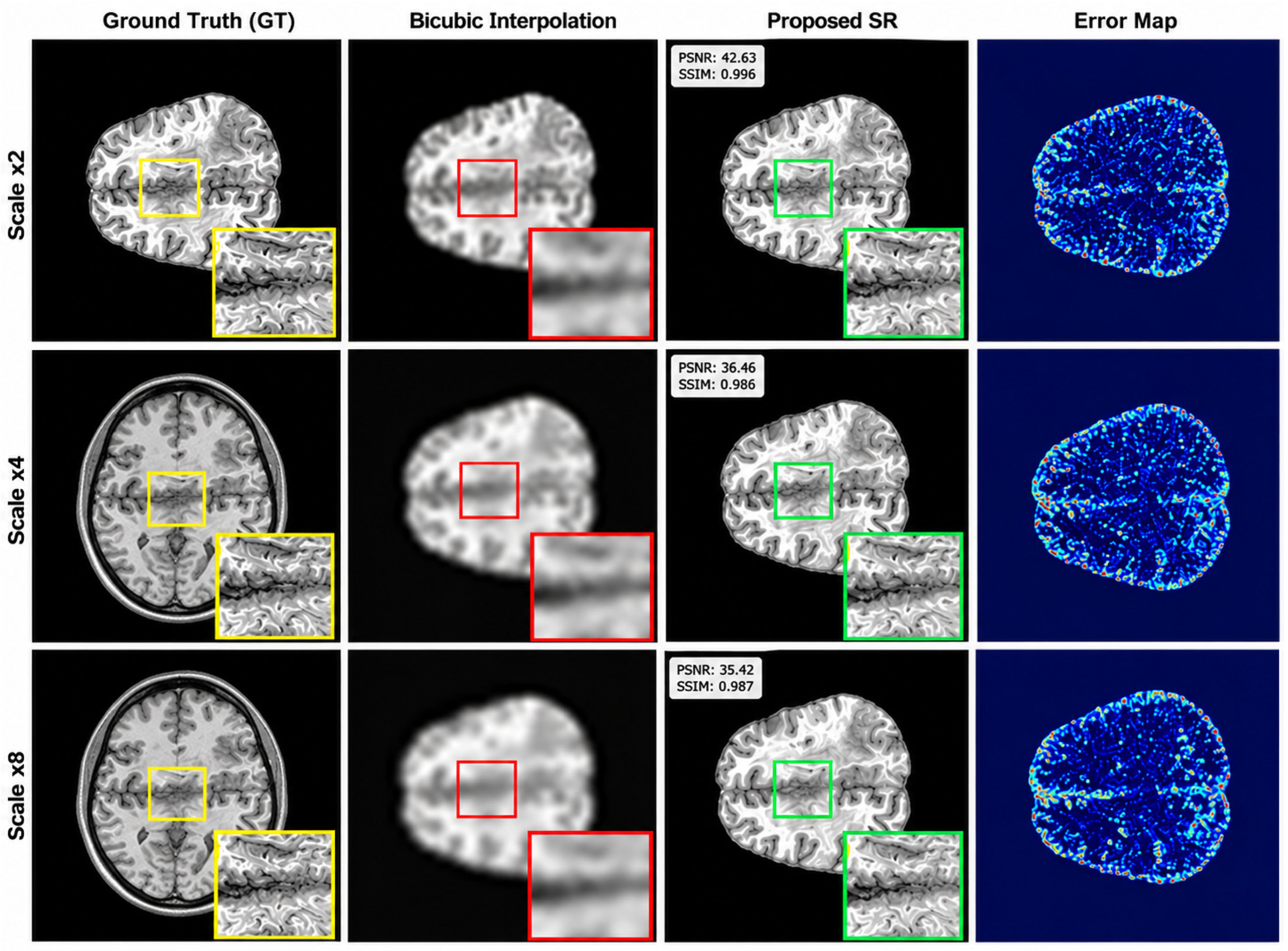
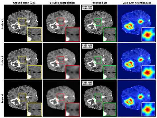
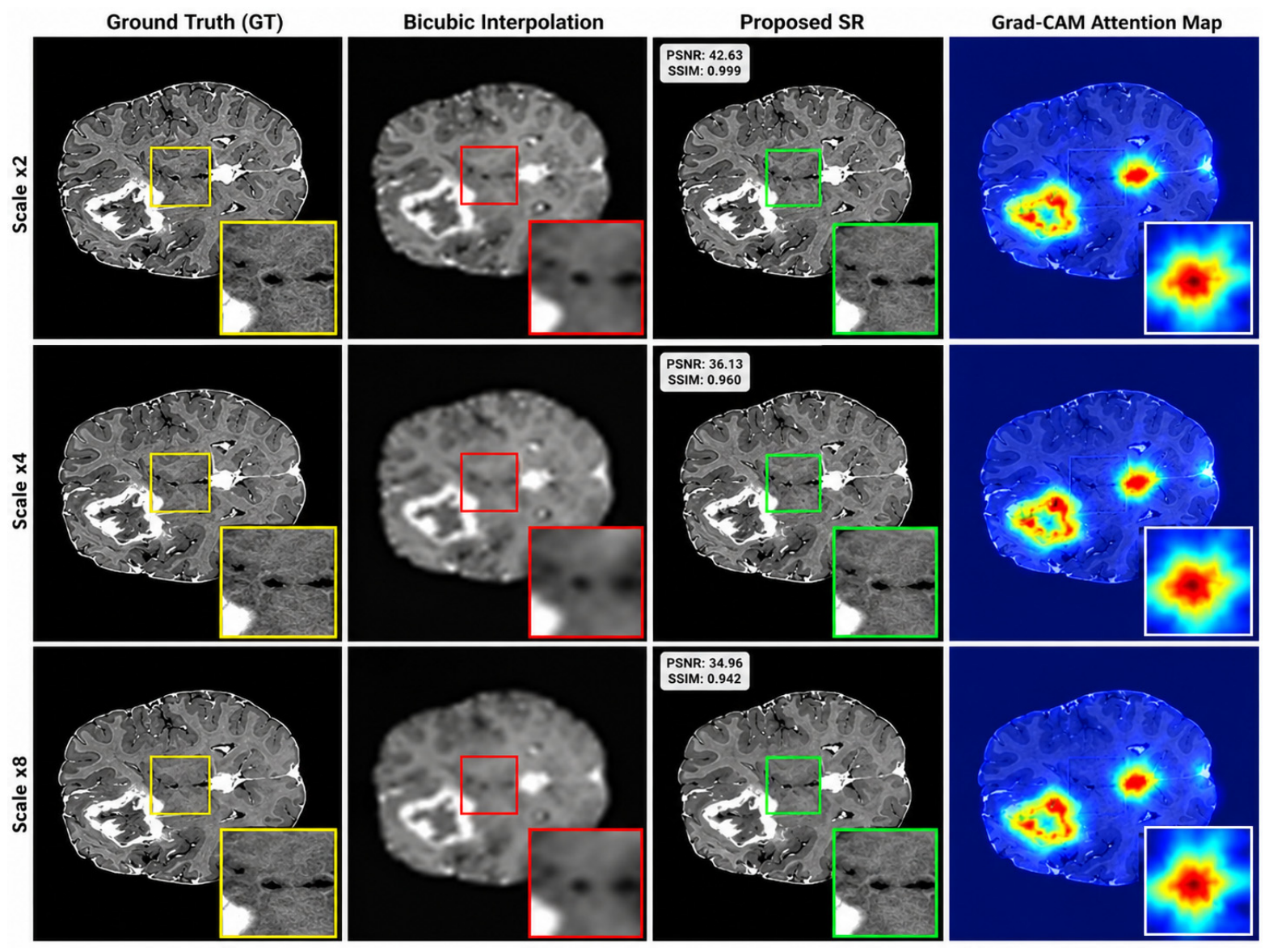
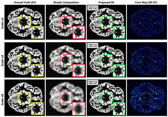
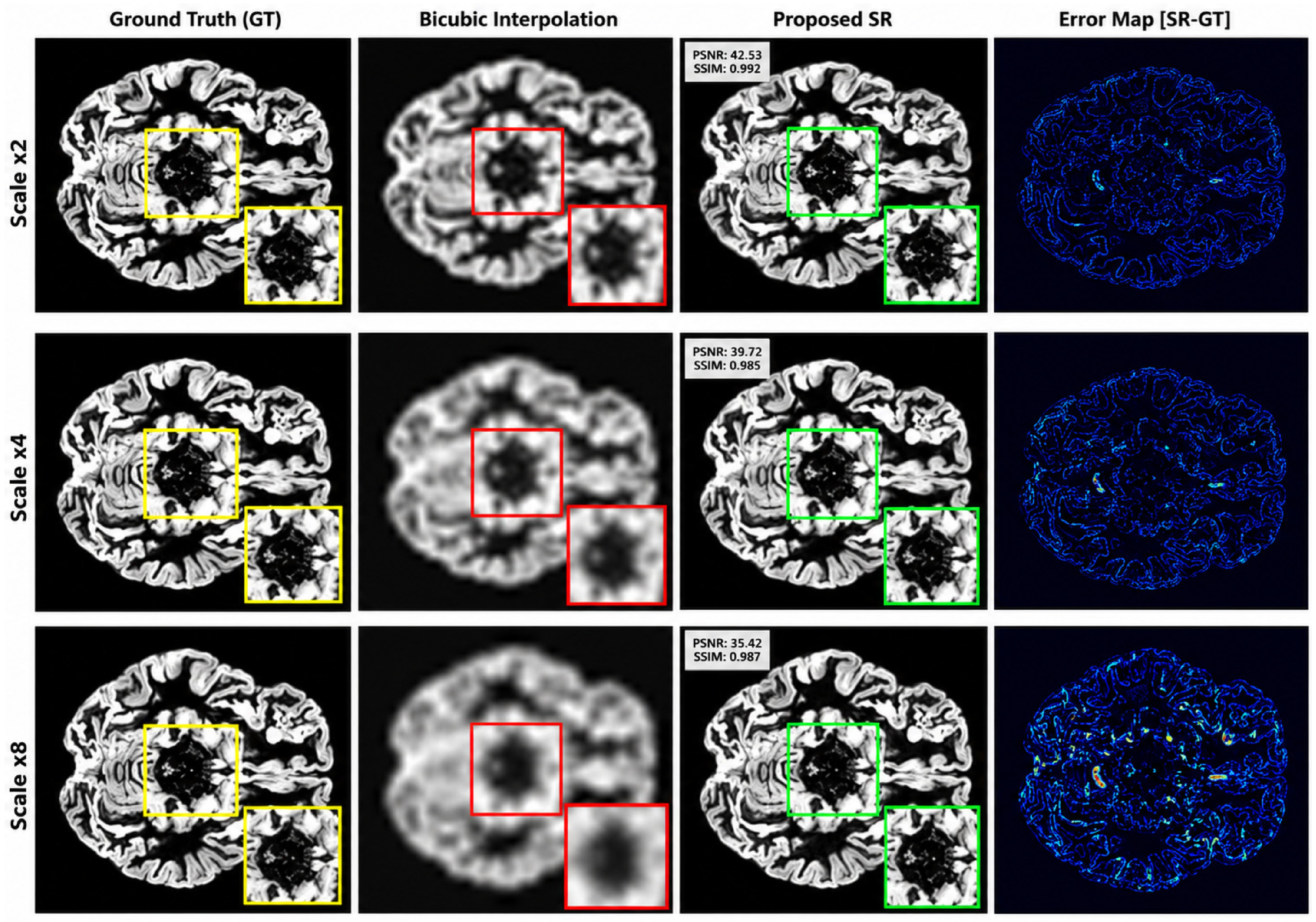

Disclaimer/Publisher’s Note: The statements, opinions and data contained in all publications are solely those of the individual author(s) and contributor(s) and not of MDPI and/or the editor(s). MDPI and/or the editor(s) disclaim responsibility for any injury to people or property resulting from any ideas, methods, instructions or products referred to in the content. |
© 2026 by the authors. Licensee MDPI, Basel, Switzerland. This article is an open access article distributed under the terms and conditions of the Creative Commons Attribution (CC BY) license.