


Abstract
Near-infrared (NIR)-emissive carbon dots (NIR-CDs) have emerged as a distinctive class of ultrasmall carbon nanomaterials for biomedical imaging because their optical absorption/emission can be shifted from the visible region toward the NIR-I (~700–1000 nm) and NIR-II (~1000–1700 nm) biological windows while retaining the characteristic aqueous dispersibility, photostability, surface functionality, and favorable biocompatibility. However, it is challenging to establish a reproducible structure–composition–excited-state relationship that simultaneously provides long-wavelength excitation, high quantum yield (QY), narrow emission, photostability, appropriate pharmacokinetics, and sufficient optical absorption for multimodal imaging. This review critically examines the strategic engineering of NIR-CDs from molecular precursor chemistry to carbonized cores, surface states/defects, heteroatom/metal doping, molecular engineering, and supramolecular self-assembly. The review further examines NIR fluorescence (NIR-FL), multiphoton, afterglow phosphorescence, photoacoustic (PA), magnetic resonance (MR), and multimodal imaging. Finally, the remaining challenges such as NIR-II QY, structural heterogeneity, reproducibility, ambiguity in intrinsic/molecular fluorescence, biosafety, and insufficient clinical validation were discussed together with future prospects.
1. Introduction
Biomedical imaging requires contrast agents that combine high sensitivity with adequate spatial resolution, deep tissue penetration, low background, chemical stability, and acceptable biological safety [1]. Conventional visible fluorescence (FL) is strongly affected by tissue scattering, absorption, and endogenous autofluorescence, limiting its utility for deep-tissue imaging [2]. Moving toward the NIR region reduces these limitations, while the NIR-II window provides particularly attractive optical conditions because photon scattering and tissue autofluorescence are generally lower than in visible/NIR-I imaging [3,4].
NIR spectral range is usually divided into the first (NIR-I, range ~700–900 nm) and second (NIR-II, range ~1000–1700 nm) biological windows [5,6]. A biological window refers to the optical tissue-transparency window. Biological windows are so termed because they permit increased penetration depths of light into biological tissue within certain spectral ranges [7,8]. Imaging in the NIR-II window reduces scattering, which improves penetration depth and spatial resolution, and the weaker endogenous background raises signal-to-background ratios [9]. As a result, many NIR-II emitters have been developed, including organic dyes, quantum dots, rare-earth-doped nanoparticles, single-walled carbon nanotubes and conjugated polymers [10].
CDs provide an unusual platform for engineering such long-wavelength optical behavior. CDs are generally defined as quasi-zero-dimensional photoluminescent carbonaceous nanoparticles, commonly 1–10 nm in diameter, made of a sp2/sp3-hybridized carbon core and a surface rich in functional groups [11]. Because they are prepared by bottom-up or top-down approaches, CDs usually show a size distribution rather than a single uniform size, with most reported average diameters falling within ~2–10 nm. Their crystallinity varies; structural categories include carbon quantum dots (CQDs), graphene quantum dots (GQDs), carbon nanodots (CNDs), and carbonized polymer dots (CPDs) [11]. Due to their excellent optical and physicochemical characteristics, CDs have been widely utilized in various biomedical and energy-related applications [12,13,14,15,16,17,18,19,20,21,22,23]. The optical behavior of CDs can be tuned in several ways, such as carbonization degree, molecular precursor design, heteroatom doping, surface-state engineering, and supramolecular assembly [12]. They disperse well in water, resist photobleaching, and carry many surface functional groups. Their surfaces can contain abundant –NH2, –OH, –COOH, –C=O, –CONH2, and other functionalities, providing opportunities for covalent modification, electrostatic interactions, molecular recognition, and coordination with metal ions [24]. The development of NIR-CDs represents an important evolution from the original blue/green-emissive CD paradigm. Early CD research focused predominantly on visible photoluminescence (PL). However, precursor engineering, increased π-conjugation, heteroatom incorporation, surface-state manipulation, molecular-state engineering, and controlled aggregation progressively enabled emission toward the red and NIR regions [12]. Recent studies have extended this concept into NIR-II-emitting CDs with emission between approximately 1000 and 1700 nm, opening opportunities for deeper FL imaging and high-resolution angiography [4]. Accordingly, the central challenge is not simply to produce CDs that emit in the NIR window. It is to deliberately control the electronic and excited-state features that govern NIR absorption, radiative emission, nonradiative decay, photothermal conversion, and intersystem crossing [25]. NIR emission can come from several sources, including extended π-conjugated graphitic cores, surface-localized defect or trap states, embedded molecular fluorophores, cross-linked polymeric domains, or a combination of these [26]. As a result, CDs with nearly identical particle sizes and emission maxima may still have fundamentally different structures, photophysical pathways and, ultimately, biological performance. Rational design therefore requires mechanistic structure–property correlations rather than reliance on spectral output alone.
Previous reviews have each covered part of NIR-CDs, including their synthesis, biomedical uses, NIR-II emission, performance as multimodal contrast agents, or role in theranostics [4,12,26,27,28,29,30]. In particular, Yang et al. gave a focused account of NIR-II CDs, covering synthesis and purification, surface engineering, and biomedical applications [4]. Recently, some reports focused on CDs designed for multimodal imaging, NIR-II CDs that self-aggregate, CDs with asymmetric electronic structures, and metal-doped CDs that work as MRI contrast agents [31,32,33,34,35]. However, these advances are spread across separate reports, and addressing these within a single framework is imperative. This review provides a single framework that links structural design to NIR photophysics and biomedical imaging performance. Scheme 1 represents the schematic overview of this review, depicting the synthesis, strategic design, and biomedical imaging application of NIR-CDs.
Scheme 1.
Schematic overview of this review, depicting the synthesis, strategic design, and biomedical imaging applications of NIR-CDs. Inset images were taken from ref. [33,36,37,38,39,40,41,42].
2. Biological Imaging Windows (NIR-I vs. NIR-II) and Advantages of CDs
Although visible FL is intense, deep-tissue imaging is limited by photon scattering, absorption by endogenous chromophores, and tissue auto-FL [5,43,44]. Shifting emission into NIR-I (~700–900 nm) partly mitigates these losses, and NIR-II (~1000–1700 nm) suppresses them further [6,45]. Tissue’s reduced scattering coefficient falls steadily with wavelength, so NIR-II FL allows deeper, higher-resolution imaging [9]. Long-wavelength emission therefore brings several practical benefits such as negligible background autofluorescence, attenuated photon scattering, deeper tissue penetration, higher signal-to-background ratios, and fast temporal acquisition. Together, these make real-time, intraoperative image-guided interventions feasible [46]. Using several classes of fluorophores, NIR-II imaging has already been applied to high-fidelity vascular mapping, gastrointestinal and lymphatic tracing, tumor delineation, transcranial brain imaging, and image-guided surgical resection [47]. Notably, the field has moved beyond proof-of-concept preclinical studies toward clinical instrumentation. For example, multispectral NIR-I/NIR-II FL imaging has been used during human liver-tumor surgery, which highlights how readily long-wavelength FL imaging can be brought into clinical practice [48].
NIR-CDs offer several advantages over conventional organic NIR fluorophores and inorganic semiconductor quantum dots. Their surfaces can carry a wide range of oxygen-, nitrogen-, and phosphorus-containing groups, including –NH2, –COOH, –OH, –C=O, –CONH2, and phosphate moieties. These groups give many reactive sites for covalent conjugation, electrostatic assembly, and other post-synthetic functionalization [12]. Their ultrasmall size can facilitate tissue penetration and renal clearance, although the same property can produce short blood circulation and insufficient target accumulation [31]. Their most notable feature is that a single carbonaceous scaffold can combine diagnostic and therapeutic functions. With careful tuning of the core structure, heteroatom doping, and surface states, NIR-CDs can emit bright FL while also absorbing strongly in the NIR region. This lets them either convert light efficiently into heat (photothermal conversion) or generate reactive oxygen species (ROS) [49]. As a result, one NIR-CD nanoplatform could, in principle, support disease detection, precise localization, targeted treatment, and real-time monitoring of how the treatment is working, which makes NIR-CDs strong candidates for image-guided precision theranostics [4].
3. CDs Structural Features and Basis of NIR Emission
CDs describe a family of structurally heterogeneous materials rather than a single molecular architecture. Broadly, CDs may be classified into four categories, including carbon quantum dots (CQDs), graphene quantum dots (GQDs), carbon nanodots (CNDs), and carbon polymer dots (CPDs) (Figure 1) [11]. Graphene quantum dots (GQDs) consist of one or a few graphene layers with lateral sizes of a few nanometers. Their edges and basal planes carry oxygen-containing groups, and HR-TEM typically shows in-plane lattice fringes of ~0.21–0.24 nm. Carbon quantum dots (CQDs) are quasi-spherical particles with a crystalline, multilayer graphitic core that shows clear lattice fringes (~0.21 nm for the graphene (100) plane or ~0.33–0.34 nm for the (002) interlayer spacing) and size-dependent, quantum-confined emission. Carbon nanodots (CNDs) are also roughly spherical but are largely amorphous or only weakly crystalline, so their emission is governed mainly by surface and defect states rather than by quantum confinement. Carbonized polymer dots (CPDs) combine a small, partly carbonized core with a crosslinked polymer shell. They usually show broad XRD features and retain polymer-like functional groups that can be detected by FT-IR and NMR spectra [24]. These architectures can generate fundamentally different photophysical behavior.
Figure 1.
Classification of CD structures such as GQDs, CQDs, CNDs, and CPDs [11].
Core-state engineering is an effective strategy for tuning the optical features of CDs. Increasing the size and conjugation of sp2 domains generally decreases the effective electronic energy gap and can drive emission toward longer wavelengths [50]. This strategy is particularly relevant for GQDs and highly carbonized CQDs, in which π–π* transitions and quantum confinement contribute to optical behavior [24]. However, excessive carbonization may produce graphitic domains with increased nonradiative relaxation and reduced FL efficiency. Thus, NIR engineering requires a balance between conjugation extension and structural passivation [51].
Surface-state engineering is an effective strategy for tuning the optical features of CDs. Surface oxidation and functionalization introduce localized electronic states that can become emissive [43,51]. Oxygen-, nitrogen-, sulfur-, phosphorus-, and halogen-containing groups can alter electron density, trap-state energies, and surface-state distributions, allowing emission to be progressively shifted from blue/green toward red/NIR wavelengths [51,52]. Surface states are particularly useful because they allow wavelength tuning without necessarily requiring a very large graphitic core. However, heterogeneous surface states can also produce broad excitation-dependent emission, making precise mechanistic assignment difficult [24].
Molecular-state engineering is often effective for tuning the optical features of CDs. Molecular fluorophores or conjugated molecular fragments are formed during synthesis. These species may remain embedded in or covalently linked to the carbonized framework and can dominate the observed FL [24]. This mechanism is particularly important for bottom-up synthesis using aromatic amines, dyes, phenolic compounds, porphyrins, cyanines, and other conjugated precursors. Molecular-state engineering can produce narrow and strongly red-shifted emission, but it also creates a major purification challenge: free molecular fluorophores may be mistaken for intrinsic CD emission. Rigorous dialysis, chromatography, ultrafiltration, absorption spectroscopy, mass spectrometry, and control experiments are therefore essential [4,52].
Donor–acceptor (D–A) architectures provide a particularly powerful route to NIR absorption and emission. Electron-rich and electron-deficient domains can generate intraparticle charge-transfer states with substantially reduced transition energies. A recent example produced CDs integrating NIR-II absorption around 1033 nm and NIR-I emission around 670 nm through a D–π–A charge-transfer state coupled with a localized excited state originating from a large conjugated sp2 domain [50].
4. Synthesis and Strategic Design of NIR-CDs
The strategic engineering of NIR-CDs ultimately depends on selecting synthesis routes that provide adequate control over carbonization, conjugation, surface states, and compositional incorporation. Accordingly, the synthesis methodology represents the first level of structural control, determining the extent to which the carbon framework and its associated optical states can be tailored.
4.1. Synthesis Techniques
The principal synthetic approaches can be broadly classified into top-down and bottom-up strategies, each offering distinct advantages and limitations for constructing NIR-emissive CDs.
4.1.1. Top-Down Approaches
Top-down synthesis involves cutting or exfoliating larger carbon materials such as graphite, graphene oxide, carbon nanotubes, carbon fibers, or carbon black into nanoscale domains [24]. Oxidative cutting, electrochemical exfoliation, laser ablation, and related approaches can produce GQDs and CQDs with relatively high structural carbonization [2]. The principal advantage is that the graphitic domain is relatively well established before size reduction. Consequently, size, edge structure, and surface oxidation can become controllable variables, which influence their electronic structures and PL properties [43]. Top-down approaches utilize abundant, relatively inexpensive carbon precursors and can produce structurally robust nanomaterials with good chemical stability. Surface oxidation enables the introduction of oxygen-containing functional moieties, thereby improving aqueous dispersibility and providing versatile anchoring sites for bioconjugation. Furthermore, deliberate defect modulation and surface-state engineering offer opportunities to tailor the photophysical characteristics of CDs for biomedical imaging.
Nevertheless, achieving intense, spectrally well-defined, and reproducible NIR emission remains a significant challenge, owing to the complex interplay between carbon-core structures, surface states, and defect-associated electronic transitions. Structural heterogeneity, broad particle-size distributions, inconsistent surface chemistry, and residual impurities can further compromise purification efficiency, optical consistency, and batch-to-batch reproducibility. In addition, aggressive oxidative treatments may necessitate corrosive reagents, whereas energy-intensive processing can impede economic scalability. From a translational perspective, systematic investigations of pharmacokinetics, biodistribution, biodegradability, long-term biocompatibility, and biological clearance are indispensable for establishing their safety profiles. Consequently, despite the considerable potential of top-down synthesis for developing functional NIR-emissive CDs, precise regulation of emission characteristics, PL QY, structural homogeneity, and biological safety remains critical to advancing these nanomaterials toward reliable biomedical imaging applications. The limitations include heterogeneous particle sizes, harsh oxidation, relatively low production yields in some approaches, and difficulty generating highly water-soluble NIR-II-emissive particles with reproducible surface chemistry [53].
4.1.2. Bottom-Up Approaches
Bottom-up synthesis is currently the most versatile route for NIR-CD engineering. Small molecules, polymers, dyes, amino acids, aromatic compounds, pharmaceuticals, and biomass can undergo dehydration, condensation, polymerization, cyclization, aromatization, and carbonization to generate CDs [24]. Hydrothermal and solvothermal synthesis are particularly attractive because they combine relatively mild processing with tunable temperature, pressure, solvent polarity, and precursor ratios. Microwave-assisted, pyrolytic, and photochemical approaches can accelerate the reaction and modify the carbonization pathway [52,53]. The principal advantages of bottom-up synthesis include accessible precursors, relatively straightforward processing, structural tunability, and opportunities for integrating functional groups for bioconjugation. Nevertheless, uncontrolled carbonization, heterogeneous emissive states, and the possible formation of molecular fluorophores complicate structural characterization and mechanistic interpretation. Additional limitations include batch-to-batch variability, challenges in achieving reproducibly high NIR QY, and insufficient long-term toxicological and biodistribution evidence. For instance, bright emission often requires post-functionalization. Liu et al. reported that solvothermally-derived CDs needed PEI passivation to generate bright FL emission [36]. NIR-II QY also remains low (1.27%), as reported by Ci et al. [54]. In hydro/solvothermal methods, long reaction times and sealed autoclaves limit batch size and scale-up, and molecular fluorophore byproducts [4,52]. In microwave bottom-up techniques, limitations include non-uniform heating and no control over the degree of carbonization. Future advances should prioritize precursor-guided molecular design, reproducible reaction control, rigorous purification, and systematic structure–photophysics correlations to facilitate the development of reliable NIR-CD probes for biomedical imaging.
4.2. Strategies for Designing and Tailoring Optical Properties of NIR-CDs
While synthesis methodology determines the formation pathway, morphology, carbonization degree, and surface chemistry of NIR-CDs, achieving precisely tunable NIR optical properties requires deliberate control over their molecular and electronic structures. Accordingly, NIR-CD design has evolved from simply optimizing reaction conditions toward a more rational strategy in which precursor chemistry, heteroatom incorporation, surface states, structural defects, molecular electronic states, and supramolecular organization are systematically manipulated. These interconnected approaches provide complementary means to modulate π-conjugation, energy-level alignment, charge-transfer states, and radiative/nonradiative relaxation pathways.
4.2.1. Precursor Chemistry
Precursor chemistry determines the molecular fragments, degree of conjugation, heteroatom composition, and surface groups available during CD formation [51]. Aromatic precursors are particularly useful for generating extended π-conjugated domains, whereas amine-rich molecules can introduce nitrogen and promote donor states [43]. Theoretical investigations have demonstrated that increasing the degree of π-conjugation within the carbon framework induces a pronounced bathochromic shift in the emission spectra [55]. Thus, judicious selection of polycyclic aromatic precursors with large conjugated π-systems has been widely investigated for NIR-emissive CD design [12]. For example, Liu et al. developed CDs featuring extended sp2-hybridized π-conjugated domains by employing perylenetetracarboxylic dianhydride (PTCDA), a highly conjugated perylene-derived molecular precursor, as the carbonaceous building block (Figure 2a) [36]. Notably, subsequent surface functionalization with polyethyleneimine (PEI) markedly enhanced the PLQY, thereby promoting the NIR emissive properties of the resulting PEI-functionalized CDs. The incorporation and molecular fusion of large π-conjugated precursor units facilitated the formation of emissive states capable of generating FL centered at ~745 nm. Owing to their favorable NIR optical characteristics, these CDs demonstrated considerable potential for in vivo NIR FL bioimaging and enabled two-photon NIR angiographic visualization, highlighting the effectiveness of precursor-driven π-domain engineering for extending CD emission into the NIR spectral window (Figure 2b). The use of dye-derived precursors provides another route to NIR emission. Cyanine-derived CDs, for example, have recently been engineered to emit at 1044 nm, demonstrating that precursor-level molecular engineering can directly establish NIR-II optical states [31]. Biomass-derived precursors provide attractive advantages in terms of sustainability and scalability, although compositional variability can compromise optical reproducibility [52]. Therefore, “green synthesis” should not be equated automatically with a clinically superior material. Precursor purity, molecular composition, reproducibility, endotoxin contamination, and residual contaminants remain essential considerations [56].
Figure 2.
Strategies for designing and tailoring optical properties of NIR-CDs: (a) Precursor engineering enables formation of larger sp2-hybridized π-domains through molecular fusion of perylenetetracarboxylic dianhydride [36], (b) Corresponding NIR emission reported at 745 nm [36], (c) Depiction of metal-based heteroatom doping (Fe-dopant) strategy to originate NIR-II emission through one-pot hydrothermal synthesis [54], (d) Corresponding NIR-II emission spectra reported at ~1000 nm [54], (e) Corresponding in-vivo NIR emission intensity (λex = 808 nm; λem = 1000 nm) in mice after 120 min post oral-administration [54], (f) Defect engineering (polaron engineering) to precisely control the optical bandgap of CDs in the NIR-II window by introducing various defects [37], (g) Corresponding NIR-emission spectra [37], (h) Molecular engineering via aniline extension of p-phenylenediamine to tune CD emission toward NIR-II [33], (i) Corresponding NIR-emission spectra of NIR-CDs centered at 1080/1265 nm [33].
4.2.2. Heteroatom Engineering
Heteroatom doping is among the most widely employed approaches for manipulating the electronic structure of CDs. Nitrogen, sulfur, phosphorus, boron, fluorine, chlorine, selenium, and combinations thereof can alter electron density, surface states, defect distributions, and charge-transfer transitions [43,51].
Nitrogen is especially important because its electronic structure is compatible with the carbon framework and can generate electron-rich sites. Pyridinic–N, pyrrolic–N, graphitic–N, and amino-type nitrogen can influence the electronic gap and radiative/nonradiative relaxation pathways [57]. Nitrogen doping can therefore induce red-shifting while simultaneously increasing surface reactivity and providing coordination sites for biomolecules or metals. A representative Mn-doped CD study showed that increasing N incorporation shifted FL from 542 to 578 nm while simultaneously improving T1 MRI properties [58]. S and P introduce electronic perturbations that can promote charge-transfer states and alter defect chemistry [59]. Dual- or triple-doping can provide stronger electronic modulation than single heteroatoms, although the resulting structures become increasingly heterogeneous. Boron is electron-deficient relative to carbon and nitrogen and can act as an electronic acceptor, making B/N combinations particularly attractive for D–A engineering [60]. F, Cl, and related halogens can modify surface electronic states, polarity, and charge distribution. Fluorinated CDs can additionally provide chemically distinct coordination environments for metal incorporation. Recent Gd3+-functionalized fluorinated CDs demonstrated that metal incorporation substantially modified FL lifetime and relaxation behavior, illustrating the coupling between surface electronic structure and MRI functionality [61].
Metal-based heteroatom incorporation can introduce accessible d/f orbitals, modify charge density and HOMO-LUMO energy gaps, thereby facilitating strong NIR emission. Ci et al. developed a Fe-doping strategy through one-pot hydrothermal synthesis by introducing FeCl3.6H2O (Fe source) and dopamine hydrochloride/o-phenylenediamine as carbon precursors (Figure 2c) [54]. Fe incorporation promoted a more highly graphitized carbon framework and, critically, increased the proportions of C=O and graphitic–N functionalities, which are associated with red-shifted CD emission. Compared with undoped CDs prepared under identical conditions, Fe doping increased C=O from 32.23% to 66.44% and graphitic–N from 37.71% to 61.85%, demonstrating the key role of Fe3+ in establishing the NIR-II emissive structure. The resulting Fe-CDs exhibited NIR-II emission centered at ~1000 nm, with a QY of 1.27% (Figure 2d). Thus, Fe doping effectively tunes the electronic structure and surface-state composition toward long-wavelength NIR-II FL, enabling deeper-tissue in vivo imaging (Figure 2e).
4.2.3. Surface-State and Defect Engineering
Surface chemistry determines both optical states and biological behavior. –COOH, –OH, –NH2, –CONH2, and other groups can influence emission wavelength, lifetime, QY, colloidal stability, protein adsorption, and cellular uptake [62]. Surface passivation can suppress nonradiative trapping and improve FL. Conversely, controlled defects can generate new emissive states and extend absorption into the NIR [43]. Surface engineering is equally important after optical optimization. PEGylation can reduce nonspecific protein adsorption and prolong circulation; peptides and antibodies can provide receptor-specific targeting; and carbohydrates or zwitterionic groups can modify biodistribution and colloidal stability [4,62]. A particularly interesting strategy is to deliberately exploit aggregation. Conventional CDs can undergo rapid renal filtration because ultrasmall particles may fall below the glomerular filtration threshold [31]. Recent cyanine-derived CDs with 2.5 nm dimensions were designed to spontaneously form 30–40 nm aggregates through hydrogen-bonding interactions, increasing tumor retention while preserving NIR-II FL. These self-aggregated CDs enabled vascular imaging, tumor visualization, and dynamic intestinal imaging [31].
Defect engineering has emerged as an effective strategy for tailoring the electronic structure, charge-carrier dynamics, and excited-state behavior of CDs, thereby enabling precise modulation of their optical properties [63]. In a representative study, Zhang et al. developed a polaron-engineering approach to regulate the optical bandgap of CDs and extend their photo-response into the NIR-II region through controlled defect generation (Figure 2f) [37]. Red-emissive CDs were subjected to post-oxidation using 2-iodoxybenzoic acid (IBX), which introduced N-related vacancies and O–containing defects within the carbonaceous framework. The resulting structural perturbations promoted lattice distortion, charge redistribution/injection, and enhanced electron–phonon interactions, collectively facilitating polaron formation. Notably, O–associated polarons generated characteristic NIR optical features in the 800–840 nm range, whereas polarons coupled with N vacancies induced a broader absorption band ranging ~1035–1350 nm, thereby substantially extending the optical response into the NIR-II window. Consistent with these defect-mediated electronic transitions, the resulting nir-CQDs exhibited distinct PL in the NIR region upon excitation at 800 and 980 nm (Figure 2g), demonstrating the effectiveness of defect-induced polaron engineering for manipulating the excited-state optical characteristics of CDs.
4.2.4. Molecular Engineering
One of the most promising directions is the construction of molecularly defined electronic states within otherwise carbonaceous nanoparticles. A D–A architecture can produce spatially separated highest occupied and lowest unoccupied electronic states, reducing the energy required for optical excitation [50]. Such engineering can produce NIR absorption without requiring extreme graphitization. In one example, CDs simultaneously exhibited NIR-II absorption at 1033 nm and NIR-I FL at 670 nm. The authors attributed the NIR-II absorption to a D–π–A charge-transfer state, whereas NIR-I emission originated from a localized excited state associated with a large sp2-conjugated domain. The resulting CDs supported multiphoton imaging and photothermal therapy (PTT) [50]. The same nanoparticle can therefore be engineered for FL, multiphoton imaging, PA imaging, and PTT depending on how radiative and nonradiative pathways are balanced. In a most recent study, Yang et al. used molecular engineering via aniline extension of p-phenylenediamine to tune CD emission toward NIR-II (Figure 2h) [33]. Increasing aniline units enhances the benzene ring’s electron-acceptor ability and molecular dipole moment, strengthening intramolecular charge transfer. The central N partial charge of the precursor with higher aniline unit changes from −0.65e to −0.38e, while donor–acceptor spatial separation increases from 5.663 to 5.686 Å. The calculated molecular bandgap narrows from 4.94 to 4.01 eV. During carbonization, graphene-domain growth and pyrrolic–N enrichment further narrow the gap. Consequently, emission shifts from 480 nm (CDs-1) to 590 nm (CDs-2) and finally 1080/1265 nm (CDs-3), achieving NIR-II emission (Figure 2i).
4.2.5. Supramolecular Self-Assembly
Traditional nanoparticle design often seeks to prevent aggregation. For CDs, this pattern is being reconsidered. Ultrasmall CDs can have excellent tissue penetration and renal clearance but insufficient tumor retention. Conversely, larger assemblies may provide longer circulation and greater tissue retention but can reduce penetration. Cyanine-derived NIR-II CDs spontaneously aggregated from approximately 2.5 nm primary particles to 30–40 nm assemblies through H-bonding interactions. This aggregation improved tumor retention and enabled high-resolution vascular imaging while maintaining 1044 nm FL [31]. Molecularly engineered NIR-II CD assemblies have similarly demonstrated high signal-to-noise angiography and tumor imaging [64]. Thus, aggregation should be considered not simply as a colloidal stability problem but as a biological function-switching mechanism.
5. NIR-CDs for Advanced Biomedical Imaging
5.1. NIR FL Imaging
NIR FL is the most direct biomedical application of NIR-CDs. Compared with visible-emitting CDs, NIR-CDs can reduce autofluorescence and tissue scattering, thereby increasing the signal-to-background ratio [51,65].
Cellular imaging has been widely implemented utilizing NIR-CDs. The abundant surface functionality and ultrasmall dimensions of CDs promote cellular internalization [24]. FL microscopy can then reveal intracellular distribution, organelle localization, or cellular uptake. NIR emission is particularly useful where visible FL suffers from strong biological background. However, intracellular imaging should not be interpreted automatically as evidence of biocompatibility. FL intensity can remain high even when nanoparticles perturb cellular metabolism. Comprehensive cytotoxicity, oxidative stress, membrane integrity, and long-term clearance analyses remain necessary.
Most notably, NIR-CDs have been explored for tumor visualization because NIR FL provides improved optical contrast and can be coupled with targeting ligands [4,65]. The ultrasmall size can facilitate tissue penetration, while surface modification can improve tumor accumulation. A critical limitation is that passive tumor accumulation through the enhanced permeability and retention effect is highly variable across tumor type, size, vascularization, and animal model. Consequently, active targeting and quantitative biodistribution measurements should accompany claims of tumor specificity. Moreover, NIR-II FL is particularly suitable for vascular imaging because reduced scattering can preserve vessel contrast at increased tissue depths [3,66]. Recent molecularly engineered NIR-II CD assemblies produced high signal-to-noise whole-body angiography and tumor visualization while also enabling photodynamic/photothermal treatment [64]. Similarly, self-aggregated cyanine-derived CDs emitting at 1044 nm provided high-resolution vascular and tumor imaging and dynamic visualization of intestinal peristalsis [31].
Han et al. designed NIR-II emissive carbonized CDs (NIR-CPDs) capable of NIR-II angiography and real-time disease diagnosis of colitis in the NIR-II window (1000–1700) [38]. Figure 3a shows the schematic of the synthesis process of CPDs-CHO and NIR-CPDs. The NIR-II emissive NIR-CPDs were developed through a two-step hydrothermal/Knoevenagel strategy, achieving emission tails extending to ~1400 nm and thereby enabling NIR-II FL imaging. The molecular engineering of indole-derived emissive centers onto the carbonized polymer core substantially enhanced NIR-II output, while the carbon core functioned as an absorption/antenna center. Figure 3b shows the schematic of the molecular state emission centers of NIR-CPDs. The NIR-CPDs exhibited markedly improved photostability compared with ICG and maintained appreciable NIR-II brightness in aqueous media. Albumin complexation further enhanced their NIR-II brightness, enabling high-contrast brain and hindlimb angiography across 1000–1300 nm with relatively low excitation power and short exposure times. Encapsulation with DSPE-mPEG2000 produced ~30–40 nm DSPE-CPDs and increased FL brightness by approximately fourfold. These nanocomplexes enabled non-invasive, real-time NIR-II visualization of intestinal inflammation and provided abdominal FL signals that correlated with colitis severity. Figure 3c shows the NIR-II images of acute colitis after intravenous administration (12 h post-injection) on days 11 and 13, respectively. Figure 3d shows the NIR-II fluorescence imaging of the colon and cecum after anatomy at 12 h.
Figure 3.
NIR-CDs utilized for NIR- FL imaging: (a) schematic of the synthesis process of CPDs-CHO and NIR-CPDs, (b) Corresponding schematics of the molecular engineering of indole-derived emissive centers onto the carbonized polymer core and molecular state emission centers of NIR-CPDs, (c) Corresponding NIR-II images of acute colitis after intravenous administration (12 h post-injection) on days 11 and 13, respectively, (d) Corresponding NIR-II fluorescence imaging of colon and cecum after anatomy at 12 h [38].
5.2. Multiphoton and Deep Tissue-Imaging
Multiphoton excitation offers another pathway to deep-tissue imaging because longer-wavelength excitation can reduce scattering and photodamage relative to conventional single-photon excitation [1,50]. CDs can possess substantial nonlinear optical responses, and their small size, aqueous stability, and surface functionalization make them attractive multiphoton probes [27,50]. The previously described D–π–A CDs simultaneously possessing NIR-II absorption and NIR-I emission demonstrated multiphoton bioimaging, establishing that rationally engineered CT states can serve not only as FL emitters but also as nonlinear excitation pathways [50]. Future development should prioritize quantitative measurement of two-photon and multiphoton absorption cross sections rather than merely demonstrating multiphoton FL, because the latter does not by itself establish superiority over established multiphoton fluorophores.
Efficient multiphoton-excited (MPE) upconversion FL generally requires luminophores possessing sufficiently large multiphoton absorption cross-sections to facilitate nonlinear optical excitation processes [67]. In this context, NIR-CDs exhibiting MPE upconversion FL have been demonstrated over a broad excitation range of 800–2000 nm, encompassing several biologically relevant NIR optical windows, including NIR-I (650–950 nm), NIR-II (1100–1350 nm), and NIR-III (1600–1870 nm). Remarkably, one-, two-, three-, and four-photon-excited PL spectra were experimentally obtained, highlighting the pronounced nonlinear optical response of these carbon-based nanomaterials. The proposed mechanisms governing the two-, three-, and four-photon excitation pathways are schematically illustrated in Figure 4a. The corresponding FL images acquired under different excitation wavelengths are presented in Figure 4b. Further advancement in this field was reported by Liu et al., who developed NIR-CDs derived from large π-conjugated molecular precursors, exhibiting a high PLQY together with a distinct emission maximum at 751 nm [36]. Owing to their favorable optical characteristics, these NIR-CDs enabled two-photon NIR FL angiography of the mouse ear vasculature as well as in vivo FL imaging of the gastrointestinal tract. In particular, high-contrast two-photon NIR FL angiographic images of the mouse ear were obtained under NIR-II excitation using a 1300 nm femtosecond laser Figure 4c, demonstrating the potential of these CDs for high-resolution, deep-tissue vascular visualization. Following systemic administration, the NIR-CDs generated intense two-photon-excited NIR FL within the vascular network of the mouse ear, producing a pronounced red FL signal and thereby confirming their applicability in advanced in vivo bioimaging.
Figure 4.
NIR-CDs utilized for multiphoton and deep tissue imaging: (a) Schematic illustration of the two-, three-, and four-photon excitation pathways of CDs, together with the subsequent excited-state relaxation processes [67], (b) representative FL images acquired under different excitation wavelengths, highlighting the wavelength-dependent multiphoton emission characteristics of the CDs [67], (c) High-contrast in vivo two-photon NIR FL angiography of the mouse ear using a 1300 nm femtosecond laser as the NIR-II excitation source, demonstrating the potential of these CDs for high-resolution, deep-tissue vascular visualization and advanced bioimaging applications [36].
5.3. Afterglow Phosphorescence Imaging
Long afterglow phosphorescence imaging can effectively eliminate autofluorescence interference from biological tissues. For instance, Zheng et al. developed photooxidation-triggered ultralong NIR afterglow CDs (p-CDs) designed specifically to overcome the short lifetime and oxygen-quenching limitations of conventional phosphorescent CDs [39]. Figure 5a shows the schematic illustration of long-persistent afterglow triggered by a photooxidative reaction. Mechanistically, photoexcited CDs generate singlet oxygen, which oxidizes the carbon framework to form metastable, energy-rich dioxetane intermediates. Their gradual decomposition releases stored chemical energy, regenerating excited states and producing persistent NIR emission. The CDs exhibited NIR afterglow centered at approximately 670 and 720 nm, while increasing particle size from 1.9 to 5.8 nm extended the lifetime by ~156%, demonstrating a pronounced size-dependent persistence. After coating with amphiphilic Pluronic F127, the resulting p-CDs displayed excellent aqueous stability and an ultralong afterglow lifetime of up to 5.9 h, attributed to slower photooxidation and delayed conversion of stored chemical energy into luminescence (Figure 5b,c). Importantly, the afterglow remained detectable through 20 mm of tissue (Figure 5d), enabling high-contrast tumor imaging. In vivo, p-CDs produced an SNR of 8.3, approximately 3.6-fold higher than FL imaging (Figure 5e), and maintained detectable emission for at least 1 h (Figure 5f). This high-contrast, excitation-free imaging enabled real-time guidance of tumor localization and surgical resection.
Figure 5.
NIR-CDs utilized for afterglow phosphorescence imaging: (a) schematic illustration of long-persistent afterglow triggered by photooxidative reaction, (b) Corresponding afterglow photograph and afterglow intensity of p-CDs vs. laser irradiation time (0 to 5 min) at the excitation of 660 nm (power density ~1.5 W/cm2, conc. ~1 mg /mL), (c) Corresponding afterglow decay profile over time (life time), (d) tissue penetration depth of afterglow in chicken breast tissues of varying thickness (2–20 mm), (e) corresponding SNR of the FL and afterglow imaging, (f) Corresponding afterglow images of mouse at various time (0–60 min) post subcutaneous injection [39].
5.4. Photoacoustic (PA) Imaging
PA combines optical excitation with ultrasonic detection. It therefore retains optical absorption contrast while achieving substantially greater penetration than conventional FL imaging [68]. For PA imaging, a CD does not necessarily need high FL QY. Instead, strong NIR absorption followed by efficient nonradiative relaxation and thermal expansion is desirable [12]. For efficient PA imaging, maximized nonradiative conversion is imperative. Therefore, NIR-CDs designed for PA imaging should often have high photothermal conversion rather than exceptionally high FL QY [68]. The complementary nature of FL and PA imaging is particularly attractive. FL offers high sensitivity and molecular information, whereas PA imaging provides greater penetration and spatial localization [68].
In this regard, Bao et al. demonstrated the PA imaging performance of the sulfur- and nitrogen-codoped NIR-CDs) in tumor-bearing mice following intravenous administration [40]. The CDs possessed broad and intense absorption extending into the red-to-NIR region, with a maximum absorption coefficient around 600 nm, providing efficient optical-to-acoustic energy conversion and enabling PA imaging. In vivo, 4T1 tumor-bearing mice exhibited strong and spatially contrasting PA signals after intravenous injection of the CDs. PA maximum-amplitude-projection (MAP) and B-scan images revealed relatively uniform accumulation of CDs within the tumor through systemic circulation. The PA signal progressively increased after injection and reached its maximum at approximately 3 h post-injection, consistent with the NIR FL biodistribution results, indicating efficient passive tumor accumulation. Comparable PA imaging was also achieved in HepG2 tumor models, supporting the applicability of these CDs as NIR-light-triggered PA contrast agents (Figure 6a,b). Importantly, the combination of strong NIR absorption, high photothermal conversion efficiency (59.2%), tumor accumulation, and renal clearance established these CDs as a multimodal FL/PA imaging and photothermal theranostic platform.
Figure 6.
NIR-CDs utilized for photoacoustic imaging: (a) PA map and B-scan PA images of HepG2 tumor from mice after intravenous injection at different times, (b) 3D PA images of 4T1 tumor and HepG2 tumor at 3 h post-injection [40].
5.5. Magnetic Resonance Imaging (MRI)
MRI provides excellent soft-tissue contrast and anatomical information but generally lacks the molecular sensitivity and temporal resolution of optical imaging [69]. Combining CDs with paramagnetic metal ions therefore provides an attractive route toward complementary optical/MR imaging. Notably, most paramagnetic CDs reported for MRI so far emit in the visible-red range because metal centers add nonradiative decay and limit how far the sp2 domains can extend. However, recently Gao et al. tried to overcome this. Atomically dispersed Mn-N2O2 sites extended the sp2 conjugation, narrowed the band gap (1.68 to 1.44 eV), and suppressed defect-related nonradiative decay, giving red/NIR emission (625/675 nm) with a PLQY of 41.3% and T1 contrast (r1 ~ 5.82 m/Ms [70]. Furthermore, metal-free CDs with engineered magnetic edges reached an r1 of ~270 m/Ms (9.4 T) while absorbing strongly across 900–1400 nm [71].
Gadolinium-doped CDs have been widely utilized as MRI contrast imaging agents. Gd3+ can provide T1-weighted MRI contrast through interaction with surrounding water protons. Incorporating or coordinating Gd3+ with CDs creates a platform in which FL provides sensitive optical information while MRI provides deep anatomical localization [72,73]. The authors categorized Gd-CD structures into chelated, electrostatically associated, and encapsulated configurations, demonstrating that the molecular location and accessibility of Gd3+ are important determinants of MRI performance [72]. The central design issue is the compromise between MRI activity and FL. Metal-induced nonradiative pathways may decrease FL, while strong metal coordination can modify particle size and surface chemistry [61]. Jiang et al. developed ultrasmall gadolinium-doped CDs (Gd-CDs) with an average diameter of 2.58 nm via a facile hydrothermal synthesis route for MRI-guided photothermal–chemotherapy of triple-negative breast cancer (TNBC) (Figure 7a) [41]. The as-prepared Gd-CDs exhibited pronounced PL characteristics, delivering a QY of 26.84% with an emission maximum centered at 437 nm. In vivo MRI studies conducted using a 1.5 T magnetic field confirmed their efficacy as contrast-enhancing agents, while NIR laser irradiation induced substantial photothermal conversion, enabling efficient tumor ablation with minimal systemic toxicity. To further exploit their multifunctional imaging and therapeutic capabilities, the Gd-CDs were incorporated into a composite theranostic system, Dox@IR825@Gd-CDs, integrating MRI guidance, NIR-mediated PTT, and doxorubicin-based chemotherapy within a single nanoplatform (Figure 7b,c). This multifunctional architecture therefore provides a synergistic strategy for image-guided treatment of TNBC.
Figure 7.
NIR-CDs utilized for magnetic resonance imaging (MRI): (a) Schematic illustration of the multifunctional Gd-CDs nanoplatform designed for MRI-guided photothermal–chemotherapy of TNBC. Corresponding in vivo MR images of tumor-bearing mice acquired before and after administration of the Dox@IR825@Gd-CDs formulation via intravenous (b) and intratumoral (c) injection routes [41].
Mn2+ incorporation offers an effective strategy for developing paramagnetic CDs-based MRI contrast agents. The presence of paramagnetic Mn2+ centers can modulate the electronic structure and relaxation dynamics of CDs, enabling simultaneous optical and MRI. For example, Mn-incorporated CDs have been reported to exhibit a pronounced red shift toward orange emission together with enhanced T1 relaxation, thereby facilitating FL/MR dual-modal imaging in vivo [58]. Huang et al. developed Mn-CDs through a hydrothermal synthesis employing manganese gluconate and L-aspartic acid and subsequently investigated their utility as contrast-enhanced MRI probes [74]. The resulting Mn-CDs possessed a particle size of ~5 nm, favorable biocompatibility, and excitation-dependent PL ranging from 450 to 650 nm. Notably, a high longitudinal relaxivity (r1) of 10.8 m/Ms was measured on a 3T MRI system, resulting in pronounced positive T1-weighted contrast enhancement. In an acute kidney injury model, these Mn-CDs enabled clear delineation of renal morphology and localization of injured regions, with MRI observations exhibiting good consistency with histopathological findings. Moreover, MRI-derived functional parameters provided quantitative information regarding renal performance, highlighting the potential of Mn-CDs as gadolinium-free contrast agents for non-invasive renal imaging. Complementary cytotoxicity and in vivo biosafety evaluations further demonstrated their favorable biological compatibility, supporting Mn-CDs as promising candidates for safer, multimodal, and clinically relevant renal imaging applications.
Gomez-Blanco et al. developed Mn-incorporated CDs nanoclusters (Mn-CDs-Cs) employing MnCl2·4H2O, ethylenediaminetetraacetic acid, and ethylenediamine (Figure 8a) [75]. The resulting nanoclusters exhibited an approximate diameter of 150 nm, excellent aqueous dispersibility, a lamellar crystalline architecture, and measurable QYs of ~0.17 and 0.20. MRI investigations performed using a 1.5 T scanner demonstrated their potential as T1-weighted contrast agents, with longitudinal relaxivity (r1) values ranging from 2.3 to 3.8 m/Ms (Figure 8b). Furthermore, their stability in phosphate-buffered media underscored their suitability for aqueous biomedical environments. The integration of FL emission and Mn-associated magnetic contrast within a single nanostructured platform highlights the potential of Mn-CDs-Cs for dual-modal FL and MRI applications, offering complementary optical and magnetic information for biomedical imaging and diagnostic investigations.
Figure 8.
NIR-CDs utilized for MRI: (a) Synthesis of Mn-doped CDs utilized for MRI imaging [75], (b) Corresponding T1-weighted MR and T1-map images [75], (c) Depiction of Fe-doped CDs functionalized with DSPE-mPEG2000 and RLS for MRI [42].
Fe-doped CDs (Fe-CDs) have recently emerged as promising magneto-fluorescent nanomaterials, offering several advantageous features, including the availability of inexpensive and abundant iron precursors, high imaging contrast, favorable biocompatibility, and robust photostability [76]. Zhu et al. developed iron-doped CDs (TPFe-CDs) with integrated diagnostic and therapeutic functionalities [77]. The resulting TPFe-CDs exhibited dual-modal FL and MRI capabilities, together with PDT activity. TEM images revealed an ultrasmall particle size of ~5 nm. PL characterization showed excitation-dependent emission behavior, with a maximum emission centered at 450 nm and a FL QY of ~34.49%. Importantly, MRI relaxivity measurements yielded a T2 relaxivity of ~10.39 m/Ms, highlighting the potential of TPFe-CDs as effective T2-weighted MRI contrast agents. In vivo MRI studies in mice further demonstrated preferential accumulation of TPFe-CDs within the thoracic pulmonary region, whereas only limited signal was observed in the gastrointestinal tract and tail. Such distinctive biodistribution suggests a pronounced affinity toward the thoracic region, supporting the utility of TPFe-CDs as multifunctional nanoprobes for selective pulmonary imaging while simultaneously providing a platform for PDT. Luo et al. developed a multifunctional liposomal CDs nanohybrid, PEG-RLS/Fe-CDs, as a biocompatible and photothermally activatable platform for gene delivery, multimodal imaging, and synergistic tumor therapy (Figure 8c) [42]. The Fe-CDs were prepared through a solvothermal process employing iron (II) phthalocyanine as both the iron source and carbonaceous precursor, followed by surface engineering with the amphiphilic polymer DSPE-mPEG2000 and the cationic lipopeptide RLS. The resulting PEG-RLS/Fe-CDs nanoparticles exhibited a mean hydrodynamic diameter of ~77 nm and demonstrated favorable MRI properties, with an r1 relaxivity of 1.25 m/Ms at 3T. Consequently, these nanohybrids generated a concentration-dependent enhancement in positive T1-weighted MRI contrast, highlighting their potential as multifunctional theranostic nanoplatforms for image-guided cancer therapy.
5.6. Multimodal Imaging
Recent developments in the multimetallic co-doping of CDs have opened new avenues for constructing multifunctional nanoprobes capable of integrating complementary imaging modalities within a single nanoscale platform. By incorporating distinct contrast-generating metal ions into the CD framework, these systems can synergistically combine the high sensitivity and spatial resolution of FL imaging with the superior tissue penetration and anatomical information afforded by MRI and CT, thereby facilitating more comprehensive disease visualization and monitoring [78]. In this context, Zhao et al. reported gadolinium/ytterbium co-doped CDs (Gd/Yb@CDs) as a trimodal imaging nanoplatform that integrates FL, MRI, and X-ray CT capabilities [79]. The nanoparticles were prepared through a facile one-step hydrothermal process using Gd3+ and Yb3+ ions as metal dopants and exhibited an average particle size of ~5.3 ± 0.9 nm. The resulting Gd/Yb@CDs displayed excitation-dependent PL, with a maximum emission centered at 418 nm and a FL QY of ~16.84%. Notably, the incorporation of Gd3+ endowed the nanoparticles with pronounced MRI contrast-enhancing characteristics, yielding a longitudinal relaxivity (r1) of 6.65 m/Ms at 9.4 T, substantially exceeding that of the clinically employed Gd-DTPA contrast agent (3.69 m/Ms). This enhanced longitudinal relaxivity was primarily associated with the ultrasmall dimensions and high specific surface area of the CD nanostructure, which promote more efficient interactions between surrounding water molecules and surface-accessible paramagnetic Gd3+ centers, thereby accelerating longitudinal proton relaxation. Furthermore, in vivo studies using tumor-bearing mice demonstrated pronounced contrast enhancement in both MRI and CT examinations, confirming the potential of Gd/Yb@CDs as an integrated multimodal nanoplatform for sensitive tumor visualization and diagnosis.
Shi et al. developed Gd/Ru-doped CDs (Gd/Ru-CDs) via a facile one-step microwave-assisted synthesis for integrated FL and MR imaging-guided PDT (Figure 9a) [80]. Ru(dcbpy)3Cl2, citric acid, polyethyleneimine, and GdCl3 were employed as the molecular precursors, yielding highly water-dispersible, monodisperse spherical nanoparticles with an average diameter of ~4.2 nm. The resulting Gd/Ru-CDs displayed pronounced red FL in aqueous media, with an emission maximum centered at ~637 nm and a QY of ~29.6%. Their potential as MR contrast agents was further demonstrated by measurements of the longitudinal relaxivity (r1) using a 3T MR scanner. The nanoprobes produced a concentration-dependent enhancement of T1-weighted MR signal intensity and exhibited an r1 value of 6.38 m/Ms, substantially higher than that of the clinically utilized Gd-DTPA contrast agent (4.9 m/Ms). Importantly, both in vitro and in vivo assessments indicated minimal cytotoxicity and favorable biocompatibility, supporting their suitability for biomedical applications. The Gd/Ru-CDs enabled effective dual-modal FL/MR visualization in 4T1 tumor-bearing mice (Figure 9b,c). Moreover, upon light irradiation, these nanoprobes facilitated pronounced tumor growth inhibition, demonstrating the feasibility of integrating real-time multimodal imaging with photoactivated therapeutic intervention. Collectively, these findings establish Gd/Ru-CDs as promising theranostic nanoplatforms for image-guided precision cancer diagnosis and PDT.
Figure 9.
NIR-CDs utilized for multimodal imaging: (a) Synthesis of Gd- and Ru-co-doped CDs (Gd/Ru-CDs) utilized for multimodal fluorescent/MR bimodal imaging. (b,c) Corresponding in vivo FL images of 4T1 tumor-bearing mice acquired following intravenous administration of Gd/Ru-CDs, together with T1-weighted MR images obtained before and at various time points after intravenous injection of Gd/Ru-CDs, demonstrating their temporal distribution and tumor-imaging capability [80].
Shu et al. developed gadolinium/ytterbium co-doped CDs (Yb/Gd-CDs) via a facile hydrothermal synthesis strategy for integrated FL, MRI, and CT imaging applications [81]. Citric acid and urea served as the carbonaceous and nitrogen sources, respectively, while Gd3+ and Yb3+ ions were incorporated into the CD framework to impart multimodal imaging functionality. The resulting Yb/Gd-CDs exhibited an average particle diameter of ~5 nm and generated intense blue PL centered at 460 nm, with a QY of ~14.2%. Notably, the incorporation of Gd3+ endowed the nanodots with favorable longitudinal MRI contrast properties, yielding a T1 relaxivity (r1) of 11.16 m/Ms at 3T, which was approximately 2.8-fold greater than that reported for the clinically used Gd-DTPA contrast agent (~4 m/Ms) under comparable magnetic-field conditions. Moreover, the remarkably low r1/r2 ratio of 1.08 supported their suitability as efficient T1-weighted MRI contrast agents. In vivo MRI studies in Kunming mice further demonstrated rapid enhancement of the renal and hepatic regions following intravenous administration, with maximal signal intensities observed at ~10 min and a progressive return toward baseline within 24 h. This temporal imaging profile suggested rapid systemic elimination, predominantly through renal clearance. Collectively, these characteristics establish Yb/Gd-CDs as multifunctional nanoscale probes capable of integrating optical and radiological imaging modalities, highlighting their potential for advanced multimodal biomedical imaging.
To make the examples easier to compare, Table 1 summarizes the CDs class, synthesis, size, excitation and emission wavelengths, spectral window, QY, and key imaging metrics of the probes discussed in Section 4 and Section 5.
Table 1.
Structural, optical, and imaging parameters of representative CDs discussed in this review.
6. Summary and Conclusions
This review has outlined how the strategic engineering of CDs has driven their evolution from conventional blue/green emitters into NIR-I and NIR-II contrast agents for advanced biomedical imaging. NIR emission in CDs arises from distinct and often coexisting origins, including extended sp2 π-conjugated cores, surface and defect states, embedded molecular fluorophores, and donor–acceptor charge-transfer states. Consequently, particles with similar sizes and emission maxima may follow fundamentally different photophysical pathways. Bottom-up synthesis, combined with precursor, heteroatom/metal-doping, surface/defect, molecular, and supramolecular engineering, now provides a complementary toolbox for narrowing the optical gap, balancing radiative and nonradiative relaxation, and extending emission beyond 1000 nm. These design levers translate directly into imaging function: highly emissive NIR-CDs enable FL angiography, tumor delineation, and multiphoton deep-tissue imaging; photooxidation-triggered afterglow CDs offer excitation-free, high signal-to-noise imaging; strongly absorbing CDs with efficient nonradiative decay serve as PA and PTT agents; and Gd-, Mn-, Fe-, and Yb-incorporated CDs extend functionality to MRI and CT. Collectively, these advances establish that emission wavelength, QY, photothermal conversion, relaxivity, and biodistribution are interdependent yet independently tunable parameters. Their mechanism-guided integration within a single carbonaceous framework positions NIR-CDs as versatile multimodal probes for image-guided precision biomedical and theranostic applications.
7. Major Challenges and Future Outlook
Although NIR-II emission provides superior imaging characteristics, long-wavelength emission is difficult to obtain with high FL efficiency because lower-energy excited states are increasingly vulnerable to nonradiative relaxation. Structural heterogeneity remains a major challenge. The absence of a single molecular structure makes it difficult to establish universal structure–property relationships. More importantly, the lack of reproducibility often hinders their practical deployment. Small changes in precursor purity, heating rate, temperature, reaction time, solvent, oxygen availability, and purification can substantially alter CD composition and optical properties. Consequently, confusion between intrinsic and molecular FL and understanding the emission mechanism remain a challenge. Molecular fluorophores can coexist with CDs and contribute strongly to apparent NIR FL. Rigorous purification and controls are therefore essential. Although heteroatom doping is widely employed to tune emission toward the NIR window and metal doping expands MRI capabilities, metal doping often introduces additional regulatory and toxicological complexity. Thus, these NIR-CDs are restricted to lab-scale investigations and lack sufficient clinical validation. Most NIR-CD studies remain at the cellular or small-animal stage. Although NIR-II imaging has progressed substantially across the broader nanomaterial field, clinical translation requires reproducible synthesis, defined composition, validated toxicity, scalable production, and standardized imaging protocols.
Thus, the field of NIR-CDs is transitioning from empirical synthesis toward excited-state and systems-level engineering. Early studies primarily sought to extend CD emission toward the red/NIR region; current research increasingly manipulates molecular states, conjugated domains, D–A charge-transfer states, heteroatom coordination, supramolecular organization, and metal incorporation to create multifunctional probes. The most significant conceptual advance is the recognition that optical wavelength, QY, photothermal conversion, MRI relaxivity, targeting, and pharmacokinetics are interdependent but independently engineerable parameters. NIR-II CDs are particularly promising because the 1000–1700 nm window can enable deeper and higher-resolution FL imaging. Recent examples have demonstrated 1044 nm-emitting CDs for vascular/tumor imaging and NIR-II CD assemblies for angiography and tumor visualization. At the same time, Gd/Yb-CDs demonstrate that CD architectures can accommodate multiple inorganic functions for FL/MRI/CT imaging.
Nevertheless, clinical translation will depend less on obtaining another NIR wavelength and more on solving reproducibility, structural definition, purification, QY, pharmacokinetics, metal-ion safety, manufacturing scalability, and standardized imaging evaluation. A particularly promising future direction is therefore the development of programmable NIR-CD platforms, in which precursor chemistry defines the electronic structure, molecular engineering establishes the NIR transition, surface chemistry controls biological interactions, self-assembly regulates biodistribution, and selected inorganic components provide MRI/CT functionality. Such a hierarchical strategy could transform CDs from broadly fluorescent nanomaterials into precisely engineered multimodal molecular imaging systems.
Author Contributions
Conceptualisation, M.M.; methodology, M.M.; software, M.M.; validation, M.M. and J.K.; formal analysis, M.M.; investigation, M.M., J.K.; resources, J.K.; data curation, M.M.; writing—original draft preparation, M.M.; writing—review and editing, M.M., J.K.; visualisation, M.M.; supervision, J.K.; project administration, J.K.; funding acquisition, J.K. All authors have read and agreed to the published version of the manuscript.
Funding
This research was supported by the Korea Basic Science Institute (National Research Facilities and Equipment Center) grant funded by the Korean government (MOE) (RS-2026-25536099).
Data Availability Statement
No new data were created or analyzed in this study. Data sharing is not applicable to this article.
Conflicts of Interest
The authors declare no conflicts of interest.
References
- Krasley, A.T.; Li, E.; Galeana, J.M.; Bulumulla, C.; Beyene, A.G.; Demirer, G.S. Carbon Nanomaterial Fluorescent Probes and Their Biological Applications. Chem. Rev. 2024, 124, 3085–3185. [Google Scholar] [CrossRef] [Scilit]
- Shi, X.; Meng, H.; Sun, Y.; Qu, L.; Lin, Y.; Li, Z.; Du, D. Far-Red to Near-Infrared Carbon Dots: Preparation and Applications in Biotechnology. Small 2019, 15, 1901507. [Google Scholar] [CrossRef] [Scilit]
- Fan, F.; Zeng, K.; Zhu, Y.; Zhang, Y.; Chen, Y.; Lv, D.; Jia, D.; Chai, Y.; Chen, X.; Li, Q. Second Near-Infrared Window Fluorescence Materials for In Vivo Dynamic Multiplexed Imaging. Adv. Funct. Mater. 2025, 35, 2422693. [Google Scholar] [CrossRef] [Scilit]
- Yang, M.; Han, Y.; Bianco, A.; Ji, D. Recent Progress on Second Near-Infrared Emitting Carbon Dots in Biomedicine. ACS Nano 2024, 18, 11560. [Google Scholar] [CrossRef] [Scilit]
- Hong, G.; Antaris, A.L.; Dai, H. Near-Infrared Fluorophores for Biomedical Imaging. Nat. Biomed. Eng. 2017, 1, 0010. [Google Scholar] [CrossRef] [Scilit]
- Wang, F.; Zhong, Y.; Bruns, O.; Liang, Y.; Dai, H. In Vivo NIR-II Fluorescence Imaging for Biology and Medicine. Nat. Photonics 2024, 18, 535–547. [Google Scholar] [CrossRef] [Scilit]
- Hemmer, E.; Benayas, A.; Légaré, F.; Vetrone, F. Exploiting the Biological Windows: Current Perspectives on Fluorescent Bioprobes Emitting above 1000 Nm. Nanoscale Horiz. 2016, 1, 168–184. [Google Scholar] [CrossRef] [Scilit]
- Stoliarov, D.; Koviarov, A.; Galiakhmetova, D.; Rafailov, E. Compact Femtosecond Fiber Laser Tunable from 800 to 850 Nm with Pulse Energy Exceeding 5 NJ. Sci. Rep. 2025, 15, 18867. [Google Scholar] [CrossRef] [Scilit]
- Wang, F.; Ren, F.; Ma, Z.; Qu, L.; Gourgues, R.; Xu, C.; Baghdasaryan, A.; Li, J.; Zadeh, I.E.; Los, J.W.N.; et al. In Vivo Non-Invasive Confocal Fluorescence Imaging beyond 1,700 Nm Using Superconducting Nanowire Single-Photon Detectors. Nat. Nanotechnol. 2022, 17, 653–660. [Google Scholar] [CrossRef] [Scilit]
- Xu, B.; Xu, K.; Li, S.; Sheng, H.; Li, H.; Zhang, X.; Wang, Y.; Feng, S.; Chen, J. Recent Advances in NIR-II Fluorescence Imaging for Biomedical Applications. Adv. Interv. Mater. 2026, 1, 100016. [Google Scholar] [CrossRef] [Scilit]
- Xia, C.; Zhu, S.; Feng, T.; Yang, M.; Yang, B. Evolution and Synthesis of Carbon Dots: From Carbon Dots to Carbonized Polymer Dots. Adv. Sci. 2019, 6, 1901316. [Google Scholar] [CrossRef] [Scilit]
- Moniruzzaman, M.; Kim, J. Precursor Diversity-Guided Strategic Designing of NIR-Responsive Carbon Dots with Tailorable Photochemical Properties for Precision Photomedicine. Coord. Chem. Rev. 2026, 565, 218110. [Google Scholar] [CrossRef] [Scilit]
- Moniruzzaman, M.; Maity, C.K.; Kim, J.; Kim, M.J. Strategic Designing of Carbon Dots with Tailorable Properties and Their Composites for Supercapacitors and Secondary Batteries. Coord. Chem. Rev. 2026, 562, 217930. [Google Scholar] [CrossRef] [Scilit]
- Moniruzzaman, M.; Kim, J. Shape-Engineered Carbon Quantum Dots Embedded on CdS-Nanorods for Enhanced Visible Light Harvesting towards Photocatalytic Application. Appl. Surf. Sci. 2021, 552, 149372. [Google Scholar] [CrossRef] [Scilit]
- Moniruzzaman, M.; Anantha Lakshmi, B.; Kim, S.; Kim, J. Preparation of Shape-Specific (Trilateral and Quadrilateral) Carbon Quantum Dots towards Multiple Color Emission. Nanoscale 2020, 12, 11947–11959. [Google Scholar] [CrossRef] [Scilit]
- Moniruzzaman, M.; Dutta, S.D.; Acharya, R.; Lim, K.T.; Kim, J. NIR-Responsive Carbon Dots as an Oxidative-Stress Amplifier and Hyperthermia-Induced Superior Photothermal in-Vitro Anticancer Activity. Colloids Surf. A Physicochem. Eng. Asp. 2024, 696, 134266. [Google Scholar] [CrossRef] [Scilit]
- Moniruzzaman, M.; Maity, C.K.; De, S.; Kim, M.J.; Kim, J. SnO2 Nanosphere/Carbon Dot-Embedded Ti3C2Tx MXene Nanocomposites for High-Performance Binder-Free Asymmetric Supercapacitor Electrodes. ACS Appl. Nano Mater. 2024, 7, 6636–6649. [Google Scholar] [CrossRef] [Scilit]
- Dutta, S.D.; Moniruzzaman, M.; Hexiu, J.; Sarkar, S.; Ganguly, K.; Patel, D.K.; Mondal, J.; Lee, Y.K.; Acharya, R.; Kim, J.; et al. Polyphenolic Carbon Quantum Dots with Intrinsic Reactive Oxygen Species Amplification for Two-Photon Bioimaging and In Vivo Tumor Therapy. ACS Appl. Mater. Interfaces 2023, 15, 52083–52099. [Google Scholar] [CrossRef] [Scilit]
- Moniruzzaman, M.; Kim, J. Synthesis and Post-Synthesis Strategies for Polychromatic Carbon Dots toward Unique and Tunable Multicolor Photoluminescence and Associated Emission Mechanism. Nanoscale 2023, 15, 13858–13885. [Google Scholar] [CrossRef] [Scilit]
- Moniruzzaman, M.; Dutta, S.D.; Lim, K.T.; Kim, J. Perylene-Derived Hydrophilic Carbon Dots with Polychromatic Emissions as Superior Bioimaging and NIR-Responsive Photothermal Bactericidal Agent. ACS Omega 2022, 7, 37388–37400. [Google Scholar] [CrossRef] [Scilit]
- Moniruzzaman, M.; Deb Dutta, S.; Lim, K.-T.; Kim, J. Wet Chemistry-Based Processing of Tunable Polychromatic Carbon Quantum Dots for Multicolor Bioimaging and Enhanced NIR-Triggered Photothermal Bactericidal Efficacy. Appl. Surf. Sci. 2022, 597, 153630. [Google Scholar] [CrossRef] [Scilit]
- Moniruzzaman, M.; Dutta, S.D.; Hexiu, J.; Ganguly, K.; Lim, K.-T.; Kim, J. Polyphenol Derived Bioactive Carbon Quantum Dot-Incorporated Multifunctional Hydrogels as an Oxidative Stress Attenuator for Antiaging and in Vivo Wound-Healing Applications. Biomater. Sci. 2022, 10, 3527–3539. [Google Scholar] [CrossRef] [Scilit]
- Dutta, S.D.; Hexiu, J.; Kim, J.; Sarkar, S.; Mondal, J.; An, J.M.; Lee, Y.K.; Moniruzzaman, M.; Lim, K.T. Two-Photon Excitable Membrane Targeting Polyphenolic Carbon Dots for Long-Term Imaging and PH-Responsive Chemotherapeutic Drug Delivery for Synergistic Tumor Therapy. Biomater. Sci. 2022, 10, 1680–1696. [Google Scholar] [CrossRef] [Scilit]
- Liu, J.; Li, R.; Yang, B. Carbon Dots: A New Type of Carbon-Based Nanomaterial with Wide Applications. ACS Cent. Sci. 2020, 6, 2179–2195. [Google Scholar] [CrossRef] [Scilit]
- Du, F.; Chen, R.; Feng, E.; Dai, H.; Yu, F. Near-Infrared I/II Fluorescent Carbon Dots: Tailoring Synthesis and Optical Properties for Biomedical Applications. Coord. Chem. Rev. 2026, 551, 217460. [Google Scholar] [CrossRef] [Scilit]
- Hu, J.; Zheng, Z.; Yang, Y.; Chen, L.; Kang, W. Advance of Near-Infrared Emissive Carbon Dots in Diagnosis and Therapy: Synthesis, Luminescence, and Application. Adv. Healthc. Mater. 2024, 13, 2401513. [Google Scholar] [CrossRef] [Scilit]
- Shi, Y.; Li, M.; Zhang, R.; Xia, Y.; Zhou, M.; Cao, R.; Zhang, Y. The Application of Carbon Dot-Based Nanoprobes in Advanced Optical Imaging and Multimodality Imaging. Coord. Chem. Rev. 2026, 549, 217256. [Google Scholar] [CrossRef] [Scilit]
- Sturabotti, E.; Sierra-Serrano, B.; Apresto, S.M.; Cesco, M.; Comparini, L.; Cardo, L.; Prato, M. Carbon Dots as Multi-Modal Contrast Agents: Opportunities and Open Challenges for in Vivo Bioimaging. Adv. Drug Deliv. Rev. 2025, 224, 115659. [Google Scholar] [CrossRef] [Scilit]
- Patyal, R.; Warjurkar, K.; Sharma, V. Carbon Dots for Next-Generation Cancer Theranostics: From Advanced Imaging to Targeted Therapy. ACS Appl. Bio Mater. 2025, 8, 10576–10602. [Google Scholar] [CrossRef] [Scilit]
- Bian, J.; Wang, Z.; Ge, J.; Gong, W.; Nan, K.; Liu, Z.; Guo, J.; Zhao, Q.; Wang, X.; Wan, L. Recent Advances of Fluorescent Carbon Dots for Biological Imaging and Biosensing. Chem. Eng. J. 2025, 519, 165135. [Google Scholar] [CrossRef] [Scilit]
- Guo, S.; Liu, W.; Lv, C.; Li, J.; Yang, M.; Du, J.; Fan, J.; Peng, X. Self-Aggregated Carbon Dots Enable High-Contrast NIR-II In Vivo Bioimaging. Adv. Funct. Mater. 2025, 36, 23460. [Google Scholar] [CrossRef] [Scilit]
- Chen, T.; Wang, J.; Yao, T.; Wu, L.; Peng, H.; Whittaker, A.K.; Zhu, S.; Wang, Z. Tailored Asymmetric Electron Structure Triggered Efficient NIR-II Dual Emission of Carbon Dot Nanogel for Precise Bioimaging and Efficient Anti-Angiogenesis. Adv. Funct. Mater. 2026, 36, e75259. [Google Scholar] [CrossRef] [Scilit]
- Yang, L.; Li, M.; Peng, Y.; Zhou, Y.; Zou, J.; Liu, H.; Zhang, W.; Liu, J.; He, P.; Dai, F.; et al. Engineering NIR-II Carbon Dots through Aniline Extension with Graphene and Nitrogen Enrichment for Hepatobiliary Theranostics. Nat. Commun. 2026, 17, 3336. [Google Scholar] [CrossRef] [Scilit]
- Ren, K.; Li, X.; Zhu, J.; Yang, H.; Yang, Z.; Zhang, F.; You, C.; Xiang, S.; Tong, Y.; Chen, J.; et al. Alkyl Chain Engineering for High-Brightness Near-Infrared-II Aggregation-Induced Emission Nanoprobes toward High-Resolution In Vivo Bioimaging. ACS Nano 2026, 20, 1132–1146. [Google Scholar] [CrossRef] [Scilit]
- Cesco, M.; Martínez-Parra, L.; Apresto, S.M.; Wetzl, C.; Echeverría-Beistegui, B.M.; Ruiz-Cabello, J.; Cardo, L.; Prato, M. Manganese-Doped Carbon Nanodots as Next-Generation MRI and Fluorescence Imaging Agents. ACS Nano 2026, 20, 21137–21154. [Google Scholar] [CrossRef] [Scilit]
- Liu, Y.; Lei, J.H.; Wang, G.; Zhang, Z.; Wu, J.; Zhang, B.; Zhang, H.; Liu, E.; Wang, L.; Liu, T.M.; et al. Toward Strong Near-Infrared Absorption/Emission from Carbon Dots in Aqueous Media through Solvothermal Fusion of Large Conjugated Perylene Derivatives with Post-Surface Engineering. Adv. Sci. 2022, 9, 2202283. [Google Scholar] [CrossRef] [Scilit]
- Zhang, T.; Wang, B.; Cheng, Q.; Wang, Q.; Zhou, Q. Polaron Engineering Promotes NIR- II Absorption of Carbon Quantum Dots for Bioimaging and Cancer Therapy. Sci. Adv. 2024, 10, eadn7896. [Google Scholar] [CrossRef] [Scilit]
- Han, T.; Wang, Y.; Ma, S.; Li, M.; Zhu, N.; Tao, S. Near-Infrared Carbonized Polymer Dots for NIR-II Bioimaging. Adv. Sci. 2022, 9, 2203474. [Google Scholar] [CrossRef] [Scilit]
- Zheng, G.S.; Shen, C.L.; Niu, C.Y.; Lou, Q.; Jiang, T.C.; Li, P.F.; Shi, X.J.; Song, R.W.; Deng, Y.; Lv, C.F.; et al. Photooxidation Triggered Ultralong Afterglow in Carbon Nanodots. Nat. Commun. 2024, 15, 2365. [Google Scholar] [CrossRef] [Scilit]
- Bao, X.; Yuan, Y.; Chen, J.; Zhang, B.; Li, D.; Zhou, D.; Jing, P.; Xu, G.; Wang, Y.; Holá, K.; et al. In Vivo Theranostics with Near-Infrared-Emitting Carbon Dots—Highly Efficient Photothermal Therapy Based on Passive Targeting after Intravenous Administration. Light Sci. Appl. 2018, 7, 91. [Google Scholar] [CrossRef] [Scilit]
- Jiang, Q.; Liu, L.; Li, Q.; Cao, Y.; Chen, D.; Du, Q.; Yang, X.; Huang, D.; Pei, R.; Chen, X.; et al. NIR-Laser-Triggered Gadolinium-Doped Carbon Dots for Magnetic Resonance Imaging, Drug Delivery and Combined Photothermal Chemotherapy for Triple Negative Breast Cancer. J. Nanobiotechnol. 2021, 19, 64. [Google Scholar] [CrossRef] [Scilit]
- Luo, T.; Nie, Y.; Lu, J.; Bi, Q.; Cai, Z.; Song, X.; Ai, H.; Jin, R. Iron Doped Carbon Dots Based Nanohybrids as a Tetramodal Imaging Agent for Gene Delivery Promotion and Photothermal-Chemodynamic Cancer Synergistic Theranostics. Mater. Des. 2021, 208, 109878. [Google Scholar] [CrossRef] [Scilit]
- Wang, Y.; Li, X.; Zhao, S.; Wang, B.; Song, X.; Xiao, J.; Lan, M. Synthesis Strategies, Luminescence Mechanisms, and Biomedical Applications of near-Infrared Fluorescent Carbon Dots. Coord. Chem. Rev. 2022, 470, 214703. [Google Scholar] [CrossRef] [Scilit]
- Zhu, P.; Wang, S.; Zhang, Y.; Li, Y.; Liu, Y.; Li, W.; Wang, Y.; Yan, X.; Luo, D. Carbon Dots in Biomedicine: A Review. ACS Appl. Bio Mater. 2022, 5, 2031–2045. [Google Scholar] [CrossRef] [Scilit]
- Ren, F.; Wang, F.; Baghdasaryan, A.; Li, Y.; Liu, H.; Hsu, R.S.; Wang, C.; Li, J.; Zhong, Y.; Salazar, F.; et al. Shortwave-Infrared-Light-Emitting Probes for the in Vivo Tracking of Cancer Vaccines and the Elicited Immune Responses. Nat. Biomed. Eng. 2024, 8, 726–739. [Google Scholar] [CrossRef] [Scilit]
- Wan, H.; Yue, J.; Zhu, S.; Uno, T.; Zhang, X.; Yang, Q.; Yu, K.; Hong, G.; Wang, J.; Li, L.; et al. A Bright Organic NIR-II Nanofluorophore for Three-Dimensional Imaging into Biological Tissues. Nat. Commun. 2018, 9, 1171. [Google Scholar] [CrossRef] [Scilit]
- Ullah, Z.; Roy, S.; Gu, J.; Ko Soe, S.; Jin, J.; Guo, B. NIR-II Fluorescent Probes for Fluorescence-Imaging-Guided Tumor Surgery. Biosensors 2024, 14, 282. [Google Scholar] [CrossRef] [Scilit]
- Hu, Z.; Fang, C.; Li, B.; Zhang, Z.; Cao, C.; Cai, M.; Su, S.; Sun, X.; Shi, X.; Li, C.; et al. First-in-Human Liver-Tumour Surgery Guided by Multispectral Fluorescence Imaging in the Visible and near-Infrared-I/II Windows. Nat. Biomed. Eng. 2020, 4, 259–271. [Google Scholar] [CrossRef] [Scilit]
- Bayda, S.; Amadio, E.; Cailotto, S.; Frión-Herrera, Y.; Perosa, A.; Rizzolio, F. Carbon Dots for Cancer Nanomedicine: A Bright Future. Nanoscale Adv. 2021, 3, 5183–5221. [Google Scholar] [CrossRef] [Scilit]
- Zhao, W.; Chen, D.; Liu, K.; Wang, Y.; Zhou, R.; Song, S.; Li, F.; Sui, L.; Lou, Q.; Hou, L.; et al. Near-Infrared I/II Emission and Absorption Carbon Dots via Constructing Localized Excited/Charge Transfer State for Multiphoton Imaging and Photothermal Therapy. Chem. Eng. J. 2023, 452, 139231. [Google Scholar] [CrossRef] [Scilit]
- Ding, H.; Zhou, X.X.; Wei, J.S.; Li, X.B.; Qin, B.T.; Chen, X.B.; Xiong, H.M. Carbon Dots with Red/near-Infrared Emissions and Their Intrinsic Merits for Biomedical Applications. Carbon 2020, 167, 322–344. [Google Scholar] [CrossRef] [Scilit]
- He, Q.; Yang, Y.; Li, R.; Ma, D.; Zhang, L. Near-Infrared Carbon Dots: Pioneering Emerging Frontiers in Biomedical Applications. New Carbon Mater. 2025, 40, 131–153. [Google Scholar] [CrossRef] [Scilit]
- Liu, Y.; Wang, H.; Qu, S. Review on Near-Infrared Absorbing/Emissive Carbon Dots: From Preparation to Multi-Functional Application. Chin. Chem. Lett. 2025, 36, 110618. [Google Scholar] [CrossRef] [Scilit]
- Ci, Q.; Wang, Y.; Wu, B.; Coy, E.; Li, J.; Jiang, D.; Zhang, P.; Wang, G. Fe-Doped Carbon Dots as NIR-II Fluorescence Probe for In Vivo Gastric Imaging and PH Detection. Adv. Sci. 2023, 10, 2206271. [Google Scholar] [CrossRef] [Scilit]
- Sk, M.A.; Ananthanarayanan, A.; Huang, L.; Lim, H. Revealing the Tunable Photoluminescence Properties of Graphene Quantum Dots. J. Mater. Chem. C 2014, 2, 6954–6960. [Google Scholar] [CrossRef] [Scilit]
- Liu, Y.; Zhang, L.; Cai, H.; Qu, X.; Chang, J.; Waterhouse, G.I.N.; Lu, S. Biomass-Derived Carbon Dots with Pharmacological Activity for Biomedicine: Recent Advances and Future Perspectives. Sci. Bull. 2024, 69, 3127–3149. [Google Scholar] [CrossRef] [Scilit]
- Ozyurt, D.; Al Kobaisi, M.; Hocking, R.K.; Fox, B. Properties, Synthesis, and Applications of Carbon Dots: A Review. Carbon Trends 2023, 12, 100276. [Google Scholar] [CrossRef] [Scilit]
- Sun, S.; Zhao, L.; Wu, D.; Zhang, H.; Lian, H.; Zhao, X.; Wu, A.; Zeng, L. Manganese-Doped Carbon Dots with Redshifted Orange Emission for Enhanced Fluorescence and Magnetic Resonance Imaging. ACS Appl. Bio Mater. 2021, 4, 1969–1975. [Google Scholar] [CrossRef] [Scilit]
- Pinheiro, A.A.; Tathe, C.G.; Garg, R.; Nandi, C.K.; Banerjee, M.; Chatterjee, A. P,N,S-Co-Doped Red-Emissive Carbon Dots for Microenvironment-Sensitive Organelle Imaging and Monitoring Cellular Redox Processes. Small 2026, e75813. [Google Scholar] [CrossRef] [Scilit]
- Redwan, F.M.A.; Ahmad, M.A.; Widati, A.A.; Chang, J.Y.; Fahmi, M.Z. Advances in Boron-Doped Carbon Dots: Computational Understanding on Its Structural Modification and Multifunctional Applications. ACS Omega 2026, 11, 34809–34822. [Google Scholar] [CrossRef] [Scilit]
- Topchylo, A.; Paliienko, K.; Zaderko, A.; Nychyporuk, T.; Géloën, A.; Pylypova, O.; Ivanov, I.; Skryshevsky, V.; Lysenko, V. Gd3+-Doped Biocompatible Fluorinated Carbon Dots for Bimodal Bioimaging Applications. ACS Appl. Nano Mater. 2025, 8, 19453–19463. [Google Scholar] [CrossRef] [Scilit]
- Al-Hetty, H.R.A.K.; Jalil, A.T.; Al-Tamimi, J.H.Z.; Shakier, H.G.; Kandeel, M.; Saleh, M.M.; Naderifar, M. Engineering and Surface Modification of Carbon Quantum Dots for Cancer Bioimaging. Inorg. Chem. Commun. 2023, 149, 110433. [Google Scholar] [CrossRef] [Scilit]
- Zhang, T.; Cheng, Q.; Lei, J.H.; Wang, B.; Chang, Y. Constructing Oxygen-Related Defects in Carbon Nanodots with Janus Optical Properties: Noninvasive NIR Fluorescent Imaging and Effective Photocatalytic Therapy. Adv. Mater. 2023, 35, 2302705. [Google Scholar] [CrossRef] [Scilit]
- Xue, X.; Li, J.; Chen, T.; Cui, M.; Liang, H.; Zhang, Y.; Xue, M.; Xiao, H.; Ge, J.; Wang, P. Molecularly Engineered NIR-II Emitting Carbon Dots Assemblies for Unprecedented High-Resolution Angiography and Synergistic Photodynamic/Photothermal Tumor Therapy. Chem. Eng. J. 2025, 505, 159356. [Google Scholar] [CrossRef] [Scilit]
- Hussain, M.M.; Khan, W.U.; Ahmed, F.; Wei, Y.; Xiong, H. Recent Developments of Red/NIR Carbon Dots in Biosensing, Bioimaging, and Tumor Theranostics. Chem. Eng. J. 2023, 465, 143010. [Google Scholar] [CrossRef] [Scilit]
- Zhao, Y.; Su, M.; Wu, Z.; Yang, W.; Du, Y.; Pang, Y.; Li, N.; Li, Y.; Xing, B.; Zhang, J.; et al. Review of Second Near-Infrared Biosensing, Bioimaging, and Imaging-Guided Therapy with Quantum Dots. ACS Appl. Nano Mater. 2024, 7, 23368–23396. [Google Scholar] [CrossRef] [Scilit]
- Liu, K.; Song, S.; Sui, L.; Wu, S.; Jing, P.; Wang, R. Efficient Red/Near-Infrared-Emissive Carbon Nanodots with Multiphoton Excited Upconversion Fluorescence. Adv. Sci. 2019, 6, 1900766. [Google Scholar] [CrossRef] [Scilit]
- Patyal, R.; Warjurkar, K.; Sharma, V. Advancements in Tumor Diagnostics through Carbon Dot-Assisted Photoacoustic Imaging. Adv. Opt. Mater. 2025, 13, 2402343. [Google Scholar] [CrossRef] [Scilit]
- Carp, O.E.; Uritu, C.M.; Coroaba, A.; Filipiuc, S.I.; Ania, C.O.; Marangoci, N.L.; Pinteala, M. Carbon Dots Meet MRI: Metal Doping for a Smart Contrast Agent Design. Int. J. Mol. Sci. 2026, 27, 1436. [Google Scholar] [CrossRef] [Scilit]
- Gao, F.; Fu, Q.; Ruan, Y.; Li, C.; Wang, Y.; Li, H.; Li, J.; Jiang, Y. Elucidating Manganese Single-Atom Doping: Strategies for Fluorescence Enhancement in Water-Soluble Red-Emitting Carbon Dots and Applications for FL/MR Dual Mode Imaging. Adv. Sci. 2025, 12, 2414895. [Google Scholar] [CrossRef] [Scilit]
- Zhang, T.; Cheng, Q.; Cheng, H.; Wang, Q.; Wang, B.; Zhang, B.; Sun, H.; Deng, C.; Tang, Z. Manipulation of Magnetic Edge States in Carbon Quantum Dots for Magnetic Resonance Imaging and NIR-II Photo-Thermoelectric Therapy. Nat. Commun. 2025, 16, 5867. [Google Scholar] [CrossRef] [Scilit]
- Lv, X.; Chen, L.; Guo, R.; Yang, Y.; Liu, X.; Yu, S. Gadolinium Functionalized Carbon Dot Complexes for Dual-Modal Imaging: Structure, Performance, and Applications. ACS Biomater. Sci. Eng. 2025, 11, 2037–2051. [Google Scholar] [CrossRef] [Scilit]
- Madni, M.; Ullah, Z.; Jiang, H.; Roy, J.; Yan, Y.; Chen, D.; Jin, Z.; Wu, X.; Lan, Y.; Wang, S.; et al. Unraveling the Hidden Balance between Quantum Confinement and Structural Modifications in Near-Infrared Fluorescent Carbon Dots. Chem. Biomed. Imaging 2026, 4, 1214–1247. [Google Scholar] [CrossRef] [Scilit]
- Huang, X.; Wang, Z.; Li, S.; Lin, S.; Zhang, L.; Meng, Z.; Zhang, X.; Sun, S.K. Non-Invasive Diagnosis of Acute Kidney Injury Using Mn-Doped Carbon Dots-Based Magnetic Resonance Imaging. Biomater. Sci. 2023, 11, 4289–4297. [Google Scholar] [CrossRef] [Scilit]
- Gomez-Blanco, N.; Prato, M. Microwave-Assisted One-Step Synthesis of Water-Soluble Manganese-Carbon Nanodot Clusters. Commun. Chem. 2023, 6, 174. [Google Scholar] [CrossRef] [Scilit]
- Huang, Q.; Liu, Y.; Zheng, L.; Wu, L.; Zhou, Z.; Chen, J.; Chen, W.; Zhao, H. Biocompatible Iron(II)-Doped Carbon Dots as T1-Weighted Magnetic Resonance Contrast Agents and Fluorescence Imaging Probes. Microchim. Acta 2019, 186, 492. [Google Scholar] [CrossRef] [Scilit]
- Zhu, T.; Cao, L.; Li, X.; Du, Y.; Yan, H.; Chang, Z.; Dong, W.F.; Li, L. Multifunctional Iron-Doped Carbon Dots: Integration of Fluorescence and Magnetic Resonance Imaging for Enhanced Photodynamic Therapy. Sens. Actuators B Chem. 2025, 424, 136812. [Google Scholar] [CrossRef] [Scilit]
- Li, C.; Wang, Y.; Nong, H.; Hu, X.; Wu, Y.; Zhang, Y.; Liang, C.; Chen, K.; Li, S. Manganese and Dysprosium Codoped Carbon Quantum Dots as a Potential Fluorescent/T1/T2/CT Quadri-Modal Imaging Nanoprobe. Nanotechnology 2022, 33, 025101. [Google Scholar] [CrossRef] [Scilit]
- Zhao, Y.; Hao, X.; Lu, W.; Wang, R.; Shan, X.; Chen, Q.; Sun, G.; Liu, J. Facile Preparation of Double Rare Earth-Doped Carbon Dots for Mri/Ct/Fi Multimodal Imaging. ACS Appl. Nano Mater. 2018, 1, 2544–2551. [Google Scholar] [CrossRef] [Scilit]
- Shi, Y.; Xia, Y.; Zhou, M.; Wang, Y.; Bao, J.; Zhang, Y.; Cheng, J. Facile Synthesis of Gd/Ru-Doped Fluorescent Carbon Dots for Fluorescent/MR Bimodal Imaging and Tumor Therapy. J. Nanobiotechnol. 2024, 22, 88. [Google Scholar] [CrossRef] [Scilit]
- Shu, G.; Jiang, T.; Zhang, X.; Zhao, H. Gadolinium and Ytterbium-Doped Carbon Dots for Fluorescence/Magnetic Resonance/Computed Tomography Multimodal Imaging. Chem. Eng. J. 2024, 494, 153086. [Google Scholar] [CrossRef] [Scilit]
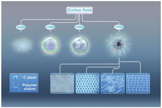
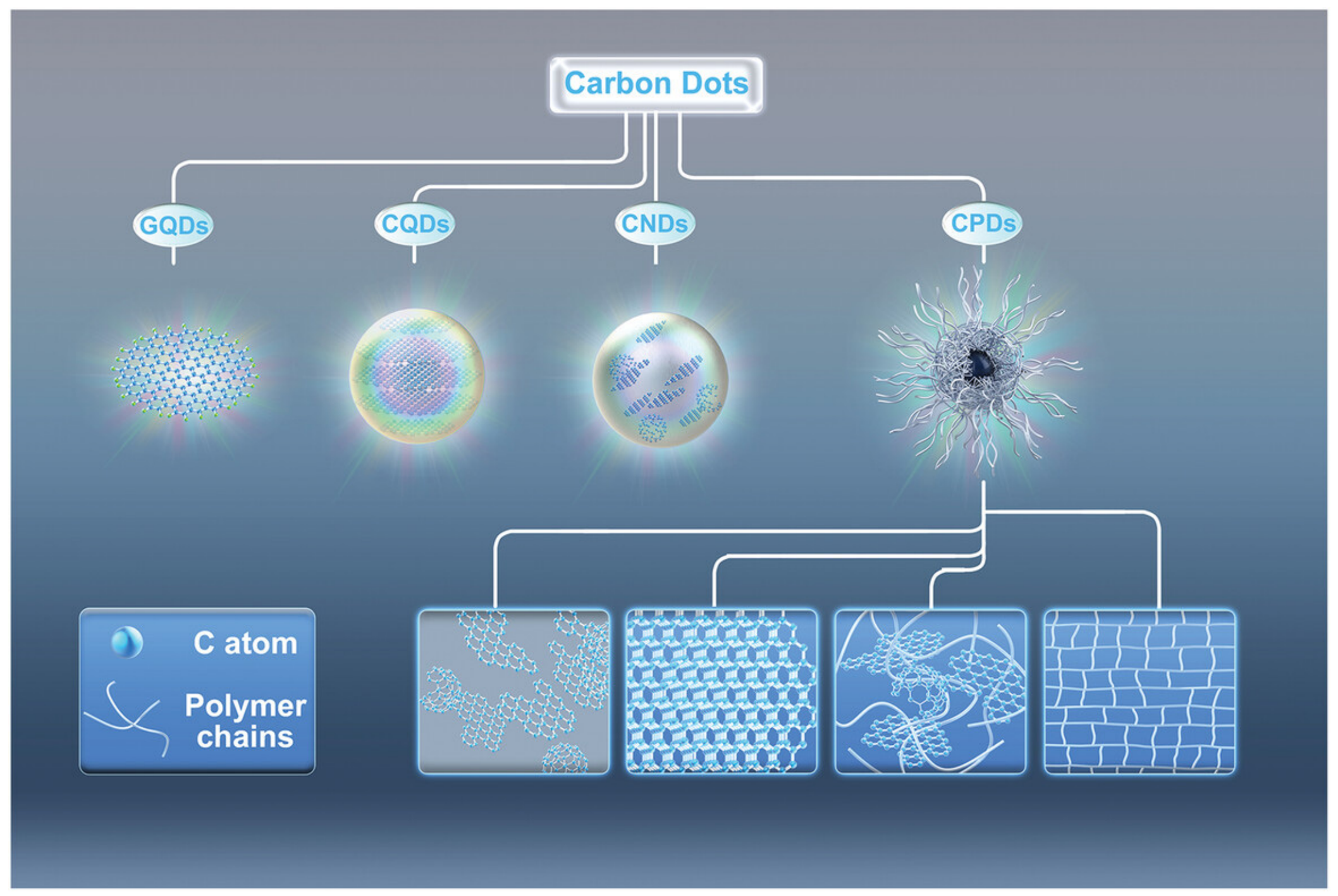
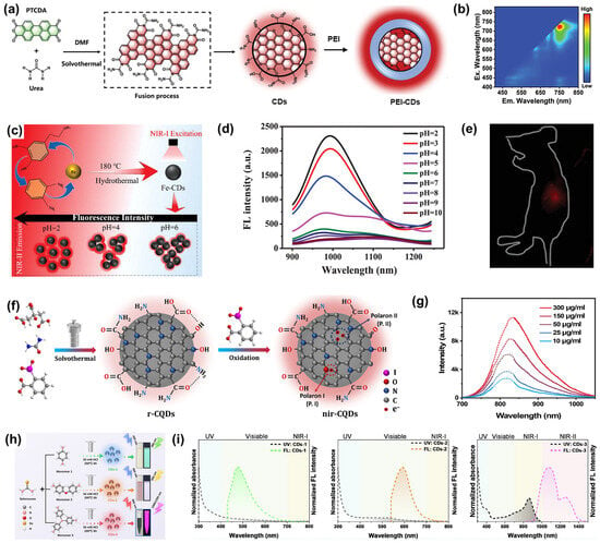
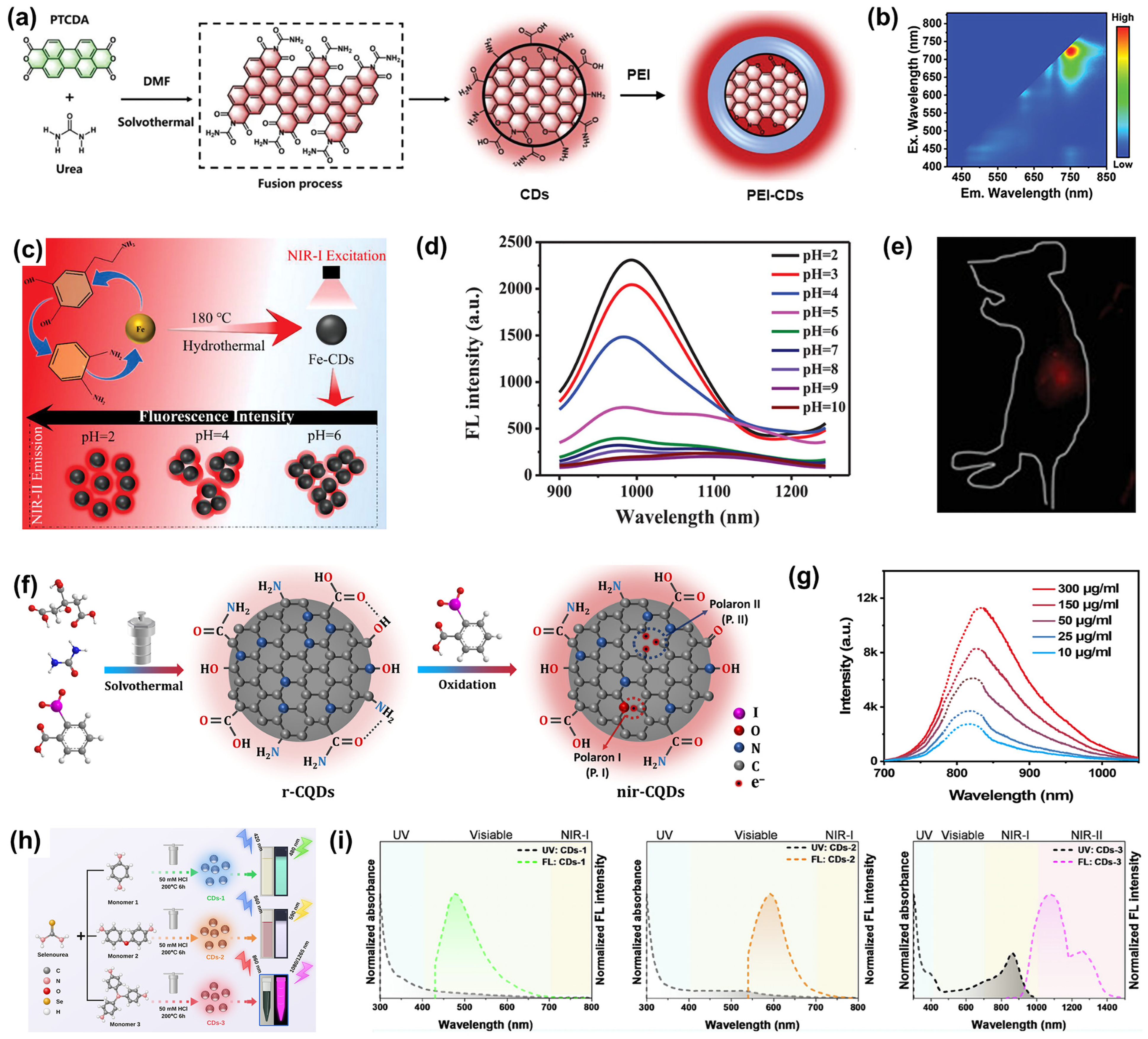
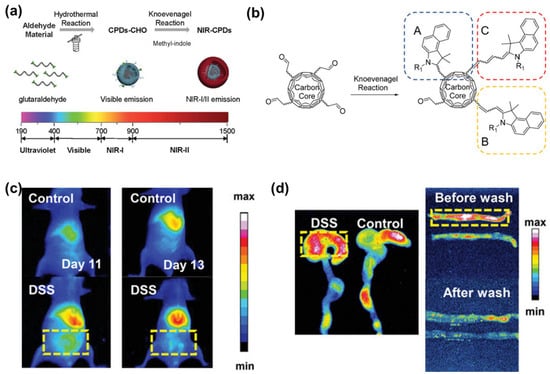
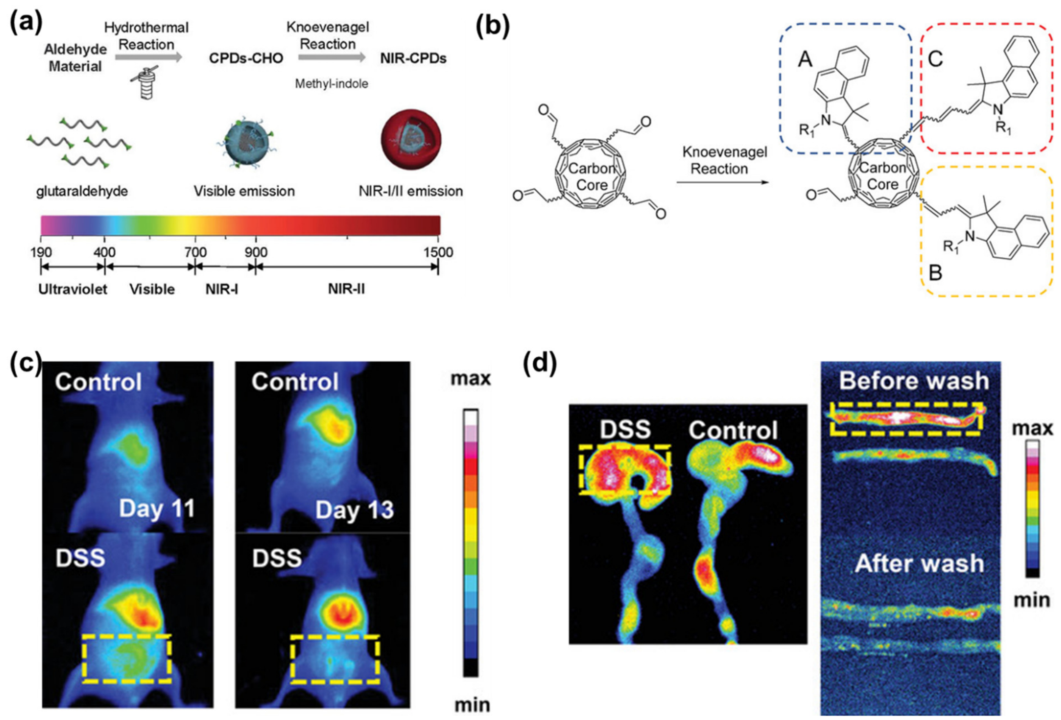
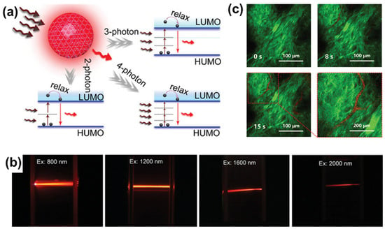
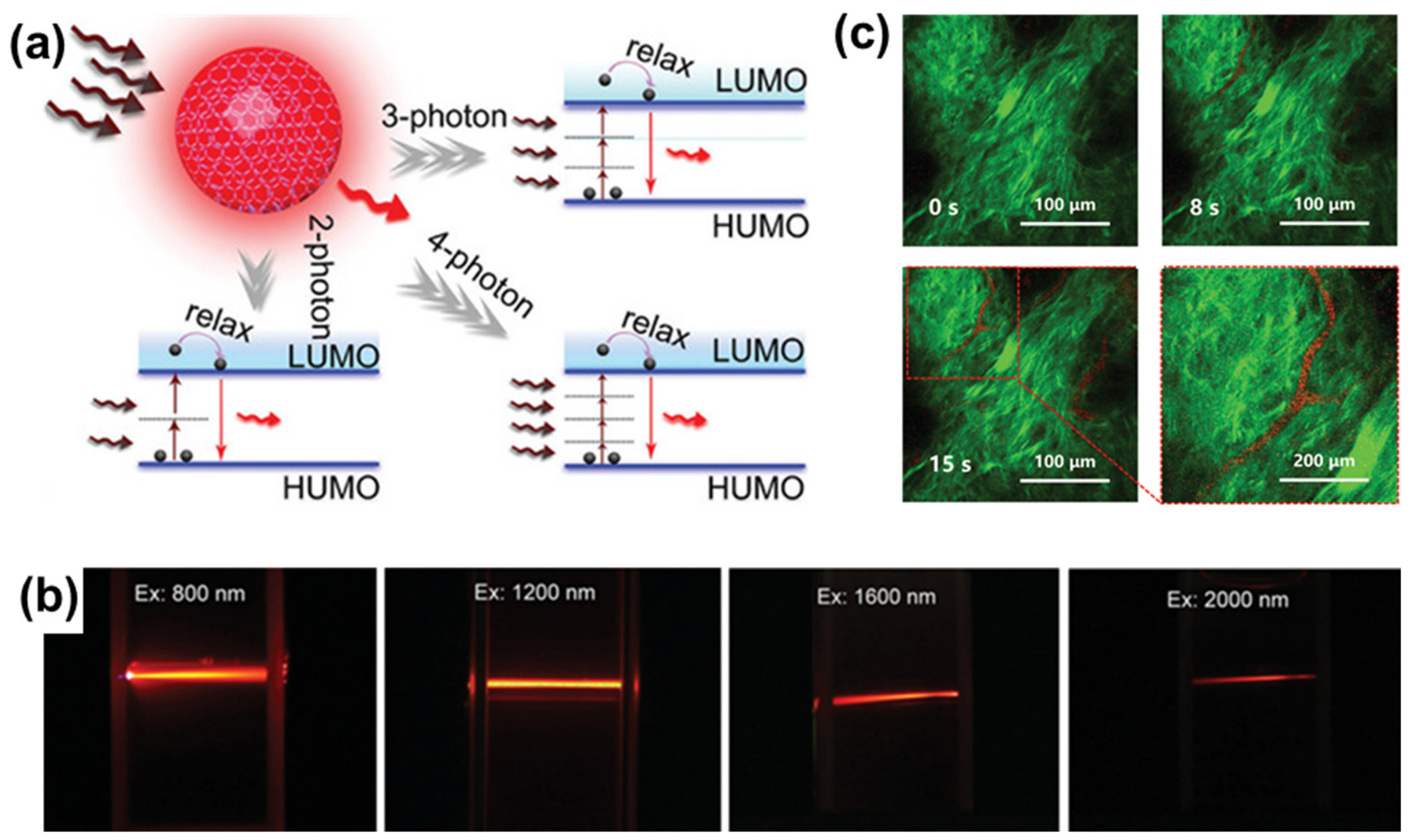
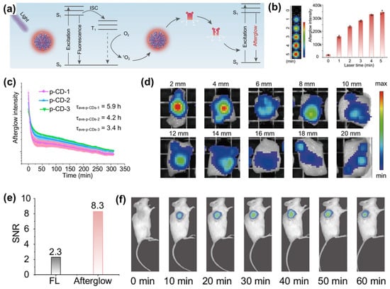
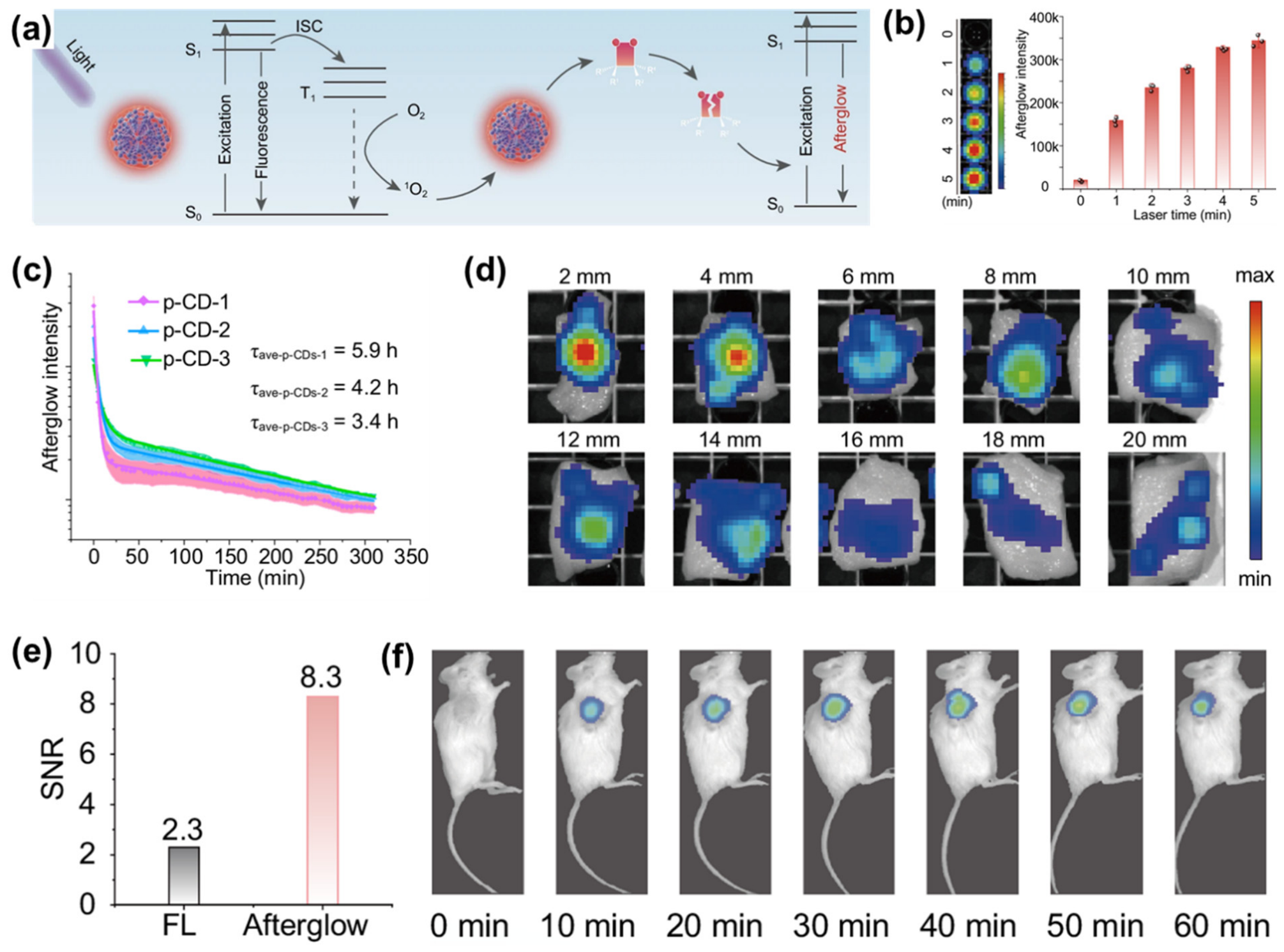
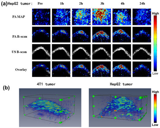
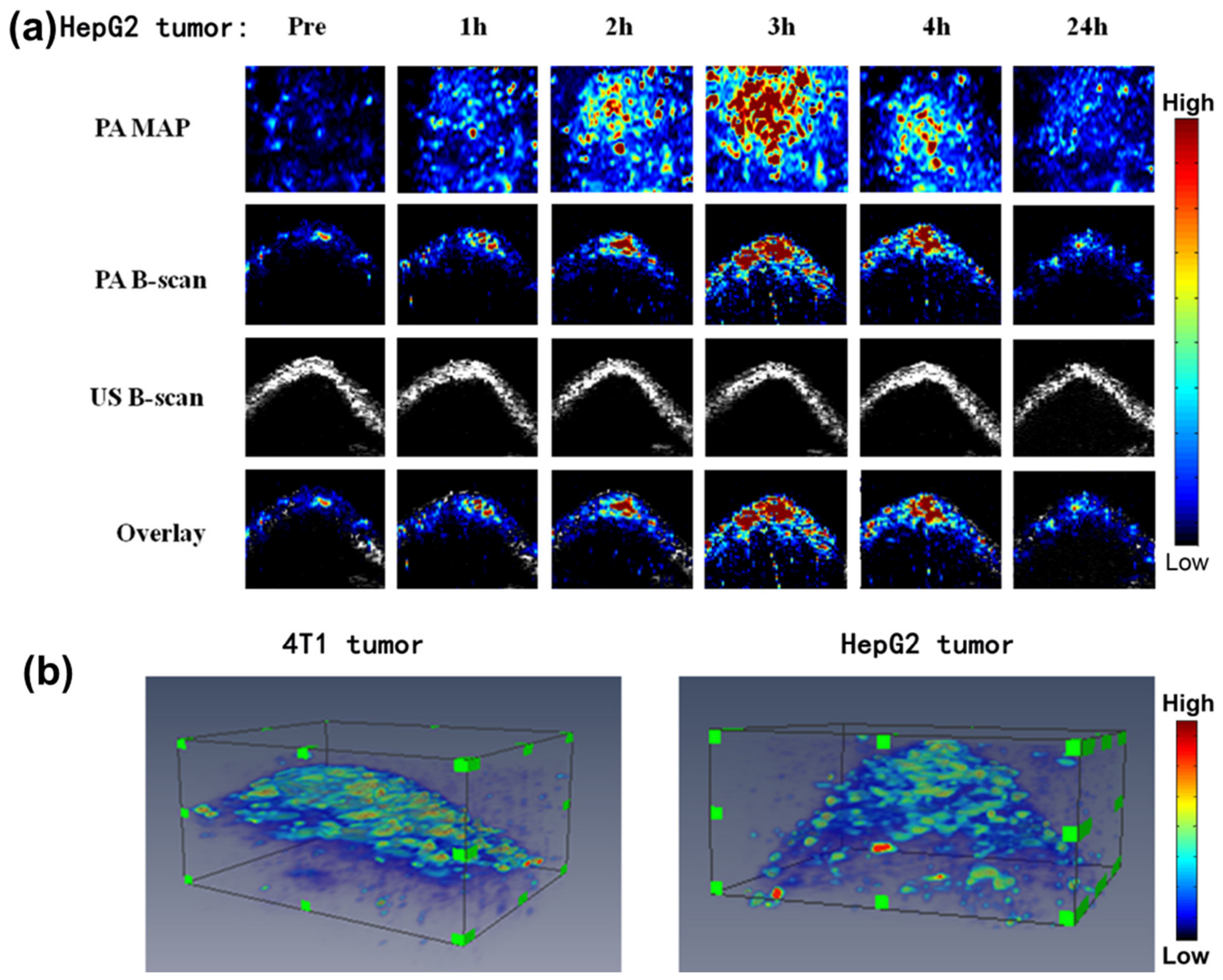
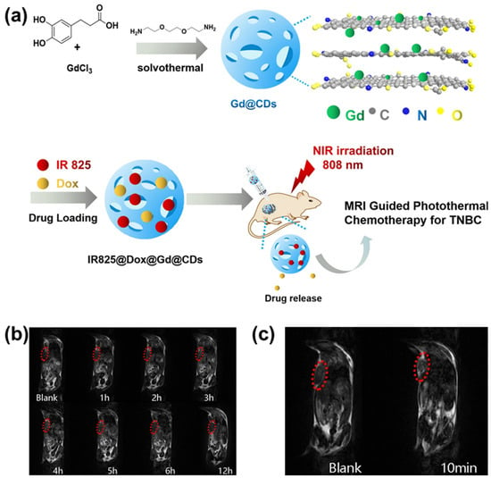
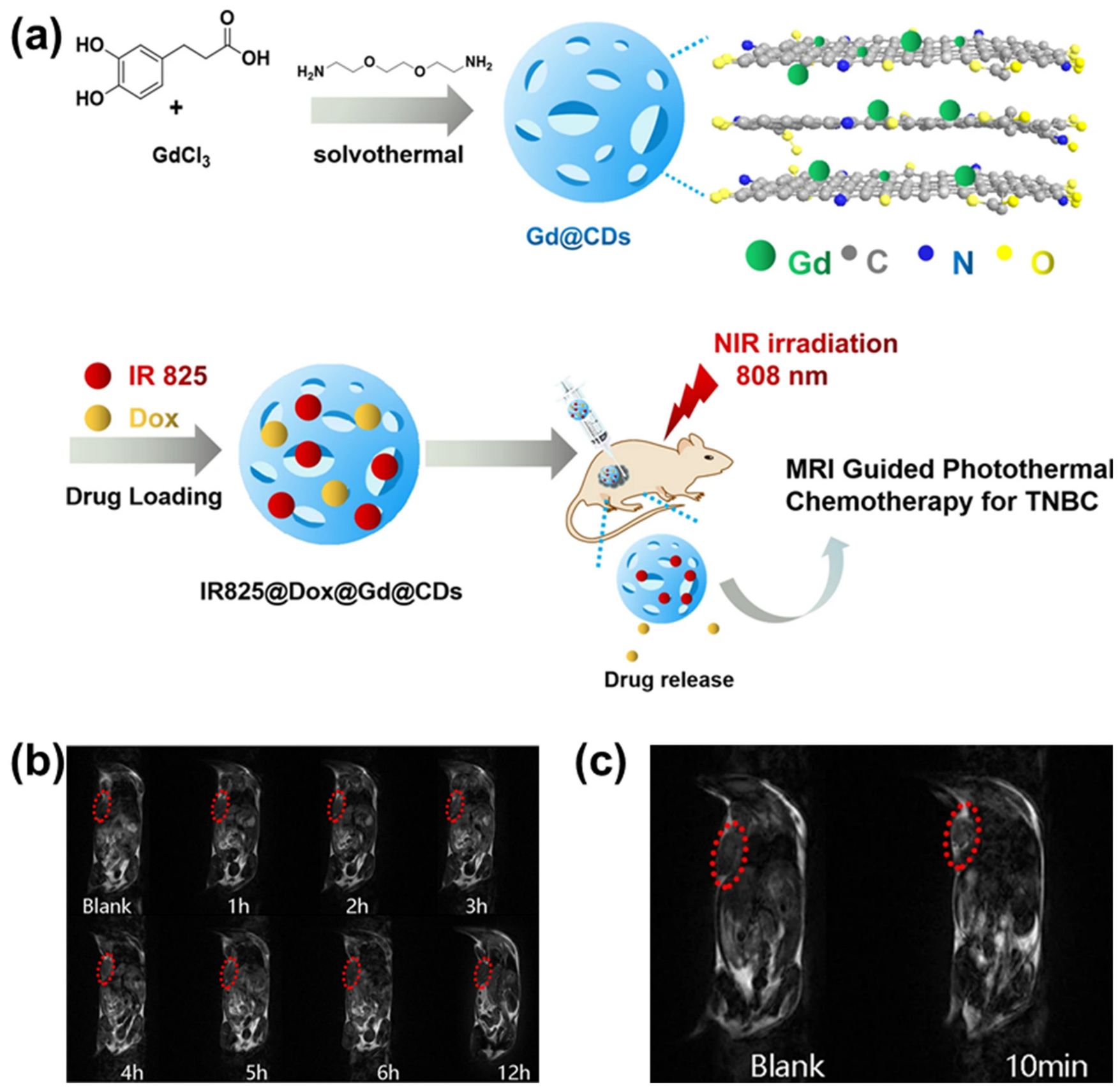
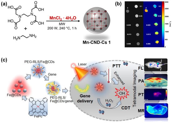
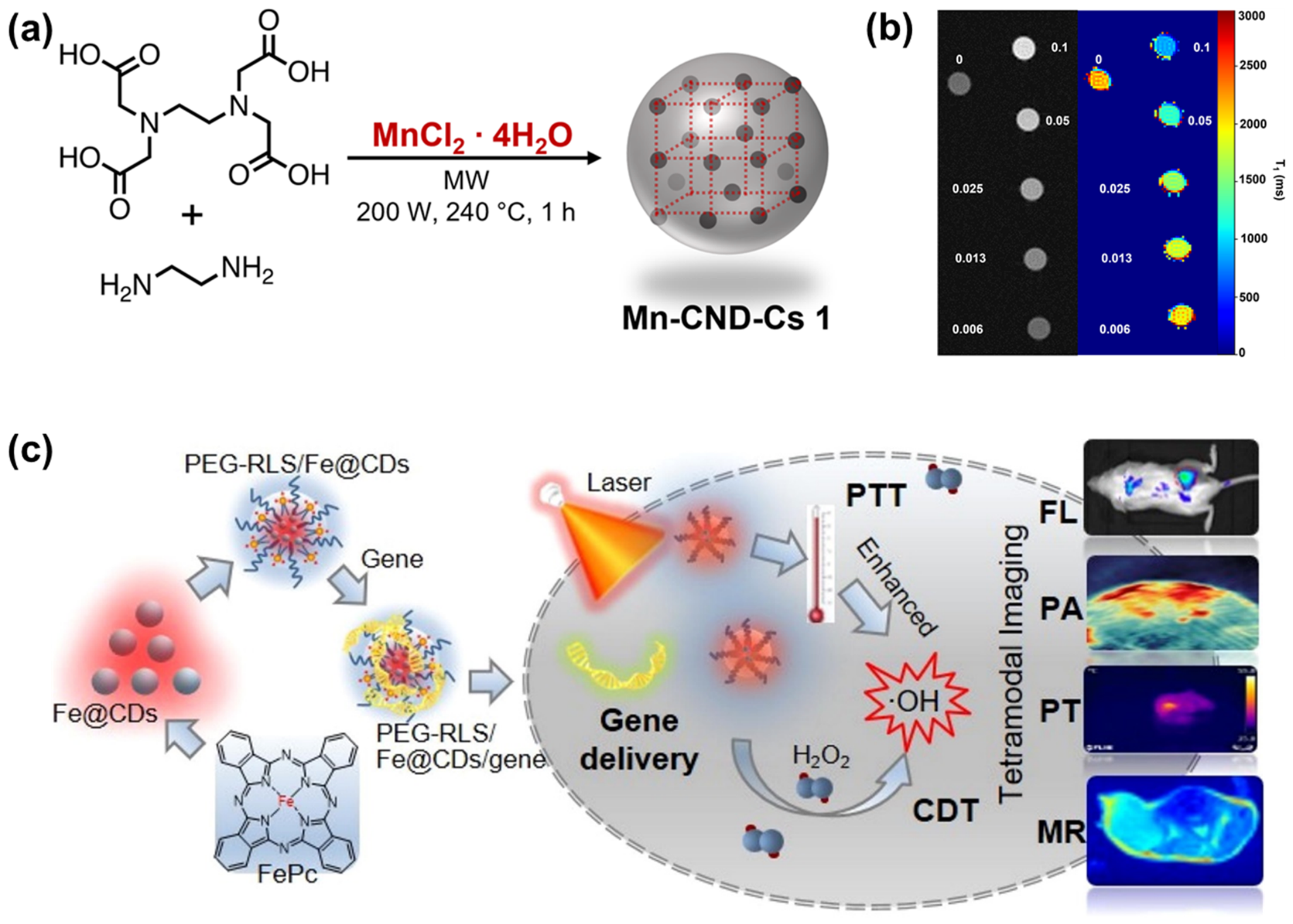
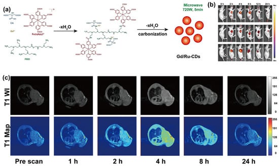
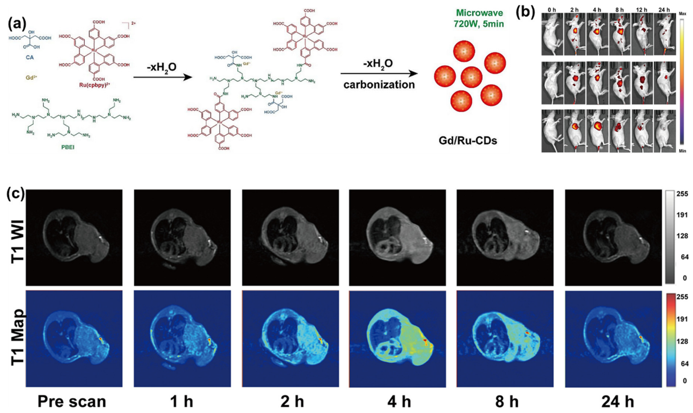
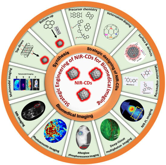
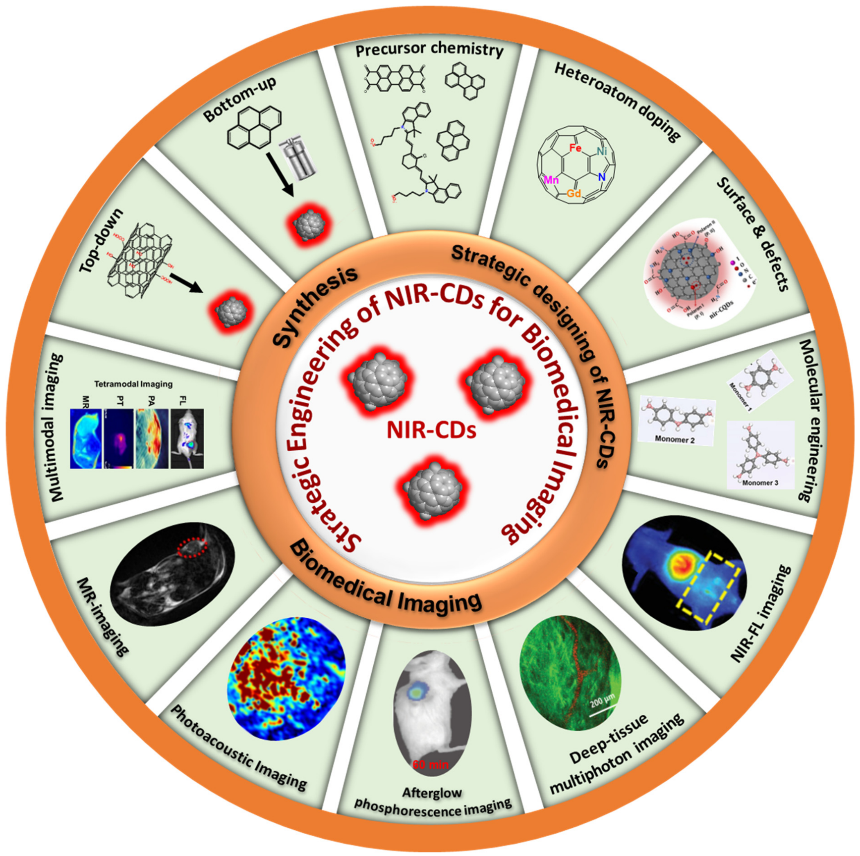
Disclaimer/Publisher’s Note: The statements, opinions and data contained in all publications are solely those of the individual author(s) and contributor(s) and not of MDPI and/or the editor(s). MDPI and/or the editor(s) disclaim responsibility for any injury to people or property resulting from any ideas, methods, instructions or products referred to in the content. |
© 2026 by the authors. Licensee MDPI, Basel, Switzerland. This article is an open access article distributed under the terms and conditions of the Creative Commons Attribution (CC BY) license.