


Abstract
Background: The purpose of this study was to determine whether magnetic resonance imaging findings are accurate and can be confirmed surgically. Methods: Surgical correlation of preoperative magnetic resonance imaging findings of trauma to tendons and ligaments of the foot and ankle were retrospectively studied in 28 patients who had undergone surgical repair for tears of the Achilles tendon, posterior tibial tendon, peroneal tendons, collateral ligaments, and other pathology. Results: This study corroborates that magnetic resonance imaging findings are confirmed surgically 83% of the time for tears of tendons and ligaments of the foot and ankle. Larger tendons (Achilles, posterior tibial tendon) have a 94% sensitivity and 6% specificity, respectively. Collateral ligament and high ankle sprains have a 73% sensitivity. There is a lower sensitivity of 57% for peroneus brevis and longus ruptures. Conclusion: This study revealed that peroneal trauma can be an isolated event and in some cases can occur with other traumatic pathology such as collateral ligament tears. (J Am Podiatr Med Assoc 98(5): 370–373, 2008)
Typically, clinical examination and standard film radiographs are all that are needed to determine whether a tendon or ligament has been ruptured in the foot and ankle.[1-4] However, unclear clinical situations exist in which further diagnostic workup is necessary to confirm the practitioner’s suspicion that there may be complete or partial rupture of tendons and ligaments of the foot and ankle. The advent of new software and sequences has substantially increased the role of magnetic resonance imaging (MRI) in determining foot and ankle pathology. Although comparison images may be helpful in certain cases, most of the time they are rarely indicated. Positioning of the patient during scanning is critical in providing consistent visualization of the foot and ankle.[4,5] Patients are scanned supine with the ankle in 10° to 20° of plantarflexion, which provides comfort and improved visualization of the collateral ligaments and sinus tarsi. All three orthogonal imaging planes are necessary to clarify ankle and foot pathology. Axial images are probably the most useful in evaluating the ankle. For the peroneal or posterior tibial tendon abnormalities, a mild obliquity provides a more perpendicular view through the tendons as they course about the malleoli of the ankle. In other cases, axial images are prescribed perpendicular to the distal tibia. Axial images provide evaluation of the medial and lateral ligamentous complexes as well as detailed views of every major tendon of the ankle including the Achilles tendon. The tarsal tunnel is also seen on axial views. Coronal images are prescribed perpendicular to the tibiotalar joint, and they provide accurate assessment of the tibiotalar joint and possible osteochondral lesions, as well as the posterior talofibular and calcaneofibular ligaments. Sagittal images are necessary for the assessement of the Achilles tendon and plantar fascia. The sinus tarsi, subtalar joint, and cases suspected of tarsal coalition are best seen in this plane.[5]
Tears of the tendons of the foot and ankle are classified as type 1 or partial tears with tendon enlargement and increased signal intensity on MRI. Type 2 tears are also partial tears with diminished width of the tendon at the site of the tear. Type 3 tears are complete tears. In type 1 tendon tears, there is often enlargement and the tendon appears to be edematous at the site of the suspected injury. The posterior tibial tendon is normally twice the size of the adjacent flexor digitorum longus tendon; but, with a type 1 tear the posterior tibial tendon can be up to five times the size of the flexor digitorum longus.[5-9] Other medial ankle tendons are much less frequently detected. The flexor hallucis longus is the next more commonly injured tendon and is typically caused by forced plantarflexion such as that experienced by ballerinas. These injuries occur at the level of the medial malleolus and are often difficult to detect clinically. The lateral aspect of the foot and ankle —the peroneal tendons—are sometimes injured during foot and ankle sprains. The peroneus brevis is more frequently injured than the longus. A common injury pattern is the split of the peroneus brevis following forced dorsiflexion that drives the peroneus longus into the brevis, splitting the brevis, with the longus preventing reapposition of the split halves. Complete tears of the peroneus longus are uncommon, but when detected they are found beneath the cuboid coursing to its insertion medially.[10] If peroneal subluxation is suspected, often there is soft-tissue edema and thickening of the peroneal retinaculum seen on MRI.[4-9,11-14] Larger tendons of the foot and ankle such as the anterior tibial tendon and Achilles tendon are frequently injured, and especially with middle-aged athletes, the Achilles tendon is commonly ruptured.[2] Tears of the Achilles are seen as the presence of fluid signal intensity within the tendon and may indicate partial or longitudinal tears.[5,6]
Lateral collateral ligaments of the ankle are more commonly injured than the deltoid ligament. Inversion injuries more commonly occur than eversion type injuries. The deltoid ligament is thicker and stronger than the collateral ligaments. High ankle sprain involves the syndesmotic ligments joining the distal tibia and fibula.[1,3] Ligamentous injures of the ankle are seen on MRI as edema, abnormal laxity, or discontinuity within the affected ligament or soft-tissue thickening and edema around the ligament in cases of partial tears. If the calcaneofibular ligament is suspected to be ruptured, the axial images are often key to accurate diagnosis of injury to this ligament seen coursing deep to the peroneal tendons. A deltoid sprain or tear is seen as a thickened and edematous ligament. The most serious deltoid injuries are usually present with other ankle pathologies such as fractures and high sprains or tears.[15] Lastly, in cases of sinus tarsi syndrome there is soft-tissue thickening and edema within the sinus tarsi, often obliterating the ligamentous anatomy.[5,6,8]
Recently, Lamm et al[4] found that MRI proved to be 83% sensitive and 75% specific in predicting peroneus brevis tears. Sensitivity was defined as the percentage of correct MRI findings of a peroneus brevis tear among the surgically confirmed cases. Specificity was defined as the percentage of incorrect MRI findings of a peroneus brevis tear among the surgically confirmed cases. Rosenburg et al[9] found that MRI was the most accurate in identifying posterior tibial tendon tears and was confirmed surgically as compared to computed tomography.[9] Rademaker et al[11] found 100% accuracy in identifying peroneus longus tears in nine patients. Radiologists suggest that MRI is superior to other diagnostic modalities such as computed tomography scans and nuclear scans for various traumas, infections, and soft-tissue tumors.[5,6,8] To prove or disprove these views, this study was undertaken to determine how accurate MRI is when suspected trauma to tendons and ligaments of the foot and ankle had occurred and surgery was done to repair any tears.
Methods
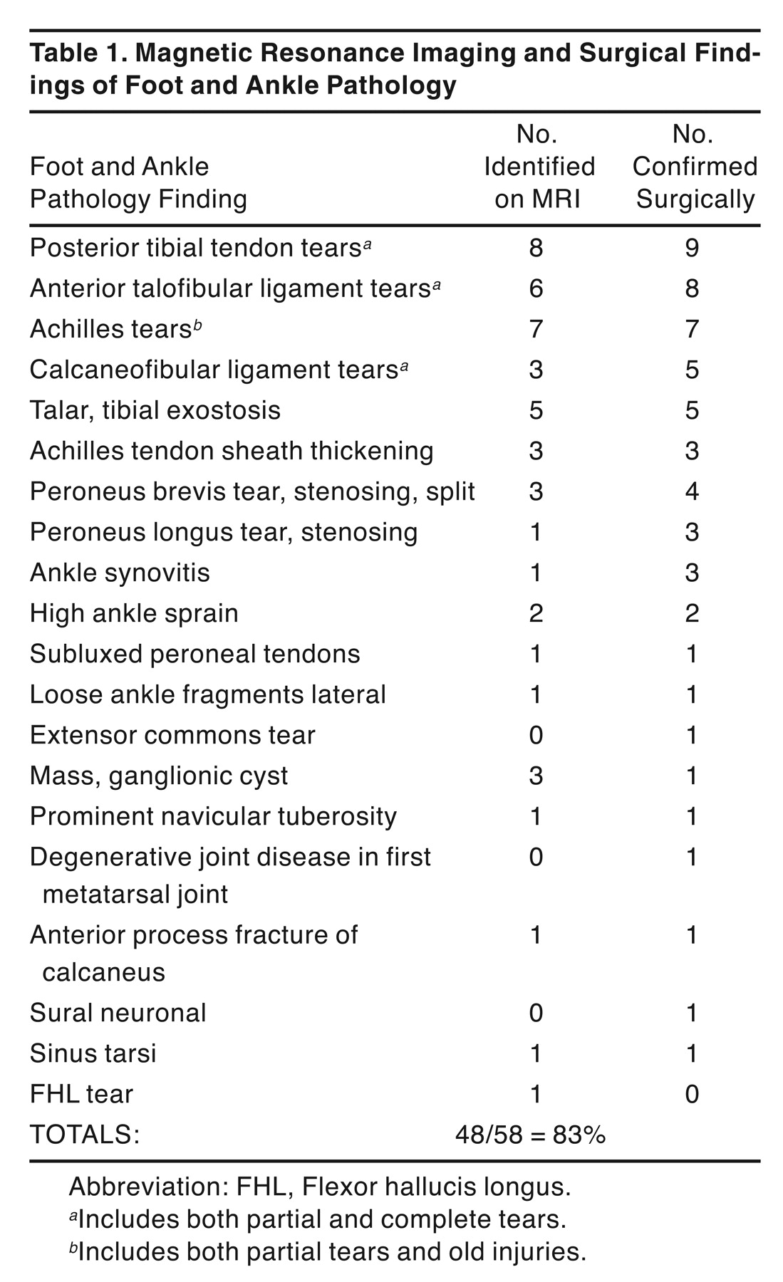
Twenty-eight patients who had MRI performed preoperatively and had undergone surgery for tendon repair and ligamentous repair of the foot and ankle were included in this study. Six radiologists interpreted the 28 magnetic resonance images. They were told prior to their interpretation which foot and ankle was injured, the suspected sites of injury, and whether the injury was acute or a chronic condition. All of the surgical procedures were performed on the 28 patients by the same surgeon (G.T.K.). Magnetic resonance images and operative reports that noted the patient’s age, sex, MRI interpretations, and operative findings from the patient’s charts were reviewed. Surgical findings included any other pathology not noted on MRI. Table 1 lists the specific pathology and how many were found preoperatively by MRI and how many were confirmed surgically. The accuracy or sensitivity was calculated as a percentage of MRI interpretation correlated with surgical confirmation. The specificity was calculated as a percentage of errors of MRI interpretations confirmed surgically.
Results
The most common pathology found in the 28 patients in this study was confirmed posterior tibial partial tears; eight were found by MRI and nine were confirmed surgically. Six anterior talofibular ligament tears were found by MRI and eight anterior talofibular ligament tears were confirmed surgically. Three calcaneofibular ligament tears were found on MRI and two more were confirmed surgically. Out of 13 collateral ligament tears, nine (70%) were found on MRI and two more were confirmed surgically. Two high sprains were found on MRI and confirmed surgically. The diagnosis of Achilles tendon partial tears were 100% correct on MRI and surgical confirmation; all seven tears were found on MRI and were confirmed surgically. Three Achilles tendon sheath stenoses were found on MRI and all were confirmed surgically. One peroneal longus tendon was detected by MRI and two others were detected surgically. Three peroneus brevis tendon injuries were detected by MRI and four were found surgically.
In the case of one patient, an MRI was interpreted as a 6-cm peroneus brevis split with an intact peroneal longus tendon. Surgery confirmed the opposite; the peroneal brevis was intact and the peroneal longus was ruptured. Sural neuroma was found surgically but not detected by MRI. A ganglionic cyst was identified on MRI but not confirmed surgically; the cyst was revealed to be fibrous adipose tissue from an old injury to the foot. Degenerative changes to the first metatarsophalangeal joint were interpreted on MRI but were found surgically during correction of a bunion deformity. In another case, an MRI was interpreted as a partial tear of the flexor hallucis longus tendon, but this was not confirmed surgically. However, noting the pathology in Table 1, the accuracy rate of MRI findings confirmed surgically is 83% or 48 out of 58.
Discussion
The results of this study indicate that MRI is a valuable diagnostic tool and is accurate in detecting traumatic injury to larger tendons and collateral ligaments of the foot and ankle. In this study, tears of the posterior tibial tendon and Achilles tendon were very accurate with 100% of Achilles tendon tears identified on MRI and only one posterior tibial tendon not identified by MRI. Ruptures of the collateral ligaments were not as easily detected; only 70% were found on MRI. Stress radiographs continue to be the recommended diagnostic test for determining ruptures of the collateral ligaments. In this study, despite having had six radiologists reading 28 MRIs, some radiologists focused their review on the patients’ area of chief complaint whereas others listed every finding of the foot and ankle, both normal and pathologic. This might be interpreted as the radiologist “over reading” the MRI. As for surgical intervention, MRI was very useful in planning procedures and preoperative surgical approach. As with any diagnostic tool available today, no one technology is 100% accurate and reliable. This study does not confirm that MRI is superior to other diagnostic modalities. However, when the diagnosis is unclear and when the physician has a suspicion that there may be more pathology, MRI can be helpful in clarifying foot and ankle conditions such as in partial tears of tendons and ligaments. Additional information from MRIs may shift treatment from surgical to conservative depending on the patient’s level of activity and type of work. The most difficult trauma to identify consistently by MRI are traumatic injuries to the peroneus brevis and longus. The radiologists were unable to determine the presence of tears in three of seven cases that were found surgically but not read on MRI. The peroneus longus tear was found in one case by MRI and surgically in two other patients. These findings might be due to low-lying muscle belly of the peroneus brevis tendon, or a slip of the peroneus tertius found in the same anatomic location, which makes interpretation of peroneus longus tears difficult. There was one case in which the MRI interpretation was read as a 6-cm split of the peroneus brevis and normal peroneus longus, which was found to be the opposite upon surgery. These findings contradict the findings of Rademaker et al[11] of 100% sensitivity for peroneus longus ruptures. However, with regard to posterior tibial tears, the results of this study are similar to that of Rosenburg et al.[9] The present study did not compare MRIs with computed tomography scans as did Rosenburg.
Conclusion
Magnetic resonance imaging is very accurate in detecting large tendon pathology such as partial to complete tears of the Achilles tendon (100%), and posterior tibial tendons (89%). Magnetic resonance imaging is accurate in detecting both partial and complete collateral ligament ruptures in 70% of the cases, although the use of stress x-rays as the primary diagnostic tool to determine collateral ligament injury is recommended. Overall, regarding traumatic events to the tendons and ligaments of the foot and ankle, the accuracy rate is 83%, which coincides with the findings of Lamm et al[4] of 83% sensitivity. The most difficult pathology to identify appears to be the peroneal injuries, with a 57% sensitivity. The accuracy was below what Lamm et al[4] and Rademaker et al[11] found in their study of peroneal injuries. The fact that there were six radiologists who reviewed 28 magnetic resonance images is a variable and may have influenced the accuracy percentage. As better imaging technology develops and better positioning becomes standardized as well as better training for radiologists in interpretation of magnetic resonance images of foot and ankle pathology, we can expect further improvement in the accuracy of MRI in detecting traumatic events to tendons and ligaments of the foot and ankle, including smaller tendons that insert into the forefoot. This study supports the notion that MRI is a valuable diagnostic tool and as technology and accuracy improve, MRIs may prove to be superior to other diagnostic modalities for trauma of the foot and ankle. This study disproves that MRIs for trauma of the foot and ankle are unnecessary and yield little to no valuable information. The discrepancy in sensitivity for peroneal injuries and tears between this study and others is probably due to several factors. First, interpretation of the MRIs in this study by the six radiologists may have played a role in the low sensitivity findings compared to other studies due to differences in the training and experience of the radiologists. Second, positioning of the patient may have also played a role in why some peroneal tears were found and others were not. The positioning may have been caused by the patient as they involuntarily moved during the MRI. Lastly, anatomical differences of the patients’ foot and ankle make it impossible to position the patient to capture the pathology in every case.

Table 1
.
Magnetic Resonance Imaging and Surgical Findings of Foot and Ankle Pathology
Financial Disclosure: None reported. | |
Conflict of Interest: None reported. |
References
- Cox HA, Kuwada GT: Transchondral fractures of the talar dome. .Clin Podiatr Med Surg12::667. ,1995. .
- Kuwada GT: An update on repair of Achilles tendon rupture, acute and delayed. .JAPMA89::302. ,1999. .
- Kuwada GT: Current concepts in the diagnosis and treatment of ankle sprains. .Clin Podiatr Med Surg12::653. ,1995. .
- Lamm BM, Myers DT, Dombek M, et al: Magnetic resonance imaging and surgical correlation of peroneus brevis tears. .J Foot Ankle Surg43::30. ,2004. .
- Awh MH, Stadnick ME: MR imaging assumes primary role in evaluation of foot and ankle. .Diagnostic Imaging12::8. ,2001. .
- Anzilotti K Jr, Schweitzer ME, Hecht P, et al: Effect of foot and ankle MR imaging on clinical decision making. .Radiology201::515. ,1996. .
- Rosenburg SZ, Bencardino J, Astion D, et al: MRI features of chronic injuries of the superior peroneal retinaculum. .Am J Roentgenol181::1551. ,2003. .
- Schneck CD, Mesgarzadeh M, Bonakdarpour A: MR imaging of the most commonly injured ankle ligaments. Part II: ligament injuries. .Radiology184::507. ,1992. .
- Rosenburg ZS, Cheung Y, Jahss MH: Rupture of the posterior tibial tendon: CT and MR imaging with surgical correlation. .Radiology169::229. ,1988. .
- Khoury NJ, el-Khoury GY, Saltzuran CL, et al: Peroneus longus and brevis tendon tears: MR imaging evaluation. .Radiology200::833. ,1996. .
- Rademaker J, Rosenburg ZS, Delfaut EM: Tear of the peroneus longus tendon: MR imaging features in nine patients. .Radiology214::700. ,2000. .
- Rosenburg ZS, Beltran J, Cheung YY, et al: MR features of longitudinal tears of the peroneus brevis tendon. .Am J Roentgenol168::141. ,1997. .
- Schweitzer ME, Eid ME, Deely D, et al: Using MR imaging to differentiate peroneal splits from other peroneal disorders. .Am J Roentgenol168::129. ,1997. .
- Major NM, Helms CA, Fritz RC, et al: MR imaging appearance of longitudinal split tears of the peroneus. .Foot Ankle Int21::514. ,2000. .
- Schuberth JM, Collman DR, Rush SM, et al: Deltoid ligament integrity in lateral malleolar fractures: a comparative analysis of arthroscopic and radiographic assessments. .J Foot Ankle Surg43::20. ,2004. .
© 2008 American Podiatric Medical Association