


Abstract
Hematoma refers to the collection or extravasation of blood, usually clotted, in a closed tissue space. It is caused by leakage from local vessels damaged by blunt trauma, local injury, or surgical dissection. In the postoperative phase, a hematoma often results in edema, pain, wound dehiscence, infection, and scarring of the surgical wound. We describe a 44-year-old woman who developed severe complications, including hematoma, abscess, failure of internal fixation, and loss of soft-tissue structures, after hallux abducto valgus surgery. Hospitalization was required for infection control, soft-tissue coverage through negative-pressure wound therapy, and first metatarsophalangeal joint stabilization through external fixation. Early recognition of the signs of infection and hematoma can help decrease the incidence of postoperative complications.
The management of operative wounds in most clean and clean-contaminated surgical procedures is important in preventing complications. This process begins preoperatively and continues through the intraoperative and postoperative phases of surgical case management. Preventing complications, most notably infection, is a primary goal. The identification of potential risk factors in the preoperative phase, implementation of operative techniques that minimize injury to tissues, and preserving local physiology have been proved to decrease the occurrence of postoperative complications [1]. During the first 24 hours of a postoperative hematoma, clinical symptoms are observed that consist of pain, swelling, and possible dehiscence, with little histologic change observed. This is followed by migration of large numbers of polymorphonuclear leukocytes and mononuclear phagocytes into the site to begin cleanup. Infection is the result of increased bacterial contamination, multiplied by the virulence of an organism relative to the resistance of the host [2,3]. Recent data accumulated by the Centers for Disease Control and Prevention indicate that the postoperative rate of infection and hematoma for clean and uncontaminated surgery is 2.5% [4]. The goal for elective surgery is a rate lower than 2.5%.
This case report describes a patient in whom elective bunion surgery resulted in significant postoperative complications. Negative-pressure wound therapy and external fixation were used to restore stability to the first metatarsophalangeal joint.
Case Report
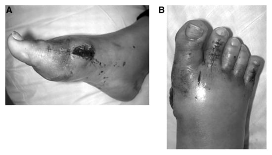
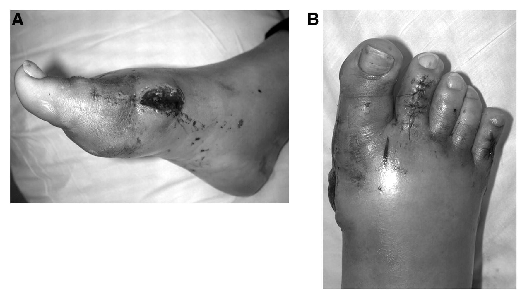
A 44-year-old woman presented to the emergency department at Staten Island University Hospital after right-foot hallux abducto valgus surgery performed at another institution 4 days earlier. Significant erythema and edema of the right foot were noted, and the patient complained of pain out of proportion to the surgical procedure. Simple sutures were intact in digits 2 and 5 as a result of hammer-toe correction, which displayed an adequate postoperative course with no signs of infection. Mild necrosis of the wound edges, with dehiscence, sanguineous drainage, and purulent discharge, was noted in the medial hallux (Figure 1). On the basis of findings from physical examination and the patient history, a diagnosis was made of postoperative hematoma with abscess of the first metatarsophalangeal joint. The patient was admitted to the hospital for intravenous antibiotic drug administration, evacuation of the hematoma, and abscess drainage.
Figure 1.
Wound dehiscence (A) and erythema and edema (B) after correction of hallux abducto valgus.
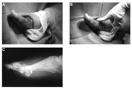
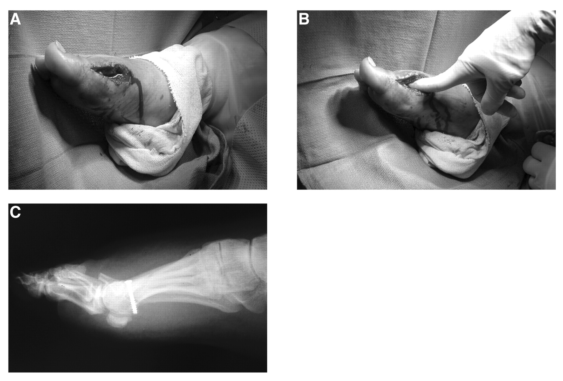
On completion of the preoperative workup, the patient was taken to the operating room. An incision was made along the original surgical site extending 4.0 cm distally, starting from the end of the wound dehiscence site and ending just distal to the first metatarsophalangeal joint (Figure 2A). A dorsal and plantar pocket of hematoma and abscess just proximal to the first metatarsal head was noted. This area was thoroughly drained and decompressed (Figure 2B). All non-viable tissue, including the necrotic capsule of the first metatarsophalangeal joint, was removed. The surgical site was then flushed with 1,500 mL of sterile normal saline solution with bacitracin and polymyxin. The screw fixating the previous surgical osteotomy was found to be loose (Figure 2C), and it was removed using a periosteal elevator. The wound was packed, SteriStrips (3M Co, St. Paul, Minnesota) were applied, and a Jones compression dressing was applied along with a posterior splint for immobilization. A staged procedure was planned to stabilize the osteotomy site, with application of external fixation on resolution of the soft-tissue infection.
Figure 2.
A, Incision and drainage of abscess; B, evacuation of hematoma and infection; C, loosened hardware after correction of hallux abducto valgus.
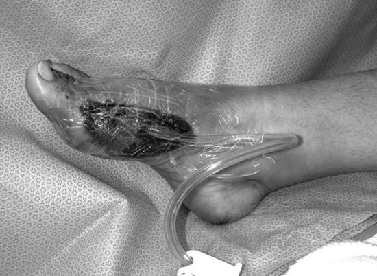
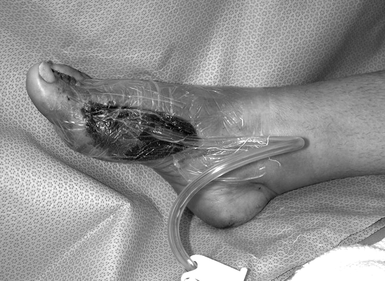
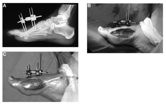
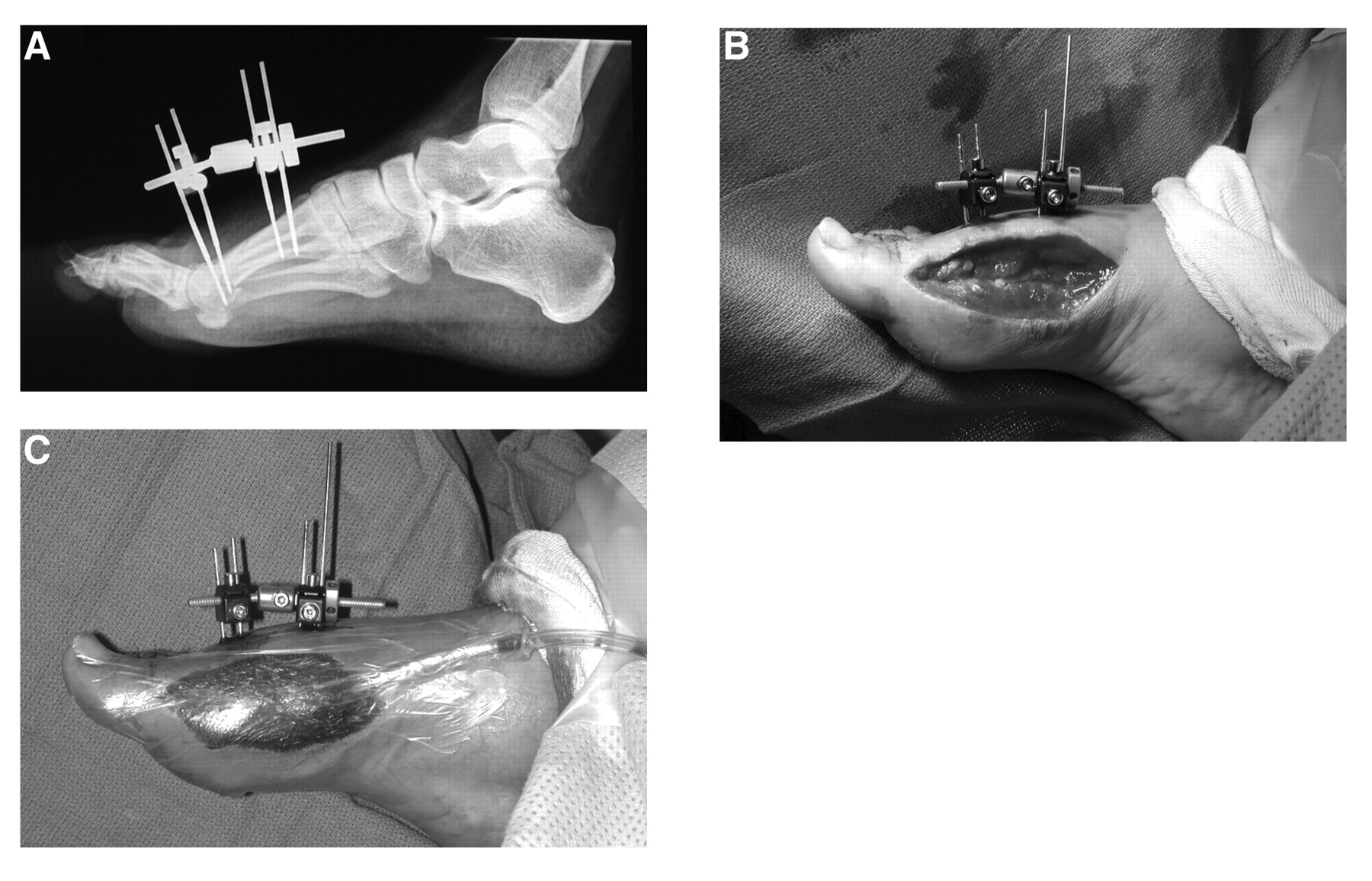
On postoperative day 1, negative-pressure wound therapy (KCI, San Antonio, Texas) was used at 125 mm Hg of continuous therapy in an effort to obtain soft-tissue coverage. The dressing remained in place for 72 hours (Figure 3). On postoperative day 3, the patient returned to the operating room for stabilization of the osteotomy site with external fixation. The DFS Mini-Fixator (EBI Medical, Parsippany, New Jersey) was used with two threaded wires placed in the metatarsal head and two in the proximal portion of the metatarsal shaft (Figure 4 A and B). The negative-pressure wound therapy dressing was reapplied, with a posterior splint for immobilization (Figure 4C). The patient remained hospitalized for 7 days for negative-pressure wound therapy and intravenous antibiotic drug therapy. She was subsequently discharged with vancomycin therapy for oxacillin-resistant Staphylococcus aureus, with local wound care and an equalizer brace for ambulation.
Figure 3.
Negative-pressure wound therapy postoperatively.
Figure 4.
A and B, Radiographic and intraoperative views of osteotomy stabilization with external fixation; C, negative-pressure wound therapy with external fixation postoperatively.
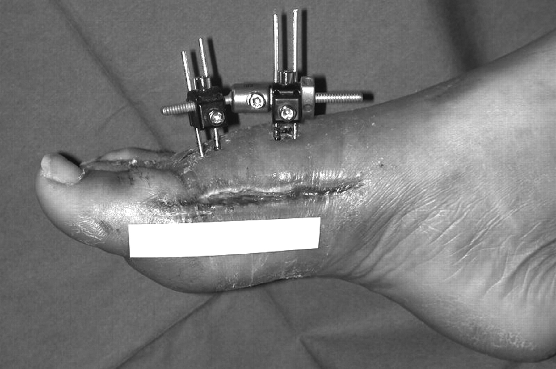
Removal of the external fixation was completed as an ambulatory procedure 6 weeks after the patient was discharged (Figure 5). The 2-week postoperative visit after fixation removal revealed significant reduction in range of motion of the first metatarsophalangeal joint: 5° of plantarflexion and 10° of dorsiflexion. The patient was referred to a physical therapist for range-of-motion exercises, was instructed to obtain a Jobst stocking for edema control, and was encouraged to walk in sneakers to allow for motion of the first metatarsophalangeal joint.
Figure 5.
The wound 6 weeks after surgery, before removal of external fixation.
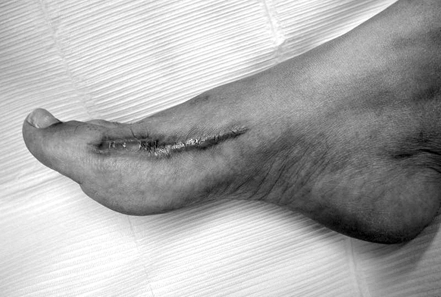
The 3-month follow-up visit revealed a hyperpigmented and slightly hypertrophied scar overlying the medial aspect of the first metatarsophalangeal joint that was asymptomatic (Figure 6). Range of motion after physical therapy improved to 15° to 20° of plantarflexion and 25° to 30° of dorsiflexion. No pain or crepitus on active or passive range of motion was noted. The patient was walking in sneakers without difficulty.
Figure 6.
The wound at 3-month follow-up.
At the 1-year follow-up visit the patient complained of pain emanating from below the second metatarsal head. Radiographs revealed a slight elevatus of the first metatarsal. Treatment included anti-inflammatory medications and custom-made orthoses. The patient returned to her usual daily and recreational activities without further complication.
Discussion
Inadequate attention to hemostasis, extensive tissue disruption, excessive bone bleeding, and patient-dependent factors may all lead to postoperative hematoma. Dead space that has not been closed or drained allows for the accumulation of blood or serum [5]. A higher incidence of hematoma formation is seen in patients with myeloproliferative disorders, polycythemia vera, and coagulation defects. Arteries that are severed and not ligated during tourniquet hemostasis are potential sources of bleeding. Compression bandaging should be used in conjunction with careful anatomical dissection to effectively reduce bleeding and the accumulation of blood in the tissues [6].
Hematoma is a great imitator of early wound infection because the body’s response to intense local inflammation is almost identical to that of infection. Patients experience local pain caused by expansion of the hematoma itself, followed by extravasation of fluid and swelling of tissues as the inflammation is established. Pain that is unresponsive to analgesics points to hematoma, infection, or dressing pressure [7]. The gross tissue edema results from the pressure obstruction of local capillaries, veins, and lymphatic vessels. This edema is usually difficult to eradicate and may result in wound dehiscence or a skin slough. Hematoma formation is also associated with an increased incidence of postsurgical infection [8]. Blood is an excellent medium for bacterial growth, especially when the incubation is provided by soft tissue.
This case report described a postoperative hematoma that led to infection, hardware failure, wound dehiscence, and soft-tissue necrosis. Extensive debridement led to significant soft-tissue deficit and instability of the surgical osteotomy. This necessitated the use of negative-pressure wound therapy for soft-tissue coverage and edema control. Negative-pressure wound therapy is indicated for chronic, acute, traumatic, and dehisced wounds [9]; partial-thickness burns; flaps; and grafts [10]. It has been shown that negative pressure accelerates the development of granulation tissue, resulting in earlier reepithelialization of wounds and ultimately wound closure [11,12]. Stannard et al. [13]. also discuss the use of negative-pressure wound therapy in treating complex traumatic wounds and hematoma, primarily by decreasing interstitial fluid.
External fixation was also used in this case to maintain the alignment of the osteotomy in an infected wound where internal hardware had failed. External fixation is designed to maximize intraoperative flexibility for the stabilization and realignment of small bone fractures [14]. Roukis and Landsman [15] discuss a similar case in which external fixation was used in conjunction with bone cement for salvage of an osteomyelitic first metatarsophalangeal joint. External fixation is a helpful tool for fracture reduction and allows for better care of skin and soft-tissue structures compared with plaster cast immobilization [16].
Conclusion
Identifying potential risk factors in the preoperative phase and preserving local physiology in the operating room are keys to preventing hematoma formation. Early recognition of the signs of infection and hematoma can help decrease the incidence of postoperative complications. Once complications have arisen, the physician must develop an armamentarium of weapons to facilitate wound healing.
Financial Disclosures: None reported. | |
Conflict of Interest: None reported. |
References
- Stapp MD, Taylor RP: “Edema, Hematoma, and Infection,” in McGlamry’s Comprehensive Textbook of Foot and Ankle Surgery, ed by AS Banks, p 1997, Lippincott Williams & Wilkins, Philadelphia. ,2001. .
- Cruse PHE, Ford R: The epidemiology of wound infection: a 10-year prospective study of 62,939 wounds. .Surg Clin North Am60::27. ,1980. .
- Hugar DW: “Management of Infection,” in American College of Foot Surgeons: Complications in Foot Surgery: Prevention and Management, ed by SA Marcus, BH Block, p 494, Williams & Wilkins, Baltimore. ,1984. .
- Culver DH, Horan TC, Gaynes RP, et al: Surgical wound infection rates by wound class, operative procedure, and patient risk index: National Nosocomial Infections Surveillance System. .Am J Med91::152S. ,1991. .
- Miller SJ: Surgical wound drainage system using silicone tubing. .JAPA71::287. ,1981. .
- Hunter RL: Transferrin in disease II: defects in the regulation of transferrin saturation with iron contribute to susceptibility to infection. .Am J Clin Pathol81::748. ,1984. .
- Roth D: “Tissue Injury Due to Extravasated Blood,” in Surgical Bleeding, ed by AW Ulin, SS Gollum, p 71, Mc-Graw-Hill, New York. ,1966. .
- Schwartz S: “Complications,” in Principles of Surgery, 4th Ed, ed by S Schwartz, p 458, McGraw-Hill, New York. ,1984. .
- Cowan KN: Vacuum-assisted wound closure of deep sternal infections in high-risk patients after cardiac surgery. .Ann Thorac Surg80::2205. ,2005. .
- KCI: KCI Wound VAC Therapy: Clinical Guidelines: A Reference Source for Clinicians, KCI, San Antonio, TX, January. 2005. , pp 4–34.
- Joseph E, Hamori CA, Bergman S, et al: A prospective, randomized trial of vacuum-assisted closure versus standard therapy of chronic non-healing wounds. .Wounds12::60. ,2000. .
- Andros G, Armstrong DG, Attinger CE, et al: Consensus statement on negative pressure wound therapy (V.A.C. Therapy) for the management of diabetic foot wounds. .Ostomy Wound Manage (June):1. ,2006. .
- Stannard JP, Robinson JT, Anderson ER, et al: Negative pressure wound therapy to treat hematomas and surgical incisions following high-energy trauma. .J Trauma60::1301. ,2006. .
- EBI Medical: EBI DFS: A Guide to Operative Techniques, EBI Medical, Parsippany, NJ. ,2002. , p 1.
- Roukis TS, Landsman AS: Salvage of the first ray in a diabetic patient with osteomyelitis. .JAPMA94::492. ,2004. .
- Seibert FJ, Fankhauser F, Elliott B, et al: External fixation in trauma of the foot and ankle. .Clin Podiatr Med Surg20::159. ,2003. .
© 2007 American Podiatric Medical Association