


Abstract
We describe a patient with a giant cell tumor in the talar head and neck of the left foot who was diagnosed as having osteochondritis dissecans and treated with arthroscopic drilling in this same location 3 years earlier. Giant cell tumors can be confused with several conditions, including giant cell reparative granulomas, brown tumors, and aneurysmal bone cysts. Giant cell tumors of bone typically occur in the epiphysis of long bones, including the distal femur and proximal tibia. They are uncommonly found in the small bones of the foot or ankle, and talar involvement is rare. Despite this rarity, the radiographic appearance and clinical signs of talar lesions should be considered in the differential diagnosis of nontraumatic conditions in the foot. (J Am Podiatr Med Assoc 97(3): 225–228, 2007)
Most giant cell tumors of bone occur in the long bones, and in skeletally mature patients, almost all of these tumors are found in the articular end of the bone. Most are located around the knee, but they can also be seen in the skull, patella, ribs, and ischium.[1] The tumor occurs more commonly in women than in men, and peak incidence is usually between the second and fourth decades of life. Giant cell tumors account for approximately 2% to 5% of all biopsied primary bone tumors of the foot and ankle.[2] They have rarely been reported in tarsal bones such as the talus.[3] Giant cell tumors can be confused with other conditions, such as giant cell reparative granulomas, brown tumors, and aneurysmal bone cysts.[1] Here we describe a patient who was found to have a giant cell tumor of the talar head and neck after being diagnosed as having osteochondritis dissecans in this same location 3 years earlier.
Case Report
A 31-year-old man was admitted to Gazi University Hospital, Ankara, Turkey, with a history of nontraumatic progressive left ankle pain, swelling, and limitation of range of motion. Approximately 3 years before hospital admission, the patient had presented to another medical center with pain in his left ankle. At that time, magnetic resonance imaging was performed, and the patient received a diagnosis of osteochondritis dissecans; as a result, he underwent arthroscopic debridement and drilling of the left talus. After that operation, the patient’s complaints of pain and swelling persisted, and range of motion and walking ability became severely limited.
Physical examination revealed pain and swelling in the left ankle and limited range of motion. Radiologic studies from the patient’s diagnosis and treatment at the other medical center were unavailable, and all images shown here are from treatment and follow-up at Gazi University Hospital. Standard radiographs of the foot and ankle demonstrated a radiolucent lesion associated with expansion of the talar head and neck (Figure 21). Magnetic resonance imaging confirmed an eccentrically located lesion at the talar head and neck that extended into the synovial tissue (Figure 2). On T1-weighted images the lesion was slightly hypointense compared with muscle (Figure 2>). The lesion had minimal and heterogeneous uptake of intravenously administered contrast material (gadolinium–diethylenetriamine pentaacetic acid) and had low intensity on T2-weighted images (Figure 2B and C). There was reactive synovitis in the adjacent soft tissues and bone marrow edema in the adjacent bones. The tibiotalar and talonavicular joints were spared. Routine blood biochemistry test results and blood parathyroid hormone levels were normal.
Anterior exploration of the ankle was performed under general anesthesia. The tibiotalar and talonavicular joints appeared normal. The lesion was located in the talar head and neck. Reactive synovial tissue and tumor were completely removed, and the part of the tumor located in the talus was completely curetted away. Results of intraoperative frozen section were consistent with a diagnosis of giant cell tumor of bone, brown tumor, or giant cell reparative granuloma. Aggressive curettage of the lesion, including electrocauterization and chemical cauterization with hydrogen peroxide, was performed. The cavity was filled with cancellous bone allograft and demineralized bone matrix. After surgery, a below-the-knee posterior splint was placed, and the lower leg was protected for 4 weeks without weightbearing. In the fourth week, gentle unloaded active motion of the ankle and mobilization with crutches were started. Partial weightbearing was started 6 weeks after surgery.
Tumor specimens were routinely processed in paraffin and stained with hematoxylin-eosin. Microscopic examination showed a mass composed of round-oval, slightly spindle-shaped mononucleated cells and widespread multinucleated giant cells between normal bony trabeculae. There were scattered areas of spindling stromal cells, hemorrhage, and necrosis. Mitotic stromal cells were seen, and none of them were atypical (Figure 3).
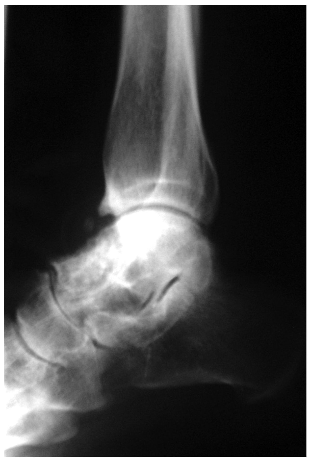
The early postoperative course was uneventful, and 25 months after surgery the patient continued to be asymptomatic, with no pain or restriction of motion. At that time, follow-up radiographs of the ankle showed a well-incorporated and remodeled graft, with no apparent recurrence of the tumor (Figure 4).
Discussion
Giant cell tumors of bone usually occur in the epiphysis of long tubular bones in young adults. The distal portion of the femur and the proximal portion of the tibia are the most common sites of involvement. In a series of 308 giant cell tumors, O’Keefe et al [4] found foot and ankle involvement in only 12 cases.
The differential diagnosis of giant cell tumor includes aneurysmal bone cyst, malignant fibrous histiocytoma, nonossifying fibroma, chondroblastoma, osteoblastoma, and osteosarcoma. However, giant cell tumors are most frequently confused with giant cell reparative granulomas or brown tumors. Histologically, the characteristic feature of giant cell tumor is that giant cell nuclei appear identical to stromal cell nuclei.[5]
Treatment of giant cell tumor in bone is surgical, with wide or marginal excision and curettage with bone grafting or reconstruction with polymethyl methacrylate. Segmental resection has been recommended in cases of pathologic fracture, in expandable bones, and for some advanced lesions. However, intralesional procedures, such as curettage, may be used when wide resection is not feasible.[6] Malawer et al [7] argue that talar localization represents a distinct clinical and radiologic entity with a less aggressive natural history than that seen in the more common sites. Talectomy is not indicated as a primary treatment because of the risk of severe functional impairment.
A multicenter study [8] of 677 giant cell tumors revealed unacceptable recurrence rates, which were as high as 45% with curettage and bone grafting alone. Shimizu et al [9] recently emphasized that giant cell tumors have high potential for recurrence and must be followed closely. To decrease tumor recurrence rates, curettage has been augmented with the use of phenol, hydrogen peroxide, ethanol, liquid nitrogen, and electrocauterization of the cavity. However, a recent multicenter study10] found that the nature of the adjuvant or filling material did not affect the recurrence rate. In the patient reported on here, after a differential diagnosis including giant cell tumor was made by means of frozen section, aggressive curettage was performed in combination with hydrogen peroxide application and electrocauterization.
The cavitary defect that remains after tumor removal can be restored using bone grafting or polymethyl methacrylate application.[11] In the present patient, allogeneic bone grafting was preferred to avoid polymethyl methacrylate–related thermal necrosis of the talar cartilage, which may result in degenerative osteoarthritis of the ankle. However, polymethyl methacrylate application after bone grafting of areas adjacent to hyaline cartilage has been performed with good results.
Although it is possible that the present patient’s giant cell tumor developed in the same site that was previously diagnosed radiologically as osteochondritis dissecans, the fact that the patient’s symptoms continued after arthroscopic debridement is consistent with the presence of an early giant cell tumor that continued to grow. The patient’s history of arthroscopic debridement and diagnosis of osteochondritis dissecans demonstrate the need to perform adequate examination of the patient and to consider giant cell tumor even in rare locations such as the talus.
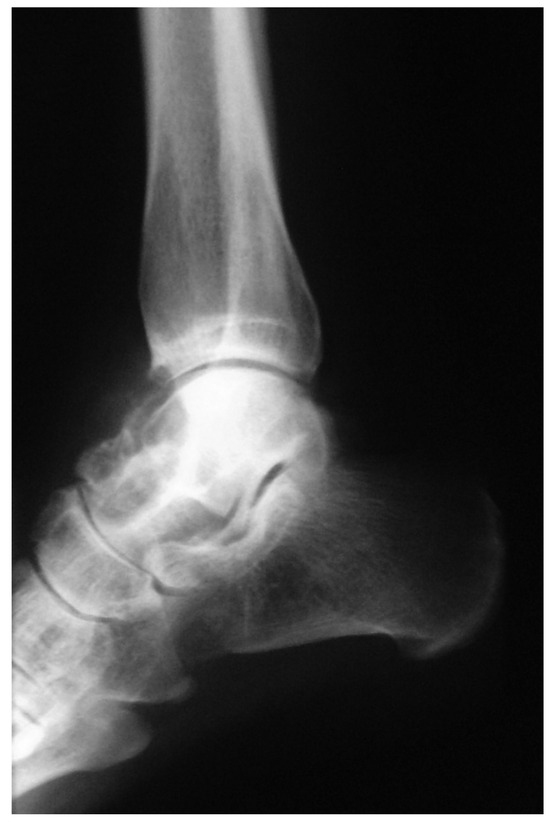
Figure 1.
Lateral radiograph obtained immediately before the operation that removed the giant cell tumor reveals an expansile, osteolytic lesion at the talar head and neck.
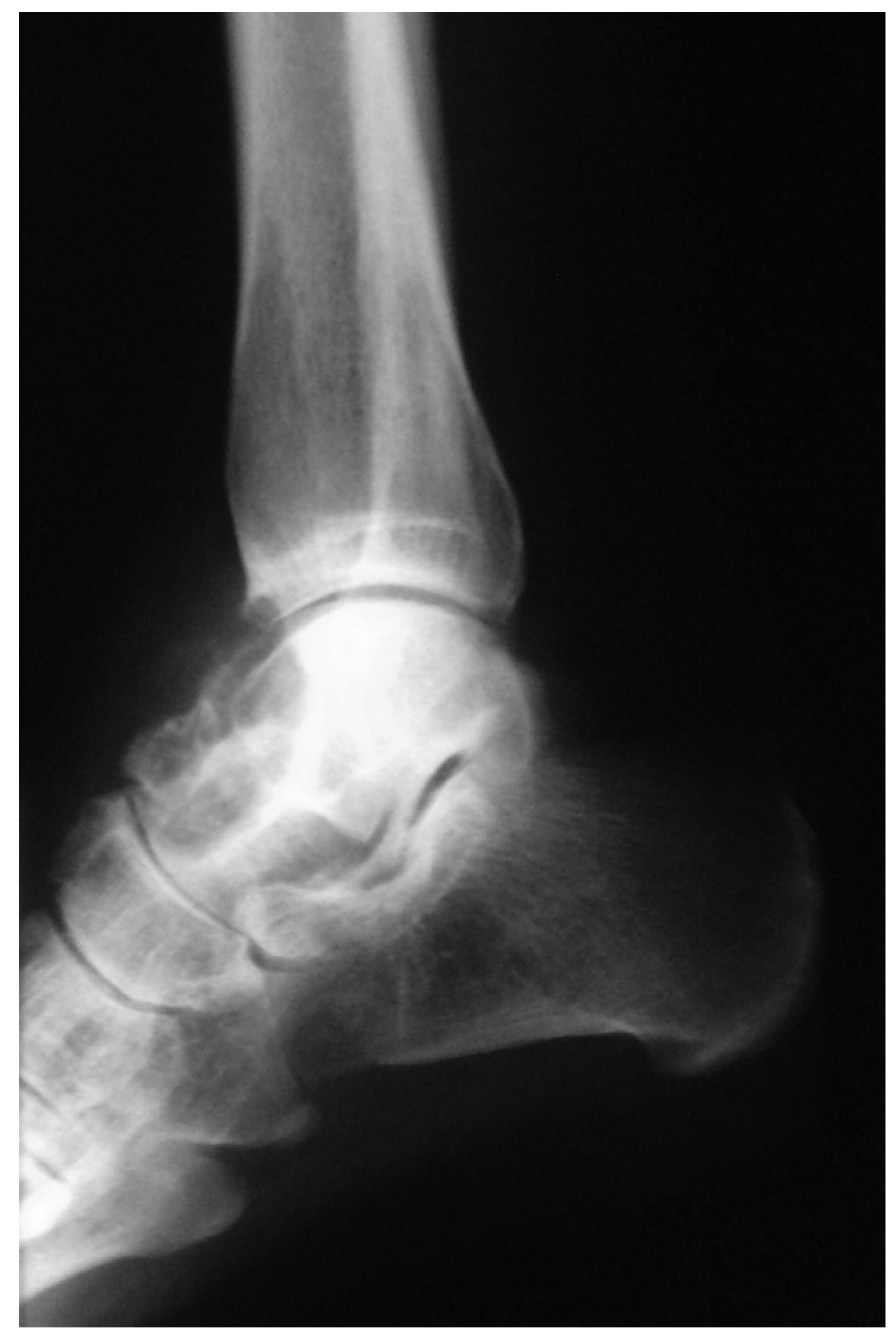
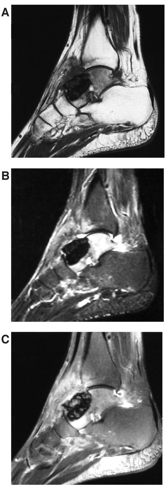
Figure 2.
A, Sagittal T1-weighted magnetic resonance image demonstrates an eccentrically located expansile lesion in the talar head and neck with low to intermediate intensity. B, Sagittal T2-weighted image shows the lesion with low intensity. C, The lesion is well visualized in this fat-saturated sagittal T2-weighted image.
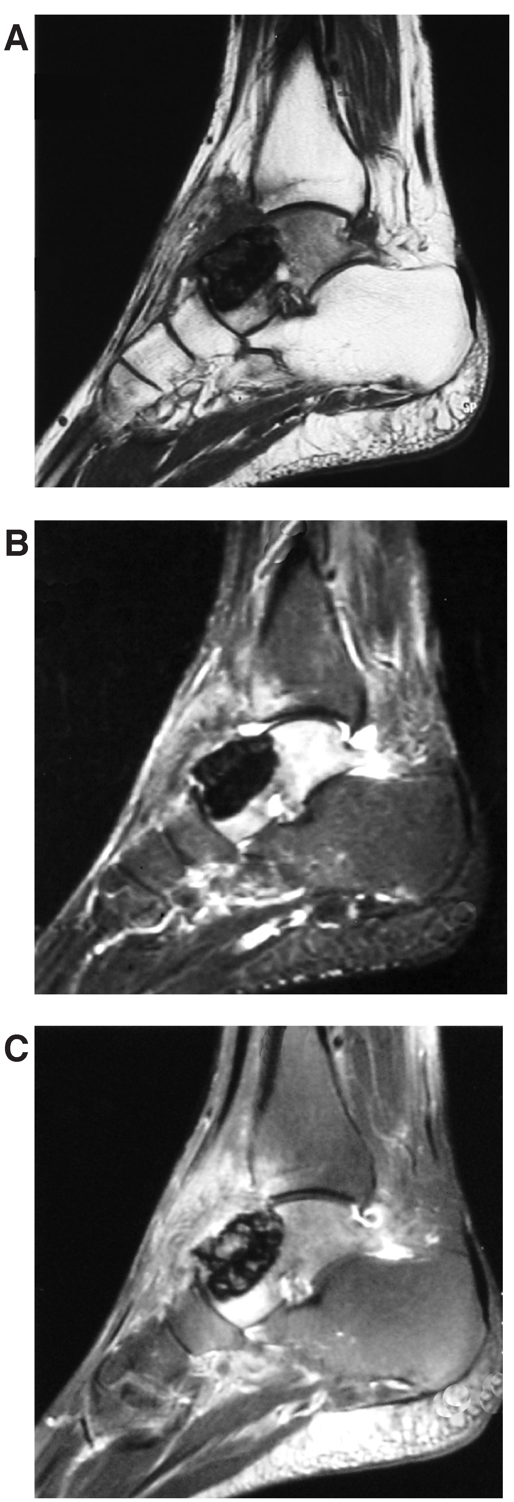
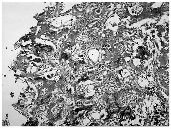
Figure 3.
Hematoxylin-eosin staining shows areas of hemorrhage with multinucleated giant cells (×25).
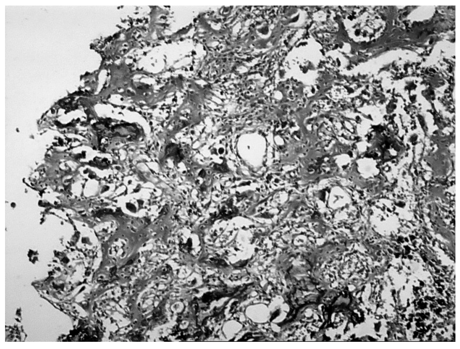
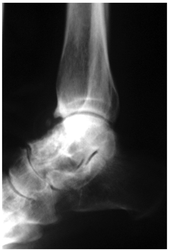
Figure 4.
Postoperative lateral radiograph 25 months after aggressive curettage and bone grafting shows a well-incorporated and remodeled graft, with no apparent recurrence of the tumor.
Financial Disclosures: None reported. | |
Conflict of Interest: None reported. |
References
- Unni KK: “Giant Cell Tumor,” in Dahlin’s Bone Tumors, ed by KK Unni, p 263, Lippincott-Raven, Philadelphia, 1996
- Temple HT, Scully SP: “Tumors,” in Orthopedic Knowledge Update: Foot and Ankle 2, ed by MS Mizel, RA Miller, MW Scioli, p 11, American Academy of Orthopaedic Surgeons, Rosemont, IL, 1998
- Saglik Y, Yildiz Y, Karakas A, et al: Giant cell tumor of bone. Bull Hosp Jt Dis58: 98, 1999
- O’Keefe RJ, O’Donnell RJ, Temple HT, et al: Giant cell tumor of bone in the foot and ankle. Foot Ankle Int16: 617, 1995
- Lackman RD: “Musculoskeletal Oncology,” in Orthopaedic Knowledge Update 8, ed by AR Vaccaro, p 197, American Academy of Orthopaedic Surgeons, Rosemont, IL, 2005
- Campanacci M, Baldini N, Boriani S: Giant-cell tumor of bone. J Bone Joint Surg Am69: 106, 1987
- Malawer MM, Bickels J, Meller I, et al: Cryosurgery in the treatment of giant cell tumor: a long term follow-up study. Clin Orthop359: 176, 1999
- Miller G, Bettelli G, Fabbri N: Joint Study of the European Musculoskeletal Oncology Society and US Musculoskeletal Tumor Society on Curettage of Giant Cell Tumor of Bone, Musculoskeletal Tumor Society, Bologna, Italy, 1990
- Shimizu T, Uehara T, Akahane T, et al: Recurrence potential of diffuse-type giant cell tumor in the foot: radiologic and pathologic features. Foot Ankle Int26: 474, 2005
- Turcotte RE, Wunder JS, Isler MH, et al: Giant cell tumor of long bone: a Canadian Sarcoma Group study. Clin Orthop397: 248, 2002
- Szendroi M, Antal I, Perlaky G: Mid-foot reconstruction following involvement of five bones by giant cell tumor. Skeletal Radiol29: 664, 2000
© 2007 American Podiatric Medical Association