


Abstract
A 66-year-old woman presented with painful gait and a large, firm, nonmobile mass in her right foot. She had undergone four previous surgeries to remove a recurring “calcified hematoma” that resulted from an injury sustained 3 decades previously. An unusually large ossific mass (7.0 × 7.0 × 2.0 cm) consistent with post-traumatic myositis ossificans circumscripta was excised. After 3 years of follow-up, there was no return of the symptoms or the mass. (J Am Podiatr Med Assoc 97(3): 229–233, 2007)
Post-traumatic myositis ossificans circumscripta is a benign condition of heterotopic bone formation.[1] It is one of three types of myositis ossificans as classified by Noble and later refined by Paterson.[2] This classification system is not considered particularly useful in that pathogenesis at the cellular level is not fully understood.[3] The other two types of myositis ossificans are myositis ossificans circumscripta without trauma and myositis ossificans progressiva.[2] Studies show that 40% to 75% of the cases of myositis ossificans are post-traumatic myositis ossificans circumscripta.[2],[4]
Here we focus on post-traumatic myositis ossificans circumscripta. The lesion generally presents with pain, swelling, and calor localized to the area of the lesion.[5] It can occur as a result of a major episode of trauma or repetitive episodes of microtrauma.[2] Trauma to a relatively avascular area, such as fascia or ligaments, results in cartilage formation (calcification). Trauma to a well-vascularized area, such as skeletal muscle, results in bone formation (ossification).[4] Some authors maintain that more proper names for these entities would be “heterotrophic calcification” and “heterotrophic ossification,” respectively.[5]
Case Report
A 66-year-old woman presented to Kern Hospital Foot Clinic and Diabetic Foot Center, Warren, Michigan, with a complaint of pain associated with a mass on the plantar aspect of her right foot. The patient reported that the mass had been present for approximately 4 years but that it had become increasingly painful in the past year. She also noted a progressive splaying (widening) between the second and third digits of the affected foot. The patient stated that the pain was limiting her overall activity level and ability to walk. She said that this was a recurrent problem and that she had previously undergone four operations to remove what she termed a “calcified hematoma.” She further related that the size of the current mass and the pain associated with it were considerably greater this time than with the previous episodes.
Physical examination revealed a well-nourished and well-developed 66-year-old woman. A right radical mastectomy had been performed 35 years previously with minimal residual scarring. The patient’s blood pressure was well maintained with medication. The remainder of the physical examination findings were essentially unremarkable except for the chief complaint.
The history of the chief complaint could be traced back to a traumatic injury that involved the right foot approximately 31 years previously. The patient related that she “kicked” a raised portion of concrete while walking barefoot. She stated that after this injury, she developed swelling and pain on the plantar aspect of the right foot. The patient initially received conservative care for the injury. Two years later she underwent the surgical removal of a “calcified hematoma.” The patient noted recurrence of the mass, with a second resection 5 years later. Two additional surgeries were performed 17 and 24 years after the injury. In all cases, the patient reported an uneventful postoperative course and temporary relief of symptoms. Pathology reports from all of the previous surgeries were negative for malignancy.
Radiographic examination revealed a radiodense mass between the second and third metatarsals of the right foot (Figure 1). On the lateral view, the mass was noted to extend from the dorsal to the plantar aspect of the right foot (Figure 2). The mass appeared to be separate from but closely aligned with the second and third metatarsal shafts. The patient presented to the clinic with a technetium triple-phase bone scan from another institution. It showed focal uptake in the region of the mass (Figure 3).
A magnetic resonance image of the right foot was ordered preoperatively in an attempt to delineate the soft-tissue and bony involvement of the mass. Multiple T1- and T2-weighted spin-echo images were obtained in the axial, coronal, and sagittal planes (Figure 4 and Figure 5). The study was significant for a large signal void abnormality, representing calcification or ossification of the plantar aspect of the right foot inferior to the second and third metatarsals. Also noted was irregular thickening of the cortex of the third metatarsal shaft but no distinct cortical disruption. There was no evidence of a soft-tissue mass or inflammation of the right foot. Differential diagnosis from this study included idiopathic tumoral calcinosis of the soft tissues, melorheostosis, and myositis ossificans. The study all but ruled out the possibility of infection or malignancy.
The patient was brought to the operating room for removal of the ossific mass in the right foot. Initially the mass was exposed by means of a plantar “lazy S” incision at the site of the previous surgeries (Figure 6). The mass was isolated using sharp and blunt dissection. It measured approximately 7.0 × 7.0 × 2.0 cm. The normal soft-tissue anatomy of the second intermetatarsal space was absent. It could not be determined whether this was from previous surgeries or from pathologic ossification of the soft-tissue structures in the area.
Attempts to remove the mass without disruption of the cortices of the second and third metatarsals from the plantar approach were unsuccessful (Figure 7). A 6-cm incision was made dorsally at the second intermetatarsal space. Gaining access to the mass from this incision, the surgeons split the mass into manageable sections using an osteotome and a mallet. These sections were then removed through the plantar incision (Figure 8).
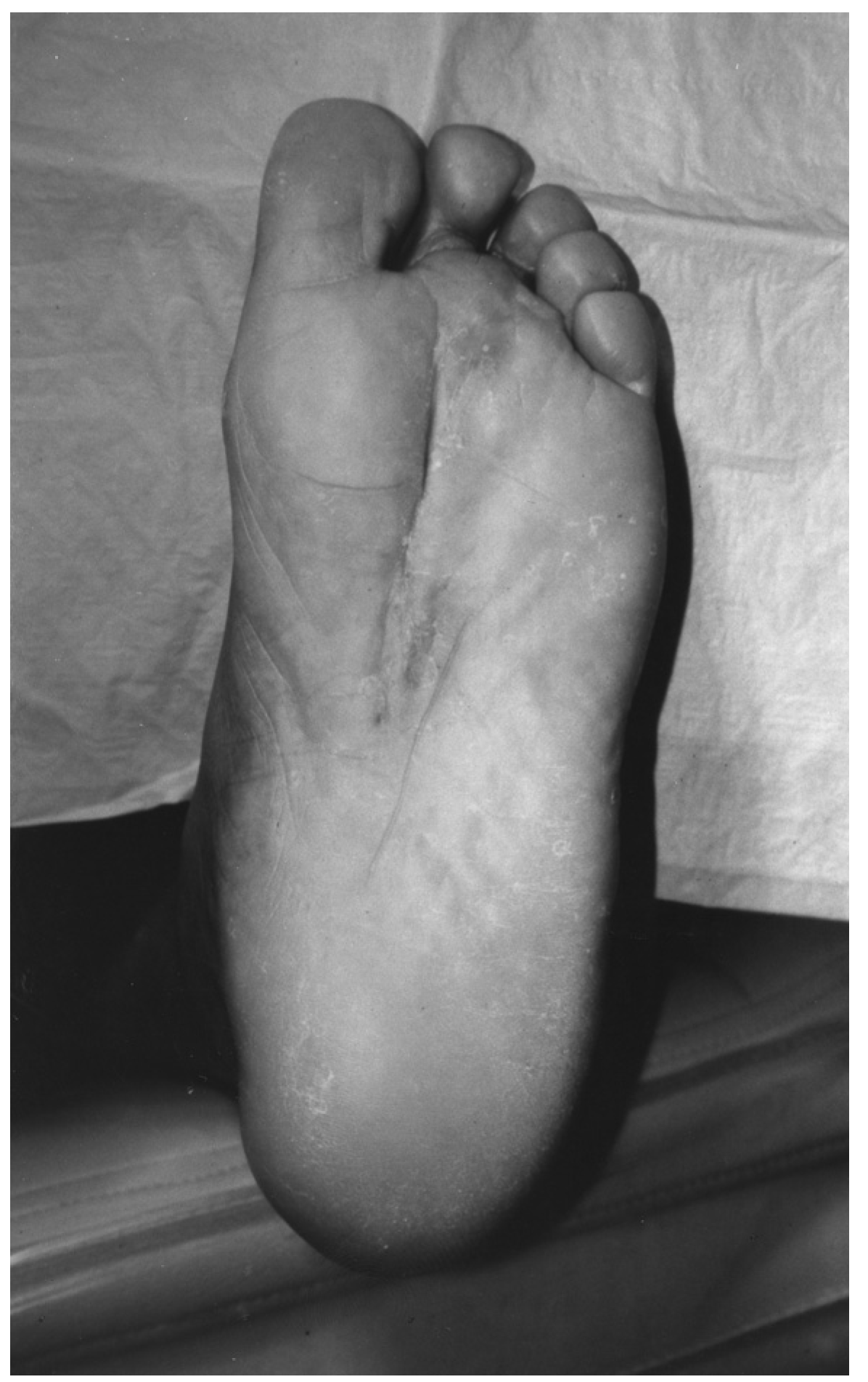
Intraoperative radiographs confirmed that the cortices of the second and third metatarsals remained intact throughout the procedure (Figure 10 and Figure 9). The radiographs also allowed inspection of the surrounding areas for any remaining ossific fragments. No additional fragments were found, and the plantar and dorsal incisions were closed in layers with absorbable sutures; 3-0 nylon simple sutures were used to approximate the skin edges. An external drain system was used in the plantar incision and was maintained for 24 hours postoperatively. The patient observed complete bed rest in the hospital for 72 hours. She was discharged with a below-the-knee cast and instructions for nonweightbearing of the right foot (Figure 11).
The patient was examined weekly for the first month after surgery. The dorsal sutures were removed at 2 weeks, whereas the plantar sutures were maintained for 3 weeks. The patient initiated partial weightbearing on the operative extremity at 6 weeks. She proceeded to full weightbearing by 8 weeks postoperatively. The patient was followed clinically for 3 years after surgery without any recurrence of the symptoms or the mass. The splaying of the digits was completely eliminated, and the incision sites have maintained a fine-line scar.
Discussion
This report describes an unusually large example of post-traumatic myositis ossificans circumscripta, an uncommonly seen entity. Less than a dozen cases in the foot have been reported in the literature. The most common sites reported are the quadriceps femoris, the brachialis, and the adductor muscles of the thigh.[4] It is critical to be able to identify post-traumatic and nontraumatic myositis ossificans circumscripta. They both can easily mimic a variety of benign and malignant conditions.[6] Note that a biopsy of the central zone of the mass can contain proliferating fibrous tissue that closely resembles a malignancy.[7] One study[8] showed that in a review of 50 cases of myositis ossificans, a malignant diagnosis was suggested by referring pathologists 23% of the time. Some pathologists have gone as far as to refer to this condition as a “pseudomalignant osseous tumor of the soft tissue.”[9] Because of this patient’s history of breast cancer and recurrences, ruling out a malignancy was extremely important. After standard radiography, bone scanning, and magnetic resonance imaging were performed, the mass was excised. The surgical team was extremely careful in isolating the entire mass and in exploring the area for any small fragments. Because of this extreme care, the patient has been free of symptoms for more than 3 years.
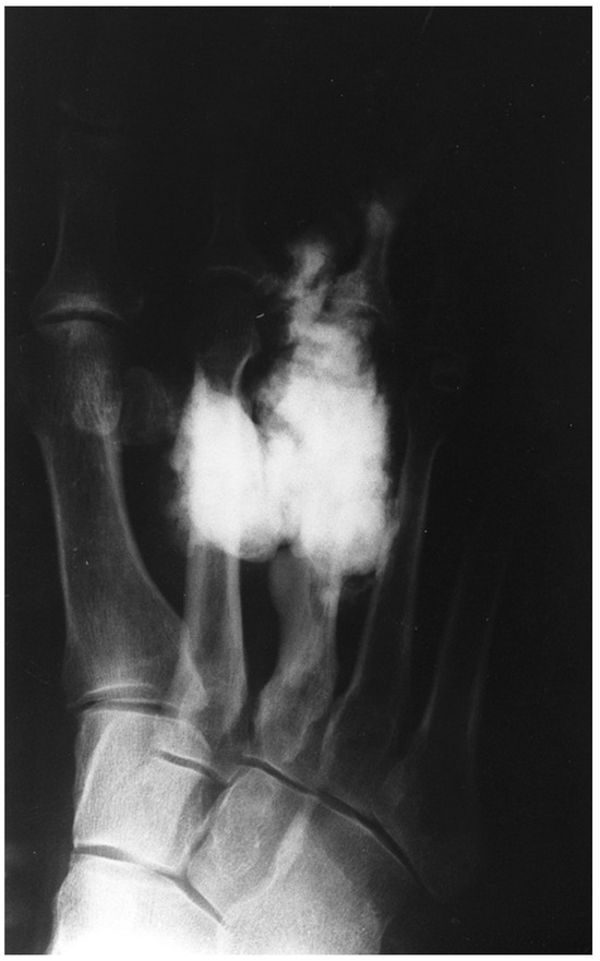
Figure 1.
Preoperative anteroposterior radiograph of the right foot.
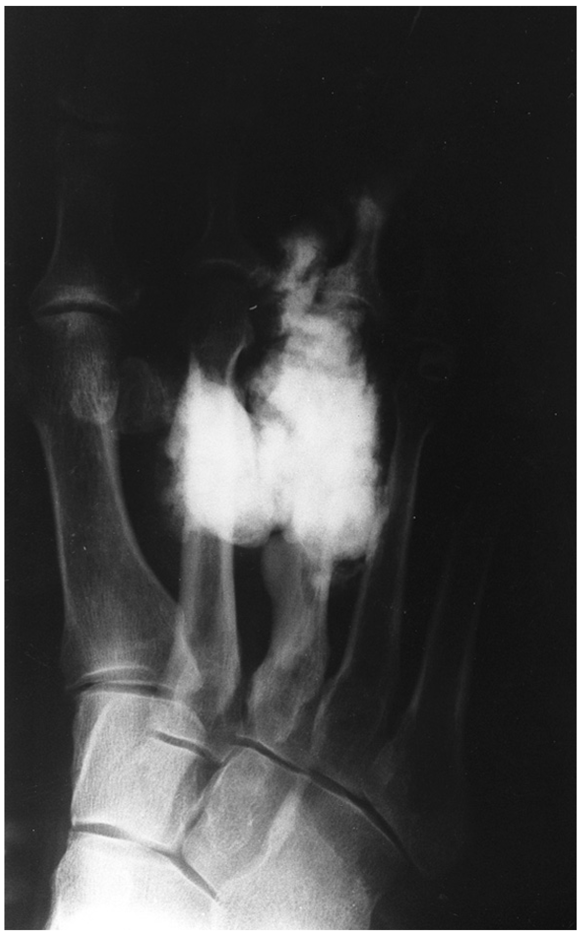
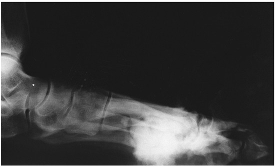
Figure 2.
Preoperative lateral radiograph of the right foot.
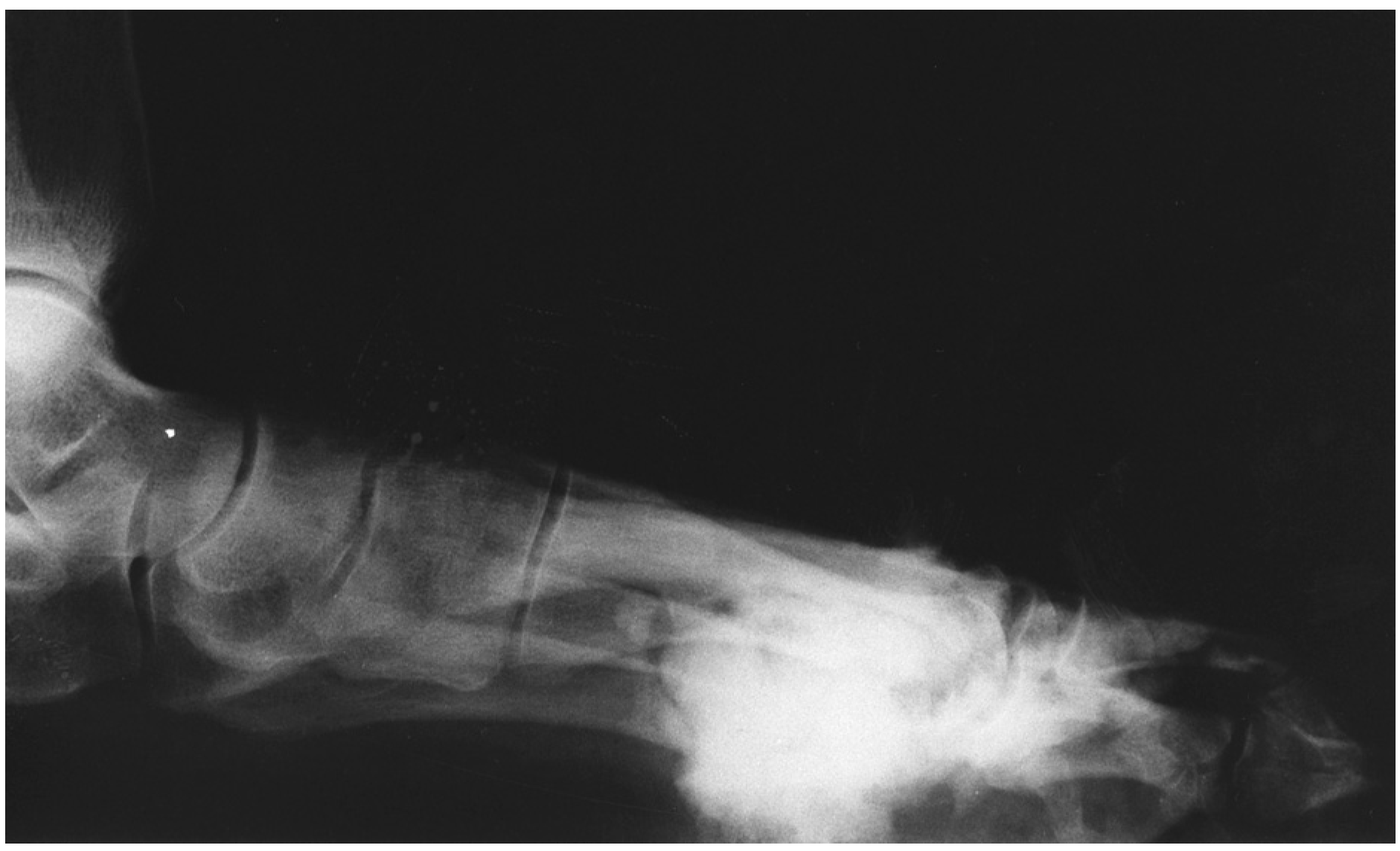
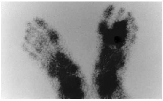
Figure 3.
Preoperative technetium bone scan of the feet.
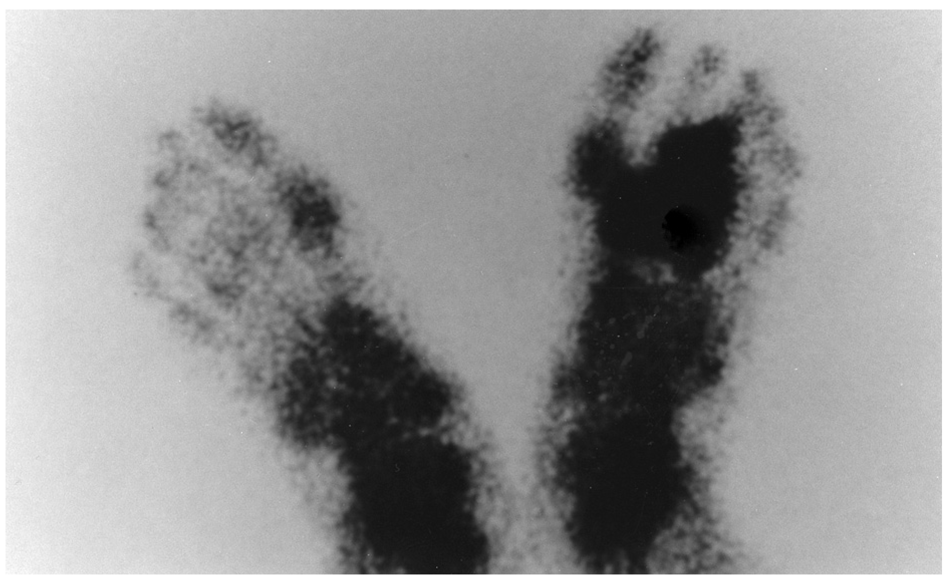
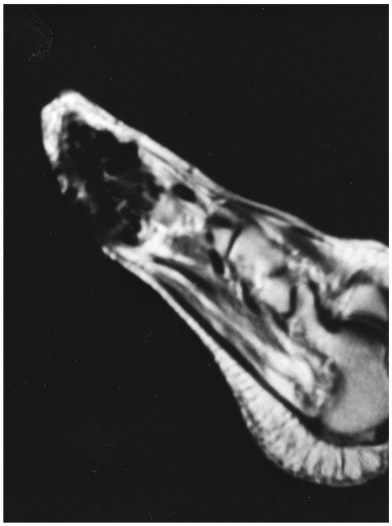
Figure 4.
Sagittal plane magnetic resonance image of the right foot.
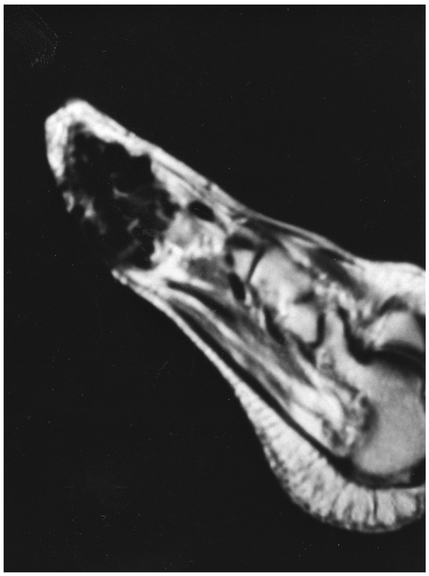
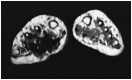
Figure 5.
Coronal plane magnetic resonance image of the right foot.
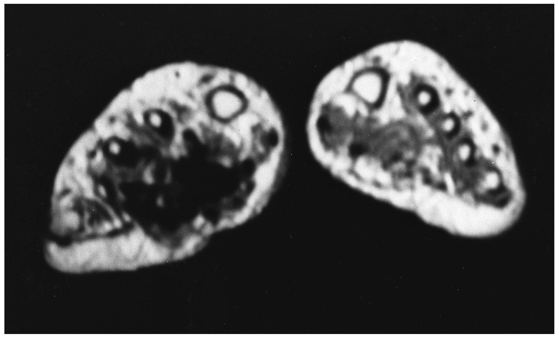
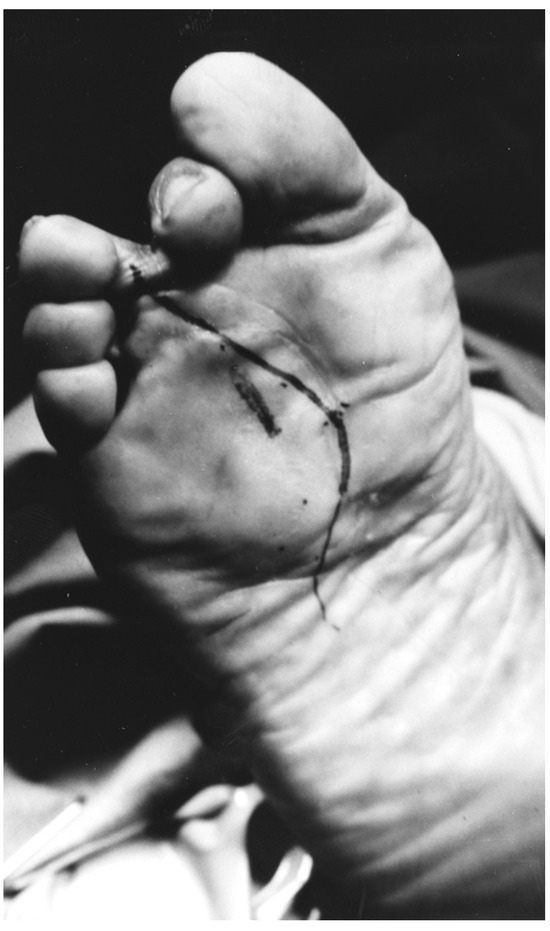
Figure 6.
“Lazy S” incision marked on the plantar surface of the right foot.
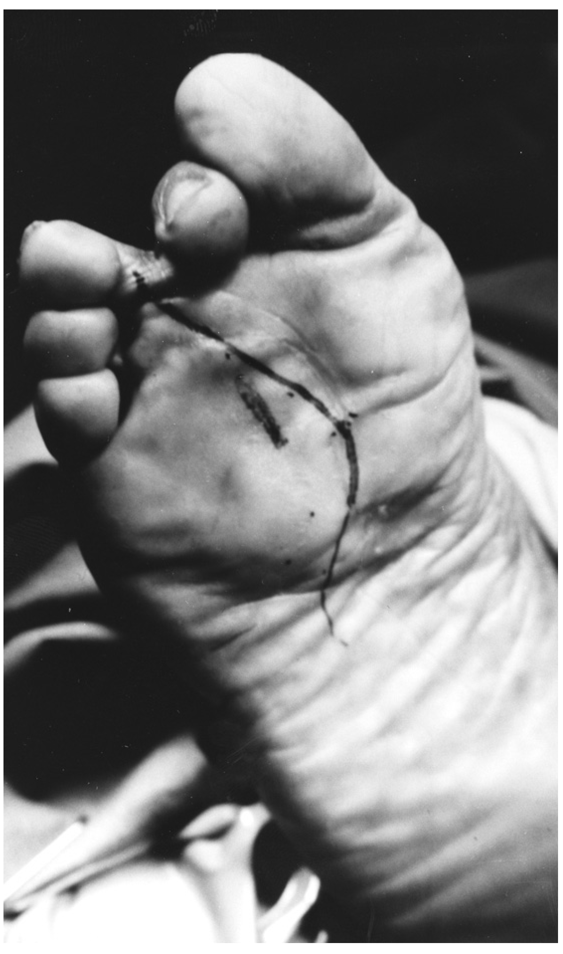
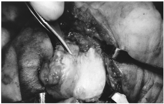
Figure 7.
The osseous mass was isolated through the plantar incision.
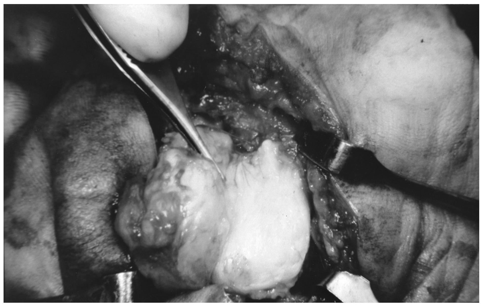
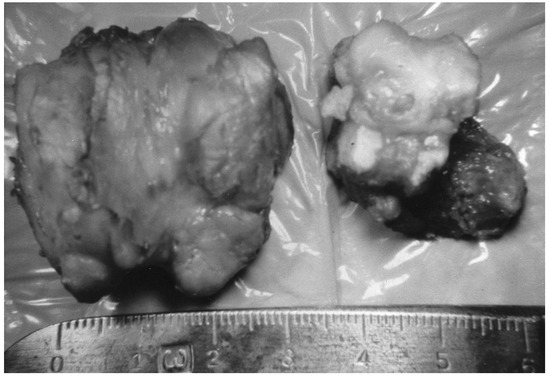
Figure 8.
The osseous mass was excised in two pieces.
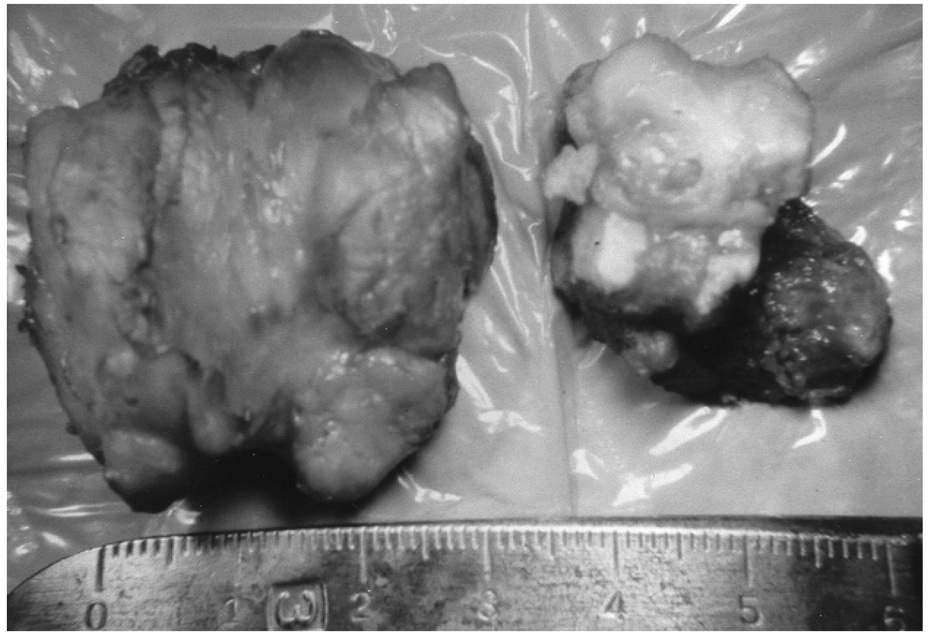
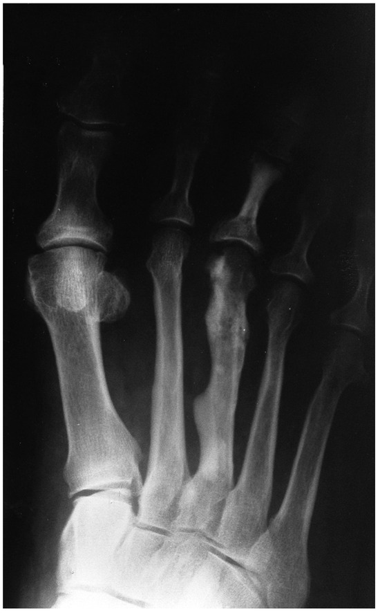
Figure 9.
Intraoperative anteroposterior radiograph of the right foot.
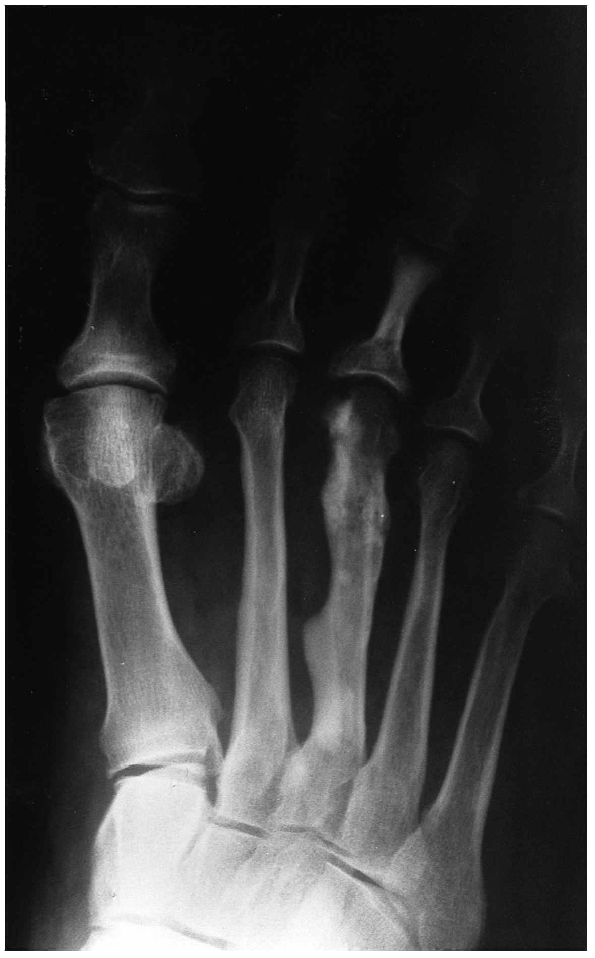
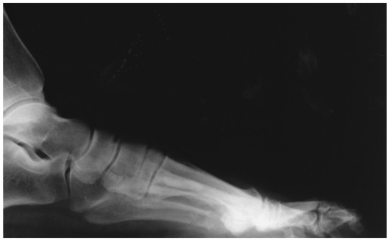
Figure 10.
Intraoperative lateral radiograph of the right foot.
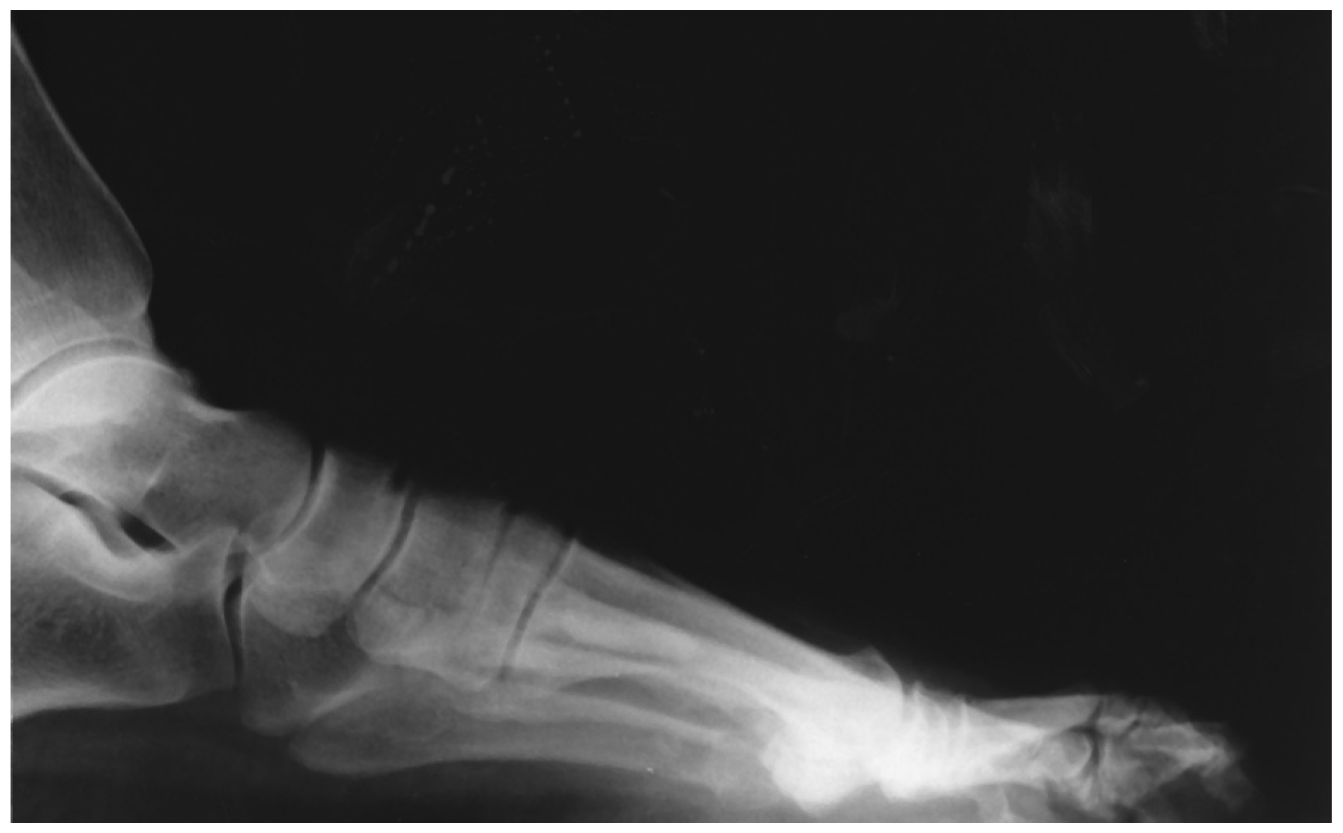
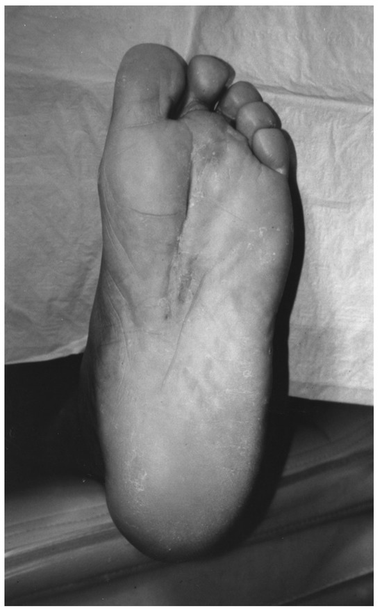
Figure 11.
Postoperative healed plantar skin incision.
Financial Disclosures: None reported. | |
Conflict of Interest: None reported. |
References
- Parikh J, Hyare H, Saifuddin A: The imaging features of post-traumatic myositis ossificans with emphasis on MRI. .Clin Radiol57: 1058, 2002
- Herring K, Levine B: Myositis ossificans of traumatic origin in the foot. .J Foot Ankle Surg31: 30, 1992
- Sirvanci M, Ganiyusufoglu AK, Karaman K, et al: Myositis ossificans of psoas muscle: magnetic resonance imaging findings. .Acta Radiol45: 523, 2004
- Allard M, Thomas R, Nicholas R: Myositis ossificans: an unusual presentation in the foot. .Foot Ankle Int18: 39, 1997
- Fuselier CO, Tlapek TA, Sowell RD: Heterotrophic ossification (myositis ossificans) in the foot: a case report. .JAPMA76: 524, 1986
- Wang X, Malghem J, Parizel PM, et al: Pictorial essay: myositis ossificans circumscripta. .JBR-BTR86: 278, 2003
- De Maeseneer M, Jaovisidha S, Lenchik L, et al: Myositis ossificans of the foot. .J Foot Ankle Surg36: 290, 1997
- De Silva MV, Reid R: Myositis ossificans and fibroosseous pseudotumor of digits: a clinicopathological review of 64 cases with emphasis on diagnosis pitfalls. .Int J Surg Pathol11: 187, 2003
- Schutte H, VanderHeul R: Pseudomalignant, nonneoplastic osseous soft tissue tumors of the hand and foot. .Radiology176: 149, 1990
© 2007 American Podiatric Medical Association