


Abstract
Chondromyxoid fibroma is a rare, benign, aggressive bone tumor whose localization in the calcaneus is even rarer. We report a case of chondromyxoid fibroma in the calcaneus of a 10-year-old Iranian boy who was treated with extended curettage. After 3 years of follow-up, the patient had experienced no recurrence. (J Am Podiatr Med Assoc 97(3): 223–224, 2007)
Chondromyxoid fibroma is among the rarest cartilaginous skeletal tumors. It represents less than 1% of all benign osseous tumors.[1] The most common location of chondromyxoid fibroma is the upper part of the tibia. Its occurrence in the foot and calcaneus is rare.[2] Because of the rarity of calcaneal involvement and the good outcome of extended curettage without the use of bone grafting, the following case is reported.
Case Report
A 10-year-old boy with a 2-month history of persistent pain and swelling of the heel area was referred to the orthopedic clinic at Ghaem Hospital Medical School, Mashad, Iran. Physical examination revealed tender swelling of the heel area. The patient was clinically well and had no relevant medical history. Ankle and subtalar motion was painful and limited. Radiography at presentation revealed a large, circumscribed osteolytic lesion without a reactive zone in the calcaneus. A segment of the plantar cortex was absent, and soft-tissue involvement was not evident (Figure 1). Chest radiographs and abdomen sonograms showed nothing. Suspecting chondroblastoma, aneurysmal bone cyst, or unicameral bone cyst, we performed an open biopsy.
Histologic examination of the lesion showed a lobular pattern with hypocellular areas in the center and hypercellular regions in the periphery of the lobules; the latter contained scattered multinucleated giant cells. The hypocellular areas had a myxoid background, with the stellate cells containing oval- to spindle-shaped nuclei and indistinct to densely eosinophilic cytoplasm with bipolar or multipolar extensions. Nuclear and cellular pleomorphism was minimal, without significant mitotic activity. Findings from histologic examination were characteristic of chondromyxoid fibroma.
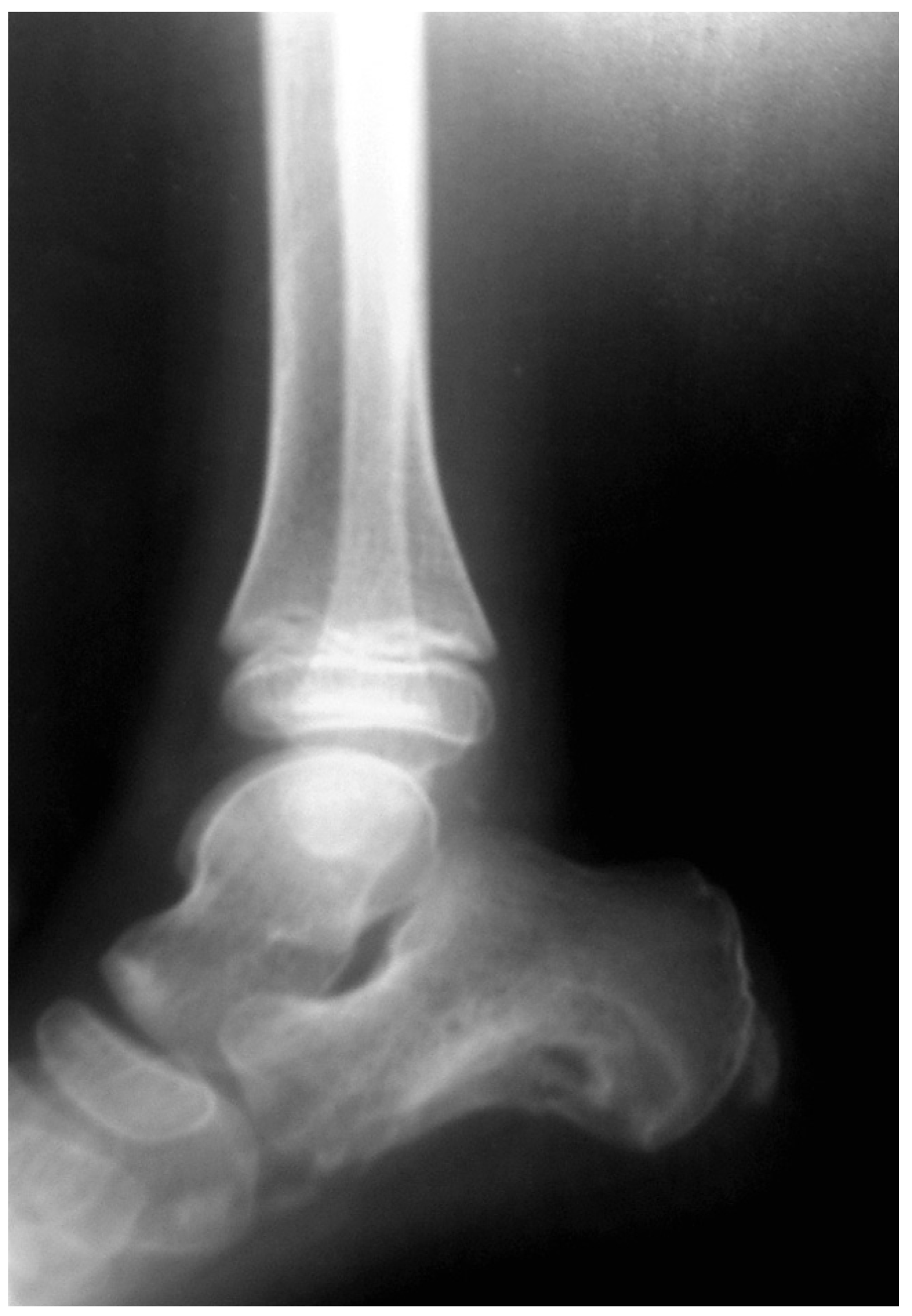
The patient then underwent open, thorough, extended curettage of the lesion with a lateral plantar approach. We extended curettage to normal cancellous tissue of the calcaneus. Grossly, the tissue was similar to cartilage tissue. We did not use bone grafting to fill out the cavity, and after irrigation we closed the wound. A well-padded short-leg cast was applied. After 3 months we removed the cast; radiographs showed bone formation into the cavity and remodeling (Figure 2). Examination 6 months after surgery showed no tenderness and no limitation of joint range of motion, and the patient did not complain of pain. In 3 years of follow-up, no recurrence was observed.
Discussion
Chondromyxoid fibroma is a benign but aggressive tumor occurring primarily in the first and second decades of life. It was first described by Jaffe and Lichtenstein in 1948.[3] The most common site of involvement is around the knee joint; the foot accounts for approximately 20% of cases. In a series of 31 cases, Beggs and Stoker[2] reported three cases of calcaneal involvement. Although it typically presents in the first and second decades of life, it has been reported in patients aged 5 to 79 years.[1,4,5]
The radiologic features of these tumors can vary, but the classic appearance is a pattern of central bone destruction with a narrow, often sclerotic, zone of transition associated with septation lobulation and reactive sclerosis. Cortical expansion is often noted, particularly in tumors of small bones. There may be cortical defects, but the absence of a periosteal reaction is thought to be characteristic. Calcification is rarely seen.[2,6]–[8]
The diagnosis is made by histologic assessment, which reveals a structure consisting of various combinations of fibrous, cartilaginous, and myxoid tissue. A variety of surgical treatments have been reported in the literature, from intralesional curettage to en bloc resection.[1,2,6,7,9,10] The present case was treated by curettage within a margin of normal bone without filling out the cavity by means of bone grafting. The patient was closely observed for 3 years, and there was no recurrence of the lesion. The cavity healed with normal bone in the first 6 months.
There have been cases reported in the literature of chondromyxoid fibroma recurrence in late childhood and early adolescence, but we believe these recurrences are attributable to inadequate margins of excision. Generous excision extended to normal tissue is a very important factor in preventing recurrence.
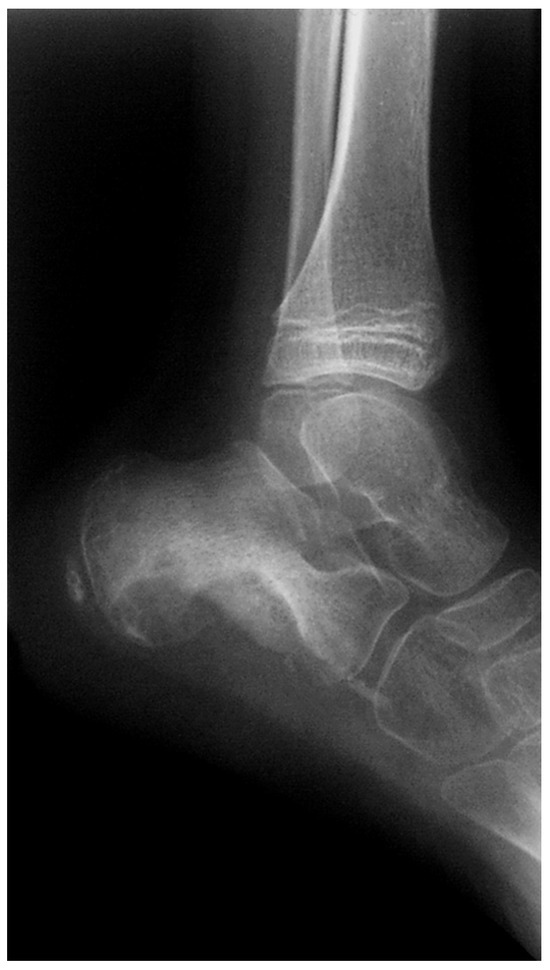
Figure 1.
The lesion evident on the lateral radiograph of the calcaneus of a 10-year-old boy has a well-defined area of bone destruction, and the plantar cortex is destroyed, without a reactive zone.
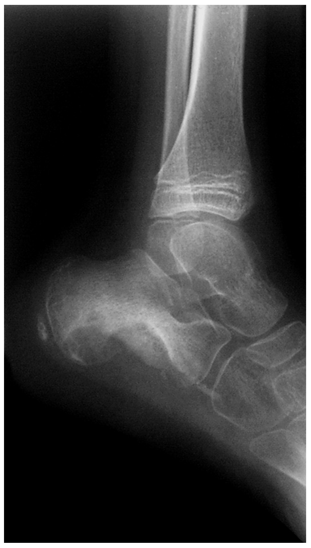
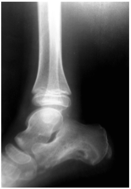
Figure 2.
Three-month postoperative lateral radiograph shows osseous formation and healing of the cavity.
Financial Disclosures: None reported. | |
Conflict of Interest: None reported. |
References
- Baron RL, Galinski AW, Vlahos M, et al: Chondromyxoid fibroma. JAPMA86:212, 1996
- Beggs IG, Stoker DJ: Chondromyxoid fibroma of bone. Clin Radiol33:671, 1982
- Jaffe HL, Lichtenstein L: Chondromyxoid fibroma of bone: a distinctive benign tumor likely to be mistaken especially for chondrosarcoma. Arch Pathol45:541, 1948
- Feldman F, Hecht H, Johnson A: Chondromyxoid fibroma of bone. Radiology94:249, 1970
- Wu KK: Differential diagnosis of pedal osseous neoplasm:. Clin Podiatr Med Surg10:683, 1993
- Feit EM, Dobbs BM: Chondromyxoid fibroma of the fourth metatarsal:. JAPMA90:211, 2000
- O’Connor PJ, Gibbon WW, Hardy G, et al: Chondromyxoid fibroma of the foot. Skeletal Radiol25:143, 1996
- Wilson AJ, Kyriakos M, Ackerman LV: Chondromyxoid fibroma: radiologic appearance in 38 cases and in a review of the literature. Radiology179:513, 1991
- Wu KK: Chondromyxoid fibroma of the foot bones. J Foot Ankle Surg34:513, 1995
- Rouvillain JL, Jouannelle A, Delattre O, et al: Apropos of a case of chondromyxoid fibroma of the talus: a review of the literature. Rev Chir Orthop Reparatrice Appar Mot83:372, 1997
© 2007 American Podiatric Medical Association