


Abstract
The biomechanical effects of talectomy on the foot were investigated in seven fresh below-the-knee amputation specimens using pressure-sensitive films placed on the facets of the calcaneus, footprints, and loading-pattern diagrams in the intact foot and after talectomy with anterior and posterior displacement of the foot. Both talectomy techniques distorted the loads carried by the facets of the calcaneus. In the intact foot, 65.6% of the loads were carried by the posterior facet of the calcaneus and 34.4% by the anterior and middle facets. After talectomy with anterior displacement of the foot, although the loads carried by the anterior and middle facets decreased significantly (P = .018), the increase in the loads carried by the posterior facet was not significant compared with the intact foot (P = .176). Similarly, the loads carried by the posterior facet decreased significantly after talectomy with posterior displacement of the foot (P = .028), but the increases in the loads carried by the anterior and middle facets were not significant (P = .735). Comparing the two types of talectomy, the loads carried by each facet changed significantly (P = .018). Talectomy with posterior displacement of the foot also changed the loading patterns and resulted in significant pronation of the foot. These results suggest that talectomy should be performed only as a salvage procedure and that talectomy with anterior displacement of the foot may be preferred when talectomy is indicated. (J Am Podiatr Med Assoc 96(6): 495–498, 2006)
Talectomy is sometimes indicated as a salvage procedure for severe foot deformities in patients with myelomeningocele,1-4 arthrogryposis multiplex congenita,1-3,5-8 postpoliomyelitic paralysis,1,9 or severe fracture dislocations of the talus.2,10-13 Review of the literature revealed two techniques for total talectomy without fusion. In the first technique, the foot is displaced posteriorly to make contact between the distal tibial articular surface and the anterior and middle facets of the calcaneus.3,12 In the second technique, the foot is displaced anteriorly to make contact between the distal tibial articular surface and the posterior facet of the calcaneus.2,10,11,13
Although satisfactory results were reported for each technique, there is no study in the literature, to our knowledge, investigating the biomechanical effects of talectomy on the foot. Therefore, we conducted the present study of the biomechanical effects of talectomy on the foot in seven fresh below-the-knee amputation specimens. In our opinion, such an approach also clarifies the function of the talus.
Materials and Methods
Seven fresh below-the-knee amputation specimens were studied. The specimens were from six men and one woman who underwent amputation for vascular reasons. Any deformity, previous operation, osteoarthritis, or previous fracture was excluded by gross examination and radiography. Skin and subcutaneous tissue around the ankle were removed, and using a curved medial incision, the ankle and talocalcaneal joints were exposed.
The specimens were mounted in the loading apparatus with an angle of 90° between the foot and the tibia (Fig. 1). Then two pressure-sensitive film transducers (Fuji Prescal Film; Fuji Photo Film USA Inc, Valhalla, New York) were inserted into the talocalcaneal joint, one on the posterior facet and the other on the anterior and middle facets of the calcaneus. The super-low film (0.5–2.5 MPa) was used throughout the study. Each specimen was loaded by 700 N for 40 sec. Footprints of the specimens during loading were also obtained.
The medial malleolus was then osteotomized transversely at the level of the talotibial joint line and turned down on the deltoid ligament. The whole of the talus was resected at the talotibial, subtalar, and talonavicular joints, leaving no bony fragments behind. When talectomy was complete, the foot was displaced posteriorly to make contact between the distal articular surface of the tibia and the anterior and middle facets of the calcaneus. Then the medial malleolus was reduced and fixed with a screw,3,12 and pressure measurements were repeated. In the second technique, the foot was displaced anteriorly to make contact between the distal articular surface of the tibia and the posterior facet of the calcaneus. Then the medial malleolus was displaced laterally to create a new joint with the calcaneal sulcus and fixed with a screw,2,10,11,13 and the study was repeated.
After the loading period, pressure transducers were removed and scanned and digitized at 300 dpi and 256-color resolution with the calibration strip, and pressure maps were produced by means of a computer program (Lucia, version 4.21; Laboratory Imaging Ltd, Prague, Czech Republic). Then the percentage of the load carried by each facet of the calcaneus was calculated. The overall mean pressure was calculated from the following equation as described by Diab et al14:
Pm = Σ (Ai ˙ Pi)/At,
where Pm is overall mean pressure (in kilopascals); Ai, area of zone i (in square millimeters) (area between any two calibration values); Pi, mean pressure in zone i (in kilopascals); and At, total contact area (in square millimeters). Mean force was determined by dividing overall mean pressure (Pm) by total contact area (At).
Footprints were also analyzed using the method of Staheli et al15 in which the width of the foot in the area of the arch is divided by the width of the heel measured. Values greater than 0.8 were considered flattening of the foot. The Wilcoxon signed rank test was used for statistical analysis.
Results
Mean loads carried by each facet of the calcaneus and the contact area in the intact foot and after two types of talectomy are displayed in Table 1. In the intact foot, 65.6% of the loads were carried by the posterior facet of the calcaneus and 34.4% by the anterior and middle facets. After talectomy with anterior displacement of the foot, although the loads carried by the anterior and middle facets decreased significantly (P = .018), the increase in the loads of the posterior facet was not significant (P = .176) compared with the intact foot. Similarly, the loads carried by the posterior facet decreased significantly after talectomy with posterior displacement of the foot (P = .028), but the increases in the loads of the anterior and middle facets were not significant (P = .735). When the two types of talectomy were compared, the loads carried by each facet changed significantly (P = .018).
The contact area in the anterior and middle facets after talectomy with posterior displacement of the foot and in the posterior facet after talectomy with anterior displacement of the foot did not increase significantly compared with the intact foot (P = .398 and P = .498, respectively). Other comparisons of the contact area showed significant changes (P = .018).
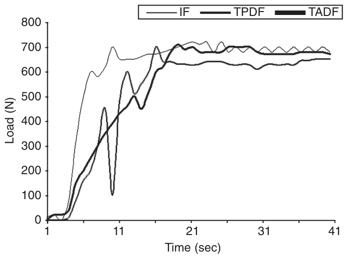
Typical loading patterns obtained in the study are displayed in Figure 2. In the intact foot and after talectomy with anterior displacement of the foot, the loading graphics showed an increase to 700 N in approximately 10 to 15 sec, and then the foot was stable. However, after talectomy with posterior displacement of the foot, after loading approximately 400 to 450 N, there was a sudden decrease and then a change to a pattern that resembled the other two loading patterns.
The mean Staheli index of the intact foot was 0.7 (range, 0.5–0.8). Although the index remained almost the same after talectomy with anterior displacement of the foot (mean, 0.7; range, 0.6–0.8; P = .317), there was a significant increase after talectomy with posterior displacement of the foot (mean, 0.9; range, 0.8–1.0; P = .027).
Discussion
Talectomy is one of the oldest of the orthopedic surgeries, and Hildanus is credited with being the first to report talectomy performed after compound dislocation of the talus in 1607.16 Today, talectomy is used as a salvage procedure for severe deformities of the foot and fracture dislocations of the talus. Despite the 400-year history of talectomy, review of the literature revealed no reports of the biomechanics of talectomy, making the present study the first on this subject.
Currently, two types of talectomy are performed, and the difference between these techniques is the facet of the calcaneus that makes contact with the distal articular surface of the tibia. Our results indicate that both techniques (talectomy with anterior or posterior displacement of the foot) distort the loads carried by the facets of the calcaneus. As expected, the loads carried by the facet with which the distal articular surface of the tibia made contact increased, but the increase was not significant. On the other hand, although the facets that theoretically have no contact with the tibia were unloaded significantly, there were still some loads carried by these facets (Table 1). In our opinion, this shows the instability of the foot after talectomy and, in accordance with the observations of Cooper and Capello,1 that “despite an attempt to displace the foot posteriorly, postoperative patients had essentially none,” and Kirschner wires are sometimes used to maintain the position.7
In fact, after talectomy with posterior displacement of the foot, the concave distal articular surface of the tibia makes contact with the concave anterior and middle facets of the calcaneus, creating an incongruent joint. The poor fit after talectomy with posterior displacement of the foot has been recognized previously,1,3,6 and this is probably the reason for the degenerative changes1 and the spontaneous fusion.1,6,7,9 In the loading period, talectomy with posterior displacement of the foot also showed a typical decrease (Fig. 2), and the footprints revealed significant pronation of the foot. This abnormal motion is probably due to the poor fit of two concave surfaces and may explain the degenerative changes after talectomy with posterior displacement of the foot.
In talectomy with anterior displacement of the foot, there seems to be a relatively more congruent joint, as the concave distal articular surface of the tibia makes contact with the convex posterior facet of the calcaneus and is stabilized with lateralization of the medial malleolus.2,10,11,13 Loading patterns (Fig. 2) and footprints were also identical to those in the intact foot. This may explain the good results without degenerative changes and spontaneous fusion despite more than 5 years of follow-up.11 However, loading of the anterior and middle facets necessitates longer follow-up.
Review of the literature revealed several studies on the measurement of contact area in the normal ankle joint17,18 or after simulated pathologic conditions,19 but none had evaluated the facets of the calcaneus. Although the present study is primarily devoted to the biomechanics of talectomy, some data were obtained regarding the loading of the calcaneal facets in the intact foot, and this may be helpful in the treatment of calcaneal fractures.
This study has some limitations. First, no muscle forces were modeled. Second, we did not investigate the limb-length discrepancy created after each type of talectomy and its role in the loading characteristics of the foot after talectomy. However, considering the limitations of the present study, we agree that talectomy should be performed only as a salvage procedure, and talectomy with anterior displacement of the foot may be preferred when talectomy is indicated.

Table 1.
Mean Loads Carried by the Facets of the Calcaneus and the Contact Area Before and After Two Types of Talectomy
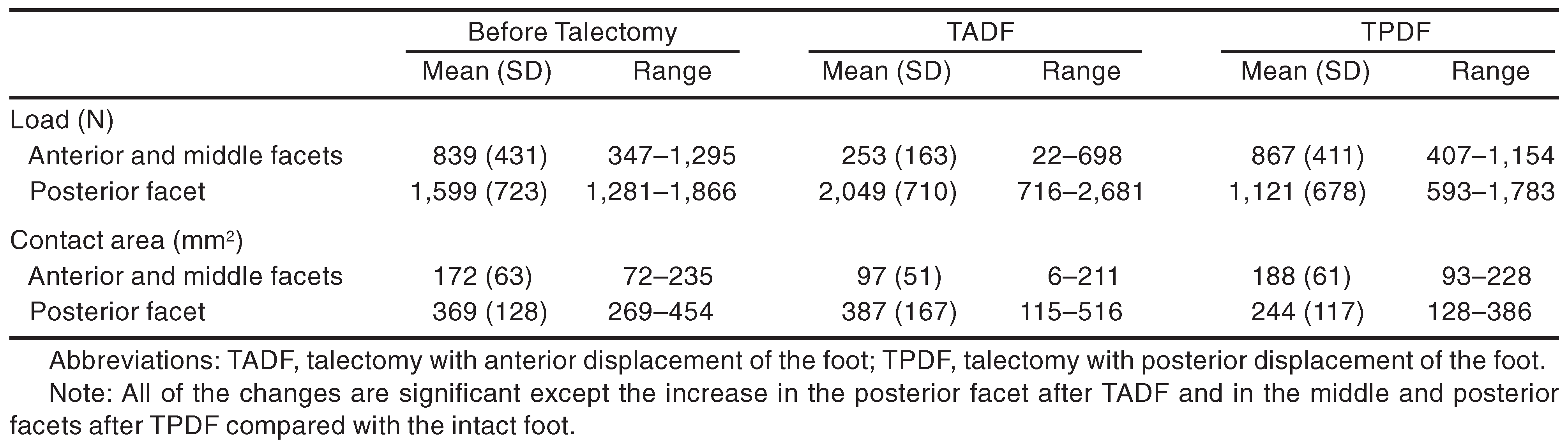
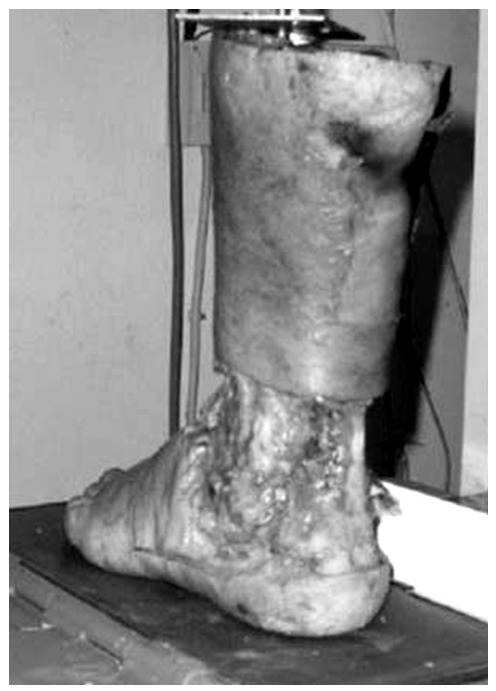
Figure 1.
Loading apparatus.
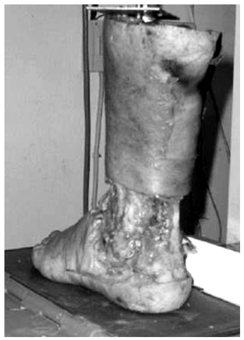
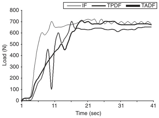
Figure 2.
Typical loading patterns in the intact foot (IF), after talectomy with anterior displacement of the foot (TADF), and after talectomy with posterior displacement of the foot (TPDF) obtained from the second specimen.
References
- Cooper RR, Capello W: Talectomy: a long-term follow-up evaluation. .Clin Orthop201::32. ,1985. .
- Gunal I: Talectomy for osteoporotic and neuropathic feet: 7 cases followed for 2–3 years. .Acta Orthop Scand65::349. ,1994. .
- Menelaus MB: Talectomy for equinovarus deformity in arthrogryposis and spina bifida. .J Bone Joint Surg Br53::468. ,1971. .
- Sherk HH, Ames MD: Talectomy in the treatment of the myelomeningocele patient. .Clin Orthop110::218. ,1975. .
- Chang C-H, Huang S-C: Surgical treatment of clubfoot deformity in arthrogryposis multiplex congenita. .J Formos Med Assoc96::30. ,1997. .
- Green AD, Fixsen JA, Lloyd-Roberts GC: Talectomy for arthrogryposis multiplex congenita. .J Bone Joint Surg Br66::697. ,1984. .
- Hushu LC, Jaffray D, Leong JC: Talectomy for club foot in arthrogryposis. .J Bone Joint Surg Br66::694. ,1984. .
- Solund K, Sonne Holm S, Kjolbye JE: Talectomy for equinovarus deformity in arthrogryposis: a 13 (2–20) year review of 17 feet. .Acta Orthop Scand62::372. ,1991. .
- Holmdahl HC: Astragalectomy as a stabilizing operation for foot paralysis following poliomyelitis: results of a follow-up investigation of 153 cases. .Acta Orthop Scand25::207. ,1956. .
- Gunal I, Atilla S, Arac S, et al: A new technique of talectomy for severe fracture-dislocation of the talus. .J Bone Joint Surg Br75::69. ,1993. .
- Gunal I, Kose N, Seber S: Die Talektomie mit Verschiebung des Fuβes nach ventral und des Innenknöchels nach lateral. .Operat Orthop Traumatol8::287. ,1996. .
- Hawkins LG: Fractures of the neck of the talus. .J Bone Joint Surg Am52::991. ,1970. .
- Papaioannou NA, Kokoroghiannis CG, Karachalios G-G: Traumatic extrusion of the talus (missing talus). .Foot Ankle Int19::590. ,1998. .
- Diab M, Poston JM, Huber P, et al: The biomechanical effect of radial shortening on the radiocapitellar articulation. .J Bone Joint Surg Br87::879. ,2005. .
- Staheli LT, Chew DE, Corbett M: The longitudinal arch: a survey of eight hundred and eighty-two feet in normal children and adults. .J Bone Joint Surg Am69::426. ,1987. .
- LeVay D: The History of Orthopaedics: An Account of the Study and Practice of Orthopaedics from the Earliest Times to the Modern Era, Editions Roche, Basel. ,1990. .
- Corazza F, Stagni R, Castelli VP, et al: Articular contact at the tibiotalar joint in passive flexion. .J Biomech38::1205. ,2005. .
- Kura H, Kitaoka HB, Luo Z-P, et al: Measurement of surface contact area of the ankle joint. .Clin Biomech13::365. ,1998. .
- Rosenbaum D, Eils E, Hillmann A: Changes in the contact stress characteristics after simulated rotationplasty. .J Biomech36::81. ,2003. .
© 2006 American Podiatric Medical Association