Leg-length discrepancy is a condition in which there is a visual difference in length between paired lower limbs. Although this condition is technically referred to as
anisomelia, many other terms are used to describe it, such as
leg-length insufficiency, leg-length inequality, and
leg-length disparity [
1,
2]. The study of leg-length discrepancy comprises a variety of controversial issues, including its prevalence, impact on pathology, and etiology. Although the condition is common in the general population,
3,
4]. its exact prevalence has not been determined. Many people with the condition are asymptomatic. This belies its true rate of occurrence [
5]. The impact of leg-length discrepancy on musculoskeletal disorders has received a great deal of attention. McCaw and Bates [
6]. suggest a direct correlation between stress fractures in the femur and tibia and the longer limb. Friburg [
7]. suggests that the condition may be a primary or contributing factor in the development of low-back pain. However, Grundy and Roberts [
8]. and Soukka et al [
9]. found no correlation between leg-length discrepancy and low-back pain. Giles and Taylor [
10]. correlated angular changes in the lumbar facets to leg-length discrepancy greater than 9 mm, whereas Papaioannou et al [
11]. reported significant scoliotic curves in patients with a discrepancy greater than 22 mm. The etiology of leg-length discrepancy has been integrated into its classification: anatomical (congenital)
versus functional (acquired) [
1,
2,
12].
Anatomical leg-length discrepancy is defined as a structural discrepancy between paired limbs. A physical shortening has occurred between the head of the femur and the ankle mortise of one limb compared with the other. Implicated causes of anatomical leg-length discrepancy include congenital hip dysplasia,
13]. vascular malformations,
14]. hypoplasia syndromes,
15]. epiphyseal injuries,
16]. healing fractures,
12]. malpositioning of prosthetic hips,
17]. and infections [
14]. Functional leg-length discrepancy is defined as an apparent discrepancy between paired limbs without a concurrent measurable difference [
1]. Implicated causes of functional leg-length discrepancy include muscle or joint tightness across the hip or knee joints,
13]. asymmetrical pressure gradients secondary to pelvic tilts,
18]. altered biomechanics,
2]. and inappropriate footwear [
12]. I have previously suggested that a functional leg-length discrepancy can be the end result of a series of pathomechanical events initiated by abnormal foot pronation [
19]. (Abnormal foot pronation is defined as any pronation occurring when the ipsilateral pelvis is externally rotating [
20]. During gait, foot pronation should occur only when the pelvis is internally rotating [
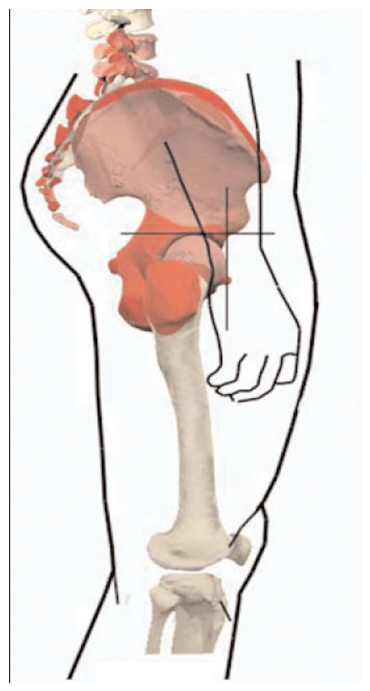
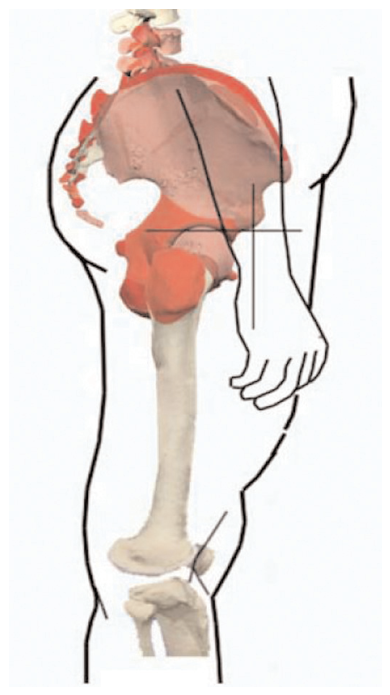
21].) Referred to as the dysfunctional pelvic model, this ascending model is delineated as follows: Abnormal pronation pulls the innominate bones anteriorly; anterior rotation of the innominate bones pulls the acetabula, and the femoral heads, cephalad (upward) and posteriorly; and this, in turn, hyperextends the knees and functionally shortens the legs, with the shortest leg associated with the more pronated foot (Figs. 1 [
and 2 [
).
The objective of this study was to determine whether a correlation exists between abnormal pronation and functional leg-length discrepancies. Three plausible null hypotheses were constructed and tested using the McNemar test for paired proportions: 1) there is no relationship between abnormal foot pronation and hip position; 2) there is no relationship between hip position and functional leg-length discrepancy; and 3) there is no relationship between abnormal foot pronation and functional leg-length discrepancy.
Methods
Methods and Subjects
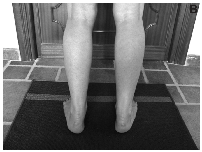
Foot pronation was graded using version 1 of the Foot Posture Index and a modified stance position (Fig. 3 [
). Hip patterns were determined by comparing the relative positions of the posterosuperior iliac spines (standing barefoot). Functional leg-length patterns were determined using a visual-assessment method combined with a pelvic thrust maneuver. All of these measurements were performed by one podiatric physician (B.A.R.) with more than 35 years of research and clinical experience. To attenuate possible experimenter bias, measurements were taken randomly: on the same patient, foot posture, hip pattern, and leg-length evaluations were performed on different days, at least 1 week apart.
The following potential sources of error were identified and controlled for: 1) because foot measurements required quiet standing for up to 5 min, fatigue could affect the position of the foot (all of the subjects tolerated this measurement without fatigue); 2) excess body fat could affect the accuracy of locating body landmarks (these subjects were excluded from this study); and 3) individual differences between subjects could generate random errors (all of the subjects were taken from a genotypically homogenous population).

The inclusion criteria were functional leg-length discrepancy (>3 mm), a positive pelvic thrust response (leg-length discrepancy disappears), and pronated feet in a modified stance position (Foot Posture Index score >2). The exclusion criteria were malocclusions, visual impairments, inner-ear or balance problems, history of trauma to the hip or lower limb, history of surgery of the lower limb, and history of neurologic deficits that affect foot or hip posture.
Three hundred seventy-three indigenous Mexicans living in Cuernavaca, Mexico, were screened; 61 met the inclusion and exclusion criteria. Fifty-six individuals (20 males and 36 females) elected to participate in this study. Their ages ranged from 10 to 67 years (mean age, 33 years), weights from 36 to 75 kg (mean weight, 59 kg), and heights from 1.40 to 1.75 m (mean height, 1.60 m). All of the measurements were taken between June and October 2003. Because this study did not entail any type of intervention, ethical approval was not required by, or requested from, the regional Mexican government. However, all of the participants provided verbal consent before initiation of this study.
Techniques
Identification of Leg-Length Pattern.
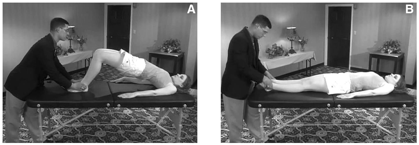
Unshod subjects were placed supine on a table. The shorter leg was identified using the direct (visual) method: with the subject supine on the table, knees extended and feet at right angles to the legs, the levels of the medial malleoli were recorded (with the more proximal malleolus identifying the shorter leg). A pelvic thrust maneuver was performed (Fig. 4 [
). This maneuver tended to level the pelvis and legs. Any remaining leg-length discrepancy after this maneuver was suspected to be anatomical, and these subjects were excluded from this study. Quantitatively, the indirect (measurement) method is considered the most accurate test for identifying an anatomical leg-length discrepancy [
4,
22,
23]. However, from a qualitative point of view, visual assessment combined with a pelvic thrust maneuver proved acceptable for filtering out potential anatomical leg-length discrepancy [
24].
Hip Position.
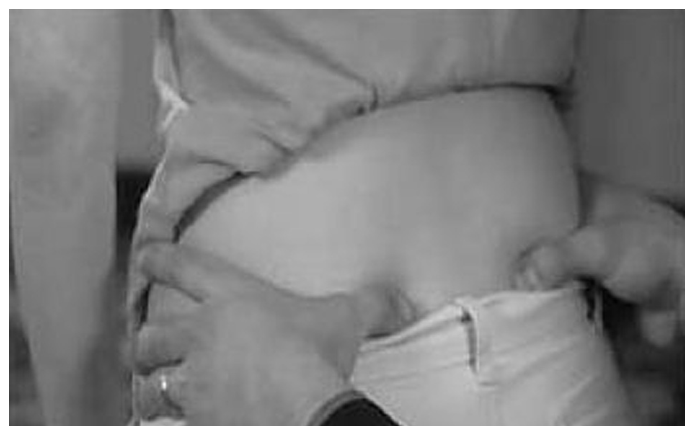
Hip position was assessed by comparing the relative position of the posterosuperior iliac spines. With the subject standing relaxed, unshod, and with vision directed forward, the posterosuperior iliac spines were located by palpation (Fig. 5 [
). Anterior rotation of the innominate bone displaces the posterosuperior iliac spines cephalad (upward). Because the innominate bones rotate independently around the sacrum,
26-
28]. the more superiorly displaced posterosuperior iliac spine identifies the more anteriorly rotated innominate bone.
Statistical Analysis
The raw data generated in this study were tested by Claudia Giacomozzi, PhD, of the Department of Biomedical Engineering, Istituto Superiore di Sanità, Rome.
A McNemar test, using a 2 × 2 contingency table, was applied to the dichotomy variables to obtain a two-sided P value: dichotomy variable 1 (VAR1), functionally short leg (left or right); dichotomy variable 2 (VAR2), greatest Foot Posture Index value (left or right); and dichotomy variable 3 (VAR3), highest posterosuperior iliac spine (left or right). P > .05 gives a high level of confidence that the paired variables are dependent events and proportionally related (ie, rejects the null hypothesis).
Results
Hypothesis 1 was that there was no relationship between VAR2 (abnormal foot pronation) and VAR3 (hip position) (P = .134). A total of 92.8% of the cases matched. Left-sided prevalence was 82.1% in the greatest Foot Posture Index value and 75% in the highest posterosuperior iliac spine.
Hypothesis 2 was that there was no relationship between VAR3 (hip position) and VAR1 (functionally short leg) (P = .248). A total of 94.6% of the cases matched. Left-sided prevalence was 80.3% in the functionally short leg and 75% in the highest posterosuperior iliac spine.
Hypothesis 3 was that there was no relationship between VAR2 (abnormal foot pronation) and VAR1 (functionally short leg) (P = 0.99). A total of 94.6% of the cases matched. Left-sided prevalence was 80.3% in the functionally short leg and 82.1% in the greatest Foot Posture Index value.
The three null hypotheses were rejected. The pronation pattern left > right occurred in 46 subjects (82%), and right > left occurred in 10 subjects (18%). The left innominate bone was rotated more anteriorly than the right innominate bone in 42 subjects (75%), and the right innominate bone was rotated more anteriorly than the left innominate bone in 14 subjects (25%). Statistically, there is a high level of confidence that a dependent relationship exists 1) between abnormal pronation patterns and sagittal plane asymmetry patterns in paired innominate bones (P = .134), 2) between sagittal plane asymmetry patterns in paired innominate bones and the functionally short leg (P = .248), and 3) between the more pronated foot and the functionally shortest leg (P = .99).
Discussion
The objective of this study was to determine whether there was a statistical correlation between abnormal foot pronation patterns and functional leg-length discrepancies. The following three hypotheses were formulated and tested using the McNemar test for paired proportions: 1) there is no relationship between abnormal foot pronation and hip position, 2) there is no relationship between hip position and functional leg-length discrepancy, and 3) there is no relationship between abnormal foot pronation and functional leg-length discrepancy. All three hypotheses were rejected. The results of this study do not suggest that all functional leg-length discrepancies are due to abnormal pronation patterns. For example, malocclusions and craniocervical dysfunctions have been linked to functional leg-length discrepancy [
29-
33]. Neither do the results of this study suggest that all abnormal pronators develop a functional leg-length discrepancy (23 abnormal pronators were excluded from this study because their legs were level). However, there is a high level of confidence that when a functional leg-length discrepancy exists, it can be the result of abnormal pronation.
Furthermore, the results of this study are consistent with my theoretical dysfunctional pelvic model [
19]. According to this model, abnormal pronation can force the innominate bones to rotate anteriorly, with the most anteriorly rotated innominate bone corresponding to the most pronated foot. Anterior rotation of the innominate bones functionally shortens the legs, with the shortest leg corresponding to the most anteriorly rotated innominate bone. Therefore, the shortest leg is ipsilateral to (on the same side as) the most pronated foot.
The dysfunctional pelvic model is an ascending model: it describes the impact that a pronated foot has on the superimposed innominate bone, which, in turn, shortens the leg. Keeling [
34]. evaluated 76 podiatric medical students for leg-length discrepancies and statistically came to a similar conclusion. However, other researchers [
5,
6,
35]. have linked the longer limb to the more pronated foot. None of these studies were statistically based, and none separated functional from anatomical leg-length discrepancy. It is conceivable that an anatomically long leg or a frontal plane torsion of the sphenobasilar articulation can force the ipsilateral foot to abnormally pronate to maintain a level pelvis (a descending model) [
33]. However, these possible “mechanical couplings” are beyond the scope of this study.
The sample sizes used in the present study and in that by Keeling [
34]. (56 and 76 subjects, respectively) were relatively small, and more statistical studies should be conducted using larger samples. Also, the present study would have been improved if measurements had been performed by two or more investigators. However, at this time, no future studies are planned.
Conclusion
Fifty-six indigenous Mexicans participated in a study to determine whether a correlation exists between abnormal pronation and functional leg-length discrepancies. All 56 subjects were evaluated for pronation patterns (which foot was more pronated), hip positions (which posterosuperior iliac spine was higher), and leg-length patterns (which leg was shorter while the subject was lying supine on a table). Plausible null hypotheses were then constructed and tested using the McNemar test for paired proportions. The null hypotheses were rejected, suggesting a high level of confidence that a positive correlation exists between the most pronated foot and the shortest leg and that the ascending dysfunctional pelvic model is valid.
Figure 1.
Level pelvis: the anatomical neutral position of the pelvis.
Figure 1.
Level pelvis: the anatomical neutral position of the pelvis.
Figure 2.
Dysfunctional pelvic model: anterior rotation of the right innominate bone resulting from a pronated foot. Note the cephalad and posterior displacement of the femoral head as the acetabulum shifts upward and backward. Pathomechanical changes include hyperextension of the knees, shortening of the legs, increased pelvic lordosis, and weakening of the abdominal muscles.
Figure 2.
Dysfunctional pelvic model: anterior rotation of the right innominate bone resulting from a pronated foot. Note the cephalad and posterior displacement of the femoral head as the acetabulum shifts upward and backward. Pathomechanical changes include hyperextension of the knees, shortening of the legs, increased pelvic lordosis, and weakening of the abdominal muscles.
Figure 3.
Modified stance position. A, The patient stands in a forward lean position with the body’s weight over the inner longitudinal arch. B, The feet are placed by the subject in a comfortable position as close to their natural base and angle of gait as possible.
Figure 3.
Modified stance position. A, The patient stands in a forward lean position with the body’s weight over the inner longitudinal arch. B, The feet are placed by the subject in a comfortable position as close to their natural base and angle of gait as possible.
Figure 4.
Pelvic thrust maneuver. With the subject supine, the relative positions of the medial malleoli are visually compared (not shown). A, With knees flexed and feet flat on the table, the subject lifts the pelvis from the table. B, With the subject returning the pelvis to the table, the subject’s knees are passively extended. The relative positions of the medial malleoli are again visually compared. This maneuver is used to facilitate pelvic symmetry. If this maneuver eliminates an apparent leg-length difference, a functional leg-length discrepancy is diagnosed. If this maneuver does not eliminate an apparent leg-length difference, an anatomical leg-length discrepancy is suspected. (From the CD-ROM accompanying Baxter RE, Musculoskeletal Assessment, p 146, Elsevier, New York, Copyright 1998. Reprinted with permission from Elsevier.)
Figure 4.
Pelvic thrust maneuver. With the subject supine, the relative positions of the medial malleoli are visually compared (not shown). A, With knees flexed and feet flat on the table, the subject lifts the pelvis from the table. B, With the subject returning the pelvis to the table, the subject’s knees are passively extended. The relative positions of the medial malleoli are again visually compared. This maneuver is used to facilitate pelvic symmetry. If this maneuver eliminates an apparent leg-length difference, a functional leg-length discrepancy is diagnosed. If this maneuver does not eliminate an apparent leg-length difference, an anatomical leg-length discrepancy is suspected. (From the CD-ROM accompanying Baxter RE, Musculoskeletal Assessment, p 146, Elsevier, New York, Copyright 1998. Reprinted with permission from Elsevier.)
Figure 5.
Palpation of the posterosuperior iliac spine. (From the CD-ROM accompanying Baxter RE, Musculoskeletal Assessment, p 146, Elsevier, New York, Copyright 1998. Reprinted with permission from Elsevier.)
Figure 5.
Palpation of the posterosuperior iliac spine. (From the CD-ROM accompanying Baxter RE, Musculoskeletal Assessment, p 146, Elsevier, New York, Copyright 1998. Reprinted with permission from Elsevier.)