


Abstract
Tarsometatarsal arthrodesis is indicated for degenerative joint disease after trauma. Deformity is often associated with post-traumatic arthropathy. Outcomes after surgical management are directly related to realignment. This article reviews the indications, preoperative evaluation, technical execution, and postoperative management of tarsometatarsal arthrodesis. Special emphasis is placed on realignment and restoration of normal foot architecture.
Preoperative Evaluation
Tarsometatarsal (TMT) joint arthrodesis is a salvage procedure recommended for patients with pain, deformity, or gross instability of the TMT joint complex. The most common problem after TMT joint injury is post-traumatic arthritis, which may result from abraded or crushed articular surfaces at the time of injury or from an unrecognized injury. The subsequent development of degenerative changes is markedly increased if the extent of the injury was initially unrecognized, if the injury has been only partially treated, or if the anatomy has not been restored. The typical symptom complex may include local pain confined to the TMT joint or pain secondary to lateral impingement syndrome when associated with adult-acquired flatfoot. Patients may describe positional changes, footwear problems, and gait disturbances. Physical examination may demonstrate deformity in the sagittal and transverse planes, where the forefoot is abducted and dorsiflexed on the rearfoot. Decreased longitudinal arch height, plantar medial eminence, uneven loading of the forefoot with resulting calluses, and altered shoe wear patterns may also be seen. Deformities may be either flexible or rigid. Clinical comparison with the contralateral extremity is helpful. Standard foot radiographs taken in the angle and base of gait should be evaluated to assess degenerative changes and underlying deformity. The quality of the dorsal skin should be evaluated if incisions have been placed for previous open reduction and internal fixation. Poor skin quality may affect decision making regarding incision placement for arthrodesis.
Special Considerations
Key issues when considering TMT joint arthrodesis include timing of surgical intervention, selection of TMT joints for arthrodesis, and type of arthrodesis, ie, in situ versus realignment. Tarsometatarsal joint arthrodesis for end-stage arthrosis may be considered at any time. However, the issue of timing after trauma remains somewhat controversial. Some authors have recommended TMT joint arthrodesis as early as 6 weeks after injury, correlating early surgical intervention to positive outcomes.[1]
Post-traumatic or primary arthritis that involves the TMT joint invariably involves the middle column (second and third rays). The medial column (first ray) is usually involved, whereas the lateral column (fourth and fifth rays) is rarely involved. Global or complete arthrodesis of the TMT joint complex has not been recommended. Arthrodesis of the lateral two rays may not contribute to a successful clinical outcome and may even be detrimental. Salvage arthrodesis of the TMT joint was evaluated by Sangeorzan et al.[1] Their results demonstrated that 69% of the patients experienced a good or excellent outcome. Open reduction and early treatment had a significant positive relation with outcome. They also determined that arthrodesis of the lateral two rays was not a significant factor relative to outcome. Komenda et al[2] and Mann et al[3] determined that arthrodesis of the fourth and fifth TMT joints was associated with poor results. Mulier et al[4] evaluated 28 patients comparing primary arthrodesis with open reduction and internal fixation. In addition, they compared complete arthrodesis and partial arthrodesis. Stiffness of the forefoot, loss of metatarsal arch, and sympathetic dystrophy occurred more frequently in the complete arthrodesis group. They concluded that open reduction and internal fixation with screws or partial arthrodesis is the procedure of choice in severe TMT fracture dislocations. Primary complete arthrodesis should be reserved as a salvage procedure. Raikin and Schon[5] performed a retrospective review of 23 patients (28 feet) after arthrodesis of the fourth and fifth TMT articulations in neuropathic and normosensate feet. Overall, they found a 74.5% improvement in pain and a 73.8% improvement in dysfunction after arthrodesis of the lateral column. The authors concluded that although inclusion of these articulations is not recommended for routine TMT arthrodesis, these joints should be included for patients with uncorrectable lateral midfoot collapse and for those whose painful arthritis involving these articulations has been resistant to adequate nonoperative management.
Berlet et al[6] reported on a procedure in which the cuboid–fourth/fifth metatarsal articulation was excised, and a tendon graft was then interposed in its place. The advantage of this procedure was maintenance of the motion at this articulation while resecting the arthritic joint. They reported a 50% improvement in pain and a 40% improvement in dysfunction at an average of 24 months of follow-up. Myerson[7] recommended mobilization of the lateral column with severe deformities. Fixation is accomplished using Kirschner wires or screws that are removed once weightbearing is started. There are some special situations, such as diabetic neuroarthropathy, in which adequate reduction of the deformity is not possible without mobilization, correction, and arthrodesis of the subluxated or dislocated lateral column.
Arthrosis of the TMT joint complex is invariably associated with significant deformity. However, a small group of patients with TMT joint arthrosis have little or no associated deformity. This can be seen in patients with a rigid cavus foot who experience compression arthropathy of the TMT joint complex. Arthrodesis in situ is indicated for this group of patients with minimal or no deformity. Otherwise, most patients who require TMT joint arthrodesis have associated deformity that will require realignment. Sangeorzan et al,[1] Myerson,[7] and Mann et al[3] reported that correction of residual deformity was the most important predictor of good outcome in TMT arthrodesis.
Preoperative considerations should include the extent of arthrodesis required, the timing of surgical intervention, the severity and reducibility of the deformity, and the condition of the dorsal skin. The extent of arthrodesis can be determined by evaluation of pedal radiographs, passive manipulation of the individual metatarsals, diagnostic intra-articular injections using image-guided delivery of local anesthesia, scintigraphy with high-resolution collimation, and advanced imaging, including computed tomography and magnetic resonance imaging. These various techniques and modalities may provide additional information to guide the surgeon to an accurate assessment as to which individual TMT joints should be incorporated into the arthrodesis.
The primary goals of TMT joint arthrodesis include pain relief, realignment, preservation of foot function, and primary arthrodesis. Although TMT joint arthrodesis is effective in improving symptoms and function, complete resolution of symptoms is unlikely even after successful arthrodesis. Therefore, patients should be thoroughly counseled that although their condition will improve, they are likely to experience residual symptoms.
Surgical Technique
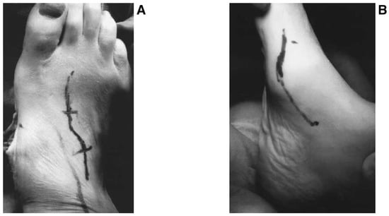
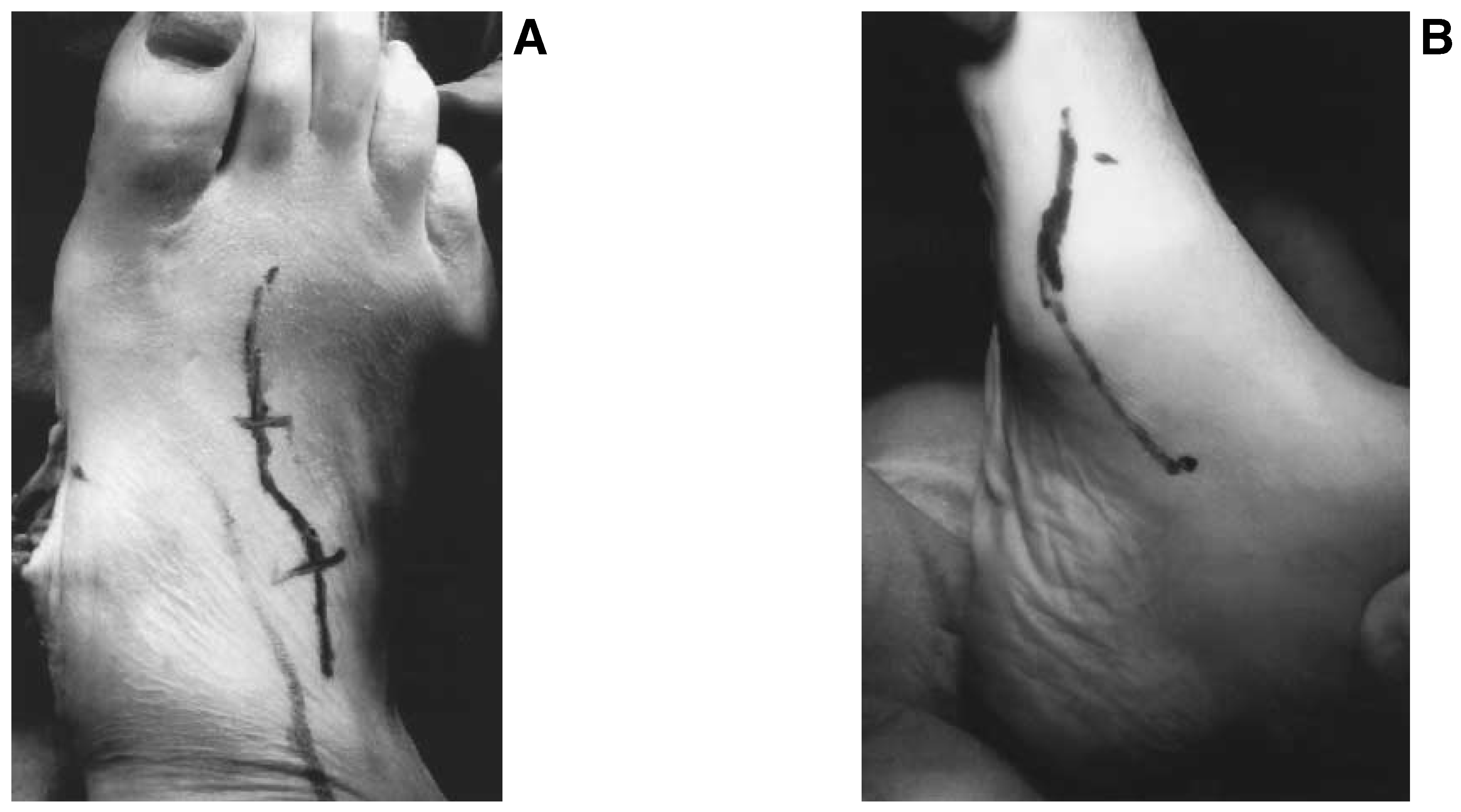
The procedure can be performed under intravenous sedation with a local nerve block using an anesthetic agent with epinephrine for hemostasis. A pneumatic tourniquet is not necessary, especially when TMT joint arthrodesis is performed as an isolated procedure. Alternatively, general inhalation or spinal anesthesia may be used with a pneumatic thigh tourniquet when ancillary procedures, such as calcaneal osteotomies, posterior muscle group lengthening, and harvesting of regional autogenous bone, are to be performed. Tarsometatarsal joint arthrodesis is usually performed through two incisions. One incision is placed medial to the first TMT joint, and a second, lateral incision is placed centrally along the dorsal aspect of the foot in a serpentine manner (Figure 1). However, the degree of correction that is required may affect the incisional approach. The medial incision is used to access the first TMT articulation. This incision provides excellent access for joint debridement and delivery of fixation. Fixation for the first articulation may include interfragmentary compression screws or screws in combination with a plate. Although screw fixation is standard, plate fixation may be considered when osteopenia is an issue or TMT arthrodesis is a component part of the Charcot joint reconstruction. The second dorsal incision is made in a serpentine fashion, with care taken to leave a relatively wide skin island between the two incisions. Dorsal cutaneous nerves, the dorsalis pedis artery, and the deep perforating artery are structures that may be encountered during dissection. Care must be taken to identify and retract these vital structures. One can easily access the second and third TMT articulations through this incision. Joint debridement may be performed with either power or hand instrumentation. Hand instrumentation, such as chisels and osteotomes, is ideal when degenerative changes are not excessive. Debridement with hand instrumentation minimizes shortening and allows a congruous fit when realignment is performed. However, if degenerative changes are extensive, power instrumentation may be preferable to access subchondral bone and develop a healthy cancellous substrate that will result in primary arthrodesis. Alternatively, a medially based wedge may be resected from the TMT joint complex to restore alignment when the deformity is severe and degenerative changes are extensive. This technique is also helpful when the deformity is rigid.
Figure 1.
A, Dorsal incision to access the second and third tarsometatarsal articulations; B, medial incision to access the first metatarsal articulation.
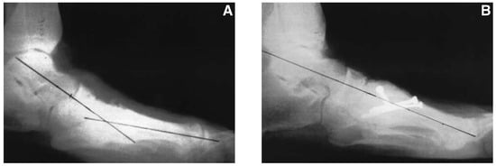
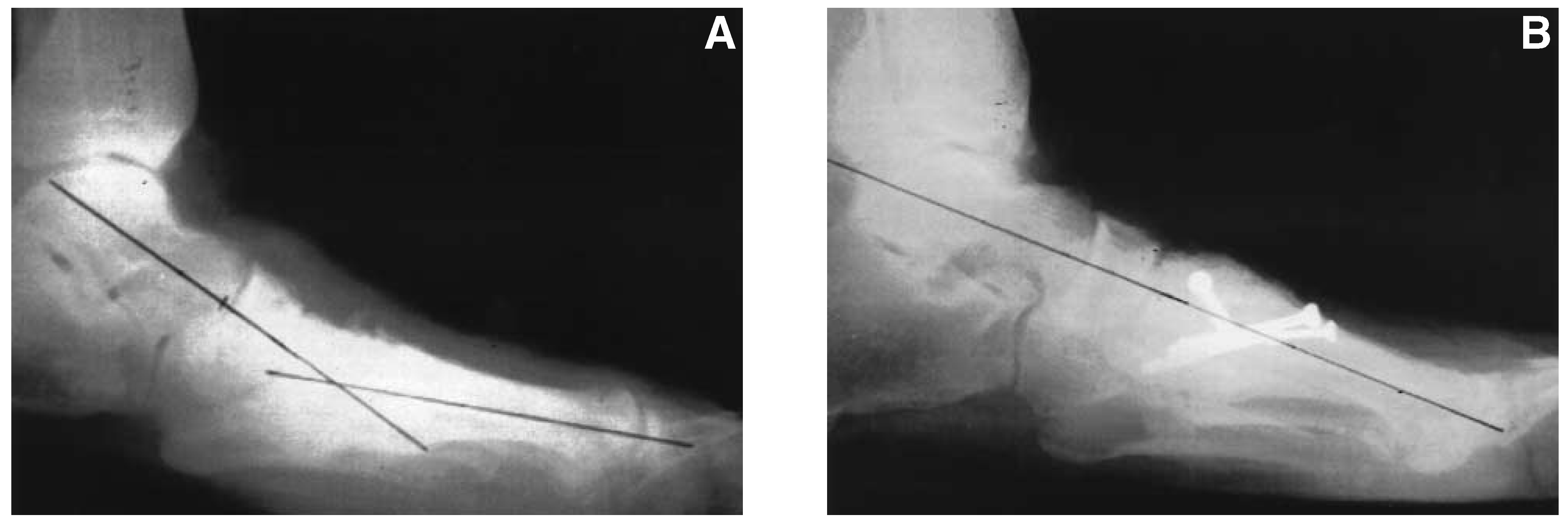
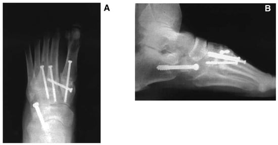
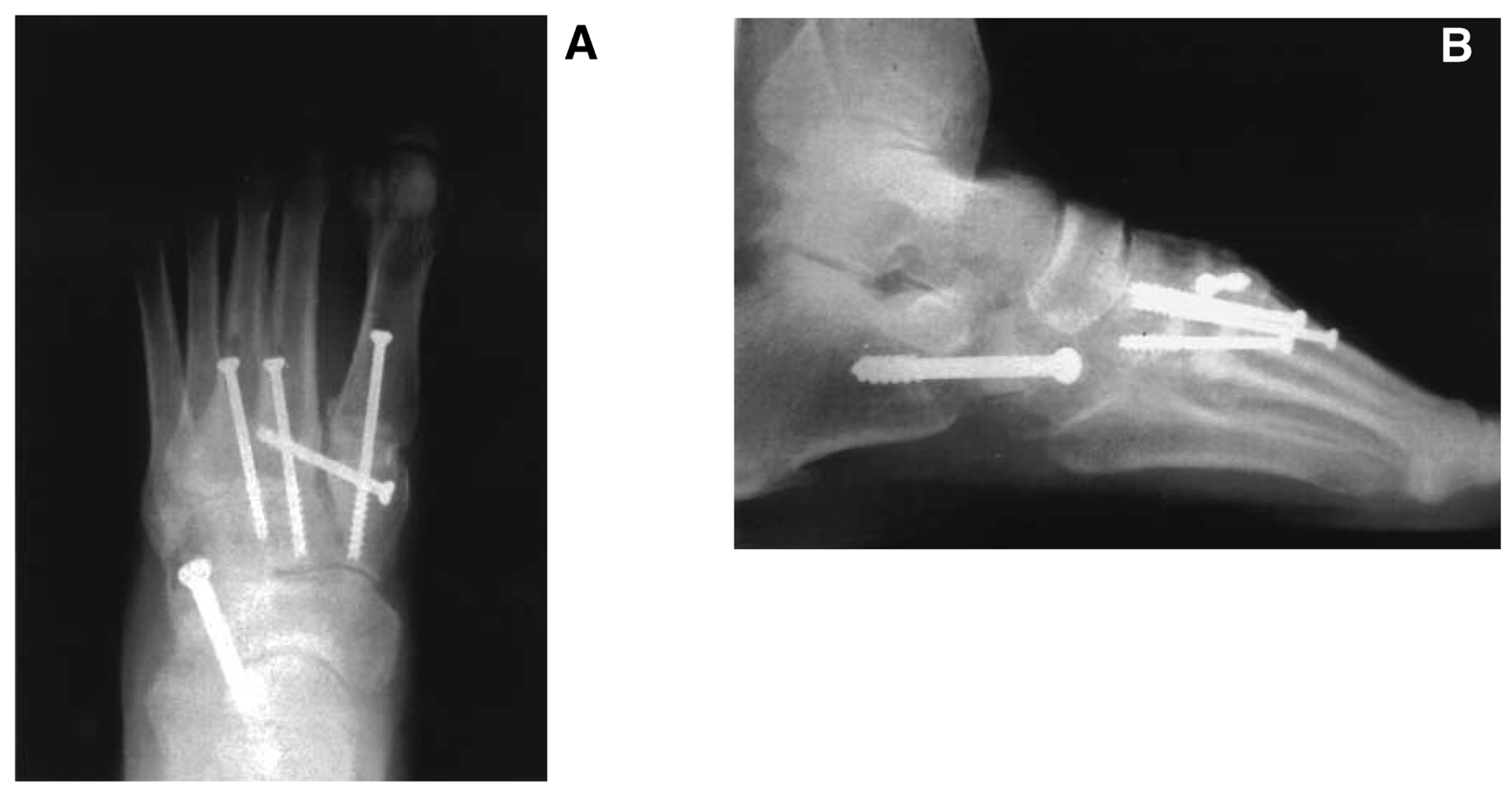
Realignment is performed after joint resection. Manual deformity reduction can be difficult with severe or fixed deformities. A laterally placed distractor may be helpful in restoring alignment in these cases. Intraoperative imaging is helpful for evaluating the reduction in deformity. The malalignment must be completely reduced in the transverse and sagittal planes. Incomplete restoration of sagittal plane alignment with dorsal angulation of the medial column can be debilitating. Sagittal plane realignment is determined on the lateral view by evaluating the talar–first metatarsal angle (Figure 2). Transverse plane realignment can be evaluated on an anteroposterior view by evaluating the alignment of the medial cortex of the second metatarsal relative to the medial cortex of the intermediate cuneiform. These two lines should be as close to one another as possible (Figure 3). Reduction can be maintained with a combination of large bone reduction forceps and cannulated-screw guide pins as provisional fixation. The first TMT articulation is fixated, followed by the second and third, which move together as one unit. Osteosynthesis is achieved with 4.0- or 4.5-mm interfragmentary compression screws. Interfragmentary compression screws are delivered in an axial fashion across the individual TMT articulations. Screw delivery should begin in the proximal one-third of the metatarsal shaft and extend in a proximal direction toward the plantar aspect of the corresponding cuneiforms. Screws can be delivered within the wound or percutaneously under image intensification. Stress risers may develop if screw delivery is too close to the articulation. Longer screws provide greater leverage and are ideal to disperse weightbearing forces during axial loading. A supplemental screw directed from the medial cuneiform into the second metatarsal base may help reduce the diastasis and also provide additional rigidity to the fixation construct (Figure 4). Any remaining defects can be packed with regional autogenous cancellous bone graft, allograft chips, or bone graft substitutes.
Figure 2.
A, Increased talar–first metatarsal angle on a lateral radiograph; B, restoration of the talar–first metatarsal angle after arthrodesis.
Figure 3.
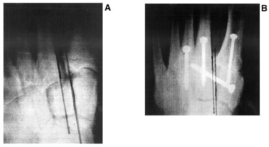
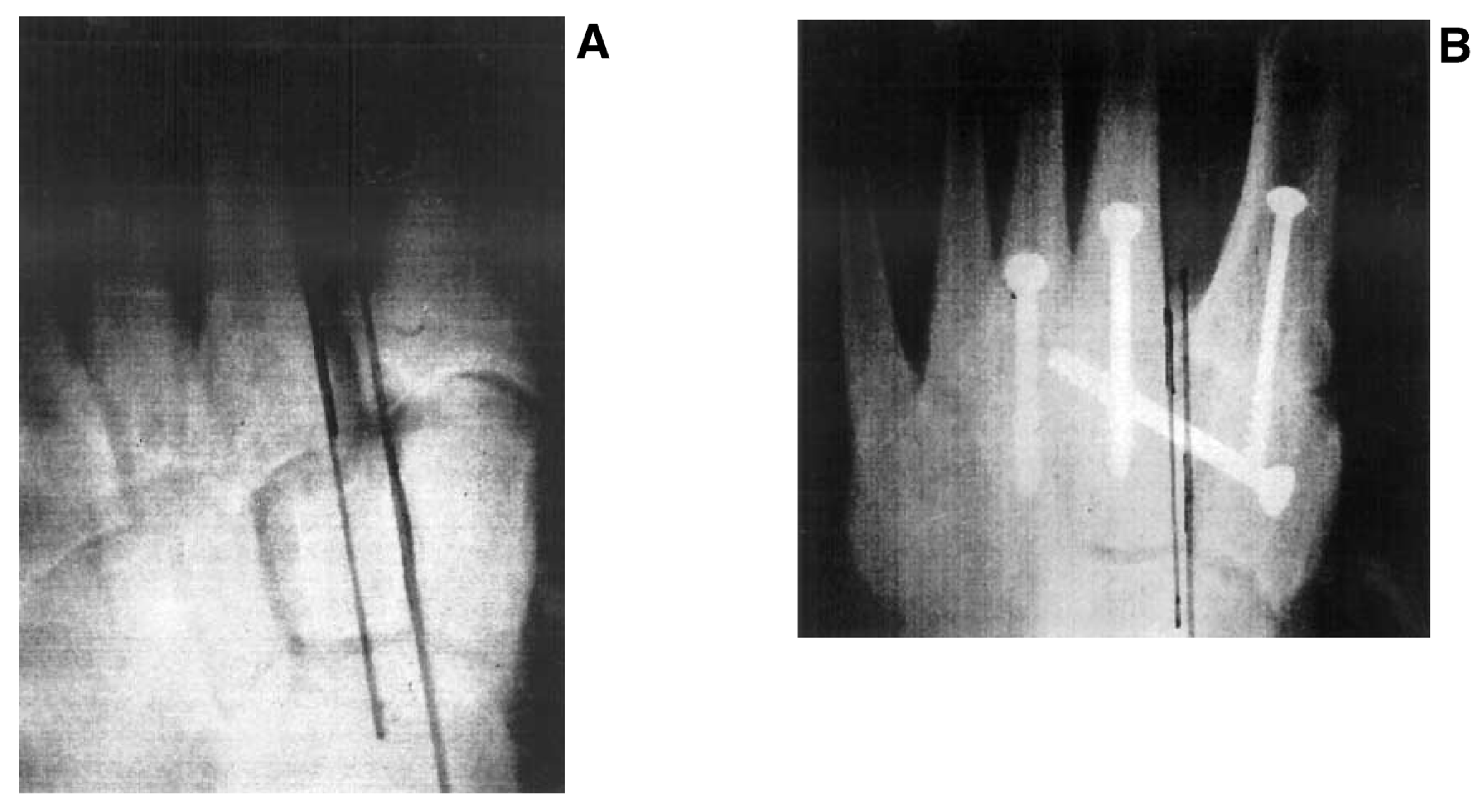
A, Diastasis between the first and second tarsometatarsal articulations demonstrated on an anteroposterior radiograph; B, diastasis is reduced.
Figure 4.
Note the supplemental screw directed from the medial cuneiform into the second metatarsal base.
Ancillary Procedures
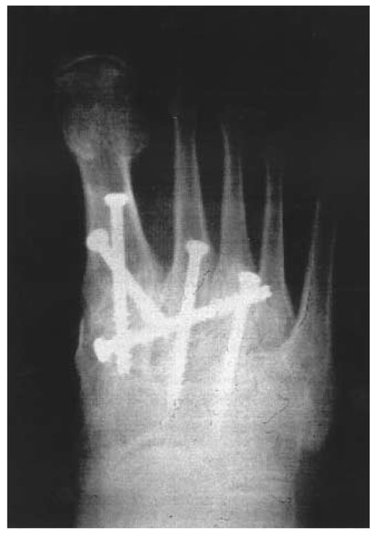
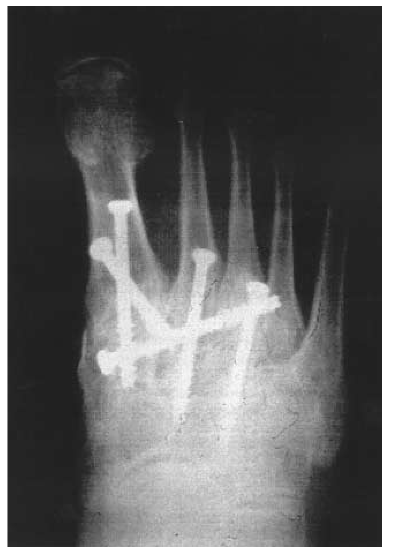
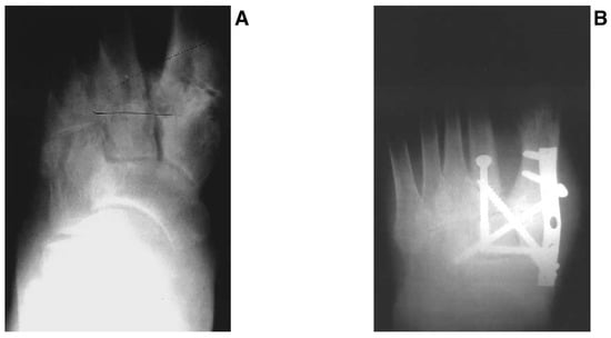
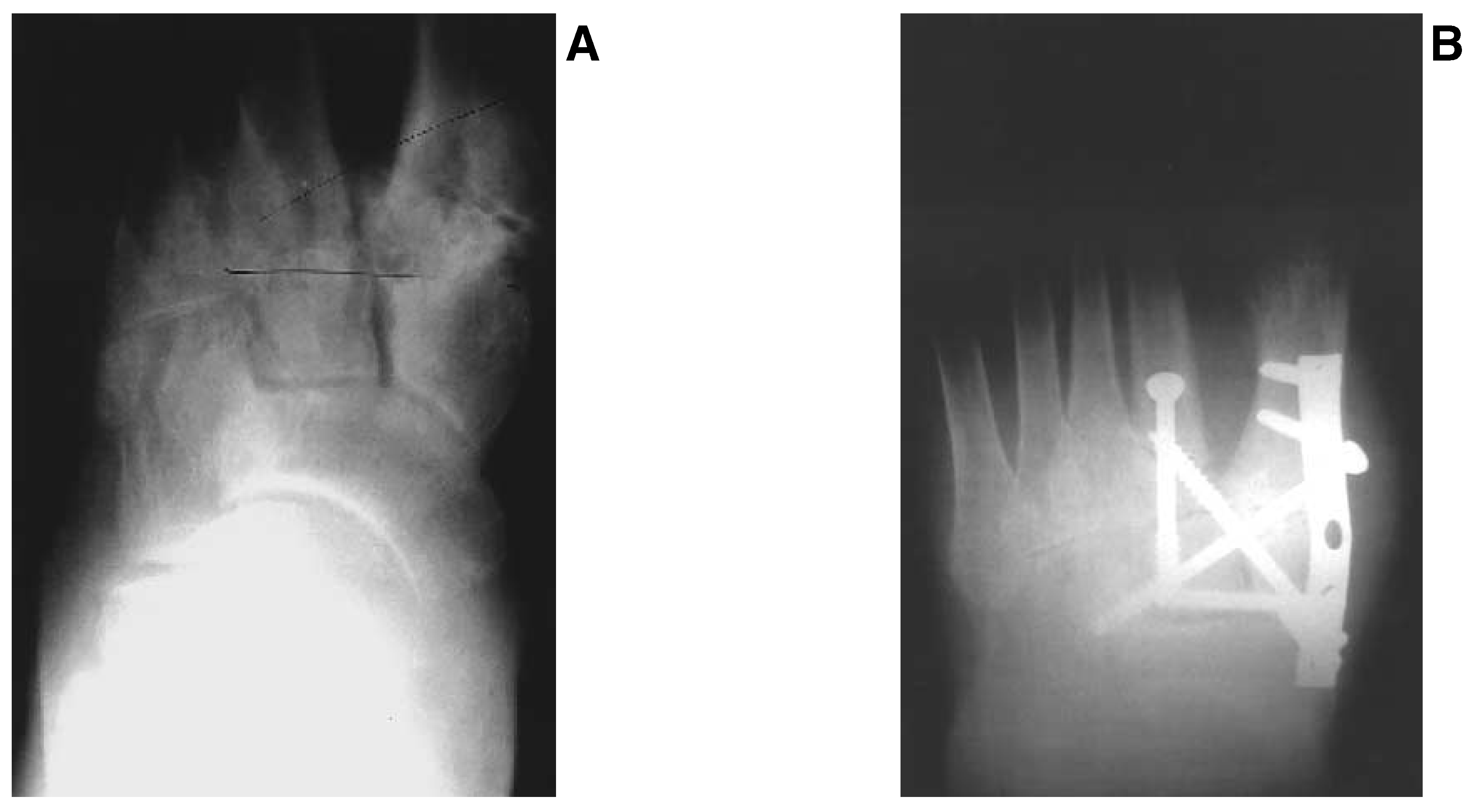
Ancillary procedures may include posterior muscle group lengthening, arthrodesis of the naviculocuneiform joint, calcaneocuboid interpositional bone-block arthrodesis, posterior calcaneal displacement osteotomies, neurectomies, nerve translocation, and hammer toe repair (Figure 5). Selection of ancillary procedures is based on the patient’s individual needs and associated deformity. Technical modifications of TMT arthrodesis should be based on the patient’s specific deformity. There may be situations in which removing a medially based wedge that incorporates multiple TMT joints is appropriate. The wedge resection technique is technically simpler and permits easier application of fixation (Figure 6).
Figure 5.
Anteroposterior (A) and lateral (B) radiographs showing tarsometatarsal arthrodesis combined with calcaneocuboid distraction arthrodesis.
Figure 6.
A and B. Anteroposterior radiographs demonstrating medial wedge resection.
Conclusion
Arthrodesis of the TMT joint complex can be beneficial when nonoperative therapy has been ineffective or when initial treatment has been delayed. Realignment is clearly a significant predictor of good outcome. There has been no correlation between the age of the patient and clinical results. Interfragmentary screws are preferred in most cases. A supplemental plate may be used to enhance the construct when osteopenia is present or when TMT joint arthrodesis is a component part of the Charcot reconstruction.
References
- SANGEORZAN, B.; VEITH, R.; HANSEN, S. Salvage of Lisfranc tarsometatarsal joint by arthrodesis. Foot Ankle Int 1990, 10, 193. [Google Scholar] [CrossRef] [Scilit] [PubMed]
- KOMENDA, G.; MYERSON, M.; BIDDINGER, K. Results of arthrodesis of the tarsometatarsal joints after traumatic injury. J Bone Joint Surg Am 1996, 78, 1658. [Google Scholar] [CrossRef] [Scilit] [PubMed]
- MANN, R.; PRIESKORN, D.; SOBEL, M. Mid-tarsal and tarsometatarsal arthrodesis for primary degenerative osteoarthrosis or osteoarthrosis after trauma. J Bone Joint Surg Am 1996, 78, 1376. [Google Scholar] [CrossRef] [Scilit] [PubMed]
- MULIER, T.; REYNDERS, P.; DEREYNAEKER, G. ET AL: Severe Lisfrancs injuries: primary arthrodesis or ORIF? Foot Ankle Int 2002, 23, 902. [Google Scholar] [CrossRef] [Scilit] [PubMed]
- RAIKIN, S.M.; SCHON, L.C. Arthrodesis of the fourth and fifth tarsometatarsal joints of the midfoot. Foot Ankle Int 2003, 24, 584. [Google Scholar] [CrossRef] [Scilit] [PubMed]
- BERLET, G.C.; ANDERSON, R.B.; DAVIS, W.H. Tendon arthroplasty for basal 4th and 5th metatarsal arthritis. Paper presented at the 15th Annual Summer Meeting of the American Orthopaedic Foot and Ankle Society, Fajardo, Puerto Rico, July 10, 1999. [Google Scholar]
- MYERSON, M.S. Tarsometatarsal arthrodesis: technique and results of treatment after injury. Foot Ankle Clin 1996, 1, 73. [Google Scholar] [CrossRef] [Scilit]
© 2005 American Podiatric Medical Association