


Abstract
Traumatic ankle conditions can lead to long-term sequelae if a pathologic process is misdiagnosed. The clinical presentation of an osteochondral lesion of the talar dome requires the clinician to have a high index of suspicion, and advanced imaging is often necessary to make the final diagnosis. Treatment should be initiated once the lesion is appropriately staged by radiologic or magnetic resonance imaging. We discuss the use of arthroscopy-assisted retrograde drilling of the medial talar dome that spares the articular cartilage within the talotibial articulation.
Ankle and foot injuries are common and involve both osseous and soft-tissue disruption. The complaints can be serious enough to require medical attention and have been reported to account for 10% or more of emergency department visits.[1] Patients with significant ankle injuries often have normal radiologic findings despite severe ligament, tendon, and bone defects.[2] Often the diagnosis of a transchondral fracture or talar dome lesion is delayed, leading to more pain. If immobilization and treatment are delayed, early osteoarthritis occurs in up to 50% of cases.[3] The clinician should have a high index of suspicion for an osteochondral lesion of the talar dome if symptoms persist after conservative treatment.
The etiology of osteochondral lesions of the talus is trauma in most cases[4]; however, embolic, hereditary, endocrine, and developmental factors have also been implicated.[5] Trauma may be direct (impaction) or repetitive (microtrauma). Bui-Mansfield et al[6] described the pathology of these lesions in three stages. Stage 1 (acute injury) features thickened and edematous intra-articular and periarticular soft tissues with adjacent metaphysis osteoporosis from active hyperemia. In stage 2, the epiphysis reveals irregular contour and a thinning of the subcortical zone of rarefaction. The blood vessels within the epiphysis are incompetent because of thrombosis or microfractures. Stage 3 is the period of repair in which granulation tissue gradually replaces the necrotic tissue. Necrotic bone may lose its structural support, resulting in compression and flattening of the articular surface. The injury to the articular cartilage allows for an influx of synovial fluid into the epiphysis, creating a subchondral cyst. This cyst, with an increase in joint pressure, can prevent normal healing.
Proper work-up of the patient should include radiologic or advanced imaging modalities to ensure accurate diagnosis. The initial evaluation includes the radiologic classification of the talus by Berndt and Harty.[4] Stage 1 is a normal radiograph with a subchondral compression fracture of the talus; stage 2, a partially detached osteochondral fragment; stage 3, a completely detached, nondisplaced fragment; and stage 4, a detached, loose fragment. Magnetic resonance imaging (MRI) allows for a more accurate determination of the presence of bone marrow edema and can enable diagnosis of an osteochondral lesion at an earlier stage.
The MRI classification of osteochondral lesions of the talus developed by Anderson et al[7] defines stage 1 as bone marrow edema. Stage 2A represents a subchondral cyst; stage 2B, an incomplete separation of the osteochondral fragment; stage 3, fluid around an undetached, nondisplaced osteochondral fragment; and stage 4, a displaced osteochondral fragment of the talus. There are a variety of grading techniques available; however, MRI classification has been shown to best correlate with surgical staging.[6]
The Ferkel and Sgaglione[8] classification system uses computed tomography (CT) to grade the talar lesion. Stage 1 is defined as a cystic lesion of the talar dome with an intact roof. Stage 2A is a cystic lesion with communication to the talar dome surface, and stage 2B is an open articular surface lesion with an overlying, nondisplaced fragment. Stage 3 is a nondisplaced lesion with lucency, and stage 4 is a displaced osteochondral fragment.
The Cheng et al[9] arthroscopic staging of osteochondral lesions of the talus is as follows. Grade A is an articular cartilage that is smooth and intact but may be soft or ballotable. Grade B has a rough articular cartilage surface, and grade C has fibrillations or fissures. Grade D features an articular cartilage flap with exposed bone; grade E, a loose, nondisplaced osteochondral fragment; and grade F, a displaced osteochondral fragment. This classification is similar to the Outerbridge[10] classification of the International Cartilage Repair Society that was popularized for use in chondromalacia of the patella. The Outerbridge classification is as follows: grade 0, normal articular cartilage; grade 1, softening or superficial fissures and cracks; grade 2, injury involving the transitional zone and extending down to less than 50% of cartilage depth; grade 3, injury involving the deep or radial zone extending to greater than 50% of cartilage depth and down to, but not through, the subchondral bone; and grade 4, subchondral bone exposed with osteochondral injuries with the lesion extending through the subchondral bone plate, or deeper defects down into the trabecular bone.
Once the lesion is appropriately classified, treatment should be instituted on the basis of the preoperative and intraoperative findings. Currently, treatment options include excision of the lesion with curettage and drilling of the subchondral bone, arthroscopic drilling of the lesion, and reduction and fixation of the fragment.[11,12,13] For a posteromedial lesion of the talus, access is limited for open and arthroscopic surgery. Later-stage lesions may require an oblique medial malleolar osteotomy.[14] In the early stages (stage 1 and 2, Berndt and Hardy[4]) of a lesion on the posteromedial talus, retrograde drilling through the sinus tarsi has shown good results and prevents additional postoperative sequelae from the medial malleolar osteotomy.[15] We report an approach to a medial talar dome lesion that is designed to be less invasive and preserve the talotibial articular cartilage.
Case Report
A 34-year-old woman presented to St. Rita’s Medical Center, Lima, Ohio, complaining of pain in her right ankle and the inability to bear full weight on the foot. One month previously, she had injured her ankle and presented to the emergency room for treatment. She was told that there was no fracture and that she had a “sprained ankle.” Compression, ice, and protected weightbearing were initiated, but the symptoms persisted.
On physical examination of the right foot and ankle, the patient had a normal neurovascular status with swelling on the anterior aspect of the ankle. Palpation of the anteromedial aspect of the ankle demonstrated tenderness, and there was pain on inversion, eversion, dorsiflexion, and plantarflexion of the ankle. The results of inversion and eversion stress tests were clinically negative, and radiographs demonstrated no fracture or varus/valgus malalignment. The patient’s foot and ankle were immobilized with a cast, and she was given crutches for gait assistance and sent for MRI. The scans revealed marrow edema within the talar dome consistent with contusion, with no evidence of a transchondral fracture. A more aggressive treatment regimen consisting of nonweightbearing was instituted, because studies have shown that bone marrow edema-like changes in the foot and ankle are predictors of long-lasting pain, often with an uncertain clinical outcome.[16]
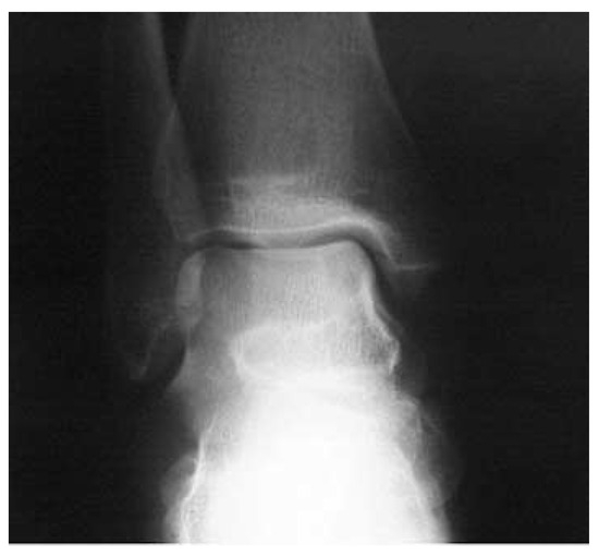
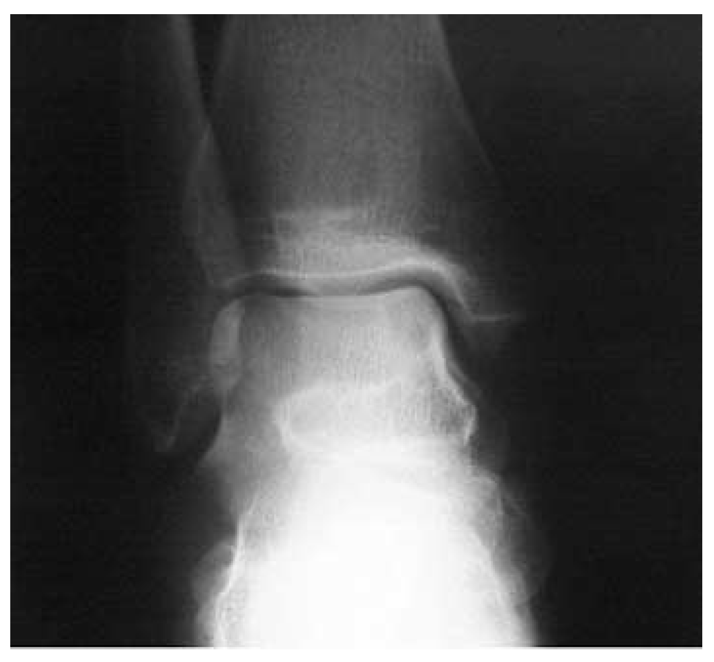
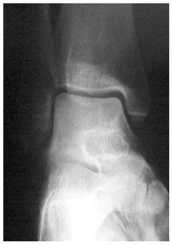
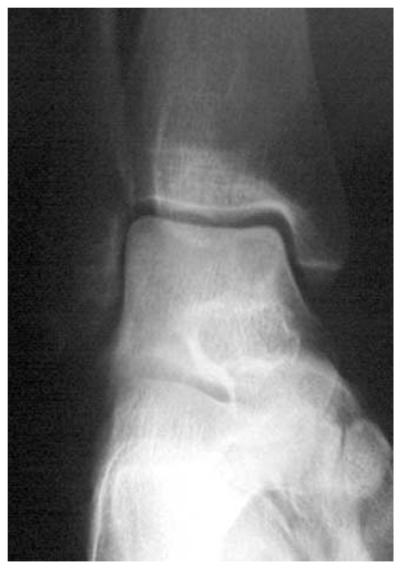
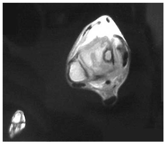
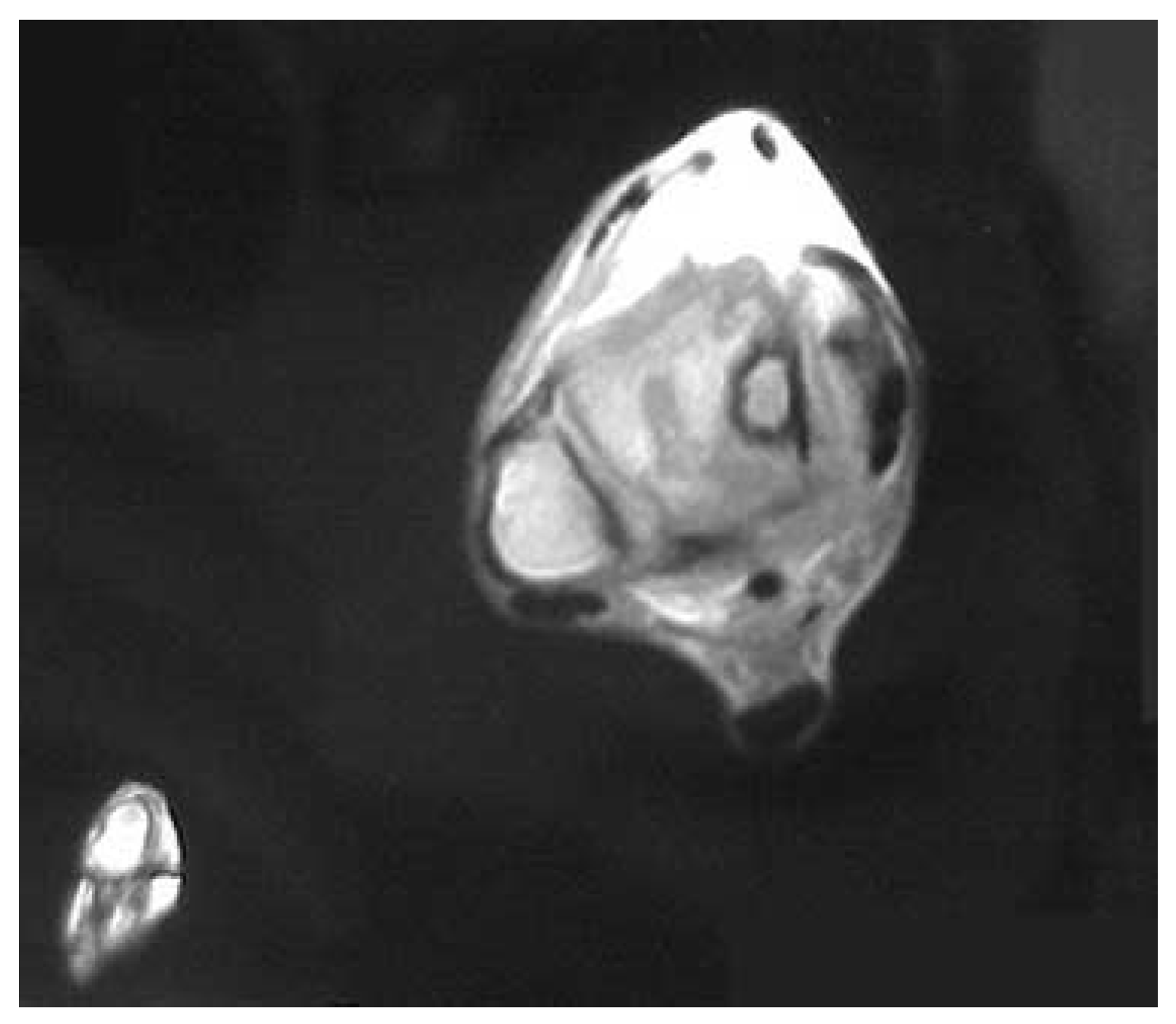
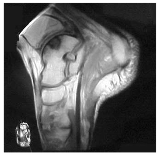
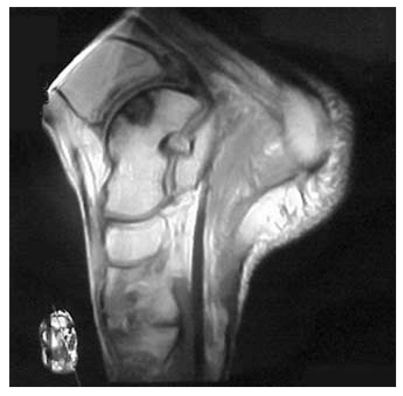
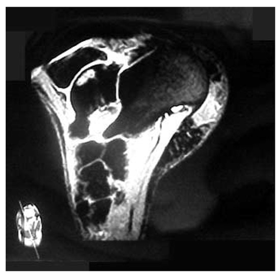
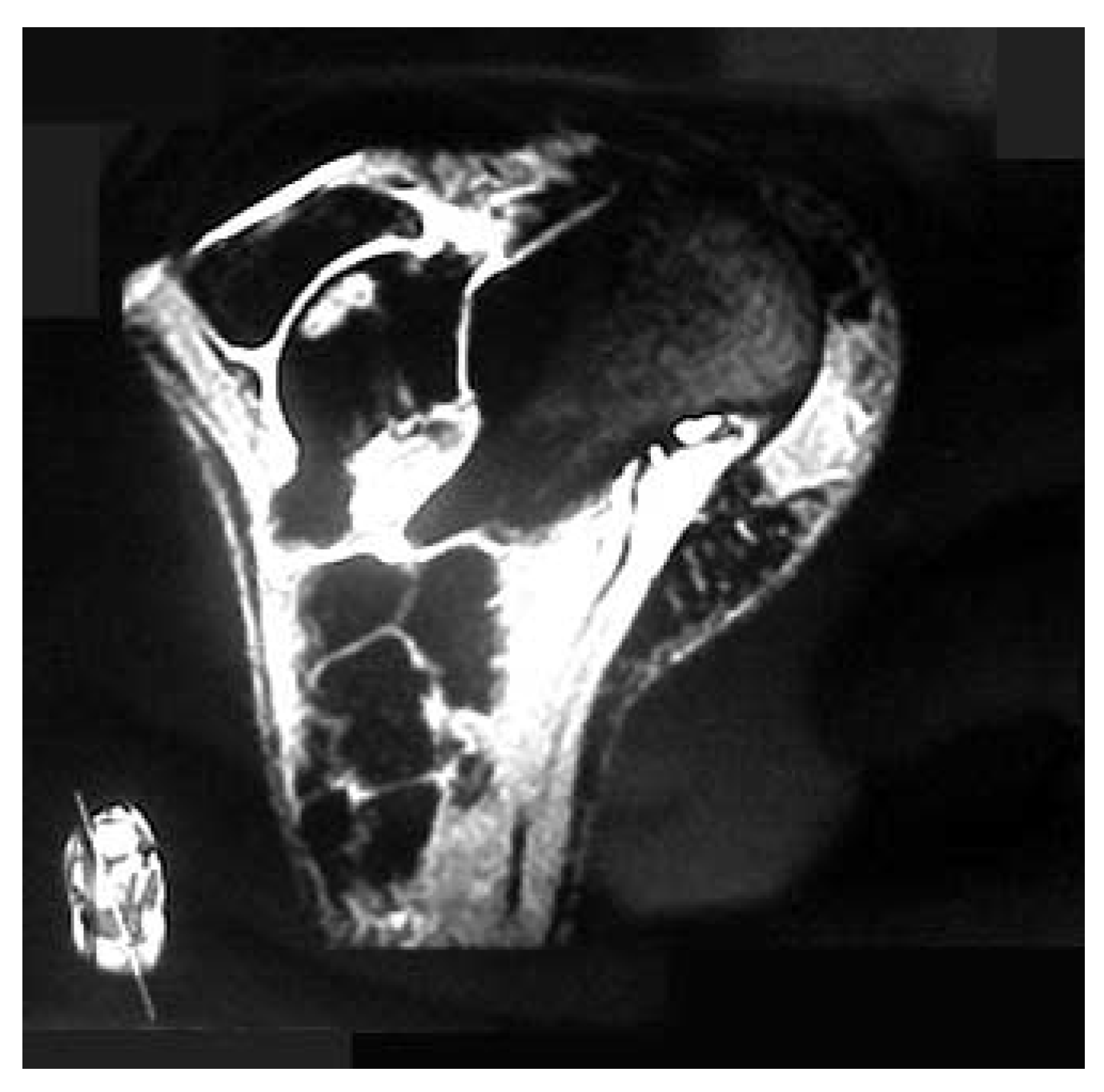
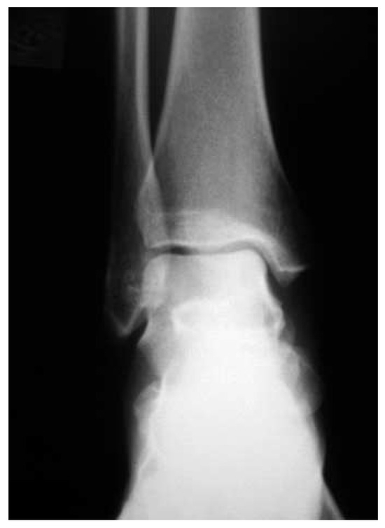
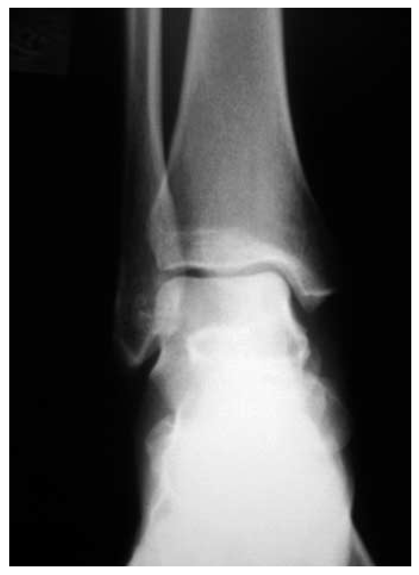
The patient underwent 6 weeks of nonweightbearing with cast immobilization followed by protected weightbearing with the use of a CAM Walker (Zinco Industries, Inc, Pasadena, California) for an additional 6 weeks. At 4 months after the initial injury, the patient was still experiencing pain with weightbearing and range of motion of the ankle joint. Radiographs revealed a talar dome lesion within the medial aspect of the right ankle (Figure 1 and Figure 2). Repeat MRI was ordered, which demonstrated marrow edema with a bony infarct within the posteromedial aspect of the talar dome (Figure 3, Figure 4 and Figure 5). The patient was then immobilized with a cast again and instructed to remain nonweightbearing for an additional 6 weeks. Despite the prolonged 6-month conservative course of nonweightbearing and immobilization, the decision was made to perform arthroscopy with retrograde drilling of the talar dome cyst and packing with autogenous calcaneal bone graft.
Figure 1.
Anteroposterior radiograph of the ankle demonstrating a talar lesion within the medial aspect.
Figure 2.
Mortise view of the ankle confirming that the lesion is confined to the subchondral bone.
Figure 3.
T1-weighted axial magnetic resonance image of the talus demonstrating bone marrow edema and a sclerotic rim.
Figure 4.
T1-weighted magnetic resonance image showing the margins and depth of the lesion.
Figure 5.
T2-weighted magnetic resonance image showing the fluid signal present within the lesion.
The procedure was performed under general anesthesia with the patient in the supine position, with the foot and lower leg prepared and draped under sterile conditions. The ankle was injected with 10 mL of 0.5% bupivacaine with 1:200,000 epinephrine. The traditional two incisions (anterolateral and anteromedial) were used to gain access to the ankle joint.[17] A partial synovectomy with a 2.7-mm, 30° arthroscope with full radius shaver was performed, and the talar dome articular surface was inspected to ensure that there was no articular cartilage damage medially. At this point, we were confident that there was no cartilage involvement, with intact articular cartilage observed on the superomedial and medial surface of the talar dome. Retrograde drilling of the talar dome cyst was determined to be the most efficacious technique to preserve the articular cartilage of the talotibial joint.
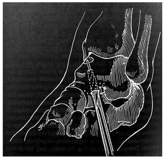
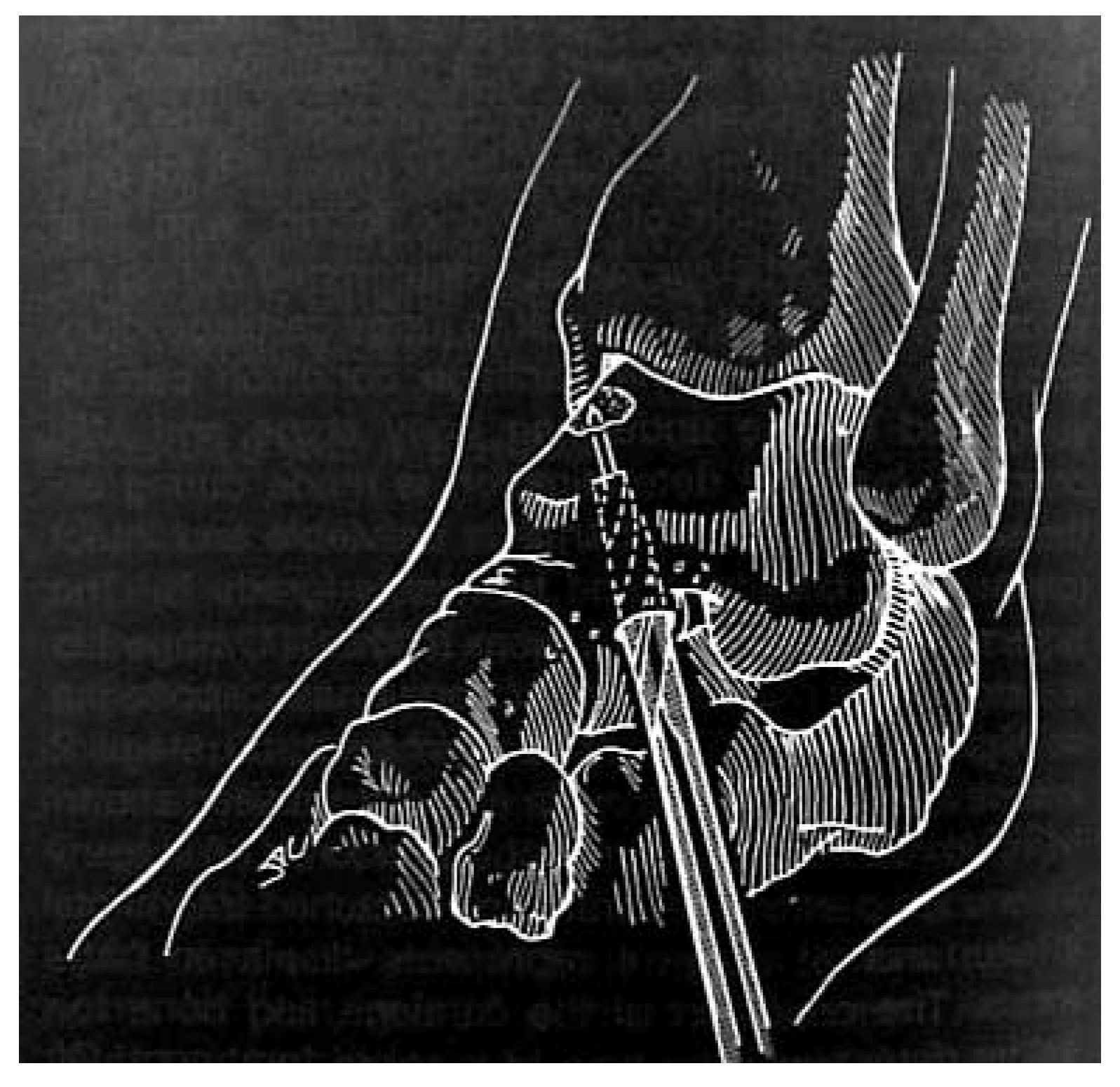
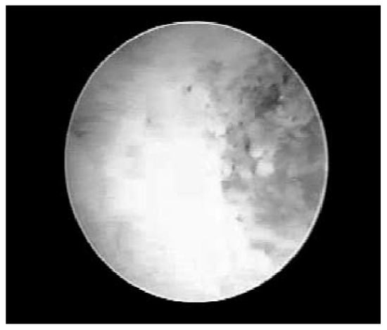
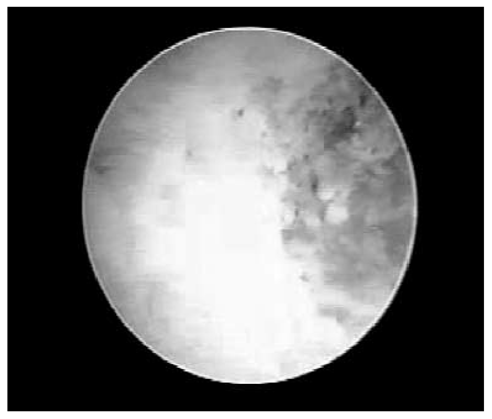
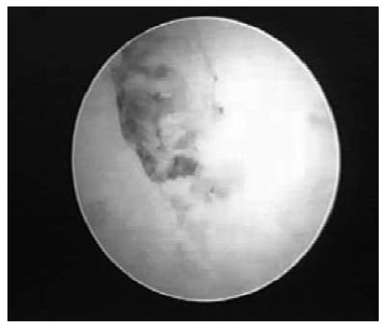
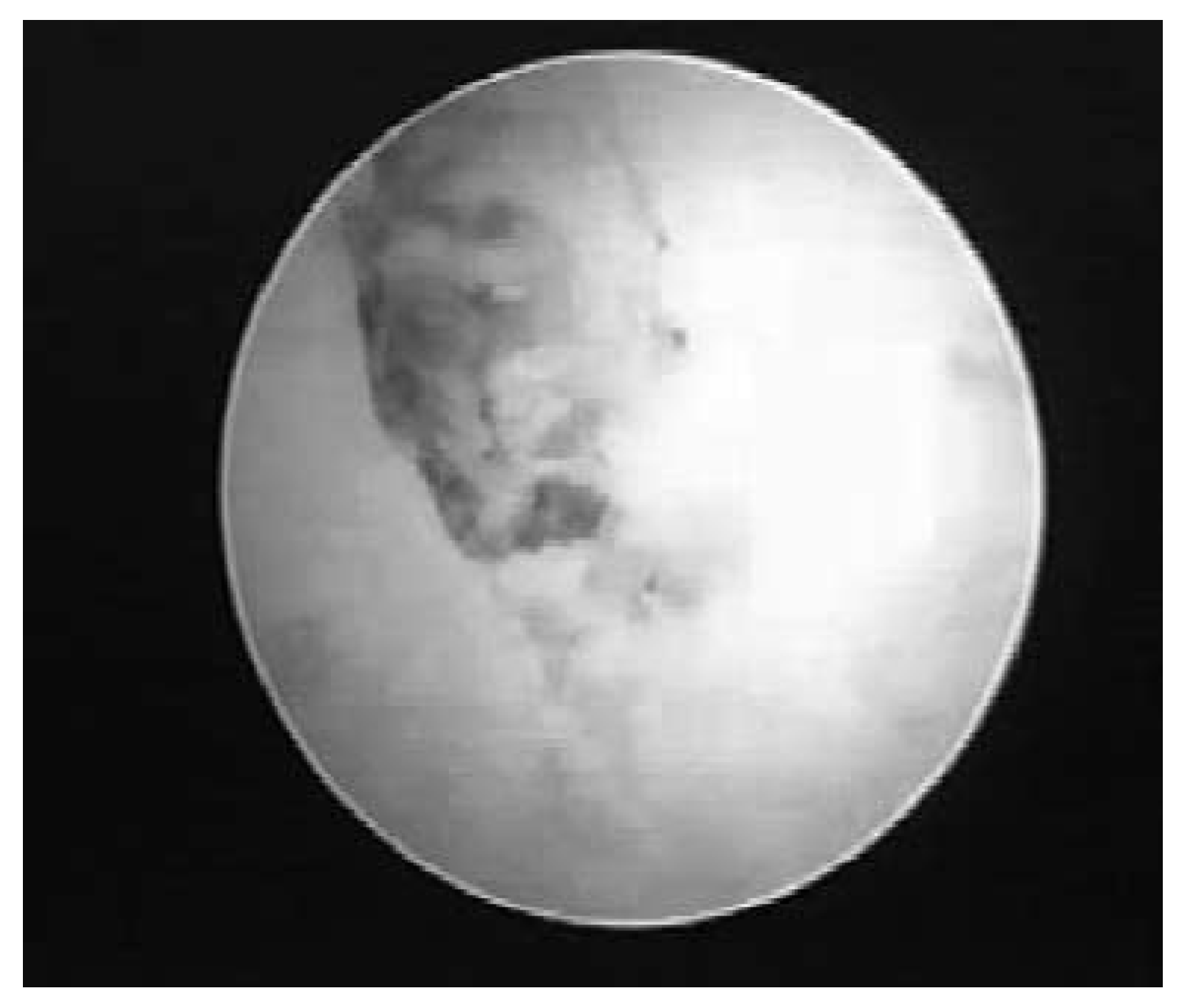
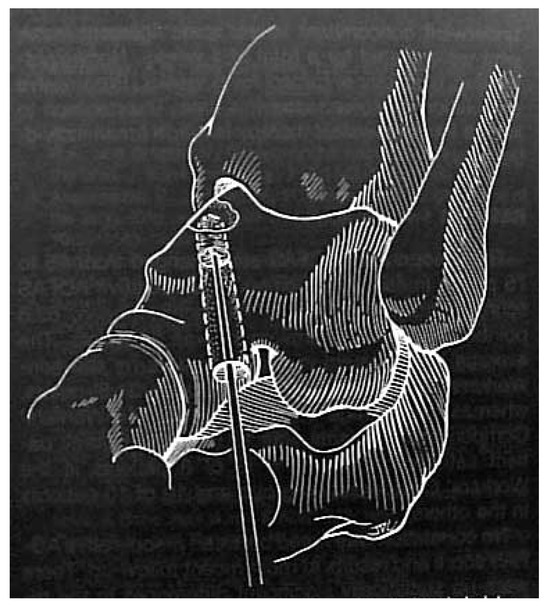
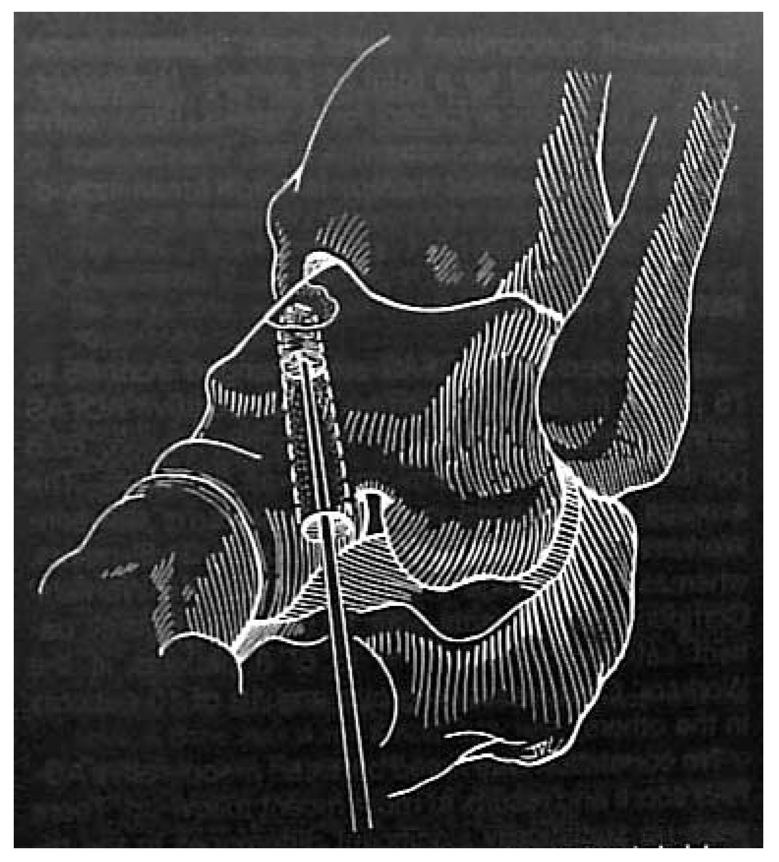
The sinus tarsi was dissected, taking care to retract the dorsal cutaneous nerve medially, and the anterolateral talus was identified. A guide pin from the 3.2-mm cannulated drill set was directed from the anterolateral aspect of the sinus tarsi to the medial talar dome using intraoperative fluoroscopy to ensure proper placement. The pin position was confirmed with the lateral and anteroposterior radiographic views. The talus was drilled with the cannulated 3.2- mm drill bit in a start-stop method to prevent thermal necrosis of the bone, and intraoperative fluoroscopy was used to prevent overaggressive drilling of the lesion (J Christiensen, DPM, personal communication, April 2002) (Figure 6). The instrumentation was removed and the 2.7-mm arthroscope inserted into the drill hole, and the bone and cyst were inspected. We identified a granular tissue within the bone (Figure 7), which broke away under the pressure from the arthroscopic fluid, and the remaining bone was inspected. An angled curette was then used to debride portions of the talar cyst again using intraoperative fluoroscopy. The area was inspected again, and a discrete lesion was observed with the 2.7-mm arthroscope (Figure 8).
Figure 6.
Diagram demonstrating the proper placement of the cannulated pin within the posteromedial talus. (Reprinted with permission from Taranow et al.[15] Copyright © 1999 by the American Orthopaedic Foot and Ankle Society.).
Figure 7.
Intraosseous arthroscopic view of the granular tissue present within the medial talus.
Figure 8.
Intraosseous arthroscopic view of the talus after curettage of the lesion demonstrating the cyst margins.
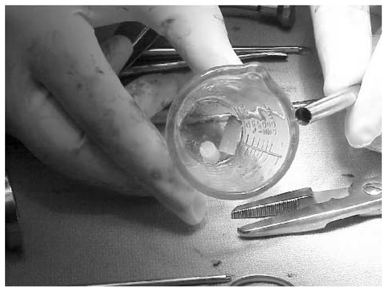
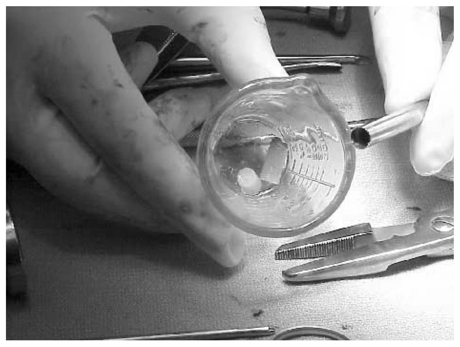
The calcaneal autogenous bone graft was harvested through a lateral incision over the calcaneus, and a 3.5-mm trephine was used to harvest two cores of cancellous bone (Figure 9). The bone graft was then packed within the talus to allow for osteoinduction (Figure 10). The calcaneal harvest site was filled with calcium sulfate pellets, and the incision was closed in layers. The sinus tarsi was irrigated and closed in layers, the foot was dressed with sterile dressings, and a modified Jones compressive dressing was applied. The patient was allowed to bear weight 2 weeks after the procedure. The postoperative course was complicated by a superficial wound infection over the calcaneal bone graft harvest site 12 days after surgery, which resolved after 10 days of oral antibiotic administration. The patient also sprained her ankle 5 months postoperatively, which required immobilization but healed uneventfully. Final postoperative radiographs demonstrated consolidation of the cyst (Figure 11).
Figure 9.
Calcaneal bone graft harvested with a 3.5- mm trephine for packing within the talus.
Figure 10.
Diagram demonstrating the method used to pack the bone graft within the talus. (Reprinted with permission from Taranow et al.[15] Copyright © 1999 by the American Orthopaedic Foot and Ankle Society.).
Figure 11.
Postoperative anteroposterior radiograph after arthroscopy-assisted retrograde drilling of an osteochondral lesion of the posteromedial talar dome.
Discussion
Osteochondral lesions and fractures of the talar dome often go undiagnosed, delaying treatment and leading to poor outcomes. We believe that it is imperative to rule out a talar dome lesion when a “sprained ankle” is unresponsive to an adequate period of conservative therapy. Proper work-up should involve several imaging modalities in a stepwise approach. If radiographs are negative, a three-phase bone scan should be considered to determine whether there is increased osteoblastic activity in the case with a healing osteochondral fracture or cyst. Magnetic resonance imaging offers a significant advantage over other modalities in determining the extent of involvement. It makes it possible to assess whether the articular cartilage is intact, the amount of bone edema, and the presence of a cyst or necrosis. If the articular cartilage is damaged or there is a loose body within the joint, CT allows better visualization of pure osseous involvement. Arthroscopic staging should also be used intraoperatively to determine whether retrograde drilling would help preserve the articular cartilage or whether other modalities are warranted.
Conclusion
We have described arthroscopy-assisted retrograde drilling of a posteromedial lesion of the talar dome classified as Berndt-Hardy[4] stage 1, Anderson[7] stage 2A, Outerbridge[10] stage 0, and Cheng[9] grade A. This procedure can be performed when the articular cartilage is intact and MRI indicates necrosis of the bone. The procedure can reduce the risk of talotibial arthritic changes postoperatively. The surgeon should be comfortable with conventional arthroscopic techniques before undertaking arthroscopy-assisted retrograde drilling of an osteochondral lesion of the posteromedial talus, and methods of preventing osseous thermal necrosis should be employed to avoid the development of more extensive bone necrosis.
References
- BERQUIST, T.H. “Foot, Ankle and Calf,” in MRI of the Musculoskeletal System,, 3rd Ed; Berquist, T.H., Ed.; Lippincott-Raven: Philadelphia, 1996; p. 467. [Google Scholar]
- MAGEE, T.H.; HINSON, G.W. Usefulness of MR imaging in the detection of talar dome injuries. AJR Am J Roentgenol 1998, 170, 1227. [Google Scholar] [CrossRef] [Scilit]
- STOLLER, D.W. “The Ankle and Foot,” in Magnetic Resonance Imaging in Orthopaedics and Sports Medicine,, 2nd Ed; Stoller, D.W., Ed.; Lippincott-Raven: Philadelphia, 1997; p. 518. [Google Scholar]
- BERNDT AL, HARTY M: Transchondral fractures (osteochondritis dissecans) of the talus. Am J Orthop 1959, 41, 988.
- DAVIDSON, A.M.; STEELE, H.D.; MACKENZIE, D.A. ET AL: A review of twenty-one cases of transchondral fracture of the talus. J Trauma 1967, 7, 378. [Google Scholar] [CrossRef] [Scilit]
- BUI-MANSFIELD, L.T.; KLINE, M.; CHEW, F.S. Osteochondritis dissecans of the tibial plafond: imaging characteristics and a review of literature. AJR Am J Roentgenol 2000, 175, 1305. [Google Scholar] [CrossRef] [Scilit] [PubMed]
- ANDERSON, I.F.; CRICHTON, K.J.; GRATTAN-SMITH, T. ET AL: Osteochondral fractures of the dome of the talus. J Bone Joint Surg Am 1989, 71, 1143. [Google Scholar] [CrossRef] [Scilit] [PubMed]
- FERKEL, R.D.; SGAGLIONE, N.E. Arthroscopic treatment of osteochondral lesion of the talus: long term results. Orthop Trans 1993, 17, 1011. [Google Scholar] [CrossRef] [Scilit] [PubMed]
- CHENG, M.S.; FERKEL, R.D.; APPLEFATE, G.R. Osteochondral lesion of the talus: a radiologic and arthroscopic comparison. Paper presented at the Annual Meeting of the Academy of Orthopaedic Surgeons, New Orleans, February 1995. [Google Scholar]
- OUTERBRIDGE RE: The etiology of chondromalacia patella. J Bone Joint Surg Br 1961, 43, 752.
- ALEXANDER, A.H.; LICHTMAN, D.M. Surgical treatment of transchondral talar-dome fractures (osteochondritis dissecans): long-term follow-up. J Bone Joint Surg Am 1980, 62, 646. [Google Scholar] [CrossRef] [Scilit] [PubMed]
- KELBERINE, F.; FRANK, A. Arthroscopic treatment of osteochondral lesions of the talar dome: a retrospective study of 48 cases. Arthroscopy 1999, 15, 77. [Google Scholar] [CrossRef] [Scilit] [PubMed]
- KUMAI, T.; TAKAKURA, Y.; KITADA, C. ET AL: Fixation of osteochondral lesions of the talus using cortical bone pegs. J Bone Joint Surg Br 2002, 84, 369. [Google Scholar] [CrossRef] [PubMed]
- MENDICINO, R.W.; LEE, M.S.; GROSSMAN, J.P. ET AL: Oblique medial malleolar osteotomy for the management of talar dome lesions. J Foot Ankle Surg 1998, 37, 516. [Google Scholar] [CrossRef] [Scilit] [PubMed]
- TARANOW, W.S.; BISIGNANI, G.A.; TOWERS, J.D. ET AL: Retrograde drilling of osteochondral lesions of the medial talar dome. Foot Ankle Int 1999, 20, 474. [Google Scholar] [CrossRef] [Scilit] [PubMed]
- ZANETTI, C.; STEINER, C.L.; SEIFERT, J.H. Clinical outcome of edema-like bone marrow abnormalities of the foot. Radiology 2002, 222, 184. [Google Scholar] [CrossRef] [Scilit] [PubMed]
- FERKEL, R.D. Diagnostic Arthroscopic Examination. In Arthroscopic Surgery: The Foot and Ankle; Whipple, T.L., Ed.; Lippincott-Raven: Philadelphia, 1996; p. p 103. [Google Scholar]
© 2005 American Podiatric Medical Association