


Abstract
Synovial sarcoma of the extremities represents 7% of all soft-tissue sarcomas. This article presents the case of a patient who was treated for a synovial sarcoma of the lateral aspect of the distal lower leg extending to the ankle and involving the fibular bone. The patient underwent a wide excision of the tumor, including the fibular bone, followed by radiation and chemotherapy, rather than undergo an amputation of the right leg. Consideration is also given to the traumatic etiology of the tumor.
Synovial sarcoma of the extremities is uncommon, representing about 7% of all soft-tissue sarcomas.[1-3] Ninety percent of these cases occur in the extremities. The most common site of occurrence is the knee,[4] followed by the ankle and foot region.[1] The tumor presents as a deep-seated mass with or without local pain and tenderness.[5]
In the past, soft-tissue sarcoma lower-extremity tumors were frequently treated with amputation[6-8] because of their tendency to recur locally after conservative resection.[9-11] Salvage of the extremity is now often possible by means of wide resection of the soft and bony tissue compromised by the tumor,[12] followed by radiation[13] and adjuvant chemotherapy,[14-16] without adverse effects on recurrence and survival rates.[17,18]
The case of a patient with synovial sarcoma who was treated with limb salvage is presented. The patient’s long duration of symptoms, confusion in diagnosis, and considerations of a traumatic etiology are reviewed.
Case Report
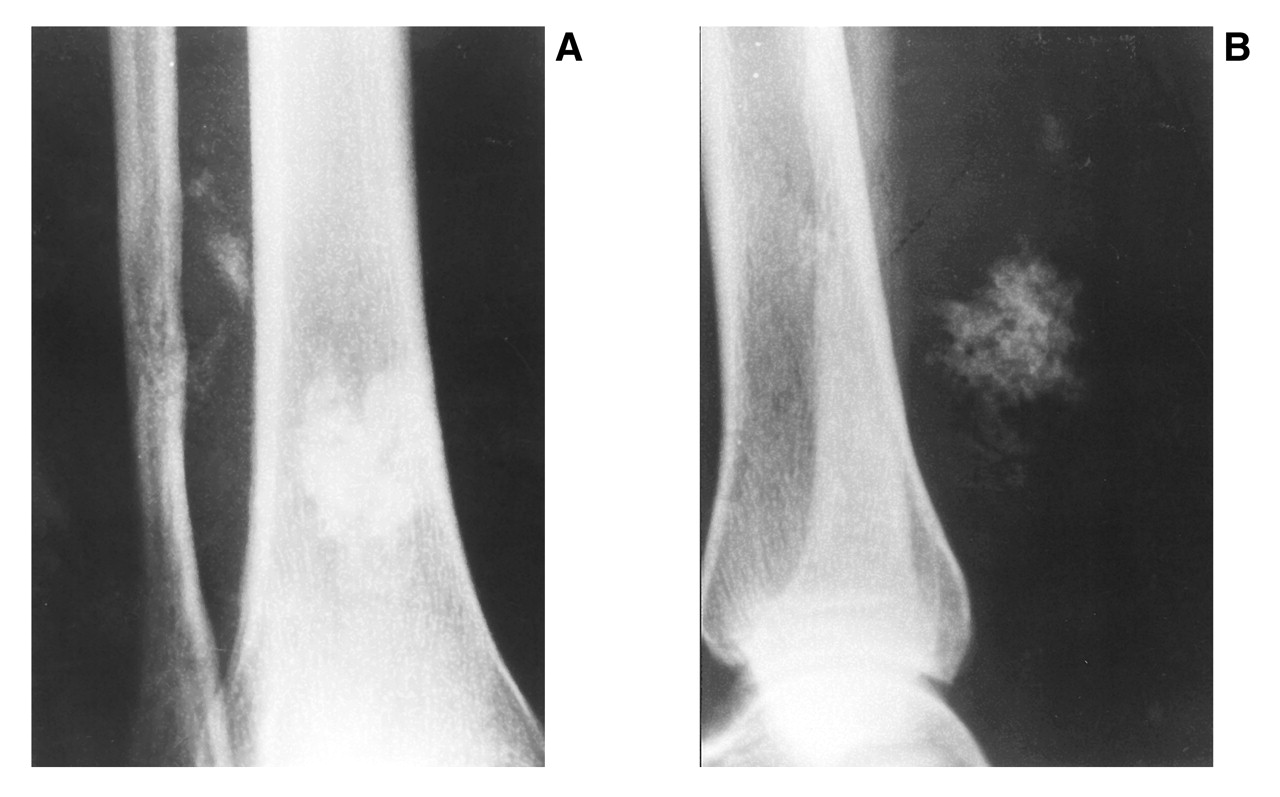
A 40-year-old woman presented with a mass posterior to her right ankle. She had been struck at the back of the lower right leg by a piece of wood when she was 10 years old. Although she had been asymptomatic for 20 years, for the last 10 years she had recurrent episodes of pain and swelling of the leg. Between 1990 and 1995, she was seen by several physicians and her leg had been investigated by means of plain radiographs. Radiographs in 1990 revealed soft-tissue calcification between the distal tibia and fibula (Fig. 1). The patient had been told that the radiographs and ultrasound reports were not suggestive of malignancy; furthermore, one radiologist had reported an exostosis of the distal right fibula.
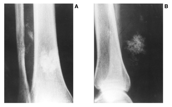
Figure 1.
A, Anteroposterior radiograph shows soft-tissue calcification between the distal tibia and fibula; B, lateral radiograph shows soft-tissue calcification at posterior aspect of tibia.
The pain and swelling of the leg had become persistent and increased during the last year. The patient’s previous physician felt that the case history was suggestive of an infective process, possibly around a foreign body. This was not supported by the findings of a normal white cell count and a low erythrocyte sedimentation rate. Three months earlier, the patient had a biopsy in her home country of Jamaica, which showed round cell–clear cell sarcoma. Her physician suggested a below-the-knee amputation, but the patient refused and came to the United States to avoid amputation.
At the time of the initial physical examination, the patient had no systemic signs, and significant findings were confined to the right leg. There was a diffuse but tense swelling just proximal to the right ankle with a surgical scar from the previous biopsy (Fig. 2). The main swelling was anterolateral but appeared to be related to a posteromedial and posterolateral swelling. There was overlying warmth and tenderness, and no nerve or vascular deficits were detected.
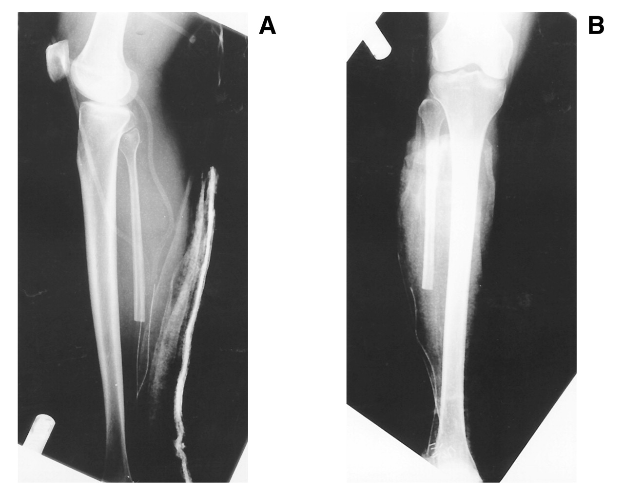
Radiographs showed permeative destruction of the distal right fibula, with some periosteal reaction (Fig. 3). Computed tomography scans revealed mature calcific soft-tissue lesions in the posterior soft tissue (Fig. 4). The leg was explored under general anesthesia, and a fleshy tumor that extended to involve the lateral aspect of the distal leg was found. The patient underwent wide excision of the tumor, including the fibular bone (Fig. 5).
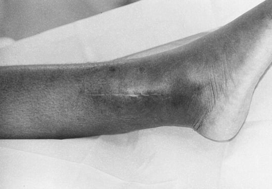
Figure 2.
Diffuse but tense swelling just proximal to the right ankle with a surgical scar from the previous biopsy.
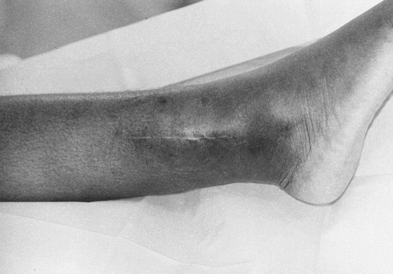
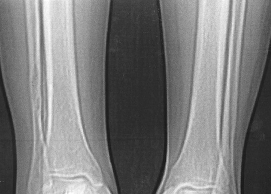
Figure 3.
Radiographs of both legs show permeative destruction of the distal right fibula with some periosteal reaction.
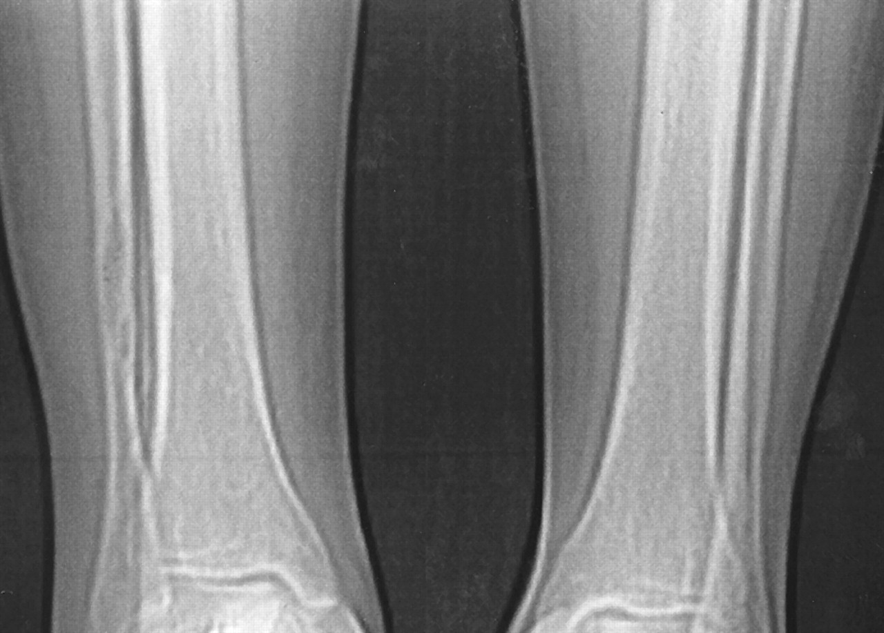
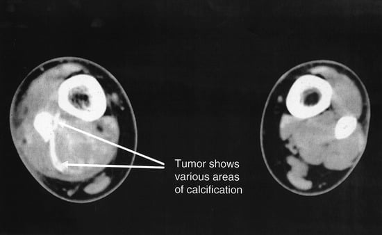
Figure 4.
Computed tomography scan shows mature calcific soft-tissue lesions in the posterior soft tissue.
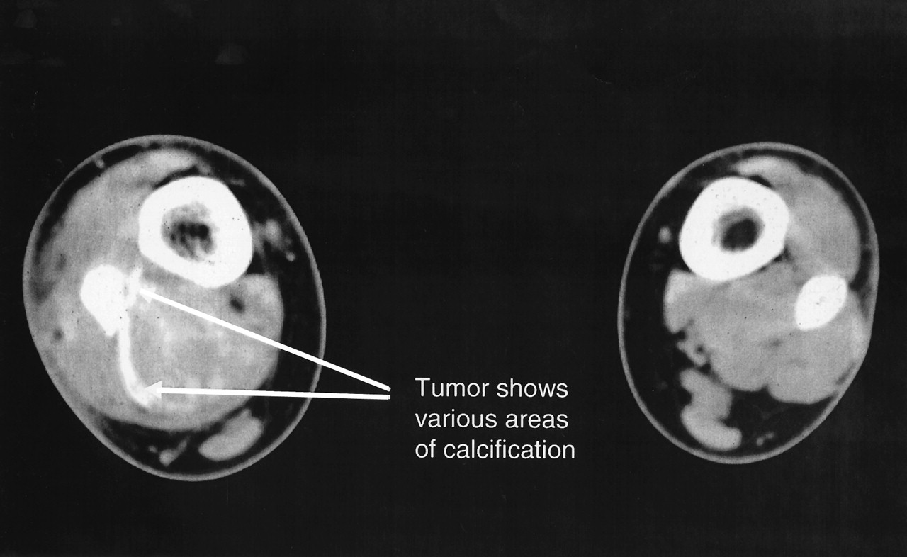
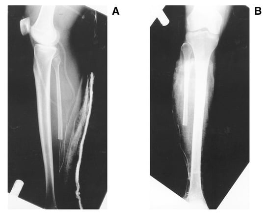
Figure 5.
A, Lateral radiograph of knee and leg shows removal of distal aspect of the fibula bone; B, anteroposterior radiograph of knee and leg shows removal of distal aspect of fibula bone.
The gross section consisted of a surgical wide excision specimen of the right leg mass, composed of an ellipse of skin 17 × 2.5 cm with underlying subcutaneous tissue, skeletal muscle, and segment of fibula. A fusiform ill-defined mass 9 × 6 × 4 cm occupied the lower end of the specimen. A cross-section of the mass revealed a nodular, tan, poorly demarcated fleshy tumor with areas of hemorrhage and necrosis, eroding through the fibula (Fig. 6). Microscopically, the tumor mass was cellular and was uniformly composed of poorly differentiated short spindle cells and round/oval cells in a solid or sheetlike growth pattern (Fig. 7). Biphasic areas were notably absent. The tumor cells had ill-defined eosinophilic or clear cytoplasm with large oval or elongated hyperchromatic nuclei that displayed pleomorphism, with some showing eosinophilic nucleoli. Mitotic figures were frequent (Fig. 8). The tumor had infiltrated the fibular bone (Fig. 9). Light microscopic features were consistent with a poorly differentiated high-grade sarcoma of the lower leg that had eroded the fibular bone with negative proximal and distal margins. According to the American Joint Committee on Cancer’s 1997 classification system of the staging of soft-tissue sarcoma, the tumor was consistent with stage III soft-tissue malignancy, giving it a relatively poor prognosis.[19] Immunohistochemistry performed on paraffin-embedded formalin-fixed tissue at a reference laboratory revealed focal positivity of tumor cells for CAM5.2/AE-1 (markers for cytokeratins) and epithelial membrane antigen, and positive staining of tumor cells for vimentin and CD99 (Ewing’s sarcoma marker). Immunohistochemical stains for S100, smooth muscle actin, human muscle actin, desmin, markers for endothelium (CD31, CD34), CD68 (macrophages), and factor XIII (fibrohistiocytic tumors) were negative. These findings favored a diagnosis of synovial sarcoma.
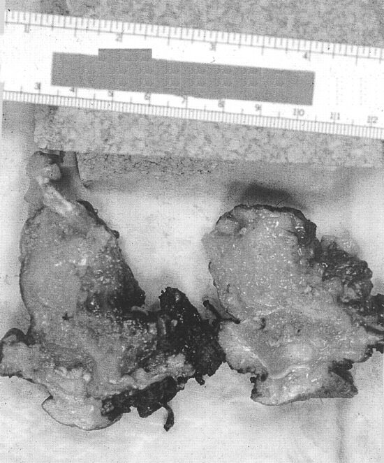
Figure 6.
A cross-section of the mass shows a nodular, tan, poorly demarcated fleshy tumor with areas of hemorrhage and necrosis, eroding through the fibula.
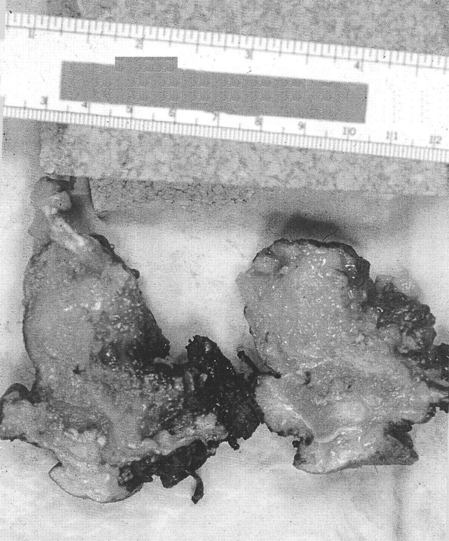
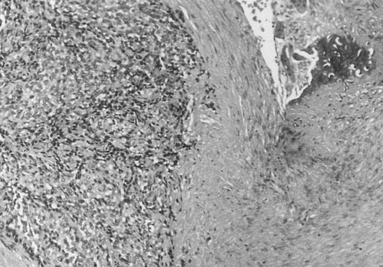
Figure 7.
H&E stain of tumor shows cellular and uniformly poorly differentiated short spindle cells and round/oval cells in a solid or sheetlike growth pattern (H&E ×125).
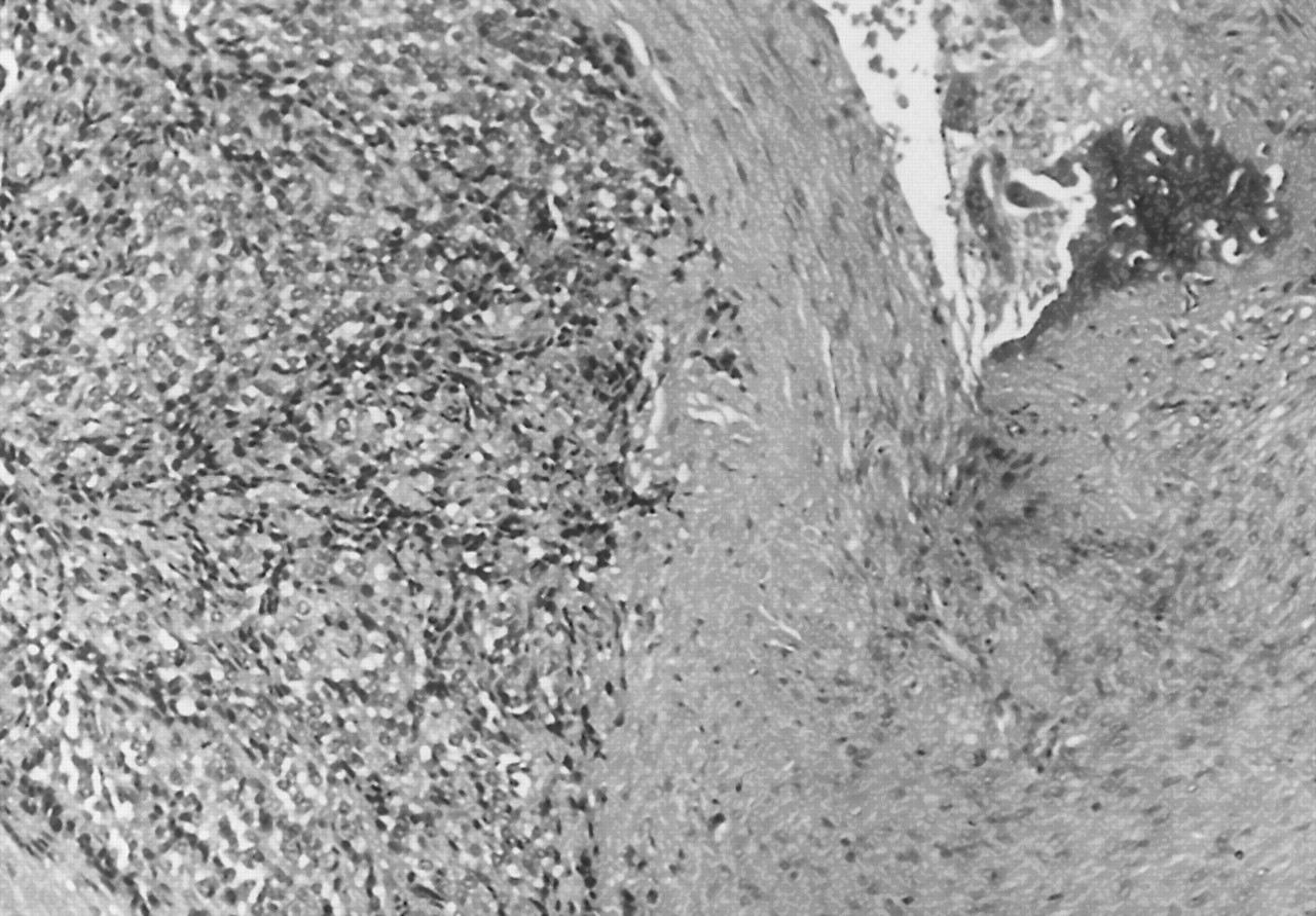
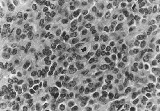
Figure 8.
Cellular pleomorphism with frequent mitoses (H&E ×500).
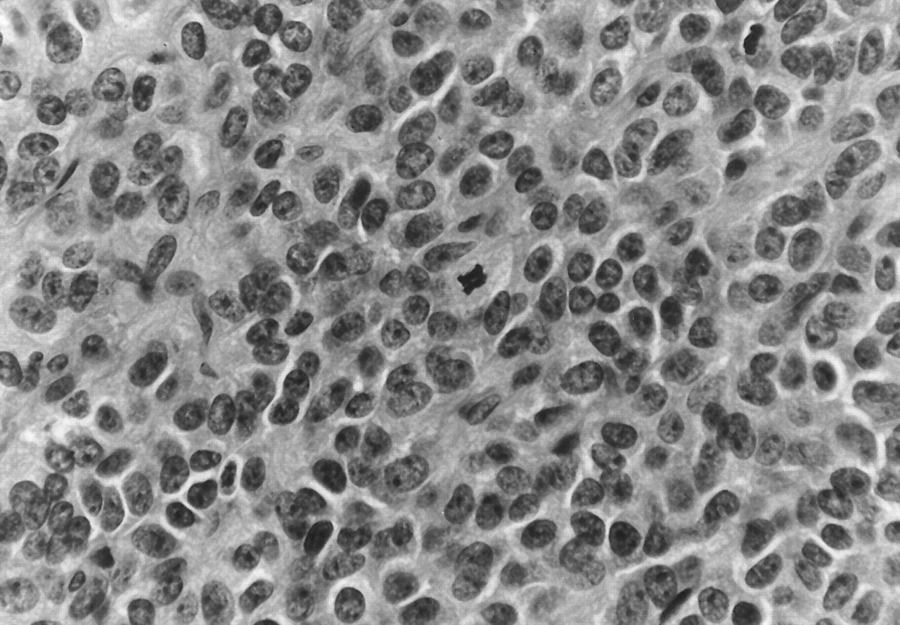
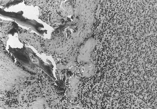
Figure 9.
Tumor eroding bone (H&E ×125).
The patient was placed on a program of adjuvant radiotherapy after surgery. Approximately 1 year later, multiple lesions of metastatic sarcoma were excised from the left lung. The patient was placed on 90 mg doxorubicin (Adriamycin [Adria Laboratories, Columbus, Ohio]) intravenously and 900 mg cyclophosphamide (Cytoxan [Bristol-Myers Squibb Oncology, Princeton, New Jersey]) in 250 mL normal saline. There was no evidence of local recurrence and the patient was able to ambulate properly with a brace and cane (Fig. 10).
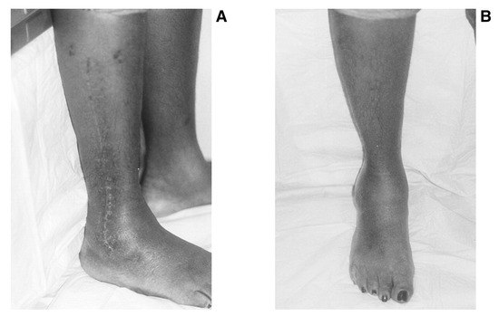
Figure 10.
A, Patient at 1-year follow-up after after limb-sparing surgery; B, the patient now has a functional weightbearing limb.
Discussion
The patient presented in this case report had a stage III poorly differentiated monophasic synovial sarcoma. Criteria for stage III synovial sarcoma include a high-grade, deeply-seated mass greater than 5 cm.[19] Poorly differentiated synovial sarcoma is characterized by high cellularity with numerous mitoses. Microscopically, there were densely cellular sheets of round/oval and spindle cells, typical of undifferentiated sarcoma. Poorly differentiated monophasic synovial sarcoma can have spindle cells with pleomorphism, as demonstrated in this patient.[20]
Immunohistochemistry was necessary in order to make a definitive diagnosis. The patient was immunoreactive for cytokeratins, epithelial membrane antigen, vimentin, and CD99, and negative for CD34, S100, and desmin. Most poorly differentiated synovial sarcomas are immunoreactive for cytokeratin, epithelial membrane antigen, vimentin, CD99, and S100; negative for CD34; and have negative staining for desmin. CD99 is particularly relevant for a diagnosis of poorly differentiated synovial sarcoma. Vimentin is present in the spindle cells of synovial sarcoma. The human hematopoietic progenitor cell antigen CD34 is virtually always negative in synovial sarcoma.[21,22]
There is a history of recent injury in about 25% of cases of common bone and joint tumors.[7] Although the role of severe trauma in the etiology of synovial sarcoma usually is considered to be incidental,[23] posttraumatic etiology is supported when the anatomical site of the final tumor and the initial injury is identical, as in this case.
Pathological mechanisms for malignant post-traumatic etiology cited in the medical literature include an inflammatory process leading to local carcinogenesis,[24] scar formation development in meningioma,[25] chronic foreign-body reaction,[26] or abnormal immune reaction of the muscular tissue to the initial traumatic injury as a causative event.[27,28] Surgical trauma was reported in five patients in which a sarcoma later developed at the site.[29]
Synovial sarcoma has been previously associated with antecedent trauma,[30-35] especially when calcification is present.[5] In a review of 60 cases of synovial sarcoma, a history of trauma was found to have occurred in most cases.[36] However, no relationship was established between the injury and the etiology of the tumor.
Local tumor control with avoidance of amputation has been the main challenge in the surgical management of soft-tissue sarcoma.[8,37,38] Karakousis et al[18] reported that in a study of 85 extremity sarcomas, only three patients were managed with amputation, whereas in the previous decade 40% of patients had been managed with amputation.
More recently, Scully et al[17] reported no difference in survival rates between patients treated with limb salvage and those treated with amputation in 14 patients with synovial sarcoma of the foot and ankle. Similarly, after limb-sparing surgery on 65 patients with sarcoma, 65% were free of disease at the 28-month follow-up, and the authors concluded that local tumor control could be achieved without amputation in 91% of patients.[39] There was no evidence of local recurrence in the patient reported in the current case, and her quality of life has been satisfactory.
The patient presented here had a tumor extending into the lateral aspect of the distal leg and involving the fibular bone. When the tumor is in the posterior compartment, resection of the entire compartment, including the tibial vessels and nerve, is well tolerated. Although there will be a lack of sensation in the plantar surface of the foot during walking, the gait is not impaired.[18] Resection of the lateral compartment and fibula is also well tolerated except for the patient’s inability to evert the foot.[18] However, removal of the peroneal muscles results in dorsal subluxation of the first metatarsal, in the experience of one of the authors (R.O.). The Kirschner wire sufficiently stabilizes the first ray.
Finally, the role of adjuvant chemotherapy and radiation in limb-sparing surgery cannot be overemphasized. Limb-sparing surgery without chemotherapy and radiation results in a high rate of local recurrence.[40,41] Adjuvant chemotherapy with doxorubicin, cyclophosphamide, and methotrexate has been proven to prolong both absence of disease and overall survival in patients with high-grade soft-tissue sarcomas of the extremities.[42-44] In a recent study of 25 children with synovial sarcoma, multimodality treatment with radiation and chemotherapy used in conjunction with limb-preserving surgery resulted in 80% survival after 5 years.[45] Success rates of patients undergoing limb-sparing surgery and radiation have ranged from 60% to 100% for all soft-tissue sarcomas of the extremities.[44,46,47]
Conclusion
The case history of a woman with lower-extremity synovial sarcoma of the lateral aspect of the distal lower leg extending to the ankle and involving the fibular bone was presented. The patient underwent a wide excision of the tumor, including the fibular bone, followed by radiation and chemotherapy, rather than undergo an amputation of the right leg.
Although lower-extremity amputation was routinely performed in the past for surgical treatment of these tumors, current emphasis is placed on the importance of preserving the extremity in patients with a high chance of distal metastasis, in which the quality of life is maintained for the duration of life, or for patients with a good prognosis and for whom the quality of life is part of the overall planning.
References
- Campanacci M: “Synovial Sarcoma,” in Bone and Soft Tissue Tumors, 2nd Ed, ed by M Campanacci, p 1109, Springer-Verlag, Vienna, Austria, 1990.
- Enzinger FM, Weiss SW: Soft Tissue Tumors, 3rd Ed, p 757, CV Mosby, St Louis, MO, 1995.
- Kransdorf MJ: Malignant soft-tissue tumors in a large referral population: distribution of diagnoses by age, sex and location. Am J Roentgenol 164: 129, 1995.
- Wu KK: Synovial sarcoma of the foot. J Foot Ankle Surg 26: 359, 1987.
- Fisher C: Synovial sarcoma. Ann Diagn Pathol 2: 401, 1998.
- Abbas JS, Holyoke ED, Moore R, et al: The surgical treatment and outcome of soft-tissue sarcoma. Arch Surg 116: 765, 1981.
- Cameron HU, Kostuik JP: A long-term follow-up of synovial sarcoma. J Bone Joint Surg Br 56: 613, 1974.
- Shiu MH, Castro EB, Hajdu SI, et al: Surgical treatment of 297 soft tissue sarcomas of the lower extremity. Ann Surg 182: 597, 1975.
- Cade S: Soft tissue tumors: their natural history and treatment. Proc R Soc Med Lond 44: 1, 1951.
- Cantin J, McNeer GP, Chu FC, et al: The problem of local recurrence after treatment of soft tissue sarcoma. Ann Surg 168: 47, 1968.
- Pack GT, Ariel IM: “End Results in the Treatment of Sarcomas of the Soft Somatic Tissues,” in Tumors of the Soft Somatic Tissues: A Clinical Treatise, ed by GT Pack, IM Ariel, p 779, Paul B Hoeber, New York, NY, 1958.
- Chou LB, Malawer MM: Analysis of surgical treatment of 33 foot and ankle tumors. Foot Ankle Int 15: 175, 1994.
- Mullen JR, Zagars GK: Synovial sarcoma outcome following conservation surgery and radiotherapy. Radiother Oncol 33: 23, 1994.
- Kampe CE, Rosen G, Eilber F, et al: Synovial sarcoma. a study of intensive chemotherapy in 14 patients with localized disease. Cancer 72: 2161, 1993.
- Rosen G, Forscher C, Lowenbraun S, et al: Synovial sarcoma: uniform response of metastases to high dose ifosfamide. Cancer 73: 2506, 1994.
- Wright PH, Sim FH, Soule EH, et al: Synovial sarcoma. J Bone Joint Surg Am 64: 112, 1982.
- Scully SP, Temple HT, Harrelson JM: Synovial sarcoma of the foot and ankle. Clin Orthop Rel Res 364: 220, 1999.
- Karakousis CP, Emrich LJ, Krishnamsetty RM: Feasibility of limb salvage and survival in soft tissue sarcomas. Cancer 57: 484, 1986.
- Cheng EY, Thompson RC: New developments in the staging and imaging of soft-tissue sarcomas. J Bone Joint Surg Am 81: 882, 1999.
- Meis-Kindblom JM, Stenman G, Kindblom LG: Differential diagnosis of small round cell tumors. Semin Diagn Pathol 13: 213, 1996.
- Suster S, Fisher C, Moran CA: Expression of bc12 oncoprotein in benign and malignant spindle cell tumors of soft tissue, skin, serosal surfaces and gastrointestinal tract. Am J Surg Pathol 22: 863, 1998.
- van de Rijn M, Rouse RV: CD34: a review. Appl Immunohistochem 2: 71, 1994.
- Buchan JH, Groman AD, Rubin ND: Synovial sarcoma: a review and pedal presentation. JAPMA 70: 342, 1980.
- Delpla PA, Rouge D, Durroux R, et al: Soft tissue tumors following traumatic injury: two observations of interest for the medicolegal causality. Am J Forensic Med Pathol 19: 152, 1998.
- Kotzen RM, Swanson RM, Milhorat TH: Post-traumatic meningioma: case report and historical perspective. J Neurol Neurosurg Psychiatry 66: 796, 1999.
- Brand KG: Diversity and complexity of carcinogenic processes: conceptual inferences from foreign body tumorigenesis. J Natl Cancer Inst 57: 973, 1976.
- Benninghoff D, Robbin R: The nature of desmoid tumor. Am J Roentgenol 91: 132, 1964.
- Choong PF, Gustafson P, Wilen H, et al: Prognosis following recurrent soft-tissue sarcoma: a staging system based on primary and recurrent tumour characteristics. Int J Cancer 60: 33, 1995.
- Dijkstra MK, Balm AJ, Gregor RT, et al: Soft tissue sarcomas of the head and neck associated with surgical trauma. J Laryngol Otol 109: 126, 1995.
- Milchgrub S, Ghandur-Mnaymneh L, Dorfman HD, et al: Synovial sarcoma with extensive osteoid and bone formation. Am J Surg Pathol 17: 357, 1993.
- Chimenti S, Calvieri S, Ribuffo M: Synovial sarcoma of the foot. J Dermatol Surg Oncol 8: 882, 1982.
- Sen S, Dooley WT: Synovial sarcoma. J Nat Med Assoc 79: 1089, 1987.
- Beasley CW: Synovial sarcoma of the foot: a case report. JAPMA 61: 485, 1971.
- Galinski AW, Vlaho M: Malignant synovioma of the foot: a case report. JAPMA 65: 175, 1975.
- Reale CD, Sinclair G: Synovial sarcoma in the foot. J Foot Surg 25: 124, 1986.
- Pack GT, Ariel IM: Synovial sarcoma (malignant synovioma). Recent Adv Surg 28: 1047, 1950.
- Paz IB, Wagman LD, Terz JJ, et al: Extended indications for functional limb-sparing surgery in extremity sarcoma using complex reconstruction. Arch Surg 127: 1278, 1992.
- Potter DA, Kinsella GJ, Kinsella T, et al: Patterns of recurrence in patients with high-grade soft-tissue sarcoma. J Clin Oncol 3: 353, 1985.
- Eiber FR, Mirra JJ, Grant TT, et al: Is amputation necessary for sarcomas? A seven-year experience with limb salvage. Ann Surg 192: 431, 1980.
- Rosenberg SA, Glatstein ES: Perspectives on the role of surgery and radiation therapy in the treatment of soft tissue sarcomas of the extremities. Semin Oncol 8: 190, 1981.
- Shiu M, Hajdu S: Management of soft tissue sarcoma of the extremity. Semin Oncol 8: 172, 1981.
- Rosenberg SA, Kent H, Costa J, et al: Prospective randomized evaluation of the role of limb-sparing surgery, radiation therapy, and adjuvant chemoimmunotherapy in the treatment of adult soft-tissue sarcomas. Surgery 84: 62, 1978.
- Rosenberg SA, Tepper J, Glatstein E, et al: Prospective randomized evaluation of adjuvant chemotherapy in adults with soft tissue sarcomas of the extremities. Cancer 52: 524, 1983.
- Potter DA, Kinsella T, Glatstein E, et al: High-grade soft tissue sarcomas of the extremities. Cancer 58: 190, 1986.
- Ferrar A, Casanova M, Massimino M, et al: Synovial sarcoma: report of a series of 25 consecutive children from a single institution. Med Pediatr Oncol 32: 32, 1999.
- Fein DA, Lee WR, Lanciano RM, et al: Management of extremity soft tissue sarcomas with limb-sparing surgery and postoperative irratiation: do total dose, overall treatment time, and the surgery-radiotherapy interval impact on local control. ?Int J Radiat Oncol Biol Phys 32: 969, 1995.
- Lindberg RD, Martin RG, Romsdahl MM, et al: Conservative surgery and postoperative radiotherapy in 300 adults with soft-tissue sarcomas. Cancer 47: 2391, 1981.
© 2002 American Podiatric Medical Association