


Abstract
In the offset V-bunionectomy used for hallux valgus repair, both the Kalish and the Vogler variations have a long dorsal arm, but the apex is more distal in the Kalish procedure. This study investigated the effect that pin orientation and location of the osteotomy apex have on weightbearing stability. The authors studied saw bone models that were loaded to failure in an Instron 4201 materials testing machine and, in addition, designed, fabricated, and used a unique jig assembly to help minimize data variability. Statistically significant differences were found between the surgical techniques and pin orientations: the Kalish osteotomy was stronger than the Vogler procedure, and in both osteotomies, the plantarly directed Kirschner wire orientation was stronger than the dorsally directed orientation.
The offset V-bunionectomy is an accepted procedure performed for hallux valgus repair. Several variations in currently practiced techniques achieve similar results: reduction in the intermetatarsal angle, reduction in the proximal articular set angle, and internal fixation stability to allow primary bone union. Many variables may contribute to the stability of an osteotomy, including the degree of displacement, geometry of the cuts, location of the osteotomy apex relative to the weightbearing load, fixation technique, and orientation.
The Austin osteotomy has a symmetrical V-configuration with dorsal and plantar arms of equal dimension.[1] The offset V-osteotomy, however, is asymmetrical, with a long dorsal wing. Both Kalish and Bernbach2] and Vogler[3] described variations that altered the osteotomy apex location (the apex is more distal in the Kalish than in the Vogler offset V-osteotomy). In addition, the two procedures used different fixation techniques. In the Kalish study, two 2.7-mm cortical screws were used in a dorsal distal to a proximal plantar orientation. In the Volger study, a single 2-mm Kirschner wire was employed from a dorsal lateral-distal to a proximal plantar-medial orientation. In contemporary practice, once a particular osteotomy has been selected, the surgeon may employ a variety of different fixation types and orientations. There does not seem to be a consensus on these parameters.
Despite the popularity of the two apex locations, no objective data are available comparing the structural properties of the offset V-osteotomy. Retrospective studies have indicated success with a variety of fixation methods. Unfortunately, objective methods are infrequently used; therefore, it is not clear if the complication rate is independent of surgical technique. Furthermore, the significance of pin orientation has not been reported in the medical literature.
The authors, therefore, developed a device to objectively determine the difference between surgical technique and pin orientation in offset V-bunionectomy construct stability. This was achieved by potting the proximal and distal aspects of the saw bone specimen within metal cubes using polymethyl methacrylate. In this manner, the base cube was mechanically grounded while the head cube was then loaded to failure in the Instron 4201 (Instron Corp, Canton, Massachusetts) materials testing machine. If the investigator elects to manually place the saw bone within the cubes, approximate the 55° osteotomy by eye, and fixate without a guide, then a wide variety of osteotomy geometries, specimen positions, and fixation orientations will occur, resulting in excessively variable structural properties. The authors designed a Plexiglas (Röhm, Darmstadt, Germany) jig that effectively controls many of these independent variables. By virtue of the separate saw and pin guide attachments, a host of osteotomies and fixation techniques can be evaluated in a repeatable manner. The authors postulated that the position of the saw bone within the test jig, as well as the repeatability of the osteotomy and fixation, is important to experimental design.
Literature Review and Historical Perspective
The chevron or horizontal V-osteotomy, first described by Corless[4] in 1976, is considered a modification of the Mitchell procedure.[5] Austin[1] popularized the technique in 1981. Many surgeons[1,4,6,7] have reported good results without internal fixation and, thus, have relied on the osteotomy’s intrinsic stability. Kalish and Bernbach[2] and Vogler[3] built upon this concept when introducing the offset V-ostetomy procedure. The two techniques both have a long dorsal arm, but differ in the location of the osteotomy apex. If the head of the metatarsal is visualized sagittally as a circle, the Kalish procedure has its apex in the center and is for the most part a metaphyseal osteotomy. The Vogler procedure has its apex on the proximal circumference of the circle (the metaphyseal/diaphyseal juncture or the metatarsal’s anatomic neck) and is therefore entirely diaphyseal in location. Unlike their predecessors, both Kalish and Vogler supplement the already stable design with internal fixation.
Shereff et al[8] provided insight into the stability of fixation for first metatarsal osteotomies. In their 1991 study, five different osteotomies (step-cut Mitchell, distal transverse, distal biplanar, chevron, and basilar) were evaluated on dried human first metatarsal bones. A variety of fixation techniques (single Kirschner wire, crossed Kirschner wires, single screw, and suture) were analyzed for each group, and a minimum of six samples were used for each fixation type. The osteotomies were created, fixated, and potted in acrylic. A sample holder fixed each metatarsal with 20° declination, and the samples were loaded to failure in an MTS (MTS Systems Corp, Minneapolis, Minnesota) servohydraulic testing machine. The load was applied in a vertical direction from beneath the metatarsal head. Three failure patterns were observed: 1) unstable preparation with poorly held fixation that yielded gradually; 2) stability up to a certain load followed by gradual failure; and 3) stability up to a certain load followed by catastrophic failure. Both load at initial failure and stiffness were reported, but no statistical analyses were performed. Thus, stability was assessed for fixation technique and osteotomy configuration. With regard to fixation technique, screws or multiple Kirschner wires were more stable than a single Kirschner wire or suture. The osteotomy configuration demonstrated no difference in stability between biplanar, transverse, or Mitchell techniques, regardless of the fixation method. Not surprisingly, the chevron osteotomy was more stable than the others, and the basal osteotomy was the least stable. Statistical analysis would have enhanced the study’s conclusion that “pin fixation may increase fragment apposition after a Chevron osteotomy.” In addition to the small sample size, a potential flaw in the Shereff et al[8] study was the use of 0.045 Kirschner wires as opposed to the clinical standard of 0.062 Kirschner wire fixation.
The relative stability of the modified Juvara osteotomy was evaluated by Landsman and Vogler[9] in their 1992 study on fresh-frozen cadaveric specimens. This osteotomy was fixated with either a single cancellous screw, two cortical screws, or two 0.062 Kirschner wires and was potted in polymethyl methacrylate. The metatarsal base was mechanically grounded in the test jig and placed in an Instron 4201 materials testing machine, in which a vertical load was applied at a rate of 500 mm/min until failure. Scaling factors based on the lever arm length and bone density were used to decrease the biologic variability in each sample group. Data analysis demonstrated no statistically significant difference in stability between one screw and two crossed Kirschner wires, yet both were found to be better than the two-screw configuration. The main drawback of the study was its small sample size of four or five specimens per group.
The aforementioned study by Shereff et al[8] illustrates the paucity of objective data for the offset V-osteotomy. The remaining literature base consists of retrospective studies that rely primarily on radiographic measurements and subjective clinical assessment.
When the numerous types of bunionectomy procedures and the variety of fixation methods are considered, it is not clear which surgical construct is inherently more stable and minimizes postoperative complications. The more commonly cited complications of hallux varus and avascular necrosis are quite varied, with reported ranges of 1% to 12.9%[10,11] and 0% to 40%,[12,13] respectively. Downey[14] has supplied a comprehensive review of potential complications of the Kalish bunionectomy, including recurring hallux abducto valgus deformity, but the incidence of complications was not quantitatively assessed.
The current investigation was designed to assess the structural properties of the Vogler and Kalish bunionectomies and the influence of Kirschner wire orientation on stability.
Materials and Methods
Polyurethane foam first-metatarsal models from Pacific Research Laboratories[1] (El Cajon, California) were used in the current saw bone study. Each model consisted of relatively homogenous material composition in the configuration of an adult first metatarsal. The base of the metatarsal model was contiguous with a polyurethane foam cube. The head of the first metatarsal was contiguous with buckled proximal and distal phalanges, which were cleaved with a Micro Aire[2] (Zimmer-Hall Surgical, Santa Barbara, California) saw prior to testing (Fig. 1). Following cleavage, the base of the first metatarsal model was placed in the jig, which incorporated a 15° declination (Fig. 2). A Plexiglas saw guide was constructed to serve as a template. This saw guide incorporated two adjacent slots at an angle of 55° in the sagittal plane and 5° of declination in the frontal plane. The saw guide was then attached to the base cube so that the offset V-osteotomies could be repeatedly performed (Fig. 3). Intermetatarsal angle correction was obtained by a 30% lateral displacement of the capital fragment with respect to the metatarsal shaft. Once the capital fragment was in its corrected position, the saw guide was replaced by a fixation pin guide for placement of a 0.062 Kirschner wire in the orientations of interest (Fig. 4). A bivalved polymethyl methacrylate mold of the metatarsal head was constructed to fit snugly around the saw bone (Fig. 5). The metatarsal head/mold system was placed within the jig (Fig. 6). The shelf connecting the two cubes ensured appropriate alignment of the whole model while the osteotomy was being performed. This shelf was removed prior to testing in the Instron device (Fig. 7). Note that the initial position or elongation of the construct corresponds to E0.
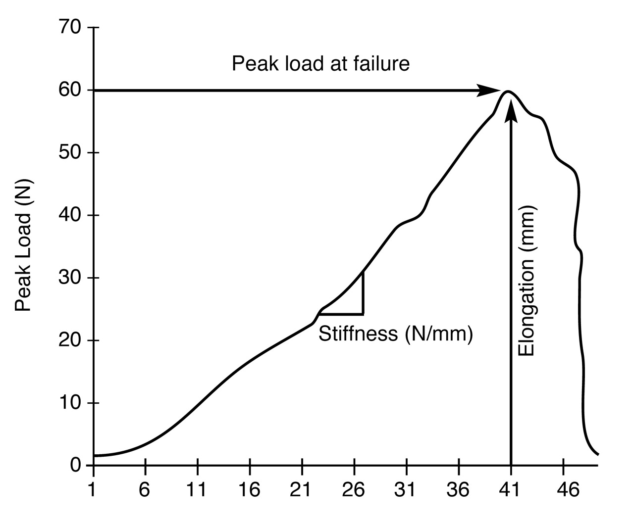
Each specimen was loaded to failure in the Instron 4201 testing machine to determine the structural properties (peak load at failure, elongation at failure, and functional stiffness) of the osteotomy (Fig. 8). The jig imparts a cantilever bending upon the surgical construct that simulates the load imparted to the first metatarsal in midstance. Figure 9] illustrates the typical force (N)-versus-elongation (mm) curve obtained from this study. Peak load (N) to failure is the maximum force measured by the load cell corresponding to the point at which the specimen failed. Elongation (mm) at failure (Ef), as illustrated in Figure 8 and defined in Figure 9, is the vertical displacement of the metatarsal head from its initial position (E0). Functional stiffness (N/mm) is the slope of the load versus the deformation curve in the elastic phase of the response (Fig. 9).
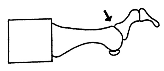
Figure 1.
Saw bone model. Arrow indicates the site of cleavage.
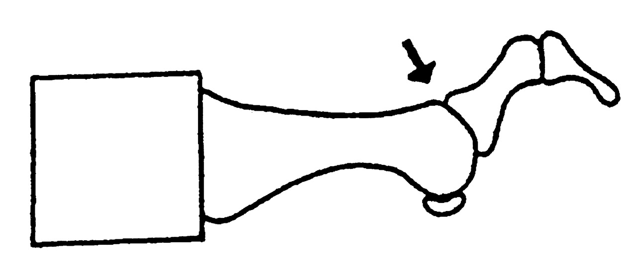
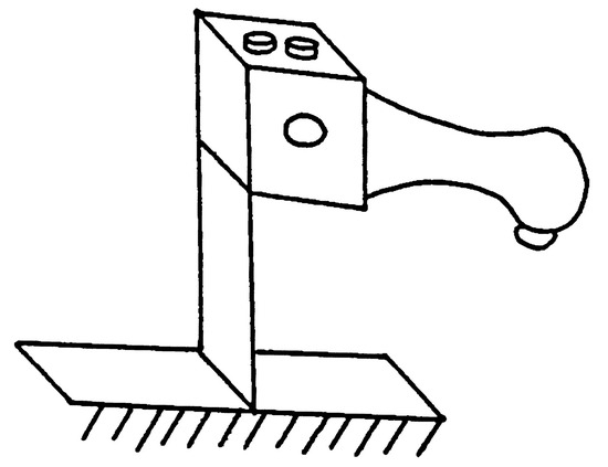
Figure 2.
Saw bone model affixed within the base cube of the jig assembly.
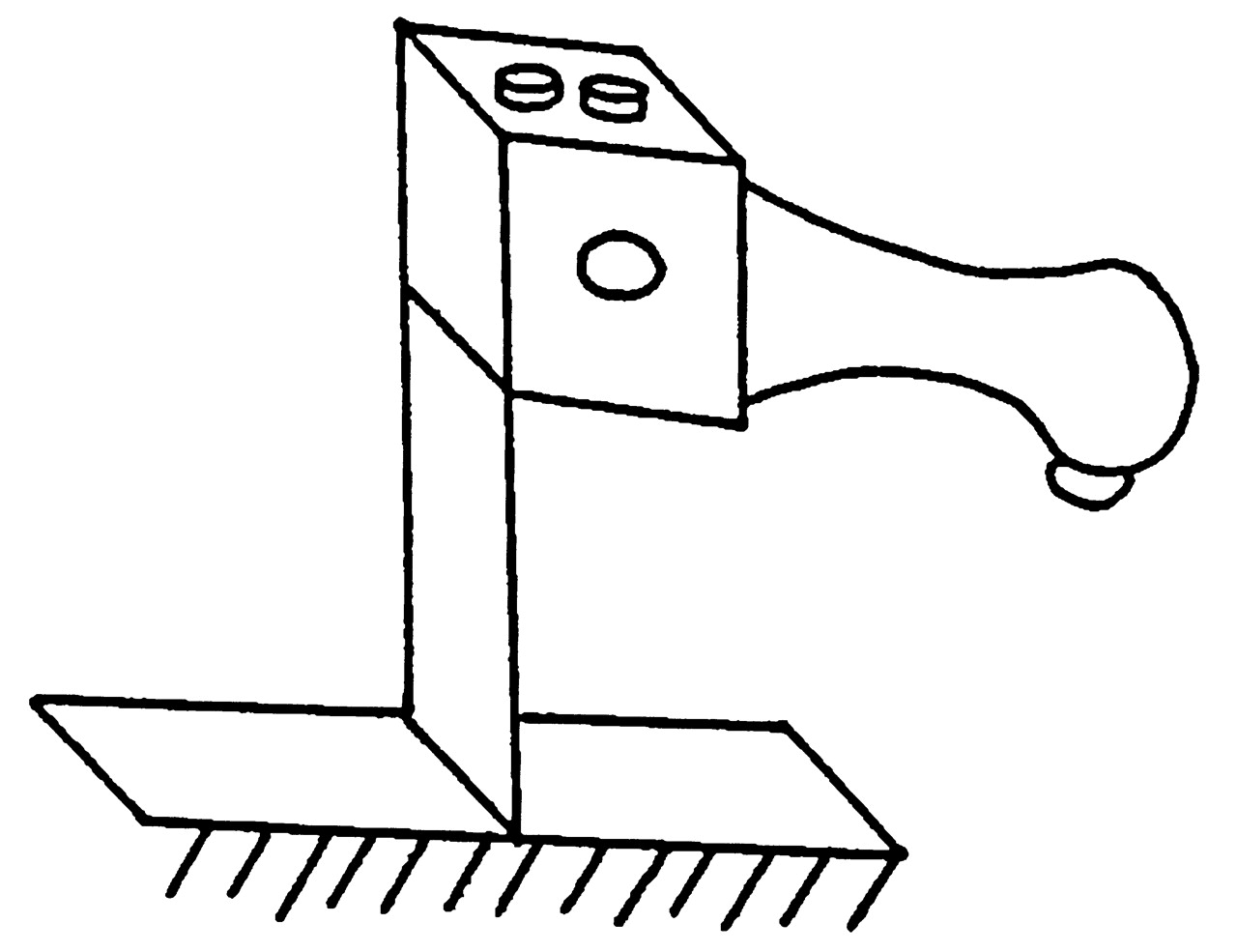
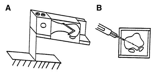
Figure 3.
A, Sagittal view of the saw guide attachment; B, frontal view showing the saw blade creating the osteotomy.
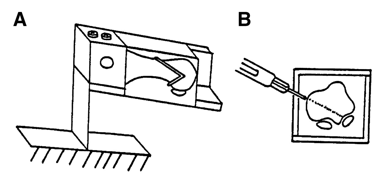
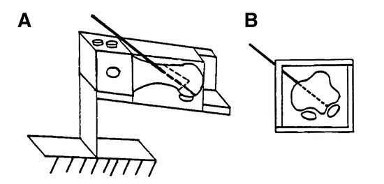
Figure 4.
A, Sagittal; B, frontal views of the pin guide attachment.
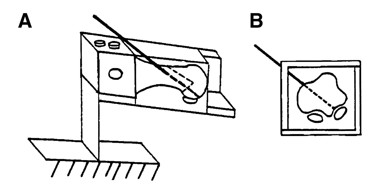
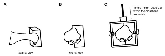
Figure 5.
Bivalve polymethyl methacrylate potting cube for mounting the distal aspect of the first metatarsal.
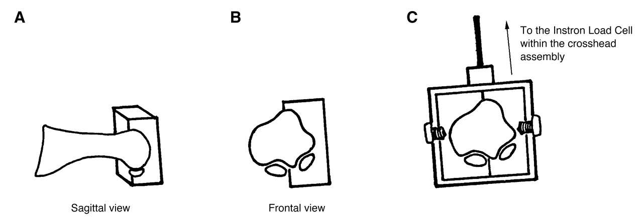
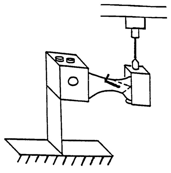
Figure 6.
Osteotomized and fixated saw bone model.
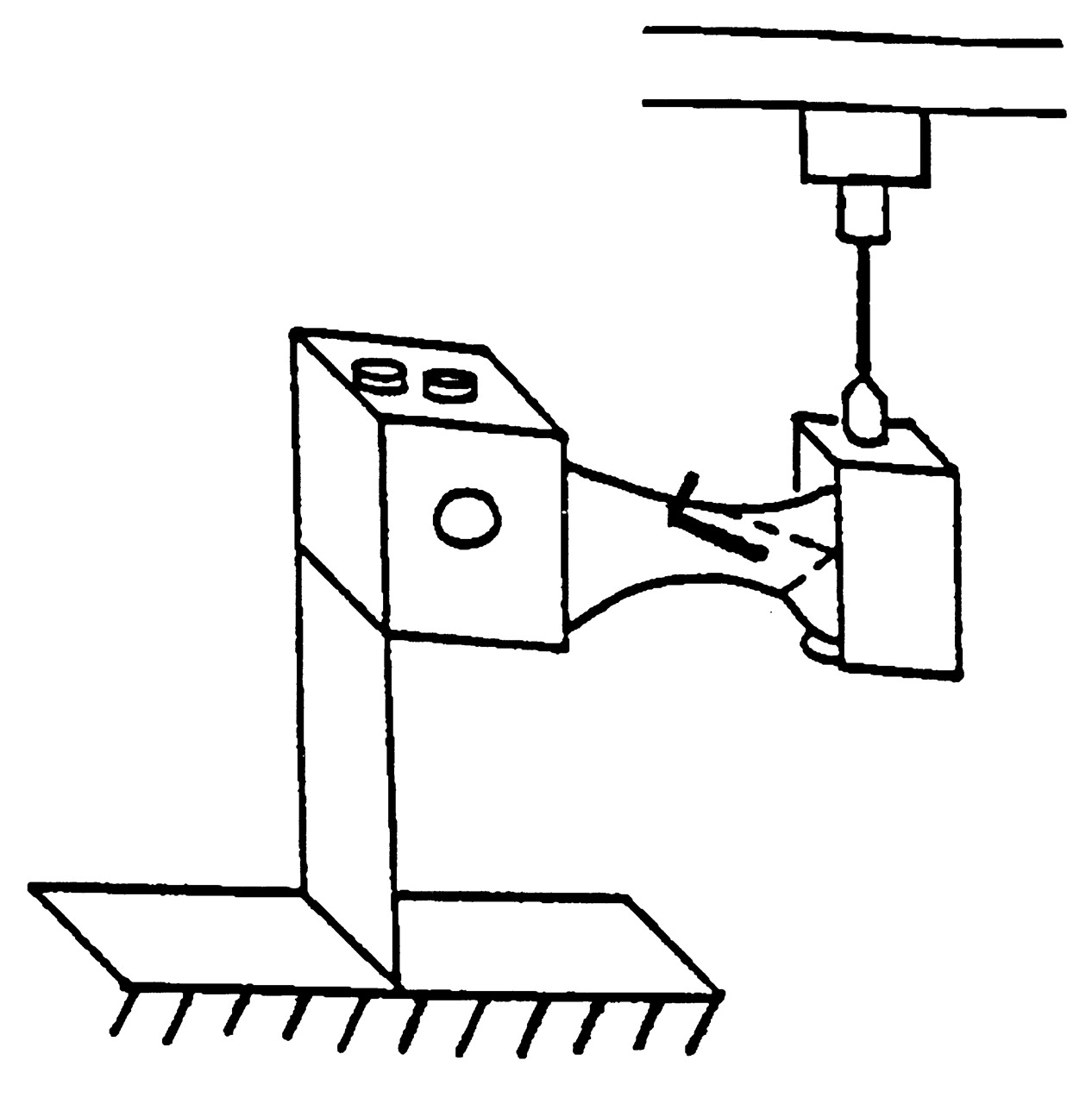
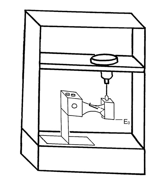
Figure 7.
Saw bone model within jig and Instron 4201 testing machine.
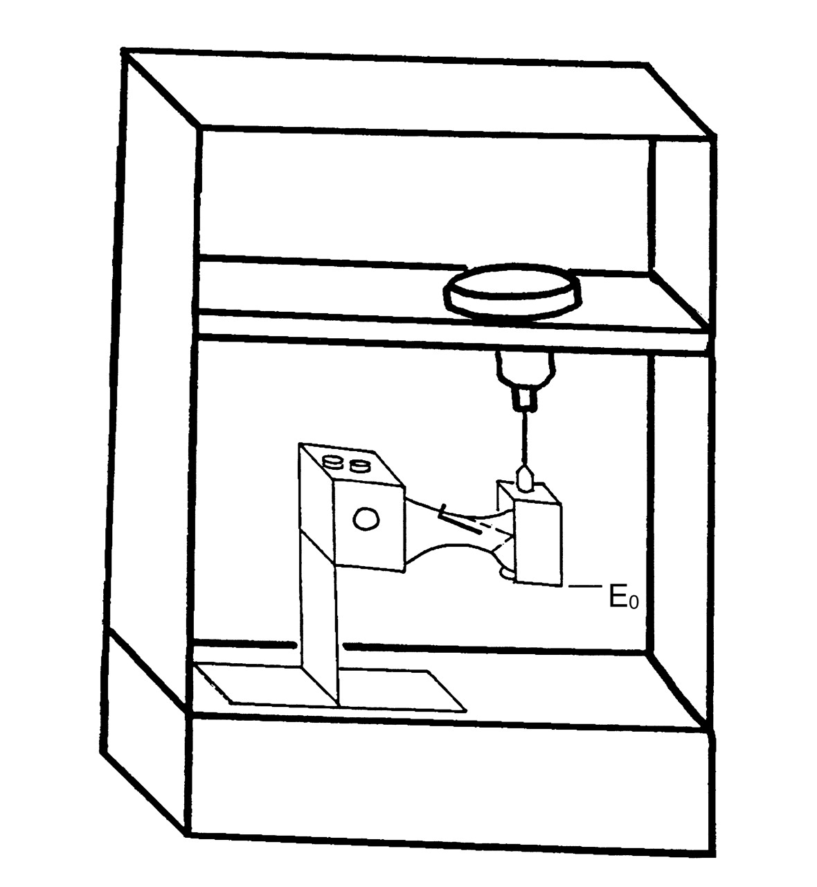
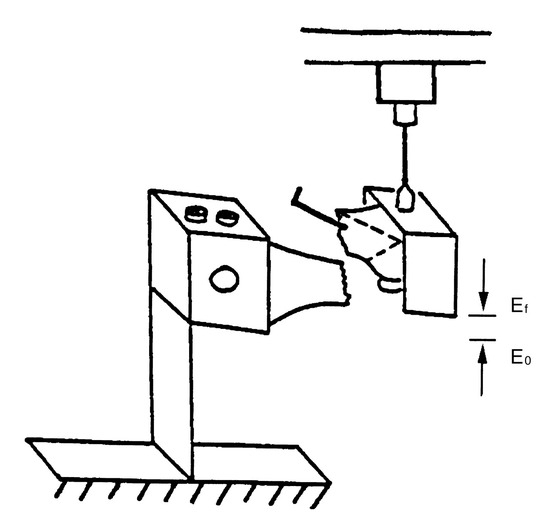
Figure 8.
Saw bone model loaded to failure.
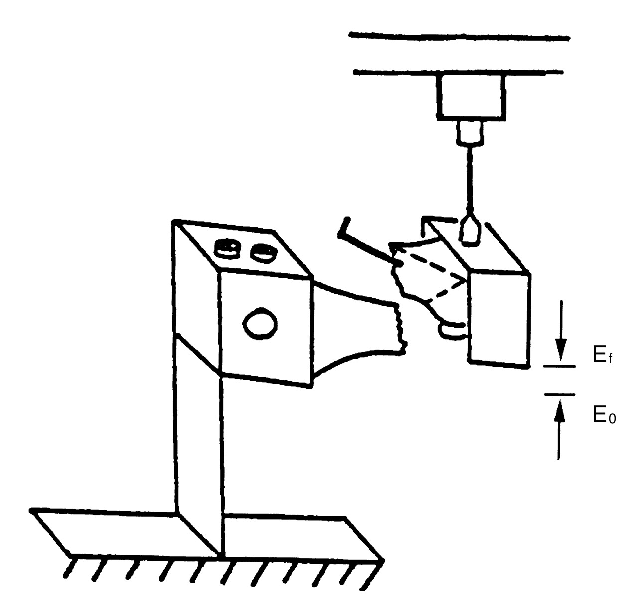
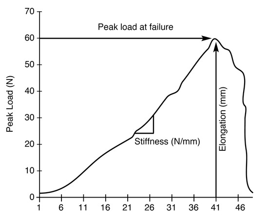
Figure 9.
Typical force versus elongation.
Four groups were included in this study, each containing 10 saw bone models. The saw bone model provided a uniform testing material that simulated the geometric shape of the first metatarsal, thus providing comparative data based solely on osteotomy location, geometry, and pin orientation. The authors selected saw bone models because they were easily obtainable and homogenous in specimen size, shape, weight, and density, and allowed the authors to compare the purely mechanical effect due to the surgical construct. Offset V-osteotomies were performed with midcapital (Kalish) and anatomic neck (Vogler) apex techniques and fixated with a single Kirschner wire in one of two different orientations. The first orientation was dorsally directed from the proximal plantar-medial aspect to the distal dorsal-lateral aspect of the metatarsal. The second orientation was plantarly directed from the proximal dorsal-medial aspect to the distal plantar-lateral aspect of the metatarsal.
The base of the first metatarsal, as secured in the test jig, was mechanically grounded. The metatarsal head was vertically loaded to failure in cantilever fashion at a rate of 500 mm/min. This loading rate and the 15° construct declination were selected to simulate the portion of stance phase between midstance and propulsion. The peak load at failure, the elongation at failure, and the functional stiffness were quantified for each specimen in each group. The resulting parameters were analyzed with a factorial analysis of variance (ANOVA) by means of Statview 5.0 software (SAS Institute, Cary, North Carolina) on a Macintosh G3 microcomputer. Group interactions and post hoc t-test results were studied as well. The alpha level of significance was set at 0.05.
Results
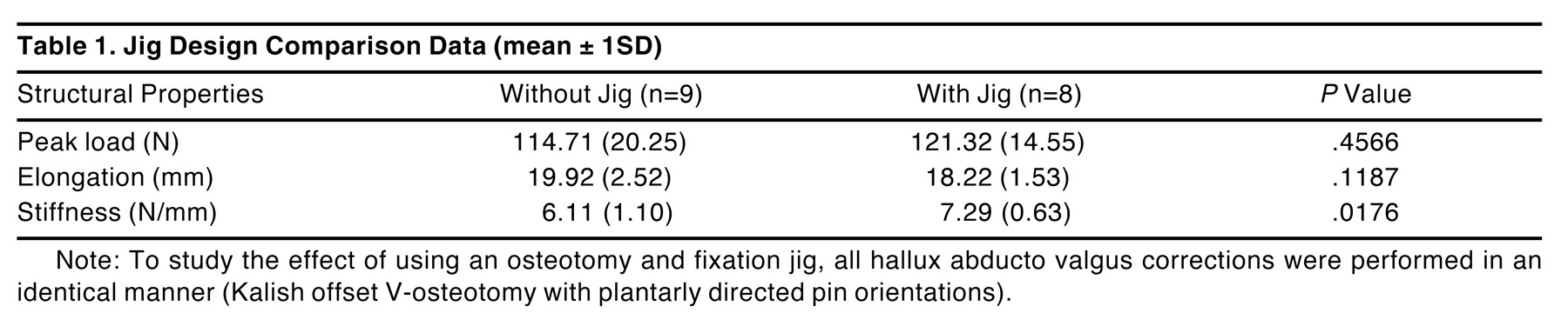
A saw bone study was performed to examine the reliability of the test jig. The Kalish offset V-osteotomy with plantarly directed pin orientation was performed with the jig and compared with the same osteotomy and fixation performed without the jig. Table 1 summarizes the data obtained for peak load, elongation, and functional stiffness with and without the jig. Note that the standard deviations for each parameter were much lower when the jig was used, signifying a reduction in methodological variability. Furthermore, a significant difference in functional stiffness (P = .0176) was obtained with the use of the jig, indicating that the structural properties were actually different without the jig.

Table 1.
Jig Design Comparison Data (mean ± 1SD)
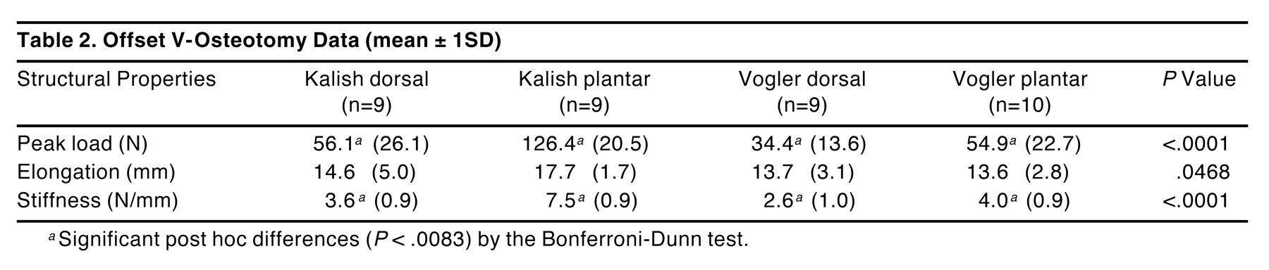
The structural properties of the osteotomized and fixated metatarsals were calculated from the load-versus-elongation data as previously described and illustrated in Figures 10] through 12. The results are summarized in histograms in which the structural properties are depicted with a mean value ±1SD error bars. Figure 10 represents the peak load at failure; Figure 11, the functional stiffness; and Figure 12, the elongation at failure. The values for peak load, elongation, and functional stiffness are summarized in Table 2. ANOVA testing revealed statistically significant differences across the four groups for peak load (P < .0001), elongation (P = .0468), and functional stiffness (P < .0001). Post hoc analyses, conducted with the Bonferroni-Dunn test, corrected for multiple comparisons by imposing a more stringent level of significance (P < .0083 in this case). For peak load, the Kalish osteotomy with a plantarly directed pin orientation was significantly greater than all other groups through post hoc analysis, whereas elongation revealed no significant differences. Stiffness was significantly greater for the plantarly directed pin orientation in the Kalish osteotomy as compared with all other groups.
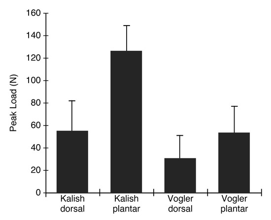
Figure 10.
Peak load at failure (mean ± 1SD).
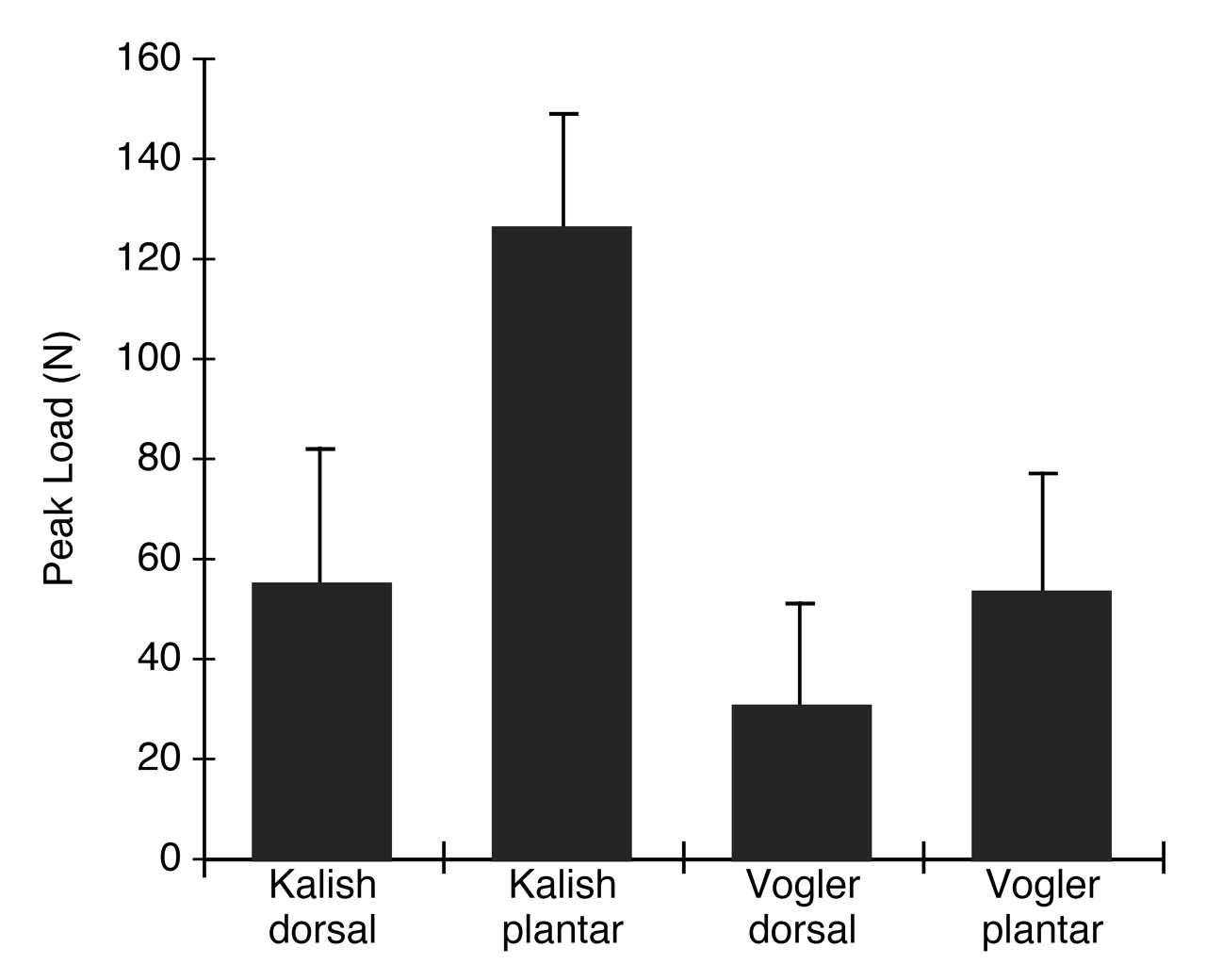
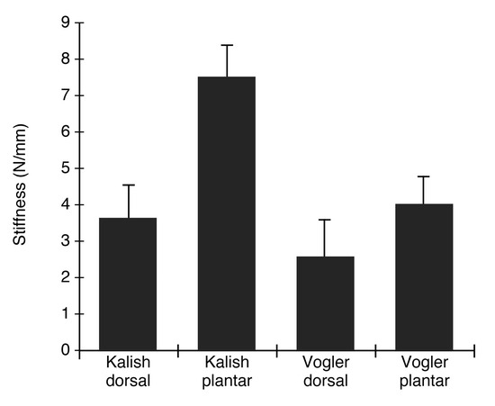
Figure 11.
Stiffness (mean ± 1SD).
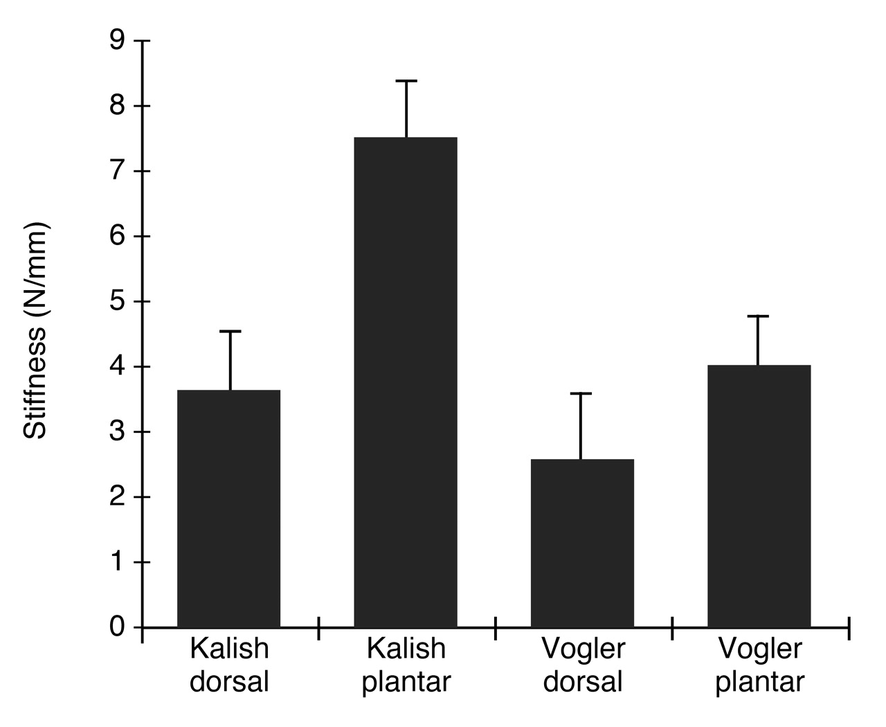
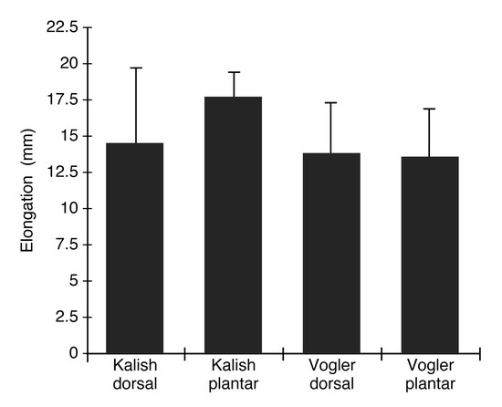
Figure 12.
Elongation at failure (mean ± 1SD).
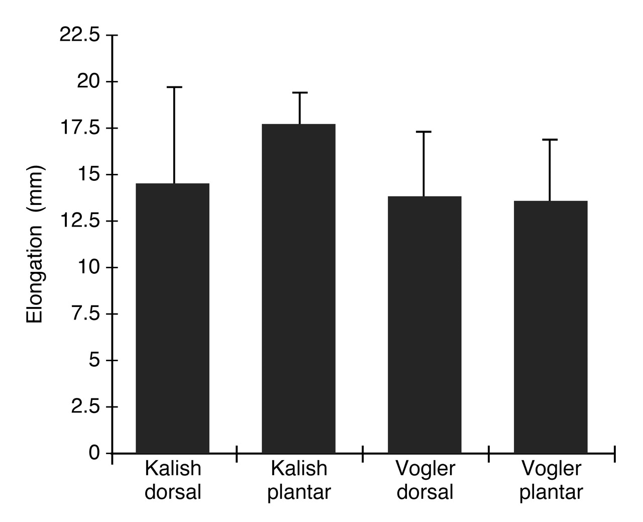
Table 2.
Offset V-Osteotomy Data (mean ± 1SD)
Discussion
In the jig validation experiment, the functional stiffness was significantly different between the data obtained with and without the jig. In all three structural parameters, the test jig effectively reduced data variability and the standard deviations decreased. This supports the authors’ postulation that saw bone position and orientation, as well as repeatability of the osteotomy and fixation, is important to experimental design when surgical constructs are studied. One possible explanation for the decreased variability in the jig data is that the relative movement between the capital fragment and the bivalved polymethyl methacrylate mold was negligible, yielding a superior mechanical grounding of the construct. In the case of fresh bone, a new polymethyl methacrylate potting is likely to be required of each specimen, owing to the variability in osseous geometries. Clearly, a different strategy for repeatedly fixating each specimen will be necessary for fresh cadaveric studies.
Pin orientation and osteotomy location significantly affect the weightbearing stability of the offset V-type osteotomy. With respect to pin orientation, the plantarly directed pin fixation behaved in a manner superior to the dorsally directed pin fixation. Under ultimate loading conditions, the pin fixation provided two failure patterns: 1) complete displacement of the osteotomy with the dorsally directed fixation, and 2) metatarsal shaft fracture with the plantarly directed fixation. These two failure patterns were not affected by the osteotomy apex location. Consequently, the failure patterns noted for both the Kalish and the Vogler osteotomies were similar, but failure occurred at different loads. Interestingly, neither dorsal wing fracture nor pin breakage were observed in any of the groups. In the dorsally directed pin fixation, the Kirschner wire served as an escape path for the capital fragment.
The similar failure patterns in the two surgical techniques illustrate the intrinsic stability of the offset V-osteotomy design. The greater ultimate strength of the Kalish osteotomy can be rationalized through mechanical engineering principles. The Kalish osteotomy apex is located in the center of the first metatarsal head, whereas the Vogler osteotomy apex is located at the anatomic neck. A shorter lever arm is present between the Kalish apex and the point of application of the weightbearing load beneath the sesamoids, thereby necessitating a greater force to reach failure. Likewise, ultimate failure is reached sooner with the Vogler osteotomy because of the longer lever arm. Perhaps the greater cross-sectional area at the metaphyseal location of the Kalish offset V-osteotomy also contributed to its stability under load. The data about this parameter will be factored into a forthcoming comparative study with fresh-frozen cadaveric specimens. Additionally, the success of the jig assembly lends itself to testing alternative fixation techniques and osteotomy designs in a highly repeatable manner.
One obvious shortcoming of this methodology, however, is that the saw bone models lack the internal architecture of bone. Additionally, a significant limitation of this study is the lack of a tension band effect, which is present in vivo. The tension band effect is supplied by the compressive forces from the capsular, ligamentous, and fascial tissues crossing the first metatarsophalangeal joint. Unfortunately, no experimental design is without error. Preserved cadaveric bone specimens may have altered structural properties because of the use of formalin. In consideration of fresh-frozen cadaveric specimens, the structural properties may change due to the freezing and thawing process. Future studies concerning the optimization of the offset V-osteotomy procedure should include the tension band effect.
Regardless of the location of the osteotomy apex, the plantar pin orientation resulted in superior peak load and functional stiffness compared with the dorsal pin orientation. In comparisons of the Kalish and Vogler osteotomies, the Kalish procedure had greater stability (larger peak load and functional stiffness). Specifically, the midcapital (Kalish) apex with a plantar Kirschner wire orientation was the most stable construct in this saw bone protocol.
References
- Austin DW, Leveten EO: A new osteotomy of hallux valgus. Clin Orthop Rel Res 157: 25, 1981.
- Kalish SR, Bernbach MR: “Modification of the Austin Bunionectomy,” in Reconstructive Surgery of the Foot and Leg, Update ’87, ed by ED McGlamry, p 86, The Podiatry Institute, Tucker, GA, 1987.
- Vogler JW: “Offset V Osteotomy in Hallux Valgus Reduction,” in Current Therapy in Podiatric Surgery, ed by RM Jay, p 158, BC Decker, Toronto, Canada, 1988.
- Corless JR: A modification of the Mitchell procedure. J Bone Joint Surg Br 58: 138, 1976.
- Hawkins FB, Mitchell CL, Hedrick DW: Correction of hallux valgus by metatarsal osteotomy. J Bone Joint Surg Am 27: 387, 1945.
- Johnson DA, Cofield RH, Morrey BF: Chevron osteotomy for hallux valgus. Clin Orthop Rel Res 157: 44, 1981.
- Lewis RJ, Feffer HL: Modified chevron osteotomy of the first metatarsal. Clin Orthop Rel Res 157: 105, 1981.
- Shereff MJ, Sobel MA, Kummer FJ: The stability of fixation of first metatarsal osteotomies. Foot Ankle 11: 208, 1991.
- Landsman AS, Vogler HW: An assessment of oblique base wedge osteotomy stability in the first metatarsal using different modes of internal fixation. J Foot Surg 31: 211, 1992.
- Feinstein MH, Brown HN: Hallux adductus as a surgical complication. J Foot Surg 19: 207, 1980.
- Hansen CE: Hallux valgus treated by the McBride operation: a follow-up. Acta Orthop Scand 45: 778, 1974.
- Meier PJ, Kenzora JE: The risks and benefits of distal first metatarsal osteotomies. Foot Ankle 6: 7, 1985.
- Williams WW, Barret DS, Copeland SA: Avascular necrosis following chevron distal metatarsal osteotomy: a significant risk. ?J Foot Surg 28: 414, 1989.
- Downey MS: Complications of the Kalish bunionectomy. JAPMA 84: 243, 1994.
Additional References
- Cain TD, Boyd D: “Defining the Limits of the Modified Austin Bunionectomy,” in Reconstructive Surgery of the Foot and Leg, Update ’90, ed by DR DiNapoli, p 128, The Podiatry Institute, Tucker, GA, 1990.
- Chang TJ, Landsman AS, Ruch JA: “Relative Strengths of Internal Fixation in Osteotomies and Arthrodesis of the First Metatarsal,” in Reconstructive Surgery of the Foot and Leg, Update ’96, ed by NS Vickers, SJ Miller, GV Yu, et al, p 120, The Podiatry Institute, Tucker, GA, 1996.
- Clancy JT, Berlin SJ, Giordano ML, et al: Modified Austin bunionectomy with single screw fixation: a comparison study. J Foot Surg 28: 284, 1989.
- Edelman RD: Iatrogenically induced hallux varus. Clin Pod Med Surg 8: 367, 1991.
- Goel AR, Vogel BI: The offset V osteotomy with screw fixation for correction of hallux valgus: a retrospective study. J Foot Surg 32: 305, 1993.
- Kalish SR, Gold RS: “Modifications of the Austin Hallux Valgus Repair (Kalish Osteotomy),” in Reconstructive Surgery of the Foot and Leg, Update ’92, ed by JA Ruch, NS Vickers, p 237, The Podiatry Institute, Tucker, GA, 1992.
- Kalish SR, Spector JE: The Kalish osteotomy: a review and retrospective analysis of 265 cases. JAPMA 84: 237, 1994.
- Kelikian H: “Hallux Valgus, Allied Deformities of the Forefoot and Metatarsalgia,” in Osteotomy, p 163, WB Saunders, Philadelphia, PA, 1965.
- Kissel CG, Unroe BJ, Parker RM: The offset V bunionectomy using cortical screw and buried K-wire fixation. J Foot Surg 31: 560, 1992.
- Malay DS: “Avascular Necrosis of the First Metatarsal Head Following Distal Reconstructive Osteotomy,” in Reconstructive Surgery of the Foot and Leg, Update ’94, ed by CA Camasta, NS Vickers, JA Ruch, p 115, The Podiatry Institute, Tucker, GA, 1994.
- Reese HW, Johnson CB: Reese osteotomy guide system. J Foot Surg 23: 386, 1984.
- Sedlin ED, Hirsch C: Factors affecting the determination of the physical properties of femoral cortical bone. Acta Orthop Scand 37: 129, 1966.
- Stokes IAF, Hutton WC, Stott JRR: Forces under hallux valgus foot before and after surgery. Clin Orthop Rel Res 142: 64, 1979.
- Vogler HW: Shaft osteotomies in hallux valgus reduction. Clin Pod Med Surg 6: 47, 1989.
© 2002 American Podiatric Medical Association