Knowledge of the innervation of the medial calcaneal region is important for the diagnosis and treatment of heel pain, tarsal tunnel syndrome, soft-tissue and bony ankle injury, and the secondary heel pain due to neuroma. This subject is inadequately covered in standard anatomy textbooks, which generally illustrate a single medial calcaneal nerve originating from the lateral plantar nerve within the tarsal tunnel [
1-
5]. For example, the 1995 edition of
Gray’s Anatomy [
4] contains two figures with a line labeled “medial calcaneal nerve” but does not show any proximal origin of this one branch, and the text does not indicate a specific origin for this other than listing it under the sciatic nerve. The 1993 edition of
Anatomy of the Foot and Ankle [
5] includes a detailed dissection of a single foot that demonstrates the medial calcaneal nerve as a single nerve originating from the tibial nerve. This medial calcaneal nerve is demonstrated as having an anterior and a posterior division, with several additional branches from the posterior division. In 1984, Mackinnon and Dellon [
6] reported three patterns of origin of the medial plantar nerve after dissecting 20 embalmed cadavers. One of these patterns had two different calcaneal nerves: one originating from the lateral plantar and another originating from the posterior tibial nerve, both within the tarsal tunnel. In 1988, Havel et al [
7] described nine patterns of origin of the calcaneal nerve after dissecting 38 pairs of feet of embalmed cadavers. Their observance of an increased number of patterns resulted from noting variations in which a calcaneal nerve originated proximal to the tarsal tunnel and also by describing an origin from the medial plantar nerve. The publications of Baxter and Pfeffer [
8], Baxter and Thigpen [
9], Schon and Baxter [
10], and Schon et al [
11] since 1984 have popularized the concept that heel pain is due to the first branch of the lateral plantar nerve. This is actually a small sensory branch arising from the motor branch of the lateral plantar nerve that innervates intrinsic muscles. Clearly, these branches exist within the lateral plantar nerve within the tarsal tunnel and do not go to the skin of the heel itself. Most recently, a microdissection of fresh cadavers has described the innervation of the skin of the medial ankle region [
12], but did not consider the medial calcaneal innervation. This article describes the variations in the origin of the medial calcaneal nerve from dissections done in living tissue during tarsal decompression in humans.
Discussion
The results of this study demonstrate a great variability in the site of origin of the medial calcaneal nerve(s). This knowledge will provide the surgeon with a previously unavailable guide during dissections in this area. For example,
Cunningham’s Textbook of Anatomy [
2] does not describe or illustrate the origin of the medial calcaneal nerve, but simply indicates that it pierces the fascia and that its branches are distributed to the skin of the heel. In text figures, neither the medial nor the lateral plantar nerves are demonstrated as giving origin to a calcaneal branch. In contrast, Pernkopf [
3] suggests that there is a branch from the medial plantar nerve to the skin of the heel area, but does not name this branch. This same illustration demonstrates the medial calcaneal branch as arising from the posterior tibial nerve, again at a proximal level. An origin for the medial calcaneal nerve from the lateral plantar nerve is not illustrated. In further contrast,
Grant’s Atlas of Anatomy [
1] demonstrates the medial calcaneal nerve as consisting of two branches arising from the posterior tibial nerve, probably proximal to the tarsal tunnel. A recent textbook demonstrates that the medial calcaneal nerve arises as a single branch from the posterior tibial nerve within the tarsal tunnel [
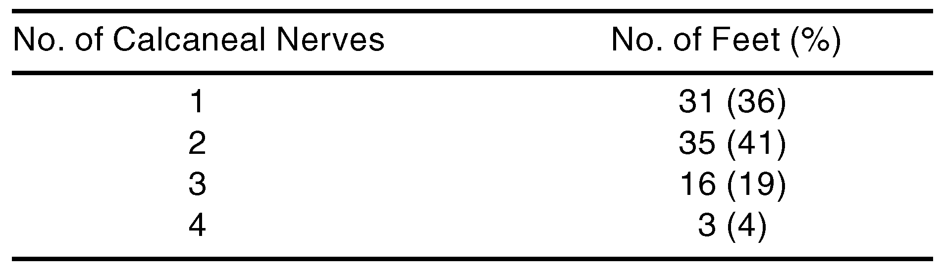
10]. The results of the present study demonstrate that 64% of feet that have surgery to decompress the tarsal tunnel will have more than one origin for the medial calcaneal nerve (
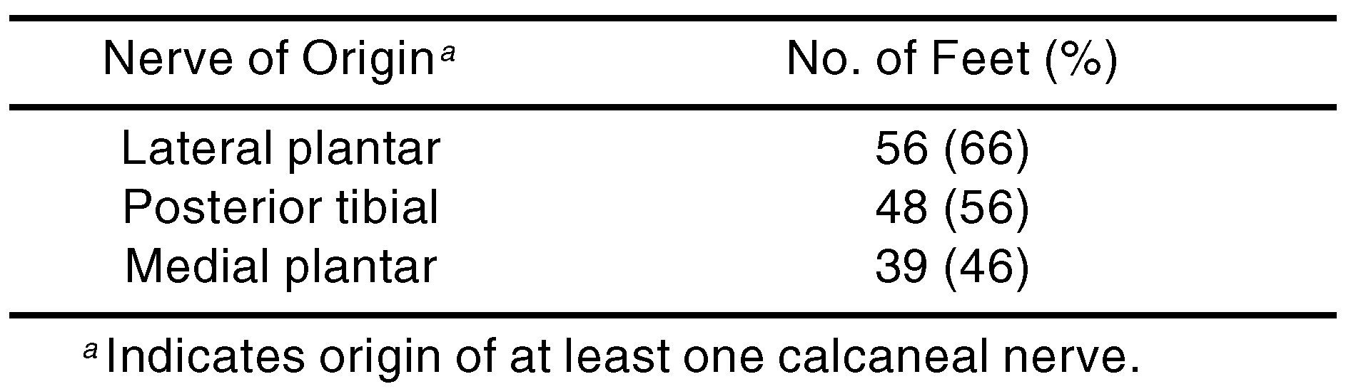
Table 1). Furthermore, these nerves may originate from either the posterior tibial nerve (56%), the lateral plantar nerve (66%), or the medial plantar nerve (46%) (
Table 2).
It is hoped that the anatomic patterns described in this study will provide a knowledge base that may be used as more surgery in this medial ankle region is done. Increased surgery in this region may be predicted from a recent article on the use of surgical decompression of the calcaneal nerve to treat recalcitrant heel pain [
13] and recent publications that suggest that decompression of the tarsal tunnel can restore sensation to diabetic feet [
14-
17].
The results of this study emphasize that the medial plantar nerve gives origin to a nerve that innervates the skin in the region in which incisions are commonly made for calcaneal spur removal or plantar fasciotomy. This nerve, which has branches on the order of 0.9 mm, is at risk for injury during these procedures. Awareness of its existence and use of magnification during surgery in this area are the only hope for prevention of painful neuromas as a complication of surgery for heel pain. In 3% of the feet in this study, the main innervation of the heel skin was from a branch of the medial plantar nerve that crossed the vessels superficially and then entered the tunnel usually occupied by a branch of the posterior tibial or lateral plantar nerve. The location of this nerve, superficial to the vessels, places it at risk for injury during division of the flexor retinaculum.
To diagnose and treat heel pain, it is probably most appropriate not to attribute heel pain to any one nerve because that may misdirect the therapy and the surgeon. Since the innervation pattern cannot be known without surgical dissection, it is most reasonable to suggest that heel pain that does not respond to nonoperative measures be approached surgically through an incision that permits the surgeon access to all of the described variations in heel innervation. It is important also to consider that the branches that innervate the medial calcaneal skin region should have a clinical correlate of a decrease in sensibility in this skin area if these nerves are impaired. This is in contrast to the nerve commonly referred to as “Baxter’s nerve,” which, arising as innervation to periosteum from a motor portion of the lateral plantar nerve [
5,
8-
11], does not innervate the skin.
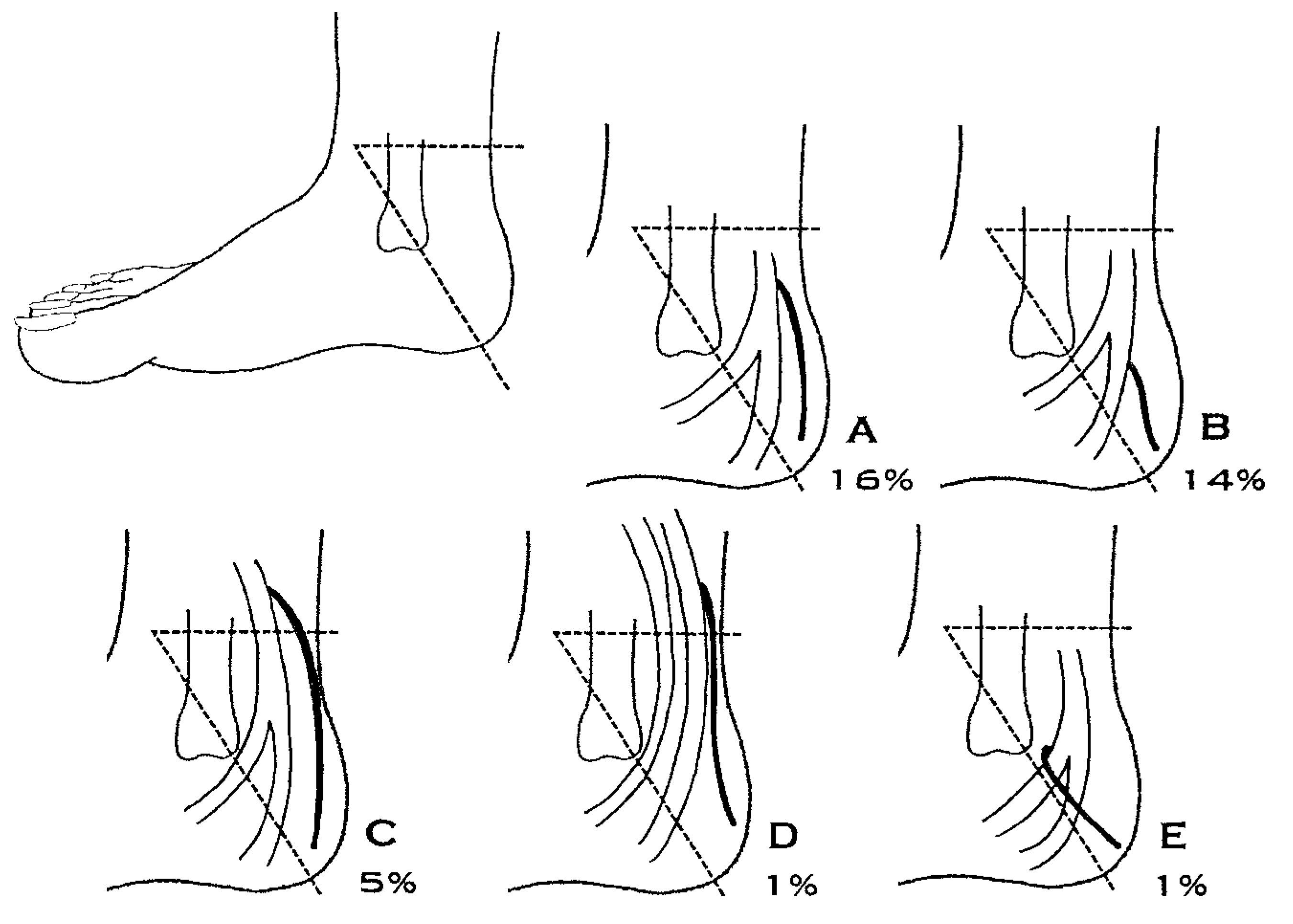
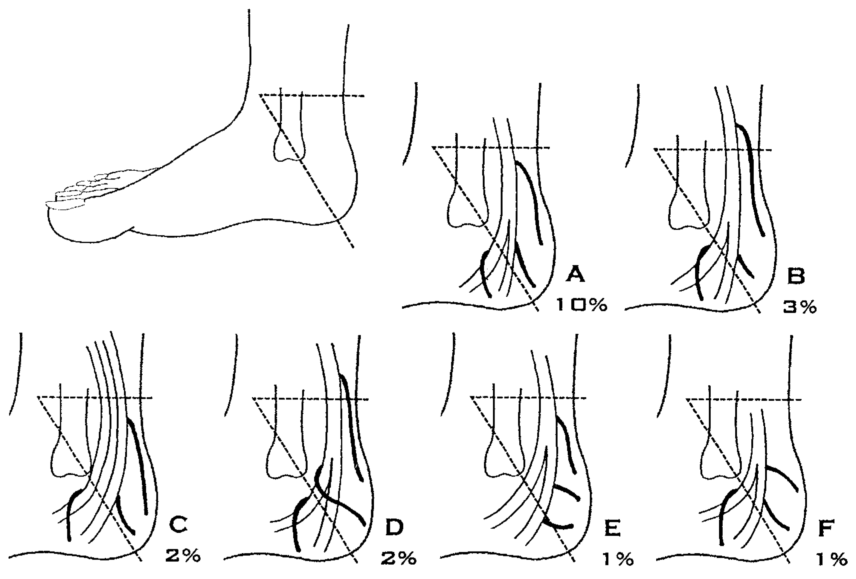
The pattern of origin of medial calcaneal nerves arising proximal to the tarsal tunnel suggests that the involvement of heel symptomatology in tarsal tunnel syndrome can vary according to the origin of this nerve. For example, based on
Figure 1C,D, it may be estimated that 6% of tarsal tunnel syndrome patients may have no heel symptomatology or have normal quantitative sensory testing of the medial heel owing to the origin of the medial calcaneal nerve proximal to the site of compression. One may predict that there will be a group of patients whose forefoot symptoms in tarsal tunnel syndrome will dominate over their heel symptoms because there is a dual innervation of the medial calcaneal region, in which a portion of the innervation does arise within the tarsal tunnel (
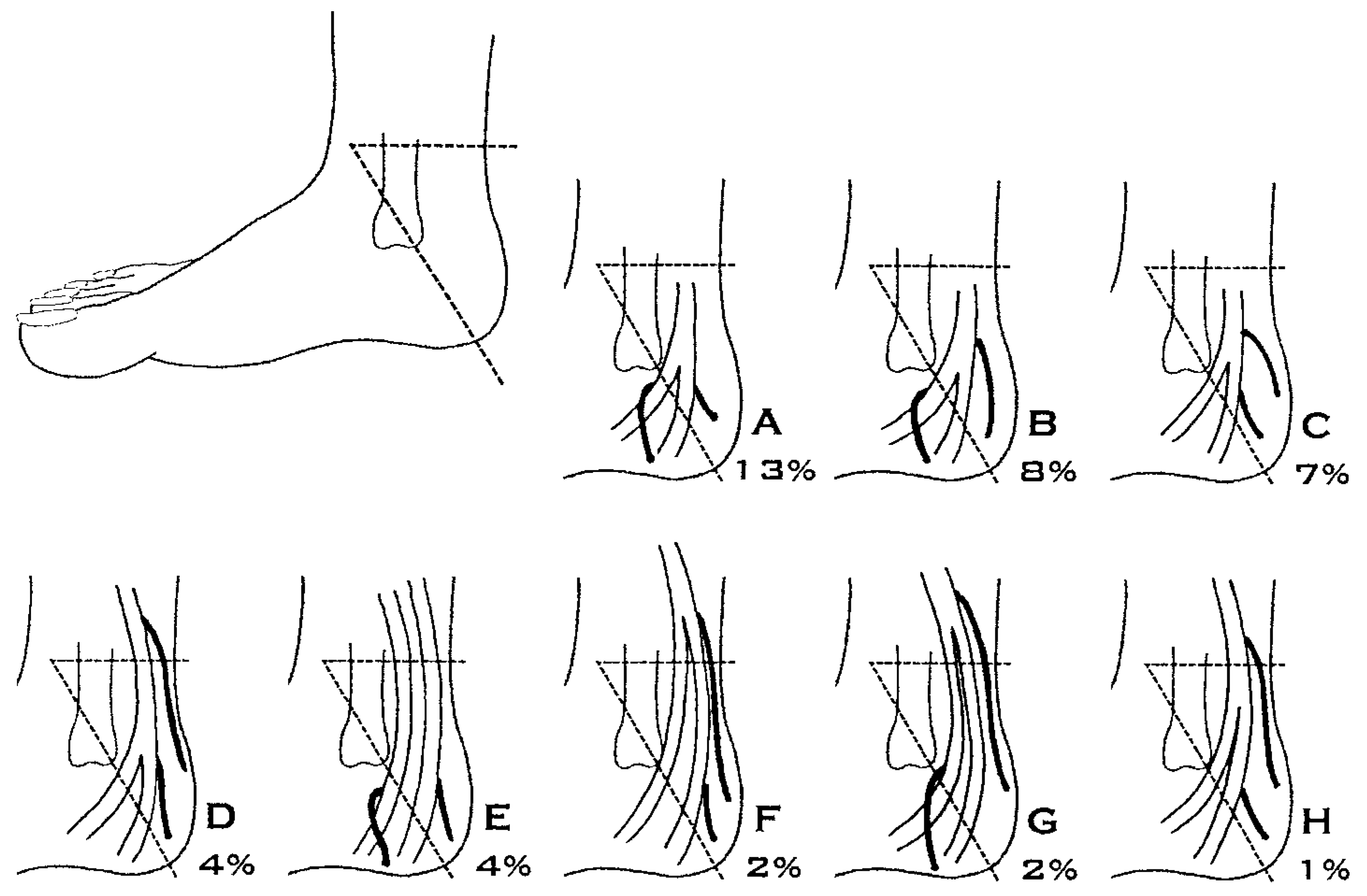
Figure 2F-H;
Figure 3B,E; and
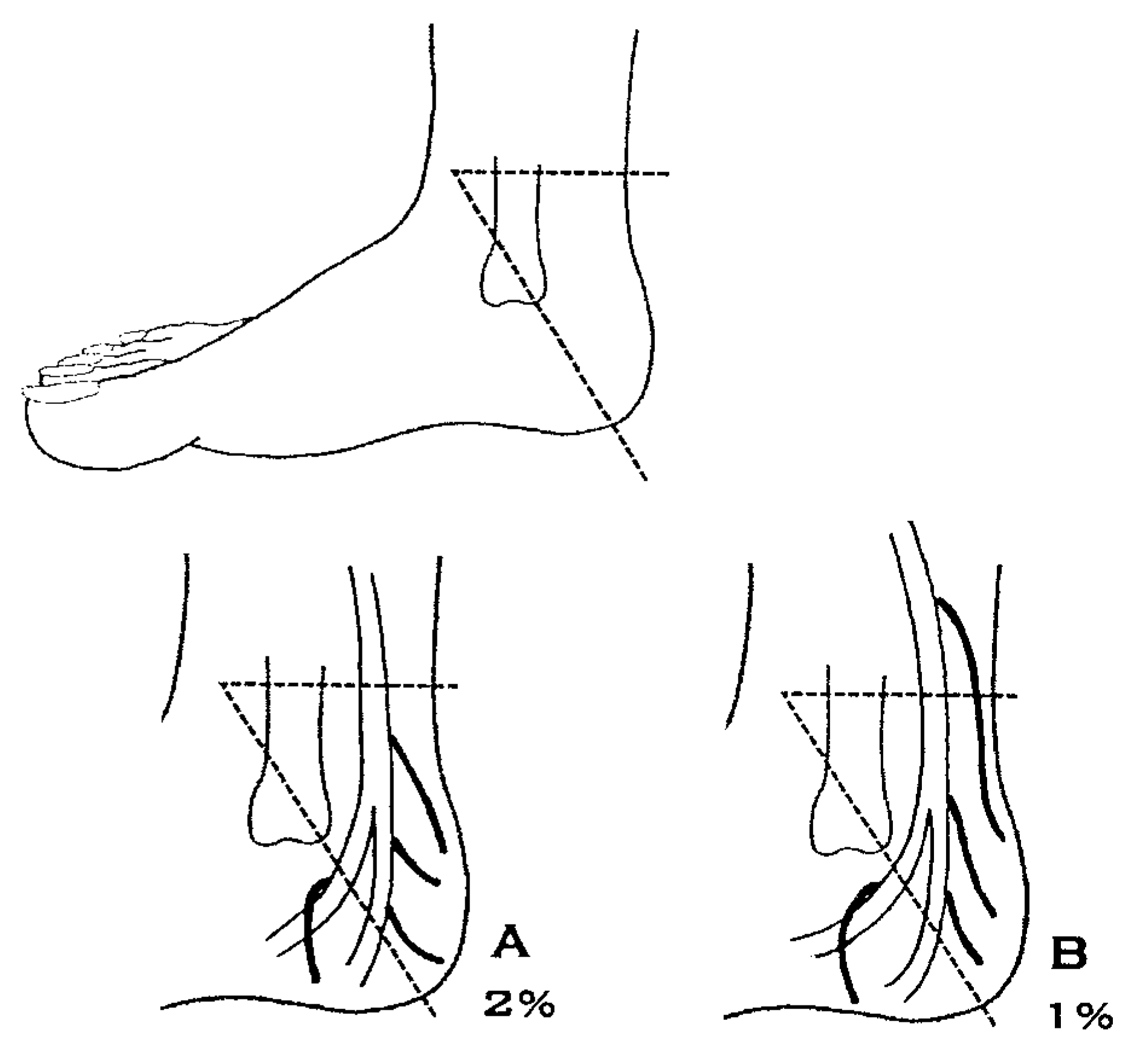
Figure 4B). On the basis of the observations reported here, this group comprises 15% of feet and, with quantitative sensory testing, may have abnormalities that are not as advanced as those found in skin innervated by the medial plantar nerve, which, in all patients will pass through both the tarsal tunnel and the medial plantar tunnel.
Figure 1.
Patterns of origin of the calcaneal nerve in which there is just one calcaneal nerve. The horizontal broken line represents the beginning of the tarsal tunnel, and the diagonal broken line represents the axis between the medial malleolus and the calcaneus. The percentage indicates the percentage of the 85 feet in which this pattern was found. Note that in panel D the medial and lateral plantar nerves have an origin from the posterior tibial nerve that is greater than 3 cm proximal to the malleolar-calcaneal axis, which is proximal to the tarsal tunnel, and that in both panels C and D the medial calcaneal nerve arises proximal to the tarsal tunnel.
Figure 1.
Patterns of origin of the calcaneal nerve in which there is just one calcaneal nerve. The horizontal broken line represents the beginning of the tarsal tunnel, and the diagonal broken line represents the axis between the medial malleolus and the calcaneus. The percentage indicates the percentage of the 85 feet in which this pattern was found. Note that in panel D the medial and lateral plantar nerves have an origin from the posterior tibial nerve that is greater than 3 cm proximal to the malleolar-calcaneal axis, which is proximal to the tarsal tunnel, and that in both panels C and D the medial calcaneal nerve arises proximal to the tarsal tunnel.
Figure 2.
Patterns of origin of the calcaneal nerve in which there are two calcaneal nerves. The horizontal broken line represents the beginning of the tarsal tunnel, and the diagonal broken line represents the axis between the medial malleolus and the calcaneus. The percentage indicates the percentage of the 85 feet in which this pattern was found. Note that in panel E the medial and lateral plantar nerves have an origin from the posterior tibial nerve that is greater than 3 cm proximal to the malleolar-calcaneal axis, and that in panels D, F, G, and H one of the medial calcaneal nerves arises proximal to the tarsal tunnel.
Figure 2.
Patterns of origin of the calcaneal nerve in which there are two calcaneal nerves. The horizontal broken line represents the beginning of the tarsal tunnel, and the diagonal broken line represents the axis between the medial malleolus and the calcaneus. The percentage indicates the percentage of the 85 feet in which this pattern was found. Note that in panel E the medial and lateral plantar nerves have an origin from the posterior tibial nerve that is greater than 3 cm proximal to the malleolar-calcaneal axis, and that in panels D, F, G, and H one of the medial calcaneal nerves arises proximal to the tarsal tunnel.
Figure 3.
Patterns of origin of the calcaneal nerve in which there are three calcaneal nerves. The horizontal broken line represents the beginning of the tarsal tunnel, and the diagonal broken line represents the axis between the medial malleolus and the calcaneus. The percentage indicates the percentage of the 85 feet in which this pattern was found. Note that in panel C the medial and lateral plantar nerves have an origin from the posterior tibial nerve that is greater than 3 cm proximal to the malleolar-calcaneal axis, and that in panels B and D one of the medial calcaneal nerves arises proximal to the tarsal tunnel.
Figure 3.
Patterns of origin of the calcaneal nerve in which there are three calcaneal nerves. The horizontal broken line represents the beginning of the tarsal tunnel, and the diagonal broken line represents the axis between the medial malleolus and the calcaneus. The percentage indicates the percentage of the 85 feet in which this pattern was found. Note that in panel C the medial and lateral plantar nerves have an origin from the posterior tibial nerve that is greater than 3 cm proximal to the malleolar-calcaneal axis, and that in panels B and D one of the medial calcaneal nerves arises proximal to the tarsal tunnel.
Figure 4.
Patterns of origin of the calcaneal nerve in which there are four calcaneal nerves. The horizontal broken line represents the beginning of the tarsal tunnel, and the diagonal broken line represents the axis between the medial malleolus and the calcaneus. The percentage indicates the percentage of the 85 feet in which this pattern was found. Note that in panel B one of the medial calcaneal nerves arises proximal to the tarsal tunnel.
Figure 4.
Patterns of origin of the calcaneal nerve in which there are four calcaneal nerves. The horizontal broken line represents the beginning of the tarsal tunnel, and the diagonal broken line represents the axis between the medial malleolus and the calcaneus. The percentage indicates the percentage of the 85 feet in which this pattern was found. Note that in panel B one of the medial calcaneal nerves arises proximal to the tarsal tunnel.
Table 1.
Variations in Number of Calcaneal Nerves (n = 85)
Table 1.
Variations in Number of Calcaneal Nerves (n = 85)
Table 2.
Sites of Origin of Calcaneal Nerves (n = 85)
Table 2.
Sites of Origin of Calcaneal Nerves (n = 85)