


Abstract
Building on previous work that was cadaver based or involved invasive techniques, this study quantifies the kinematics of an approximation of the midtarsal joint with a noninvasive method. Three-dimensional kinematic data describing the motion of the forefoot and heel during transverse plane rotation of the leg were collected from 25 subjects by means of reflective markers and four infrared cameras. The motion between these segments was assumed to be the best possible clinical approximation of the midtarsal joint. The kinematic characteristics of the midtarsal joint were described in terms of the range and direction of motion in each cardinal body plane, the ratio of the range of motion in each plane, and the orientation of the axis of rotation. The characteristics of the midtarsal joint changed during the range of motion; thus, multiple axes of rotation could be calculated and joint characteristics were varied among subjects. The results of this in vivo method were generally consistent with those of cadaver studies and invasive in vivo studies.
The kinematics of the midtarsal joint (movement of the navicular and cuboid relative to the talus and calcaneus) are poorly understood. Many foot health specialists base their midtarsal joint model on the pronation and supination movements around the oblique and longitudinal axes of rotation as described by Manter.[1] However, Manter’s kinematic assessment was restricted considerably by the technology of the time and lack of knowledge about the midtarsal joint. Furthermore, some of the experimental procedures were poorly described. More recent and scientific kinematic assessments[2-4] have improved the understanding of the individual talonavicular and calcaneocuboid joints. Recent investigators have assessed the kinematics of the rearfoot joints while the leg was rotated in the transverse plane, driving the foot into pronation and supination. However, these data are difficult to interpret clinically because the studies treat the talonavicular and calcaneocuboid joints separately, whereas in a clinical context they are generally regarded as a single functional unit. Consequently, these more relevant data have failed to alter the model of the midtarsal joint adopted by the majority of foot health specialists. The aim of this study was to build on the principles of previous research described in the medical literature by demonstrating a noninvasive method of quantifying the kinematics of the midtarsal joint functional unit.
Materials and Methods
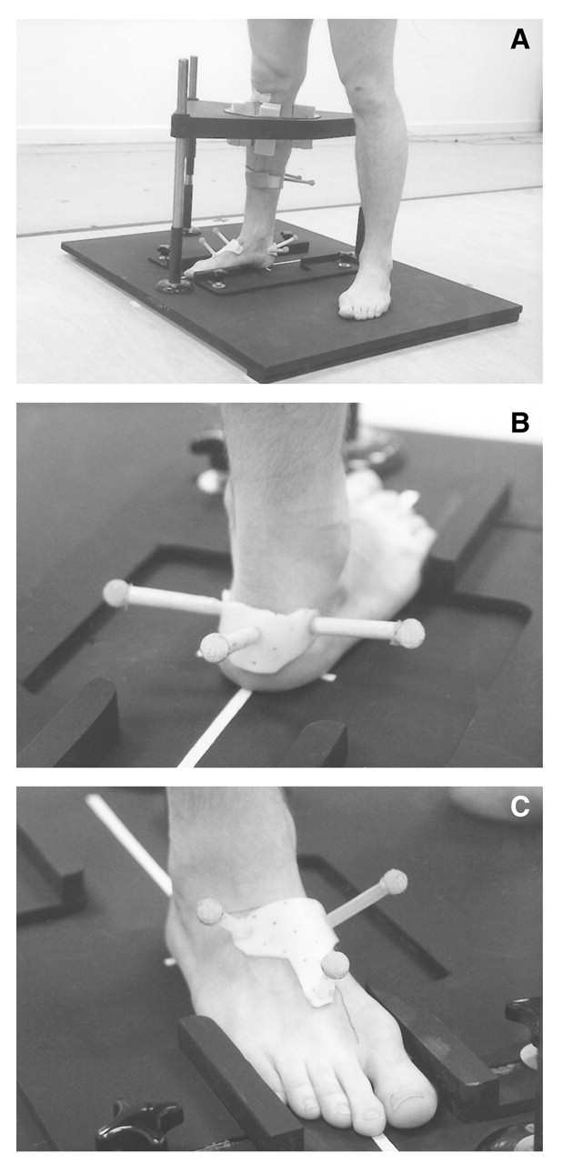
Twenty-five subjects (12 men, 13 women), from 21 to 44 years of age, participated in this study. None had a history of lower-limb injury or disease, and all gave consent to participate. No assessment of foot mobility or deformity was undertaken in the sample; such information would not have been used as inclusion or exclusion criteria since the purpose of the study was to describe the normal variations in the characteristics of the midtarsal joint. Also, as far as was known, no scientific method of assessing midtarsal joint mobility exists; therefore, any selection criteria based on such an assessment would have been flawed. Data describing the kinematics of the heel and forefoot were collected for both feet of each subject during transverse plane rotation of the leg. The relative motion between these segments was assumed to be the best possible clinical approximation of the actual midtarsal joint. To assess repeatability, data on four subjects were collected twice, 7 days apart. Kinematic data for each segment were derived by tracking the position of three reflective markers attached to the heel and the forefoot, from which local coordinate systems were subsequently defined (Fig. 1). Markers were mounted on rigid plastic bases that were attached to the foot with double-sided tape. The size of the plastic base was not varied between subjects, but was sufficiently small that the plastic base remained solely on the appropriate segment for all subjects. The forefoot base covered an area over the navicular, cuboid, and second metatarsal shaft. The heel base covered solely the calcaneus.
A rig similar to that used by Van Langelaan[2] and Benink[3] was designed, allowing the leg to move freely in the transverse plane but, as far as was possible, restricting its motion in the sagittal and frontal planes (Fig. 1). To ensure that the transverse plane motion of the leg produced motion within the foot, as opposed to motion of the whole foot, boards were secured in position on the medial and lateral sides of the forefoot.
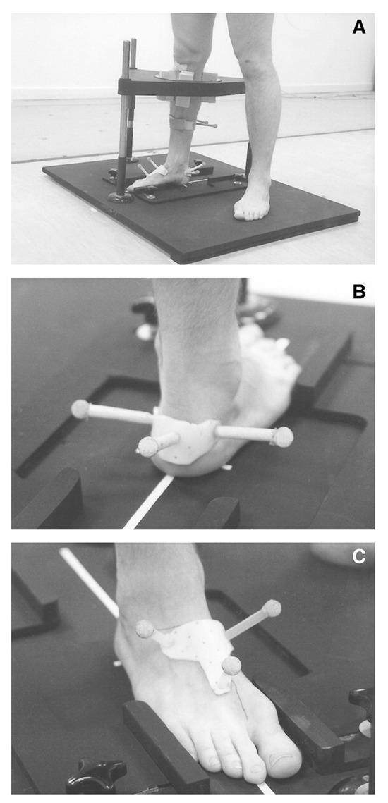
Figure 1.
A, Subject standing with one leg inside the rotating ring. The sagittal and frontal plane motion of the leg was restricted by wedging the leg against the inner surface of the ring. B, Arrangement of markers on the heel. C, Arrangement of markers on the forefoot.
Each subject moved through the following sequence of movements: The leg was in external rotation and the foot supinated to the extent that the first metatarsal was nonweightbearing. The subject next internally rotated the leg to the point of maximum internal leg rotation. The subject then externally rotated the leg to return to the original start position. Ten consecutive recordings of this motion sequence were taken. The movement sequence was performed in a smooth, continuous pattern and typically took 3 to 5 seconds to complete.
Since the literature suggests that the functional characteristics of the rearfoot joints change during the range of motion,[2,4-6] the range of motion was divided into three predefined phases: the supination, pronation, and composite phases of motion. The supination phase was defined as the period during which the leg moved from a position of 20° of external rotation to 0° (equivalent to the relaxed standing position). The pronation phase was defined as the period during which the leg moved from the 0° position to its position of maximum internal rotation. The composite phase was defined as the absolute motion of the leg from a position of 20° externally rotated to a position of maximum internal rotation.
To reduce the volume of data, the angular data obtained from the rotation of the leg from an externally rotated position to an internally rotated position and the angular data obtained from the rotation of the leg from an internally rotated position to an externally rotated position were averaged. The characteristics of rearfoot motion in these two rotations are the same, though the directions of motion are reversed.[3,6]
The characteristics of the midtarsal joint motion were described by means of the range of motion in each cardinal body plane, the ratio of the range of motion in each cardinal body plane, and the orientation of the axis of rotation. This last quantity was calculated by means of a vector summation method previously used by Downing et al.[7]
Results
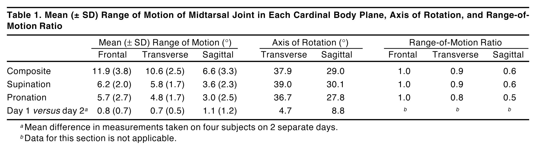
Results were based on a total of 48 limbs (two were excluded due to loss of data). The mean ranges of motion, standard deviations, axes of rotation (calculated from the mean range of motion), and ratios of the mean ranges of motion are listed in Table 1. Table 1] also shows the mean differences between the measurements taken on separate days for four subjects in order to assess repeatability. Positive values refer to the coupling of eversion, abduction, and dorsiflexion of the forefoot relative to the heel during internal rotation of the leg and to the coupling of inversion, adduction, and plantarflexion of the forefoot relative to the heel during external rotation of the leg. Negative angles indicate the opposite couplings. The mean axis of rotation is illustrated in Figure 2; the data on range of motion and the axis of rotation for each of the 48 limbs are detailed in Figure 3.
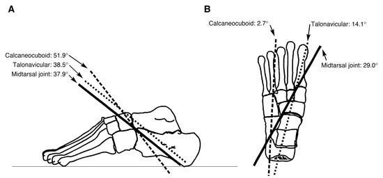
Figure 2.
Mean axis of rotation of the midtarsal joint projected on transverse and sagittal plane images of a left foot (calculated from mean range of motion). Angles relate to the angulation of the axis to the sagittal and transverse planes. Anatomical position was not calculated and is assumed from work by Van Langelaan[2] and Benink.[3] Talonavicular and calcaneocuboid axes are from Van Langelaan.[2]
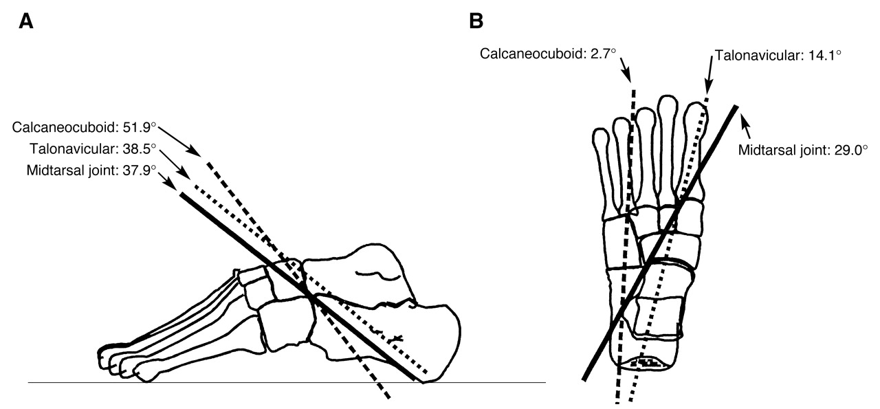
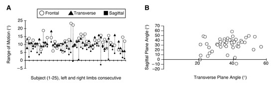
Figure 3.
A, Variations among subjects in the range of sagittal, frontal, and transverse plane motions. B, Variations among subjects in the angulation of the axes of rotation of the midtarsal joint composite phase.
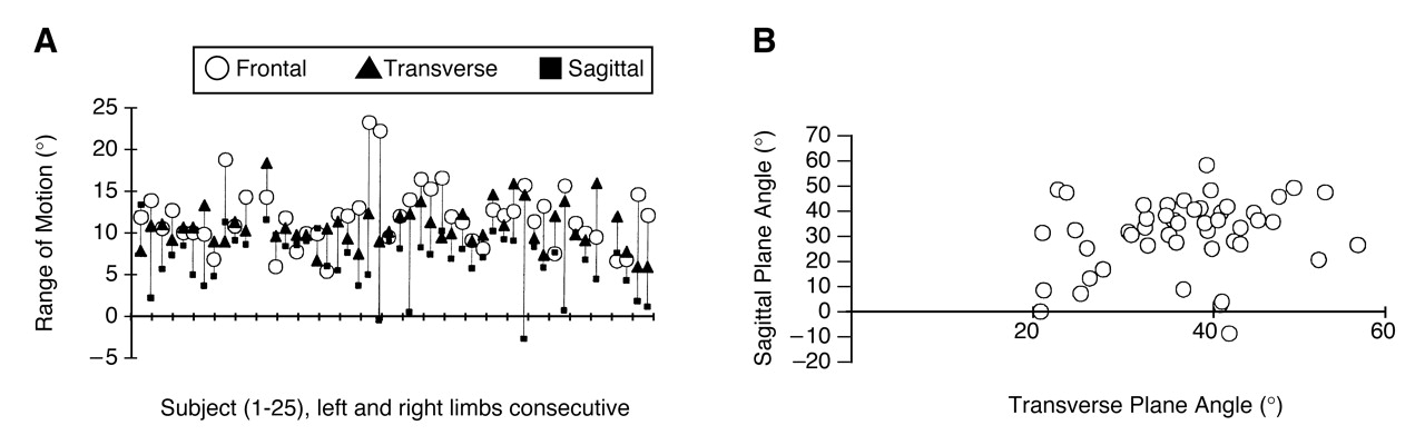

Table 1.
Mean (± SD) Range of Motion of Midtarsal Joint in Each Cardinal Body Plane, Axis of Rotation, and Range-of-Motion Ratio
Discussion
During internal rotation of the leg, the forefoot everted, abducted, and dorsiflexed relative to the heel. During external rotation of the leg, the forefoot inverted, adducted, and plantarflexed relative to the heel. There are few studies to which this can be compared. A reasonable comparison can be found in Lundberg and Svensson,[8] who described the rotations at the talonavicular joint during internal and external rotation of the leg. The direction of motion at the talonavicular joint was identical to this study’s midtarsal joint, and the proportions of frontal and transverse plane motion are similar (Lundberg and Svensson[8] reported 15.5° and 14.6° for frontal and transverse planes, compared with 11.9° and 10.6° in this study). Van Langelaan[2] described adduction and inversion motion at the calcaneocuboid joint during external leg rotation, which is in agreement with the results presented here.
The axis of rotation for the midtarsal joint calculated from the mean range of motion during the composite phase was oriented upwards and medially from posterior to anterior (Fig. 2). The angle of the midtarsal joint axis to the transverse plane varied between 56.4° and 22.0° (Fig. 3). This range reflects the variation in the predominant motion at the midtarsal joint, with some subjects having a predominance of frontal plane motion and others a predominance of transverse plane motion, and also demonstrates the wide variation that exists among subjects. The angle of the midtarsal joint axis to the sagittal plane varied from 58.6° to –9.8°. There were minimal differences between men and women; the angle of the mean axis of rotation to the transverse and sagittal planes, respectively, was 37.0° and 27.0° for men and 37.7° and 31.3° for women.
There are no studies in the literature describing an axis of rotation for the midtarsal joint as it was defined in this study. Thus, there are no studies to which these data are directly comparable. However, the upward and medial orientation for the midtarsal joint axis is consistent with the general orientation of the talonavicular and calcaneocuboid joint axes described by Van Langelaan[2] and the talonavicular axis described by Lundberg and Svensson.[8] In Van Langelaan’s work, which is based on 10 cadavers, the angle of the talonavicular axis varied from 3.8° to 21.4° (mean, 14.1°) relative to the sagittal plane and from 27.0° to 47.4° (mean, 38.5°) relative to the transverse plane. The angle of the calcaneocuboid axes varied from –15.5° to 19.9° (mean, 2.7°) relative to the sagittal plane and from 43.3° to 72.0° (mean, 51.9°) relative to the transverse plane. The angulation of the midtarsal joint axis to the transverse plane reported here is consistent with the mean axis for the talonavicular joint. One explanation of why the midtarsal joint axis is more similar to the talonavicular axis than the calcaneocuboid axis derived by Van Langelaan[2] may be that the range of motion is greater at the talonavicular joint than at the calcaneocuboid joint.[4] The principal difference between the results of this study and the work of Van Langelaan[2] and Lundberg and Svensson[8] is that the angulation of the axis to the sagittal plane is considerably greater in this study. This may be due to sagittal plane motion at the metatarsocuneiform joint, which would influence data in this study but not in the aforementioned work. Similarly, in this study, the authors may have measured more sagittal plane motion because only the heel motion was assessed proximally, whereas the invasive and cadaver studies assessed the individual talus and calcaneus.
The ratio of transverse to frontal plane motion for the mean midtarsal joint was relatively similar for the supination and pronation phases. This consistency, however, is not a true representation of the joint characteristics within the sample. Approximately 50% of the sample displayed more transverse plane motion per degree of frontal plane motion in the supination phase than in the pronation phase, while the other 50% displayed the opposite pattern. Thus, the majority of individuals did have some differences between the supination and pronation phases, but the nature of the difference was not consistent within the sample. Furthermore, these differences were cancelled out in the collation of the mean data for the sample.
Since this method of kinematic assessment is new, it is important to establish the boundaries for its repeatability. The mean difference between the ranges of motion recorded on separate days is excellent, with a maximum mean difference of 1.1°. The reason for the larger differences between days in the angulation of the axes of rotation is that even small differences in the measured range of motion can result in apparently large differences in the angulation of the axis of rotation because these small differences are large as a proportion of the total range of motion.[9,10] Given the level of repeatability in the ranges of motion measured, it would not be expected that the repeatability of results on the axes of rotation could be improved. This is an inherent difficulty when assessing joints with small ranges of motion.
Conclusion
A method has been demonstrated to quantify some of the kinematic characteristics of the midtarsal joint during transverse plane rotation of the leg. The results of the study generally agree with those of cadaver and invasive in vivo studies and add further scientific data to the evidence-based model of midtarsal joint function.
References
- Manter JT: Movements of the subtalar and transverse tarsal joints. Anat Rec 80: 397, 1941.
- Van Langelaan EJ: A kinematic analysis of the tarsal joints: an x-ray photogrammetric study. Acta Orthop Scand 54 (suppl 204).:1983.
- Benink RJ: The constraint mechanism of the human tarsus. Acta Orthop Scand 56 (suppl 215).:1985.
- Lundberg A, Svensson OK, Bylund C, et al: Kinematics of the ankle/foot complex. Part 3: influence of leg rotation. Foot Ankle 9: 304, 1989.
- Engsberg JR: Biomechanical analysis of the talocalcaneal joint: in vitro.. J Biomech 20: 429, 1987.
- Hintermann B, Nigg BM, Sommer C, et al: Transfer of movement between calcaneus and tibia in vitro.. Clin Biomech 9: 349, 1994.
- Downing BS, Klein BS, D’Amico JC: The axis of motion of the rearfoot complex. JAPA 68: 484, 1978.
- Lundberg A, Svensson OK: The axes of rotation of the talocalcaneal and talonavicular joints. Foot 3: 65, 1993.
- Panjabi MM, Goel VJ, Walter SD: Errors in kinematic parameters of a planar joint: guidelines for optimal experimental design. J Biomech 15: 537, 1982.
- Cunliffe M: Measurement, analysis and description of human arm motion [doctor of philosophy thesis]. Salford, England: University of Salford, 1997.
© 2002 American Podiatric Medical Association