


To the Editor:
Lemierre’s disease is an acute fulminant disease characterized by an anaerobic oropharyngeal infection leading to septic thrombophlebitis of the internal jugular vein, followed by metastatic foci of infection, most commonly to the lung. This disease is also known as postanginal septicemia and necrobacillosis. Distant metastatic infections that are likely caused by hematogenous seeding can often appear as septic arthritis, soft-tissue infections, meningitis, and osteomyelitis. The organism most commonly recovered from blood or pus from metastatic foci is Fusobacterium necrophorum. Other organisms isolated from patients with this syndrome include Bacteroides melaninogenicus, Eikenella corrodens, and non–group A streptococcus. In the preantibiotic era, Lemierre’s syndrome usually had a rapid and fatal course, with a mortality rate as high as 90%. However, with the advent of antibiotics, the mortality rate has dropped to 8%. Although numerous authors have described this acute condition, the syndrome was named after André Lemierre, a physician at the Claude Bernard Hospital in Great Britain who wrote a comprehensive article on the subject in 1936 that reviewed 20 patients with this condition.
The typical presentation of Lemierre’s syndrome initially begins with flu-like symptoms, including acute oropharyngeal infections. Infection of the deep pharyngeal tissue allows anaerobic organisms to drain into the lateral pharyngeal space that contains the internal jugular vein. Anaerobic organisms cause septic thrombophlebitis of the internal jugular vein. Sepsis occurs within 3 to 24 days after the initial onset of symptoms. Bacteremia associated with inadequate tissue perfusion may lead to septic shock, which is characterized by acute circulatory failure, usually with hypotension and inadequate tissue perfusion that will not satisfy nutrient demands and eventually leads to cellular death.
Septic shock is classified as a distributive shock. Hemodynamic changes occur at a cellular level, leading to abnormal volume distribution secondary to changes in permeability and vascular resistance. [1] The most common infectious agent causing septic shock is gram-negative bacteria, which is consistent with the offending agent in Lemierre’s disease, Fusobacterium necrophorum. The lipid fraction of the lipopolysaccharides released from the cell walls of gram-negative bacteria is known to initiate septic shock. Tumor necrosis factor, leukotrienes, lipooxygenase, histamine, bradykinin, serotonin, and interleukin-2 can also initiate septic shock. Ultimately, these mediators lead to the initial stages of vasodilation of arteries and arterioles, decreasing peripheral arterial resistance and increasing cardiac output. As the condition progresses, the cardiac output may be decreased and peripheral resistance increased. [2] The sepsis experienced in Lemierre’s disease is usually associated with high fevers, rigors, and elevated serum liver enzyme levels. Septic clots commonly dislodge from the internal jugular vein thrombus, resulting in infected pulmonary infarcts. Patients frequently have pleural effusions and occasionally have thoracic empyemas, with symptoms of dyspnea and pleuritic pain. Distant metastatic infections can occur as a result of hematogenous seeding. Septic arthritis, soft-tissue infections, meningitis, and osteomyelitis all have been reported. [3] A diagnosis of Lemierre’s syndrome is based on clinical presentation, the occurrence of anaerobic septicemia, radiographic evidence of thrombosis, and metastatic infection. [4]
Current treatment of Lemierre’s syndrome consists of long-term intravenous antibiotic therapy, surgical drainage of purulent collections, and removal of necrotic tissue. [4] Ligation and resection of the internal jugular vein thrombosis usually is not necessary and should be considered only in refractory sepsis or severe respiratory compromise from repeated pulmonary emboli. [5] Initial antibiotic therapy should include antibiotics with good antianaerobic coverage. Fusobacterium necrophorum typically is susceptible to penicillins, and only rarely are beta-lactamase producers isolated. Fusobacterium species also are susceptible to other antianaerobic antibiotics, including cefoxitin, clindamycin, metronidazole, chloramphenicol, ampicillin-sulbactam, and ticarcillin-clavulanate. Surgical drainage of any abscesses and the removal of necrotic tissue generally are necessary for successful treatment and may decrease the length of antibiotic therapy.
Case Report
A 36-year-old woman without significant medical history presented to the emergency department at New York Methodist Hospital because of progressive shortening of breath with fever and chills of 7 days’ duration. The patient related a history of nausea and vomiting with intermittent diarrhea during this time period as well. The patient related having a cold and sore throat with a temperature of 102°F approximately 2 weeks prior to admission. The patient thought she had the flu and took ibuprofen, which provided mild symptomatic relief. Upon her arrival in the emergency department, her vital signs were as follows: temperature, 101.5°F; blood pressure, 112/80 mm Hg; pulse, 130 per minute; respirations, 52 per minute; and an oxygen saturation level of 94%. The physical examination was remarkable for labored breathing with bilateral rhonchi and increased chest pain on deep inspiration. The patient’s lower extremity also appeared mottled but was warm on palpation. Laboratory data at the time of admission included normal electrolytes, an elevated blood urea nitrogen level of 54 mg/dL, and an elevated creatinine level of 2.0 mg/dL. An initial complete blood count was within normal limits, with a white blood cell count of 6,200/mm3. A chest radiograph revealed bilateral lobar infiltrates with bilateral pleural effusions. The patient was admitted because of sepsis and respiratory failure. Empiric intravenous antibiotic therapy was initiated, consisting of cefuroxime, 750 mg every 8 hours; erythromycin, 1 g every 6 hours; and trimethoprim-sulfamethoxazole, 200 mg every 6 hours. The patient’s condition deteriorated, leading to septic shock. On hospital day 2, dopamine and norepinephrine were initiated to control circulatory failure. The patient underwent multiple procedures for empyemas and clotted hemothorax. Culture and sensitivity testing of the empyema and blood yielded Fusobacterium necrophorum, the most common causative agent of Lemierre’s disease. Doppler studies of the internal jugular vein revealed thrombosis. The diagnosis of Lemierre’s disease was confirmed on hospital day 7, based on the clinical presentation, the culture of Fusobacterium necrophorum, and the presence of thrombosis in the left internal jugular vein. The antibiotics were changed to clindamycin, 900 mg intravenously every 6 hours; and metronidazole, 500 mg intravenously every 6 hours, to cover Fusobacterium necrophorum. The patient’s condition improved.
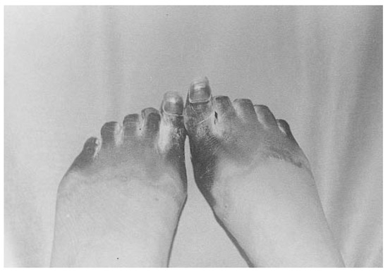
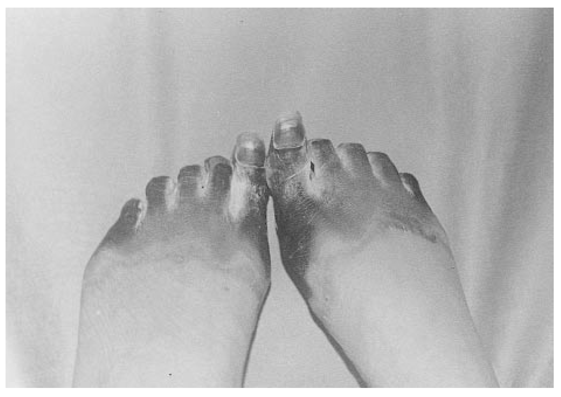
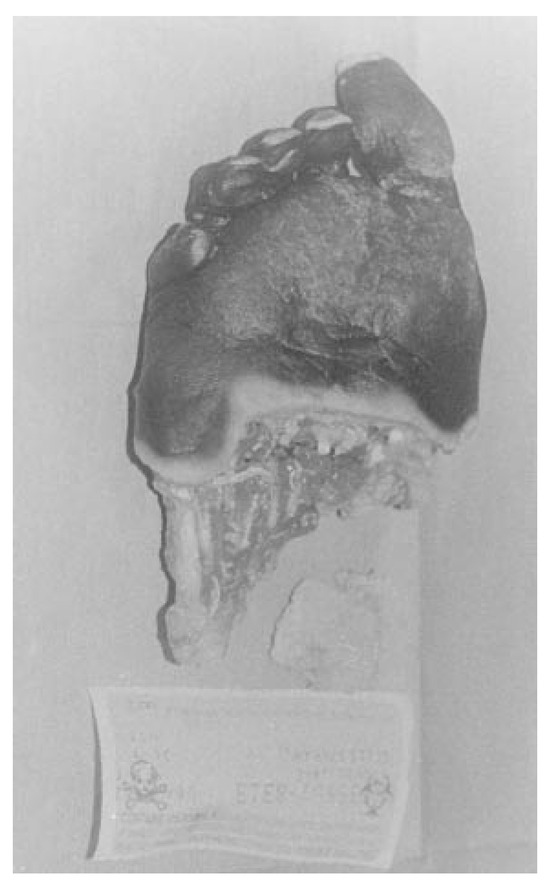
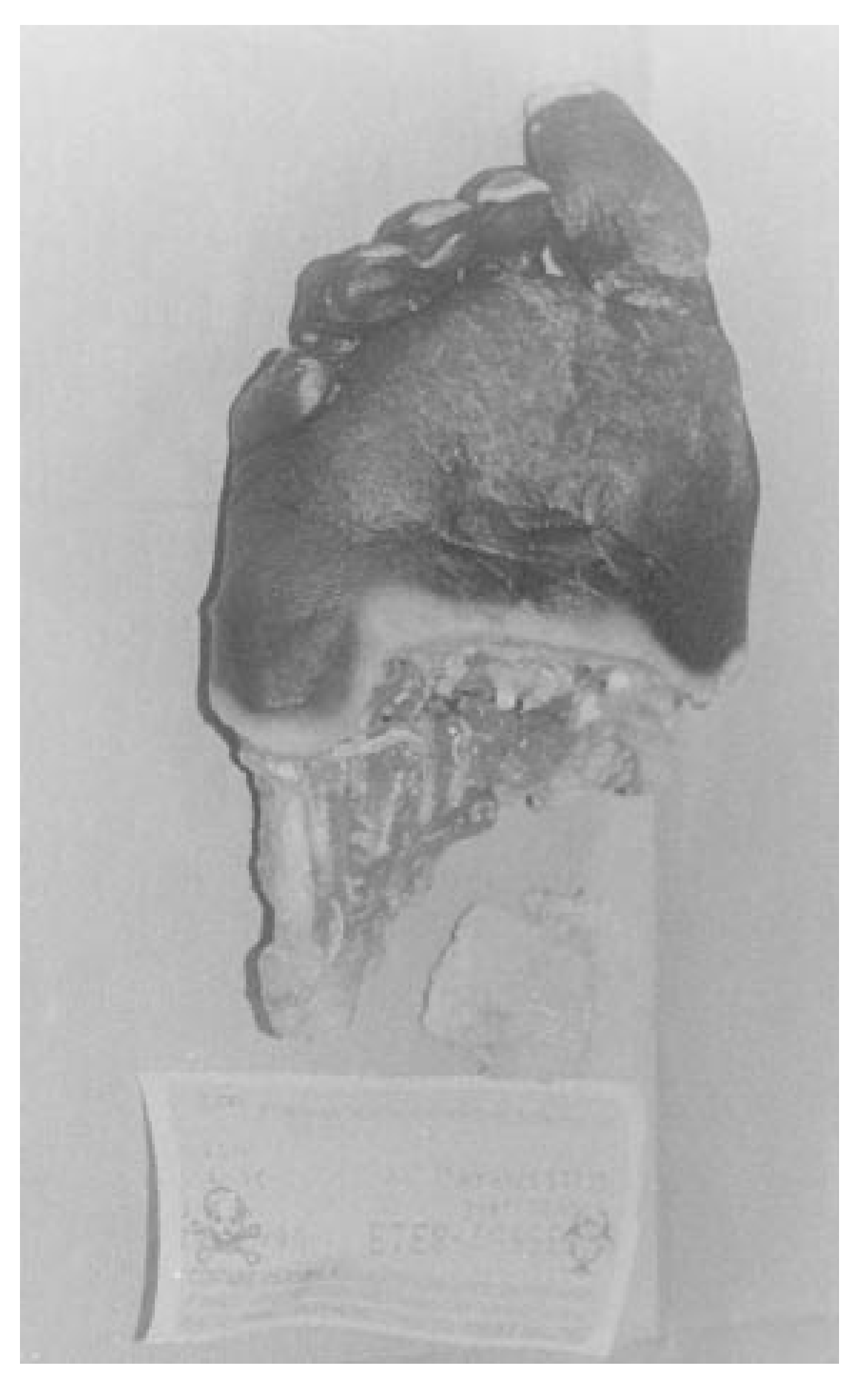
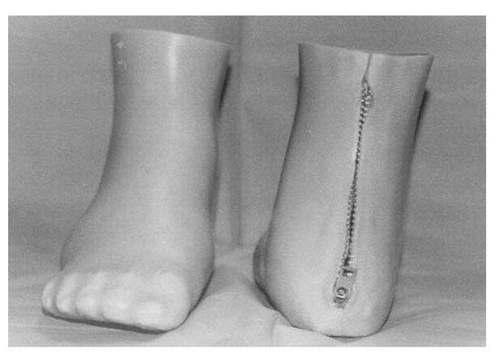
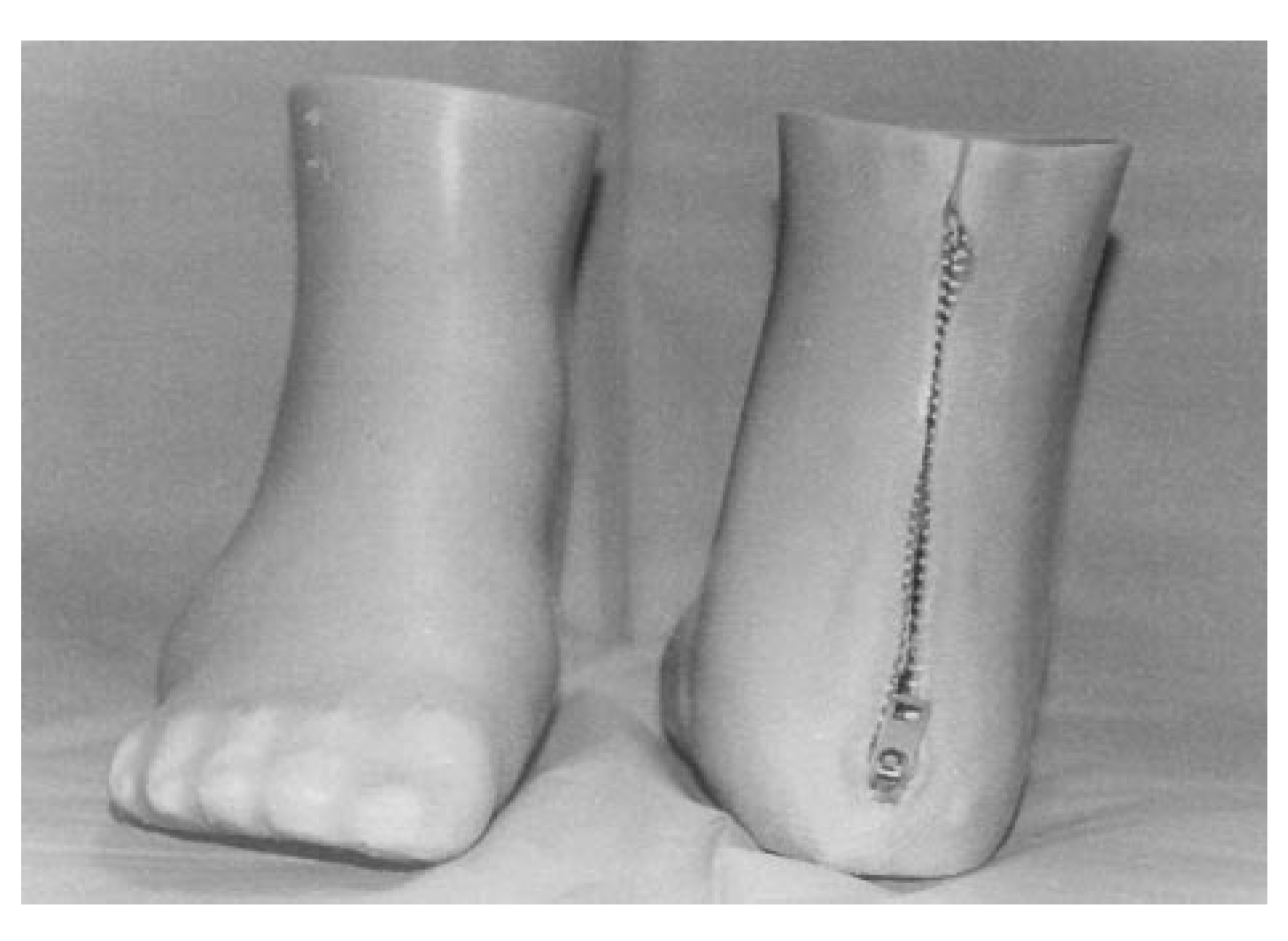
On hospital day 15, the podiatric medicine department was consulted about the patient’s bilateral forefoot gangrene (Figure 1). Upon examination, well-demarcated bilateral forefoot gangrene extending to the metatarsal heads was noted. There was no evidence of cellulitis, drainage, or purulence. Dorsalis pedis and posterior tibial pulses were 2/4 bilaterally. The patient’s legs were warm to the touch, and there was increased pain with palpation over the forefoot area bilaterally. Bilateral transmetatarsal amputations were recommended to prevent suprainfection, once the patient was medically stable. On hospital day 25, the patient underwent bilateral transmetatarsal amputations. Intraoperative findings included well-demarcated dry gangrene with healthy viable peripheral tissue and no clinical signs of infection (Figure 2). Pathologic analysis of the bilateral specimens revealed gangrenous changes of the forefoot with viable margins and no signs of osteomyelitis or peripheral soft-tissue infection. The postoperative course included intensive physical therapy. The patient continued to improve and was discharged on hospital day 50. She was then referred to a private prosthetist for custom-molded silicone prostheses (Figure 3).
Figure 1.
Bilateral foot gangrene.
Figure 2.
Intraoperative findings showing healthy viable peripheral tissue.
Figure 3.
Custom-molded silicone prostheses.
Discussion
Although distant metastatic infection secondary to hematogenous seeding is common in Lemierre’s syndrome, in this case study the gangrenous changes were secondary to the combination of the pathophysiologic effects of sepsis, including insufficient tissue perfusion leading to cellular death, and the potent vasoconstriction effects of dopamine and norepinephrine. This conclusion was based on the clinical presentation of bilateral, well-demarcated gangrene, intraoperative findings of viable intrinsic musculature, and no clinical evidence of infection. This was further substantiated by pathologic findings that included viable peripheral tissue, with no evidence of infection or emboli.
Although the incidence of Lemierre’s syndrome has decreased greatly in the postantibiotic era, many cases are still reported today. [2,3,4,5,6] As with all forms of sepsis, Lemierre’s disease is associated with high morbidity and mortality. Therefore, it is important for the podiatric physician to be familiar with this disease entity, and it should be considered in the differential diagnosis when treating patients with gangrene secondary to sepsis.
References
- ZIER B, BYRNE C: Septic shock syndrome in the surgical wound patient. J Foot Ankle Surg 1992, 31, 459.
- WEIL, M.H. “Bacteremia and Septic Shock,” in The Merck Manual; Berkow, R., Ed.; Merck Research Laboratories: Rahway, NJ, 1992; p. 70. [Google Scholar]
- LEE, B.; LOPEZ, F.; GENOVESE, M. ET AL: Lemierre’s syndrome. South Med J 1997, 90, 640. [Google Scholar] [CrossRef] [Scilit] [PubMed]
- STALLWORTH, J.; CARROLL, J. Lemierre’s syndrome: new insights into an old disease. Clin Pediat 1997, 36, 715. [Google Scholar] [CrossRef] [Scilit] [PubMed]
- HUGHES, C.; SPEAR, R.; SHINABARGER, C. ET AL: Septic pulmonary emboli complicating mastoiditis: Lemierre’s syndrome revisited. Clin Infect Dis 1994, 18, 633. [Google Scholar] [CrossRef] [Scilit] [PubMed]
- LUSTIG, L.; CUSICK, B.; CHEUNG, S. ET AL: Lemierre’s syndrome: two cases of postanginal sepsis. Otolaryngol Head Neck Surg 1995, 112, 767. [Google Scholar] [CrossRef] [Scilit] [PubMed]
© 2001 American Podiatric Medical Association