


Abstract
Many procedures have been described for the resection of plantar calcaneal spurs as treatment of heel spur syndrome and chronic plantar fasciitis. Most of these techniques involve a medial incision of between 2 and 6 cm for adequate exposure of the calcaneal spur. This article describes a new technique for resecting a calcaneal spur with a smaller medial incision using the holmium:yttrium-aluminum-garnet (Ho:YAG) laser. This laser permits adequate resection of a plantar calcaneal spur as well as coagulation of the bone and surrounding tissues. This minimally invasive procedure has been used with good results over the past year by the senior author (W.K.S.) for the resection of calcaneal spurs.
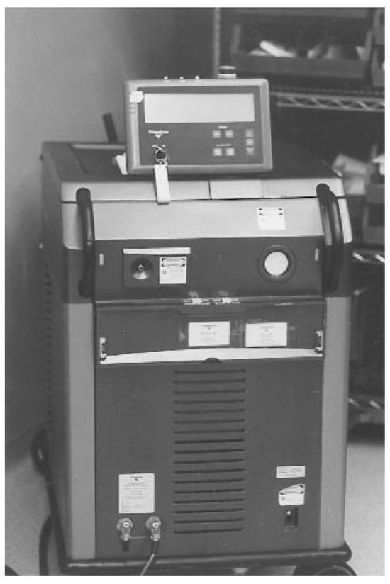
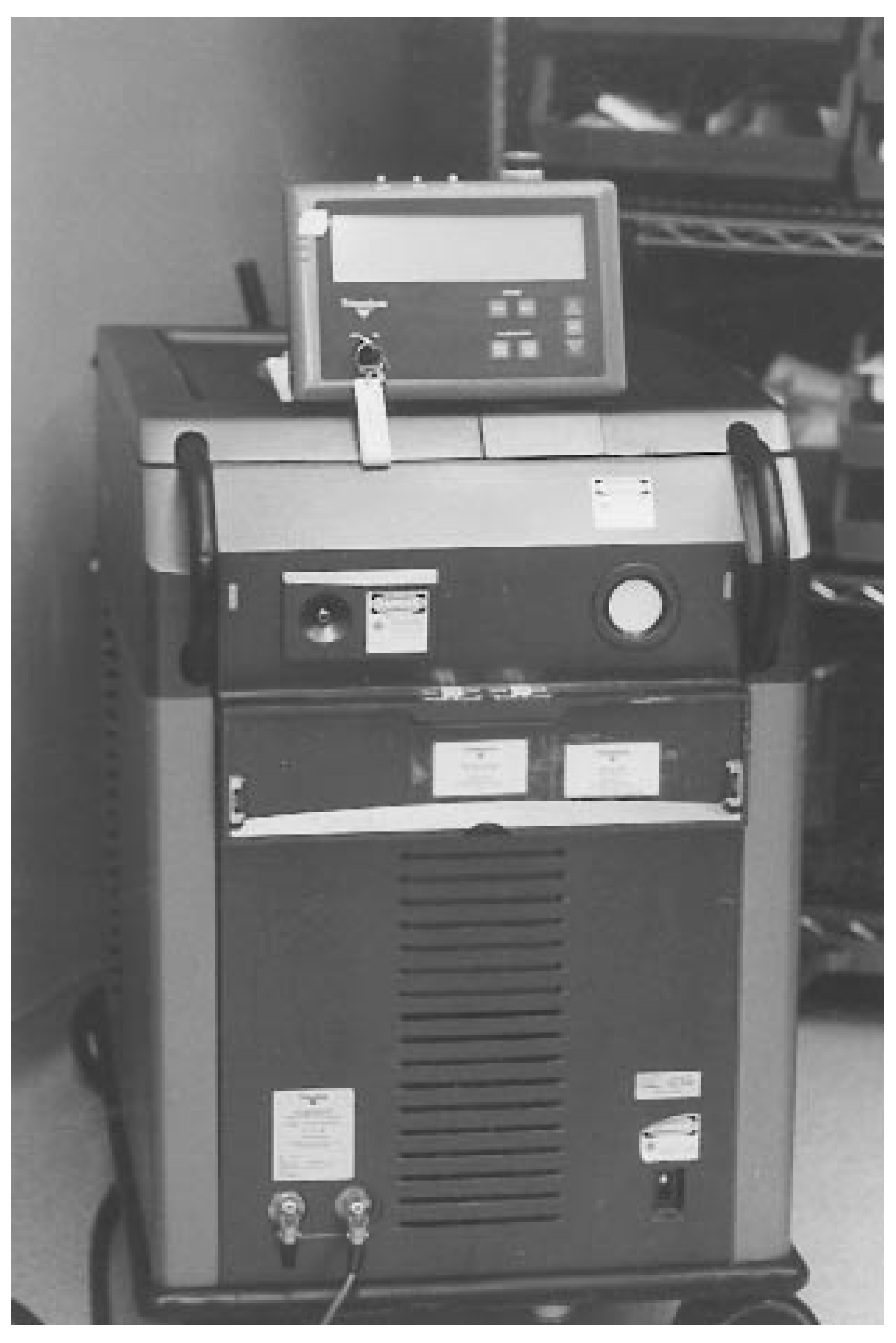
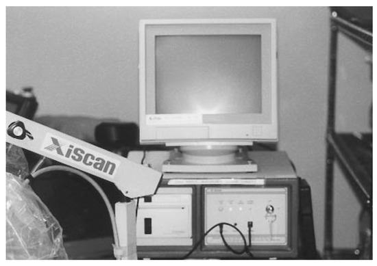
The holmium:yttrium-aluminum-garnet (Ho:YAG) laser (Trimedyne, Inc, Irvine, California) (Figure 1) is a multidimensional tool with the ability to cut, coagulate, vaporize, and sculpt tissues of varying densities. The Ho:YAG laser uses a combination of holmium, yttrium, aluminum, and garnet as its solid, active medium. [1] This combination produces an infrared radiation wave of approximately 2100 nm with a pulse width of 350 microseconds. The delivery of laser energy in a pulsed manner allows the tissues to be cooled, thereby decreasing any negative thermal effect. The average depth of penetration of the Ho:YAG laser is between 0.1 and 1.0 mm, making this wavelength safe for use around vessels, nerves, and bony tissue. The Ho:YAG laser has also shown an excellent ability to cause coagulation during cutting, thereby decreasing postoperative bleeding. [2].
Figure 1.
The Ho:YAG laser unit.
Laser Technique
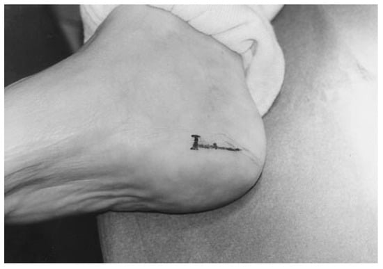
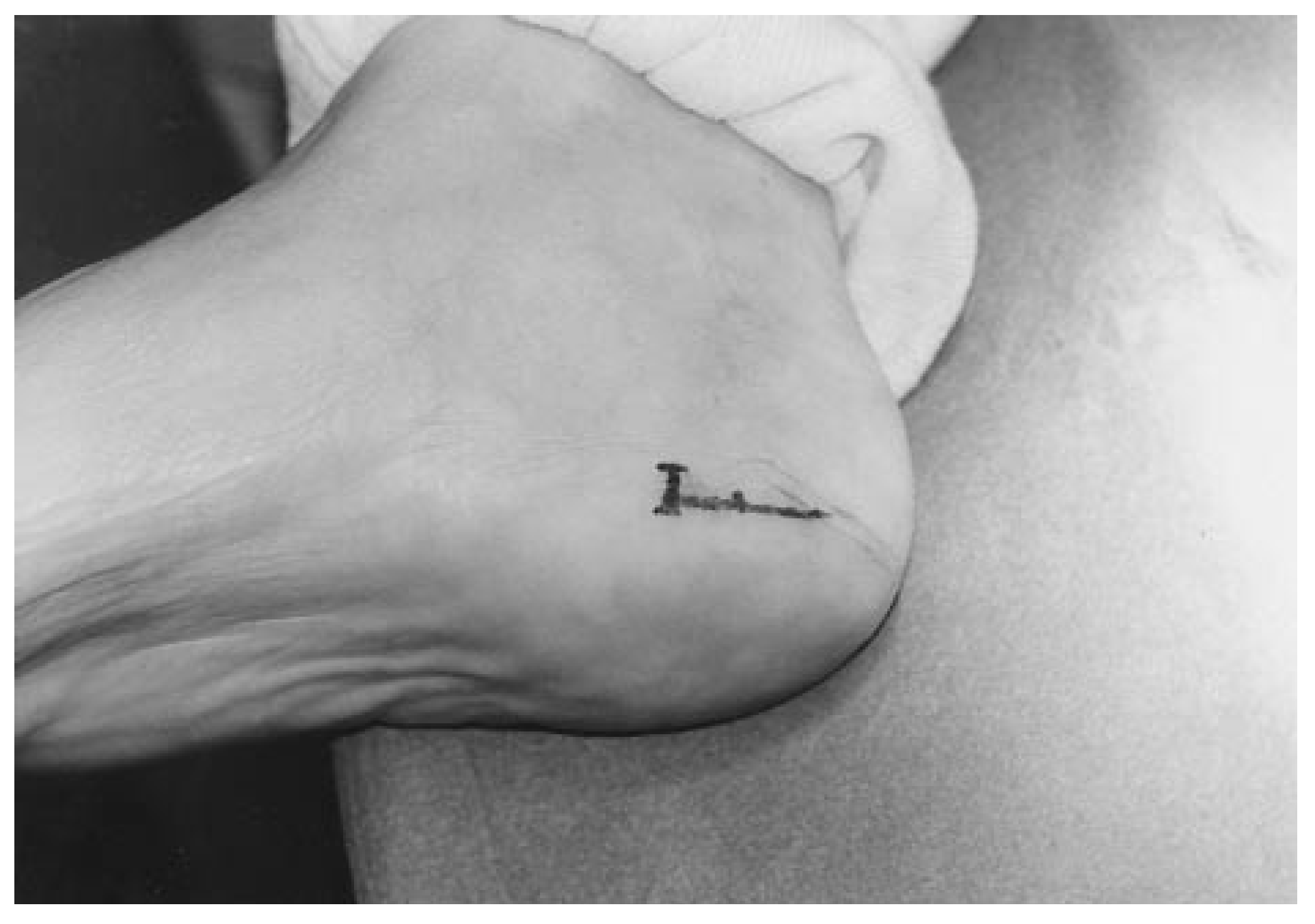
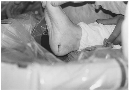
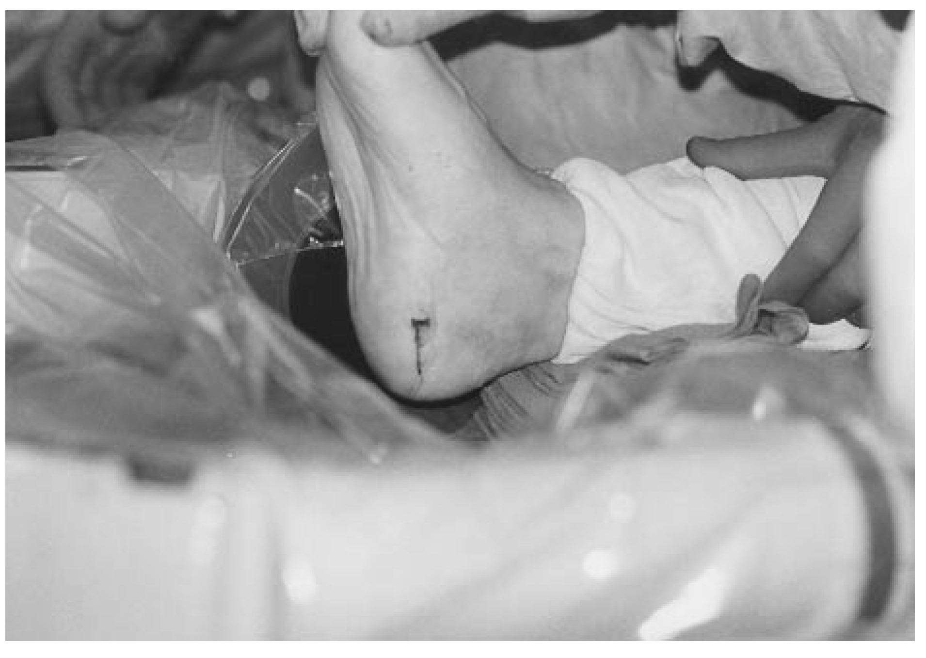
The surgical laser technique for resection of a plantar calcaneal spur requires that the patient be placed in the supine position. The patient is prepared for surgery by use of either general anesthesia or local anesthesia with intravenous sedation, and is draped in the usual aseptic manner. Hemostasis is obtained with either a thigh or an ankle cuff. The incision site is determined; the site is similar to that used for the endoscopic plantar fasciotomy as described by Barrett et al. [3] (Figure 2).
Figure 2.
The medial incision is marked with a skin marker according to the method of Barrett et al. [3].
Once the cannula is placed properly, inferior to the plantar fascia, the sections of the plantar fascia are measured and marked (Figure 3). Release of the plantar fascia is performed from lateral to medial, starting with the medial section, rather than medial to lateral as recommended by Barrett et al. [3] After the medial section is released completely, the foot is placed in dorsiflexion to check for adequate separation. If the central band must be released, this is done and then the foot is checked again. Once adequate release is obtained, the cannula is removed.
Figure 3.
The plantar fascia is marked and then compared visually with the plantar aspect of the foot to coincide with the patient’s symptoms.
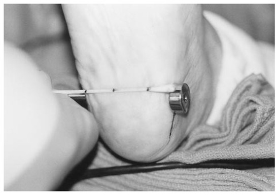
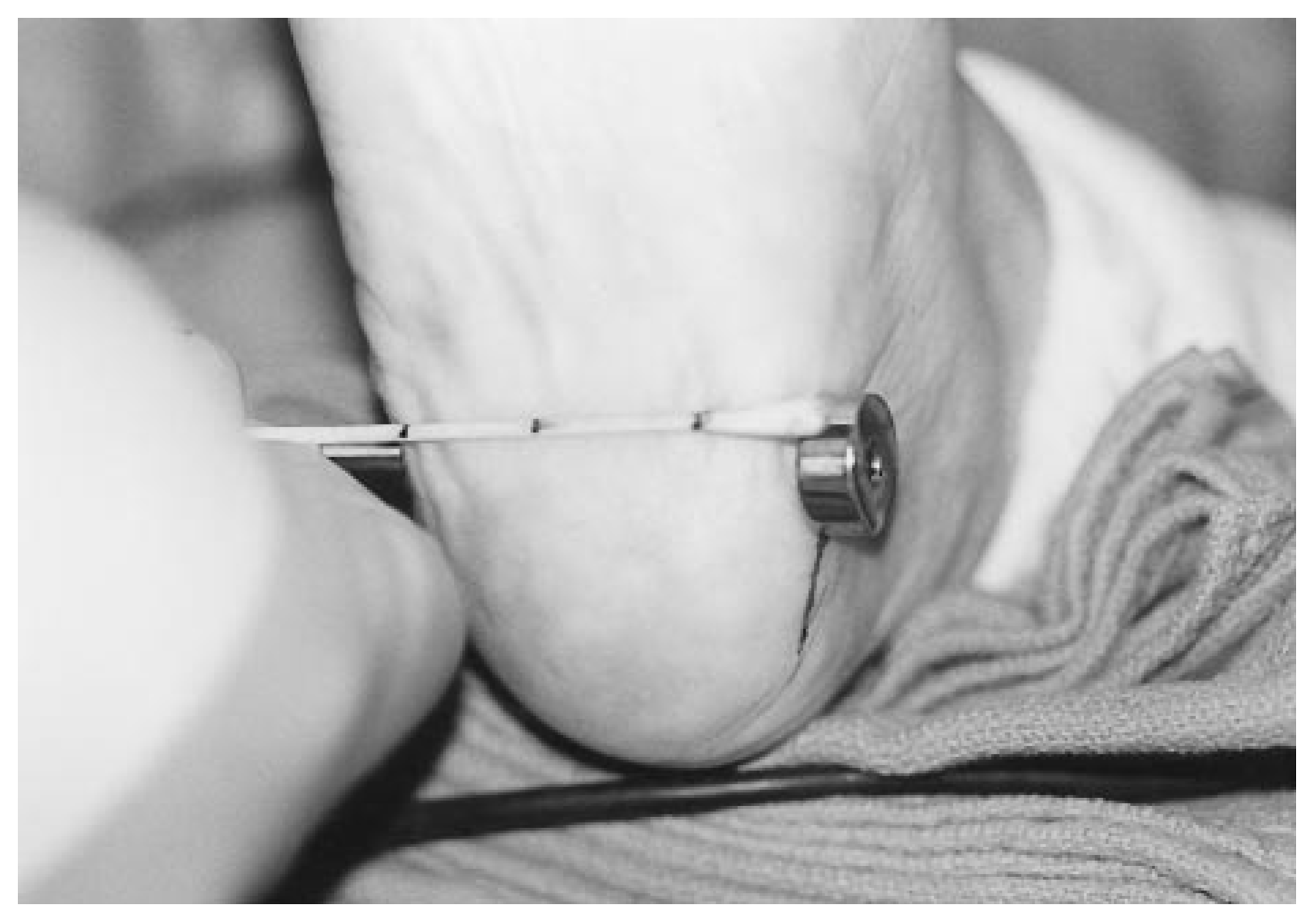
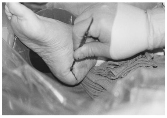
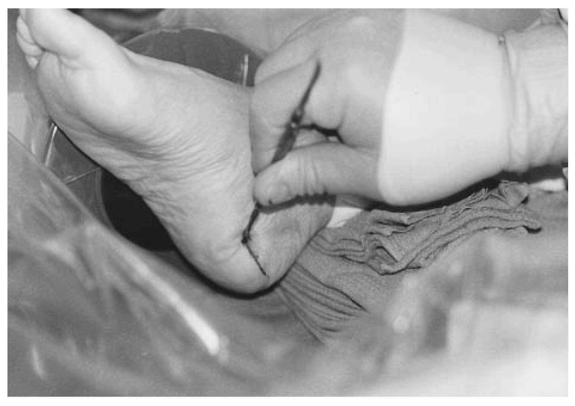
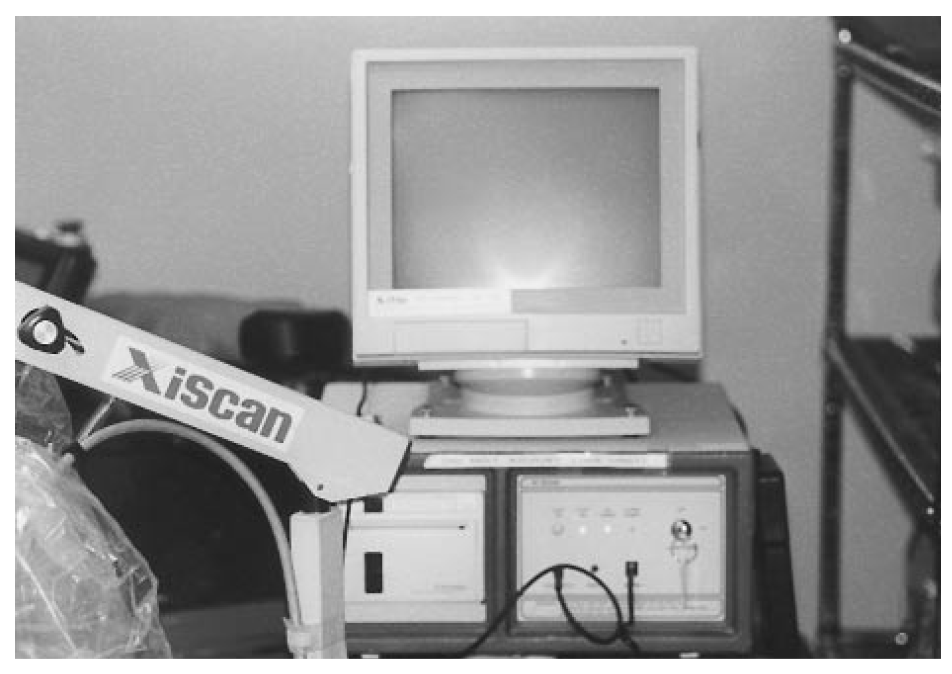
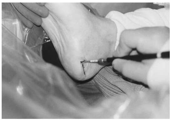
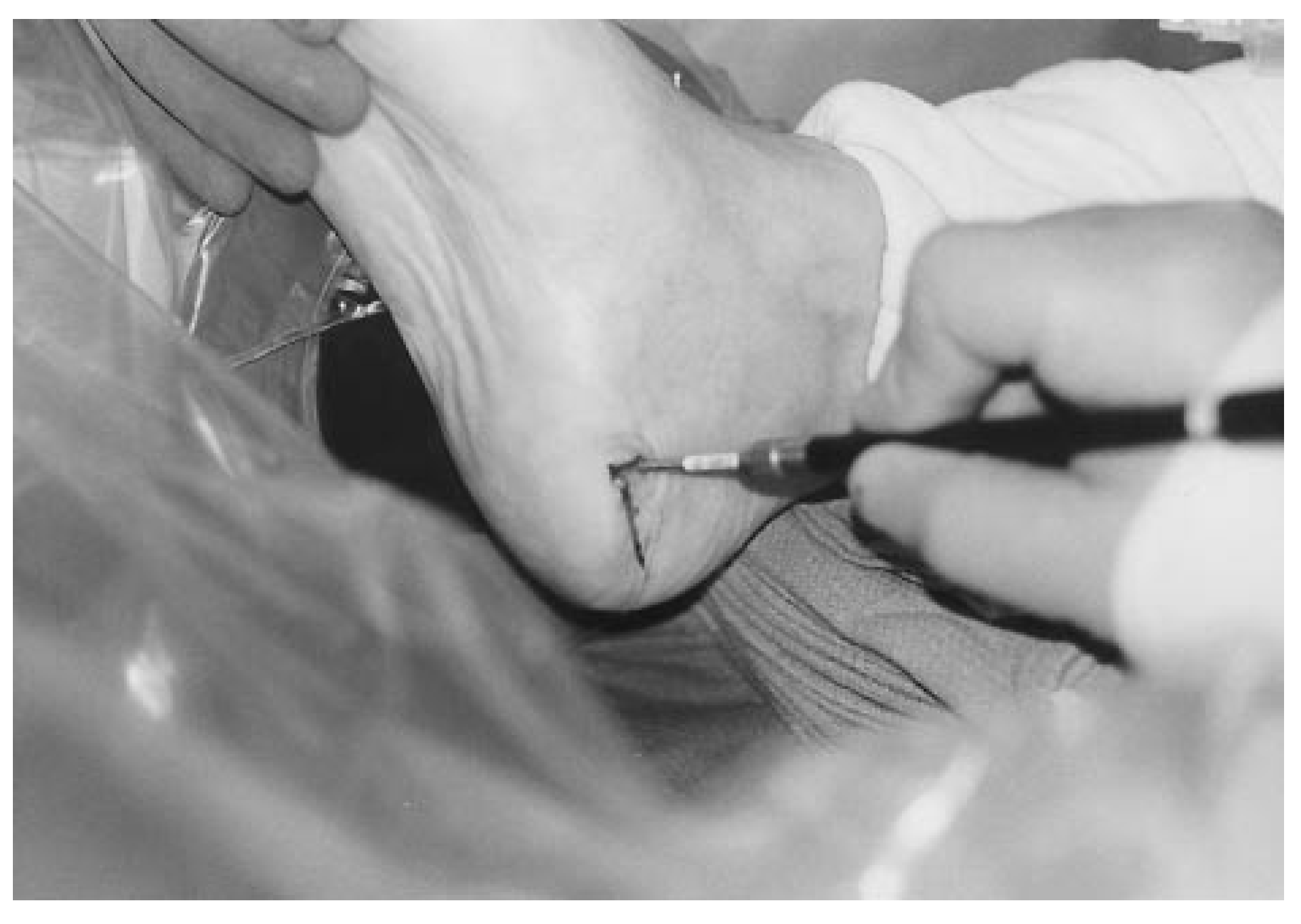
A Freer elevator is inserted from the medial side of the foot (Figure 4) and the plantar calcaneal spur is dissected free from surrounding soft-tissue attachments with the assistance of a Xi-Scan (Xitec Inc, East Windsor, Connecticut) (Figure 5). After the spur has been adequately exposed, the laser tip is introduced from the medial side (Figure 6). The laser tip is placed adjacent to the spur, which aids in the identification of the spur and ensures adequate laser penetration. The laser should be set at 1 joule at 20 repetitions using a double-pulse setting, which approaches 40 W of power.
Figure 4.
The Freer elevator is used to dissect all soft tissue from the underlying heel spur.
Figure 5.
The Xi-Scan unit.
Figure 6.
Constant radiographic monitoring through the intra-operative placement of the XiScan is used to ensure adequate resection of the heel spur.
The laser tip is moved across the spur until the spur cannot be felt with the laser tip and is not visible on the Xi-Scan (Figure 7). During the resection procedure with the laser, the site is flushed with normal saline. This helps reduce further the possibility of thermal effects and removes any particulate matter. The incision closure is performed according to the surgeon’s preference. The wound is bandaged with a large bulky dressing and compression is accomplished with a Coban dressing (3M, St Paul, Minnesota). The patient is placed in an Equalizer (Royce Medical, Camarillo, California) leg cast, which allows either partial or no weightbearing, depending on the patient’s tolerance.
Figure 7.
The laser tip is inserted into the medial aspect of the heel to begin resection.
The patient is seen for follow-up at 4 days after surgery. At that time, the patient’s dressing is replaced with a small bandage on each incision. Swelling is controlled with either Coban or a tube gauze stocking. The patient may clean the foot daily with a clean washcloth until postoperative day 7, after which the patient is allowed to shower. The skin sutures are removed 2 weeks following the procedure. Once the patient can ambulate in the cast without pain, the patient may wear a sturdy athletic shoe with orthotic inserts.
Postoperative radiographs are obtained at regular intervals to monitor for thermal necrosis or stress fractures, as well as any regrowth of the spur. Nor mal activities are resumed when the patient can tolerate the cast boot or athletic shoe with orthoses.
Case Report
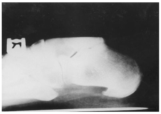
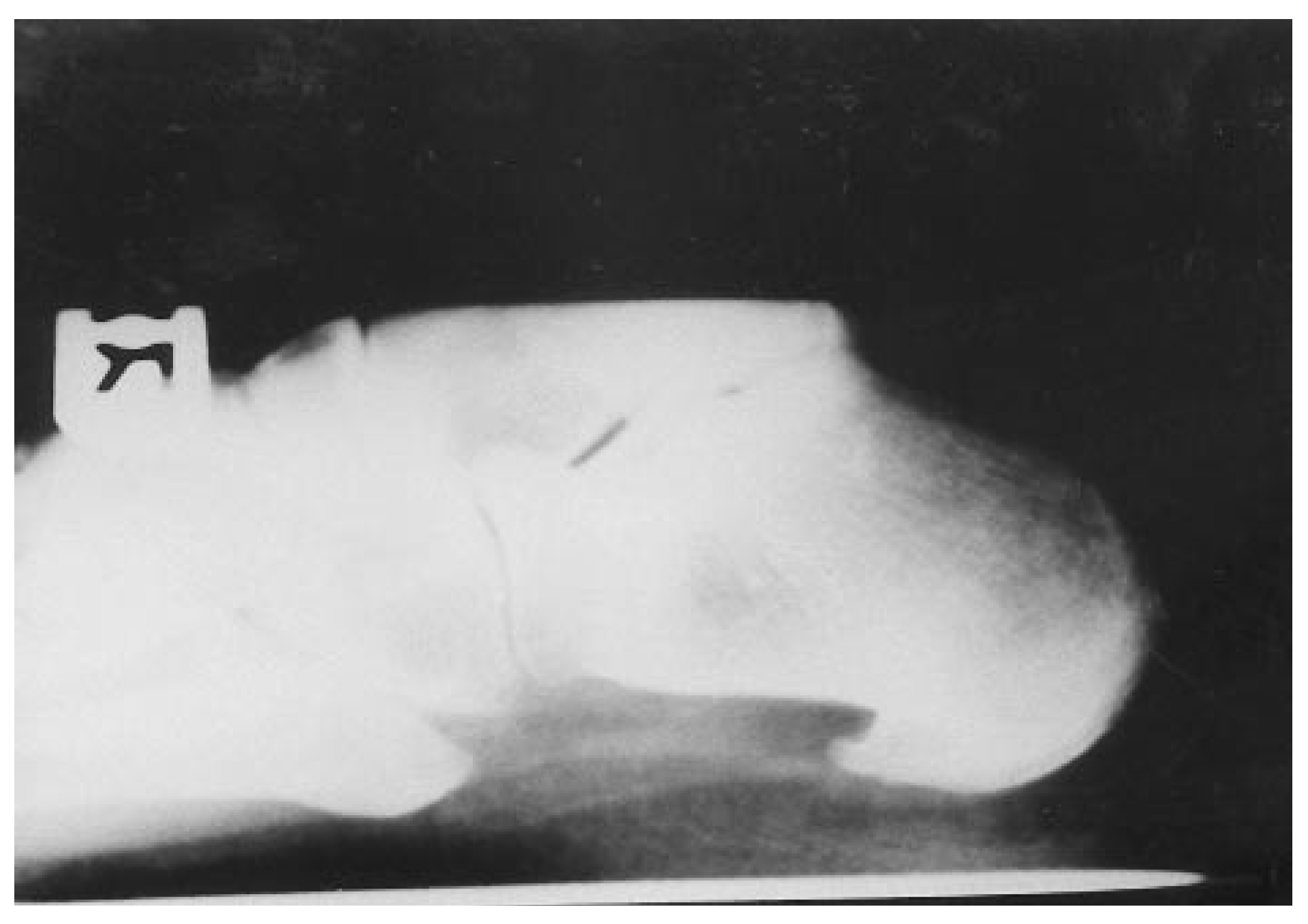
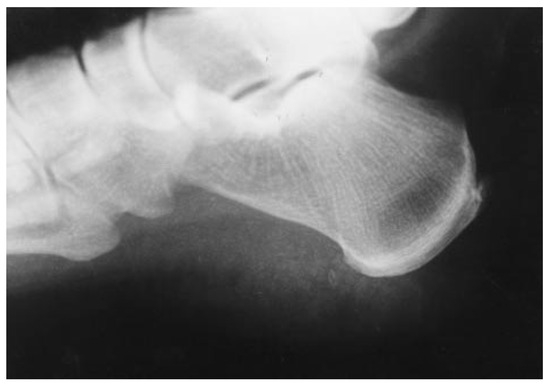
A 54-year-old woman presented to the office of the senior author (W.K.S.) with a chief complaint of heel pain of several months’ duration. The patient denied any acute trauma to her feet or legs. Her work—8 hour shifts with short breaks during the day—required her to stand on concrete all day, every day. The patient reported that similar episodes of heel pain had resolved in the past simply with rest and elevation for several days, as well as new shoes or arch supports. The previous episodes lasted no more than 1 week to 10 days and were followed by complete recovery. The patient sought medical advice because she was unable to obtain relief from the latest episode of heel pain. She related classic symptoms of pain with early-morning ambulation and after short periods of rest. Radiographic evaluation included a weightbearing lateral view, which revealed a large plantar calcaneal exostosis in the area of the plantar tuberosity (Figure 8).
Figure 8.
Preoperative radiograph showing the large plantar calcaneal spur.
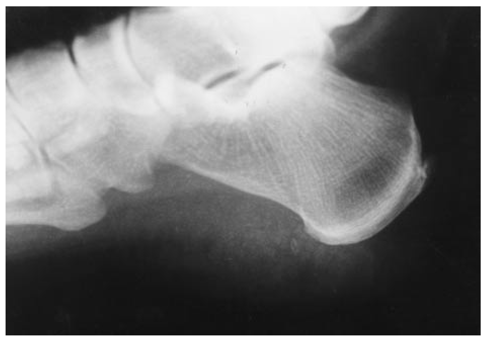
Conservative therapy, including oral anti-inflammatory medications, temporary orthoses, custommade orthoses, corticosteroid injections, and ultrasound to the area, was tried for approximately 6 months without success. The patient elected to undergo surgical correction. The surgical procedure previously described, using the Ho:YAG laser, was performed. Postoperative radiographic evaluation revealed adequate resection of the calcaneal spur (Figure 9). The patient wore a walking cast boot for approximately 2 weeks and then was instructed to wear a good, sturdy walking shoe. She returned for follow-up for approximately 3 months. At over 1 year after surgery, the patient was still asymptomatic.
Figure 9.
Postoperative radiograph showing complete ablation of the calcaneal spur that occurred with the use of the Ho:YAG laser.
Discussion
The Ho:YAG laser has been used in orthopedic surgery for many years. It is also becoming more popular in urologic surgery. In podiatric surgery, use of the Ho:YAG laser has been restricted to the ablation of hypertrophic synovium, smoothing osteochondral defects, and ablation of anterior tibial exostosis in ankle arthroscopy. The advantages of the Ho:YAG laser when used in ankle surgery are hemostasis due to the coagulative or cauterizing effect of the laser, and total ablation or disintegration of tissue, with no residual debris.
The same advantages of the Ho:YAG laser in ankle surgery apply in heel spur surgery. Past attempts to resect plantar spurs through minimal incisions have left bone dust or a residual bone paste in the wound. With the use of the Ho:YAG laser, the bone is completely ablated without residual bone particles remaining in the muscle or fascia. The coagulative effect of the laser ensures minimal bleeding during and after the procedure. Theoretically, this reduces the potential for plantar fascia reattachment, thus reducing the recurrence of heel pain.
Past studies have shown that release of the plantar fascia with resection of the heel spur creates better results than does release of the plantar fascia alone. [4,5,6] Vohra et al. [6] compared 48 open heel spur resection procedures with 20 endoscopic plantar fasciotomy procedures. The authors reported that “patients who had open heel spur surgery experienced a greater reduction of pain than patients who had endoscopic plantar fasciotomy at the time of follow up.” [6]
The use of fluoroscopy or the Xi-Scan is an effective method of visualization of the plantar spur. The laser tip is radiopaque, ensuring visualization throughout complete osseous resection.
At the time of the writing of this article, the senior author had performed more than 150 of these laser procedures. The results thus far have been very good. Pain was completely alleviated at the initial pain site in all cases. There have been some cases of pain in new areas following surgery. There were two episodes of calcaneocuboid syndrome as a result of the endoscopic plantar fasciotomy, but these were completely resolved with below-the-knee casting for 4 weeks. One patient complained of tarsal tunnel syndrome postoperatively, but this was also resolved with the use of a below-the-knee cast for 4 weeks.
Summary
This article has described a new tool for the resection of heel spurs using a minimally invasive technique. Laser excision provides a safe, efficient, and effective surgical treatment for pain from recalcitrant plantar fasciitis in selected patients. However, surgeons must realize that this surgery requires expertise in the use of the Ho:YAG laser. Further evaluation is needed to document long-term results.
Acknowledgments
David Casper, MD, Charles Coker, ORT, Andrew Parker, ORT, and John Mollett, ORT, Surgical Hospital of Oklahoma, Oklahoma City, OK.
References
- VANGSNESS, C.T., JR.; GHADERI, B.; BRUSTEIN, M. ET AL: Ablation rates of human meniscal tissue with the Ho:YAG laser: the effects of varying fluences. Arthroscopy 1997, 13, 148. [Google Scholar] [CrossRef] [Scilit] [PubMed]
- Holmium Laser Arthroscopy and Disc Decompression Reference Manual; Trimedyne, Inc, Dept of Education: Irvine, CA, 1998.
- BARRETT, S.L.; DAY, S.V.; BROWN, M.G. Endoscopic plantar fasciotomy: preliminary study with cadaveric specimens. J Foot Surg 1991, 30, 170. [Google Scholar] [CrossRef] [Scilit] [PubMed]
- CONTOMPASIS, J.P. Surgical treatment of calcaneal spurs: a three-year post-surgical study. JAPA 1974, 64, 987. [Google Scholar] [CrossRef] [Scilit] [PubMed]
- ALI E: Calcaneal spur in Guyana. West Indian Med J 1980, 29, 175.
- VOHRA, P.K.; GIORGINI, R.J.; SOBEL, E. Long-term follow-up of heel spur surgery: a 10-year retrospective study. JAPMA 1999, 89, 81. [Google Scholar] [CrossRef] [Scilit] [PubMed]
© 2001 American Podiatric Medical Association