


To the Editor:
Osteoporosis is a potentially devastating disease affecting millions of Americans, causing more than 1.5 million fractures annually. The cost of treating this disorder is projected to be more than $60 billion over the next 20 years. Heightened awareness of risk factors should help in the recognition and treatment of osteoporosis-related fractures. [1]
Osteoporosis is seen more frequently in women than in men. While the pathogenesis of osteoporosis is not completely understood at this time, certain major risk factors are emerging. Among the more important of these are family history, low calcium intake, early menopause, and sedentary lifestyle. Other possible risk factors that have been suggested include high intakes of protein, alcohol, and caffeine; low body weight; exercise-induced amenorrhea; and cigarette smoking. [2]
Recent research by Harris and Dawson-Hughes [3] has shown a relationship between caffeine consumption and bone loss in healthy postmenopausal women. This research showed that daily consumption of caffeine in amounts equal to or greater than that obtained from about two to three cups of brewed coffee may accelerate bone loss from the spine and other areas of the body in women with calcium intake below the recommended daily allowance of 800 mg. Caffeine use was linked to hip fracture in a large study involving 84,484 women aged 34 to 59 over a 6-year period. [4] The data from this study suggested that caffeine consumption increased the risk of osteoporotic fractures in middle-aged women. The Framingham Study followed 3,170 individuals for a 12-year period and found that intake of more than two cups of coffee (or four cups of tea) per day increased the risk of fracture. [5] A study conducted at the Mayo Clinic examined the influence of caffeine consumption on bone density at six skeletal sites in an age-stratified random sample of white women residing in Rochester, Minnesota. After adjustment for age, the Mayo Clinic data suggested that caffeine intake in the range of a representative sample of white women was not an important risk factor for osteoporosis. [6]
Osteoporosis is diagnosed when bone density has decreased to the point where fractures will occur with mild stress. Bone-density testing is used to diagnose osteoporosis. A bone-density test measures bone mineral content. The greater the bone mineral content, the stronger or denser the bone is. Low bone density increases one’s risk of fracture.
People who are at risk for osteoporosis should be tested. The following is a case study of a healthy premenopausal woman with osteoporosis who presented without the obvious risk factors previously mentioned.
Case Report
On September 27, 1996, a 23-year-old white woman presented to the Tower Medical Clinic in Des Moines, Iowa, with pain over the second metatarsal of her left foot. The patient first noticed the pain, which had become progressively worse, about 1 month prior to presentation. The patient recalled no history of trauma.
The patient’s medical history revealed that she had been pregnant three times, with two successful births and one miscarriage. She had fairly regular menses and did not have amenorrhea. She was found to have hypothyroidism during her last pregnancy. Her medications included levothyroxine sodium, 15 mg once daily, and medoxyprogesterone acetate as a form of birth control. The patient had no known drug allergies. She had a family history of diabetes mellitus and heart disease. She denied use of tobacco and stated that she used alcohol occasionally. The patient also mentioned that she drank one cup of coffee and up to 60 ounces of a highly caffeinated soft drink per day. The patient stated that she worked as a clerk and was on her feet 8 or more hours per day. Physical examination revealed a young, healthy woman (height, 5 feet 6 1/4 inches; weight, 146 pounds). Her blood pressure was 118/74 mm Hg; her heart rate was 78 beats per minute; and her respirations were 16 breaths per minute.
Physical examination of the foot revealed a mild nonpitting edema on the dorsum of the left forefoot. There was moderate pain with palpation of the second metatarsal just proximal to the metatarsal head. The patient had decreased range of motion of the metatarsophalangeal joints of the left foot. The neurologic and vascular examinations were unremarkable. Musculoskeletal evaluation revealed a partially flexible cavus foot bilaterally.
Plain film radiographic evaluation revealed evidence of periosteal new bone growth, located on the distal medial aspect of the left second metatarsal, just proximal to the second metatarsal head.
The patient was placed in a surgical shoe and instructed to stay off of her feet as much as possible. One week later, the surgical shoe was discontinued and the patient was placed in a CAM Walker (Zinco Industries, Inc, Pasadena, California).
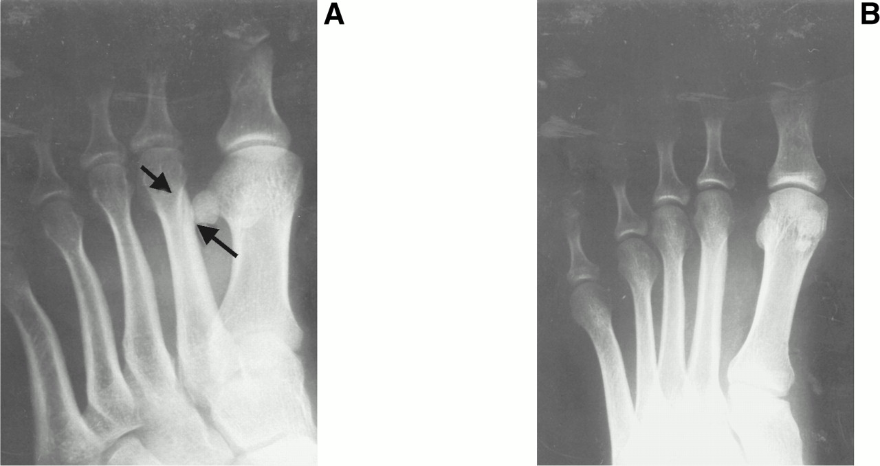
On November 11, 1996, the patient returned reporting an increase in pain in her left foot. Plain film radiographic evaluation revealed a lucent line through the distal aspect of the second left metatarsal shaft with some mild associated cortical thickening, consistent with a nondisplaced fracture (Fig. 1). The patient was placed in a below-the-knee cast and was instructed to remain nonweightbearing for 4 weeks and then to use a CAM Walker for an additional 4 weeks. The patient went on to heal uneventfully.
On October 7, 1997, the patient returned to the Tower Medical Clinic because she was concerned that she once again had a stress fracture in her left foot. The patient’s symptoms and clinical findings were consistent with a stress fracture, and she was placed in a CAM Walker.
On October 29, 1997, the patient presented with an exacerbation of her symptoms. Radiographic evaluation revealed a suspicious area of possible early periosteal reaction at the base of the second metatarsal of the left foot. F-Scan (Tekscan, Boston, Massachusetts) evaluation performed 1 week later revealed increased weight distribution under the first and second metatarsal heads of her left foot. Laboratory tests were ordered for a complete blood count with differential, rheumatoid factor profile, and chemistry panel. All laboratory values were found to be within normal limits.
On May 27, 1998, the patient presented with pain in her left foot. At this time, a diagnosis of stress fracture syndrome was made.
On December 2, 1998, the patient presented with pain in the top of her left foot that she thought might be another stress fracture. Pain was elicited with palpation of the third metatarsal of the left foot. The patient was placed in a CAM Walker and referred for bone-density measurement. The bone-density measurement revealed osteopenia or osteoporosis of trabecular bone, premature given her age. It was felt that the most likely major contributing factor to her osteopenia or osteoporosis was her caffeine intake. The patient was instructed to limit her intake of caffeine and increase her daily intake of calcium.
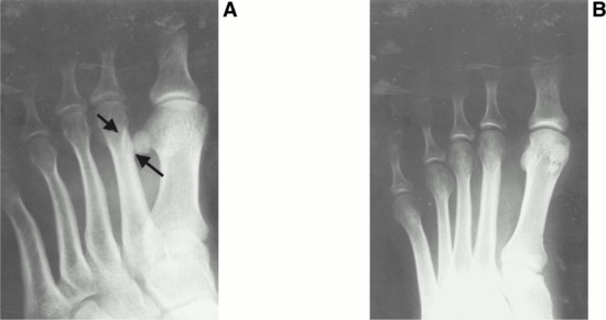
Figure 1.
A, Oblique view of the patient’s left foot showing a lucent line through the distal aspect of the second metatarsal (arrows). B, Anteroposterior view of the left foot showing mild cortical thickening of the second metatarsal.
Discussion
High caffeine consumption has been proposed as a risk factor for osteoporosis because caffeine increases urinary calcium output. However, the evidence associating high caffeine intake with low bone density is inconsistent. Some research indicates that drinking two or more cups of coffee per day is associated with reduced bone density and that drinking large quantities of coffee is associated with fracture in women. There seems to be no association between reduced bone density and intake of other caffeinated beverages, including tea and soft drinks. [3-5] Other studies showed that drinking coffee was only a minor risk factor for bone loss and fractures. [6,7]
Most of the research on the relationship between caffeine consumption and bone loss has used postmenopausal women as the subjects, and most of the research on the incidence of bone loss in premenopausal women has been done with athletes and ballet dancers. The conclusion reached by that research is that low bone density in premenopausal women is a result of amenorrhea. [8-10] The case presented here is interesting because it represents a premenopausal woman who does not have amenorrhea and has high daily caffeine consumption not from coffee, but from a soft drink. Because she did not have the classic risk factors for osteoporosis, this patient was treated for recurring stress fractures for 2 years before the diagnosis of osteoporosis as the underlying etiology was finally made.
Conclusion
A careful history considering risk factors for osteoporosis should be obtained when an insufficiency-type stress fracture is diagnosed. Bone-density measurements should be considered for patients with this type of injury. The treatment objective is two-fold: appropriate medical therapy directed at treatment of the underlying osteoporosis and management of the fracture itself. This dual approach may prevent subsequent injury.
- Tucci JR: Osteoporosis update. Med Health R I81: 169, 1998.
- Coralli CH, Raisz LG, Wood CL: Osteoporosis: significance, risk factors and treatment. Nurse Pract11: 16, 1986.
- Harris SS, Dawson-Hughes B: Caffeine and bone loss in healthy postmenopausal women. Am J Clin Nutr60: 573, 1994.
- Hernandez-Avila M, Colditz GA, Stampfer MJ, et al: Caffeine, moderate alcohol intake, and risk of fractures of the hip and forearm in middle-aged women. Am J Clin Nutr54: 157, 1991.
- Kiel DP, Felson DT, Hannan MT, et al: Caffeine and the risk of hip fracture: the Framingham Study. Am J Epidemiol132: 675, 1990.
- Cooper C, Atkinson EJ, Wahner HW, et al: Is caffeine consumption a risk factor for osteoporosis. ?J Bone Miner Res7: 465, 1992.
- Johansson C, Mellstrom D, Lerner U, et al: Coffee drinking: a minor risk factor for bone loss and fractures. Age Ageing21: 20, 1992.
- Kadel NJ, Teitz CC, Kronmal RA: Stress fractures in ballet dancers. Am J Sports Med20: 445, 1992.
- Myburgh KH, Hutchins J, Fataar AB, et al: Low bone density is an etiologic factor for stress fractures in athletes. Ann Intern Med113: 754, 1990.
- Warren MP, Brooks-Gunn J, Fox RP, et al: Lack of bone accretion and amenorrhea: evidence for a relative osteopenia in weight-bearing bones. J Clin Endocrinol Metab72: 847, 1991.
© 2001 American Podiatric Medical Association