


Abstract
The authors conducted a retrospective review of 16 patients who presented with the complaint of pain at the incision site after tarsal tunnel decompression. Specifically, the pain was located at the proximal aspect of the tarsal tunnel decompression scar. The mean duration of pain was 21 months (range, 6 to 34 months). The pain was eliminated by a block of the distal saphenous nerve, demonstrating that the pain was due to a neuroma of this nerve. The pain was treated by resection of the distal saphenous nerve in the distal leg and implantation of the proximal end of this nerve into the soleus muscle. At a mean of 18.5 months after surgery (range, 6 to 33 months), excellent relief of pain was achieved in 76% of cases and good relief of pain in 24% of cases.
Decompression of the tarsal tunnel is a relatively recent entry into the history of foot surgery, having been first described in 1962. [1,2] The most common complication of decompression of the tarsal tunnel, which is used to treat compression of the tibial nerve in the tarsal tunnel, is failure of the operation. [3-8] Incision-site pain that persists for more than 3 months following the operation and that is not successfully treated with scar massage is most likely due to a neuroma at the incision site. Pain in the distal portion of the scar that radiates to the heel following tarsal tunnel decompression is clearly related to a neuroma of one of the calcaneal branches.
When the pain is in the proximal portion of the incision, the origin of the pain is often not clear. After examining several patients with pain in this location, the authors hypothesized that the small branch of the saphenous nerve that is shown in Grant’s Atlas of Anatomy might be the source of this pain. [9] Subsequent cadaveric dissections demonstrated the cutaneous anatomy of this retromalleolar region, as well as the reliable occurrence of the posterior branch of the saphenous nerve at this location. [10]
A review of the literature yielded no previously reported case of saphenous neuroma in tarsal tunnel syndrome. [11-28] The purpose of this article is to describe a pain syndrome related to tarsal tunnel decompression that results from injury to the posterior branch of the distal saphenous nerve, and to describe an approach to its treatment.
Methods
A consecutive series of 16 patients who had undergone tarsal tunnel decompression and saphenous neuroma resection was identified by a computer search of patient records in the practice of the senior author (A.L.D.). Each of these patients complained of pain at the incision site that did not radiate into the heel or into the plantar aspect of the foot. This pain was sufficient to interfere with footwear, walking, and, in some patients, even with wearing socks.
Each patient had undergone tarsal tunnel decompression to treat tarsal tunnel syndrome. One of these patients had the complication following tarsal tunnel decompression by the senior author; the other patients were treated initially by other surgeons. During this 3-year period, approximately 100 tarsal tunnel decompressions were performed by the senior author. The 16 patients included in this study represent the total number of patients with this complication. The mean (±SD) age of the patients was 47.7 (±9.8) years. There were 3 men and 13 women. The right foot was involved in five patients, and the left foot in ten patients; one patient had this problem bilaterally. A total of 17 neuromas in 16 patients made up this series. Each patient had a minimum follow-up time of 6 months after the saphenous neuroma resection. The mean time interval between the tarsal tunnel decompression and the neuroma resection was 20.9 (±16.2) months (range, 6 to 34 months).
Three factors were used to determine that pain at the incision site was due to a neuroma of the posterior branch of the saphenous nerve. These three factors were 1) location of the painful portion of the incision site at the level of the medial malleolus or more proximally and not in the distal portion of the incision, where the pain was likely due to a branch of the calcaneal nerve; 2) identification of a distally radiating Tinel’s sign over the distal saphenous nerve at a site that was anterior to the tarsal tunnel incision; and 3) a local anesthetic block at the site of the positive Tinel’s sign that resulted in elimination of the pain in the incision site.
Treatment of the painful neuroma in all cases began with massage of a steroid-containing cream into the scar to desensitize the region. If pain persisted after 6 months of nonoperative therapy, and the patient had relief of pain following a block of the distal saphenous nerve, then surgical treatment of the painful neuroma was suggested. The most successful treatment for upper-extremity neuroma pain has proven to be resection of the neuroma and implantation of the proximal end of the nerve into an innervated muscle. [29-34] This approach has been demonstrated to be effective for painful neuromas of the plantar digital nerve [35] and of the dorsum of the foot. Specifically, for the distal saphenous nerve, the soleus muscle is the innervated muscle that is located in the immediate vicinity.
Surgical Technique
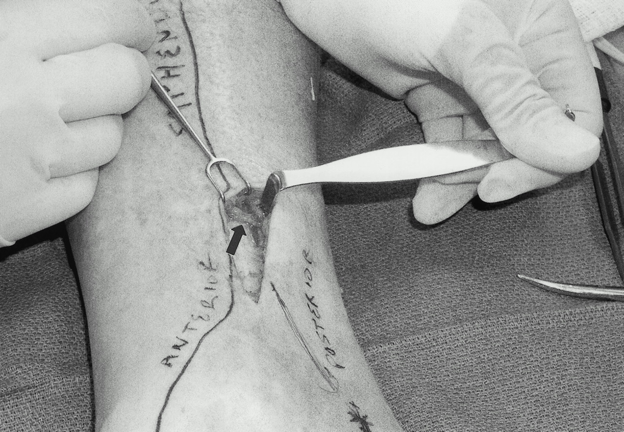
In the operating room, with a pneumatic tourniquet inflated, an incision is made over the distal saphenous nerve. The incision site is best determined by locating the Tinel’s sign and the saphenous vein (Fig. 1). An incision about 3 cm long is made longitudinally over the course of the vein. The dissection is done bluntly until the vein is encountered (Fig. 2). A bipolar coagulator and loupe magnification are used during the surgery. A search is made anterior and posterior to the vein until at least one, and usually two, small branches of the distal saphenous nerve are identified (Fig. 3). These small branches are dissected distally and divided (Fig. 4). A 5- or 6-mm segment is resected and sent for pathologic examination to document that a nerve has been resected. This piece of nerve will be a histologically normal nerve, because the neuroma is embedded in the tarsal tunnel incision site. The saphenous nerve branches are then dissected proximally for another 2 cm. A fascial opening is made over the soleus muscle just posterior to the location of the nerve. The nerve branches are turned and implanted deeply, going in a proximal direction into the muscle (Fig. 5). The curve of the nerve into this location should be done loosely. A suture should not be placed into the nerve in order to prevent pain from the suture itself causing injury to the nerve. Rather, the nerve will be held in place because the soleus muscle has little excursion and the nerve is implanted for a distance of 1 cm or more, and the curved course of the nerve into the muscle is loose and without tension. A long-acting local anesthetic, such as bupivacaine, is instilled into the wound, and the skin is closed. The patient is permitted to walk immediately. Rest for the first week is encouraged. Sutures are removed in approximately 2 weeks.
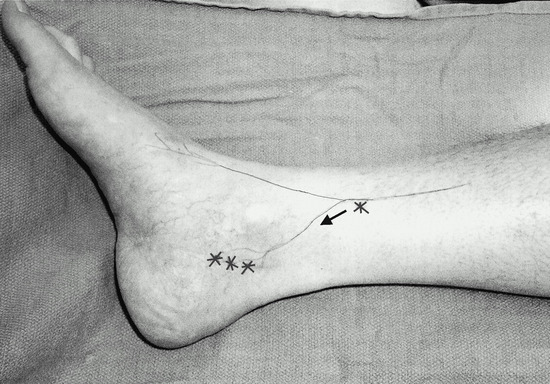
Figure 1.
Location of original tarsal tunnel incision marked with a triple asterisk at the site of the pain. Anterior and posterior branches of the distal saphenous nerve are shown with the location of the positive Tinel’s sign, indicated by a single asterisk. The arrow indicates direction of distally transmitted Tinel’s sign.
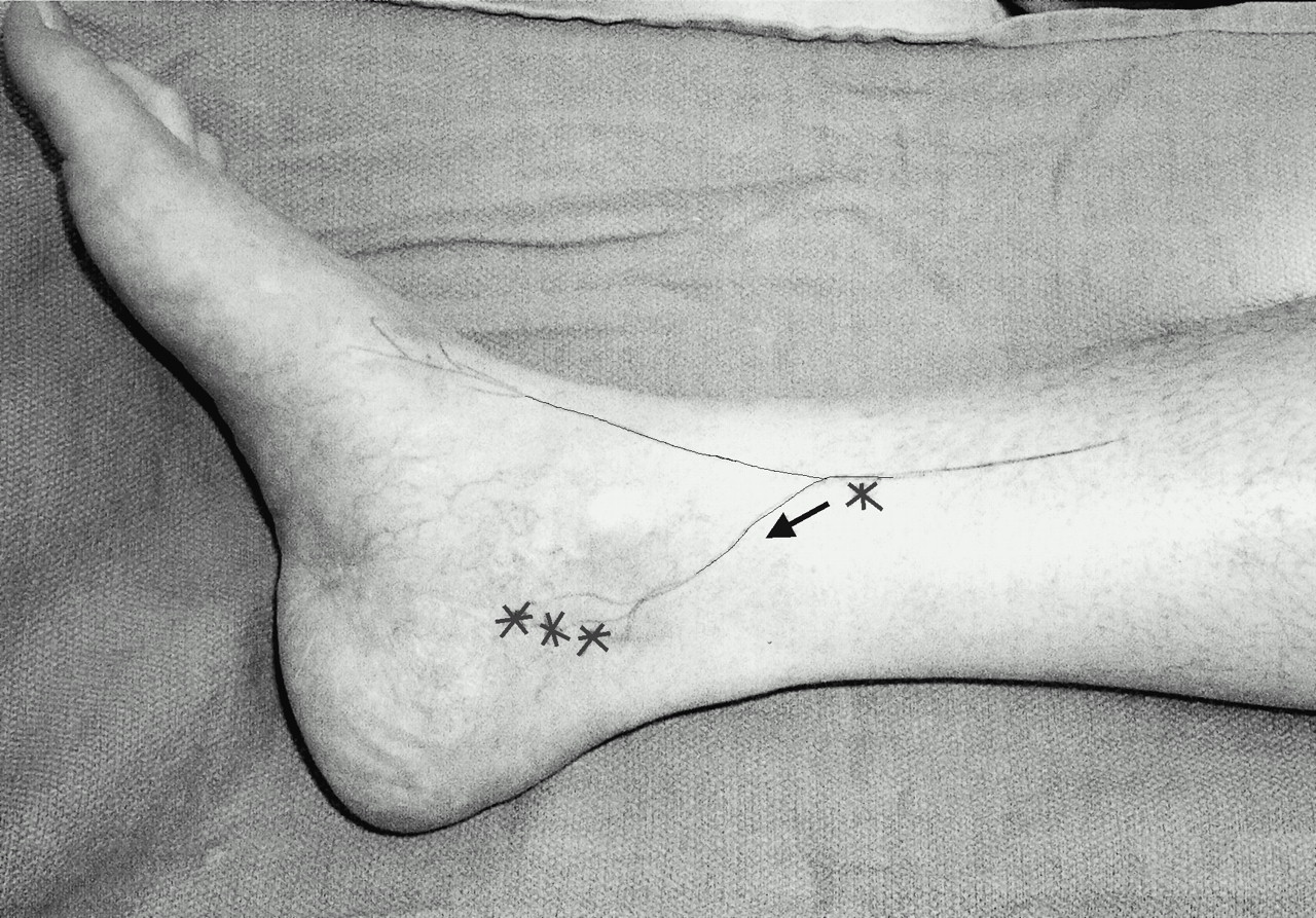
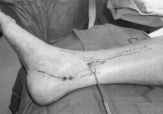
Figure 2.
Intraoperative view showing a vessel loop encircling the posterior branch of the saphenous nerve.
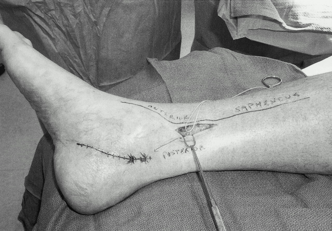
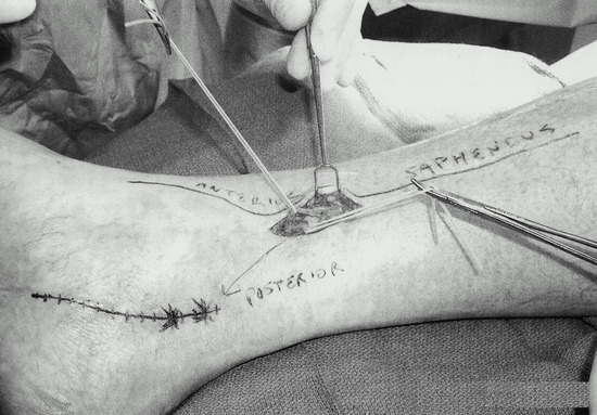
Figure 3.
The anterior and posterior branches of the saphenous nerve are both encircled with vessel loops.
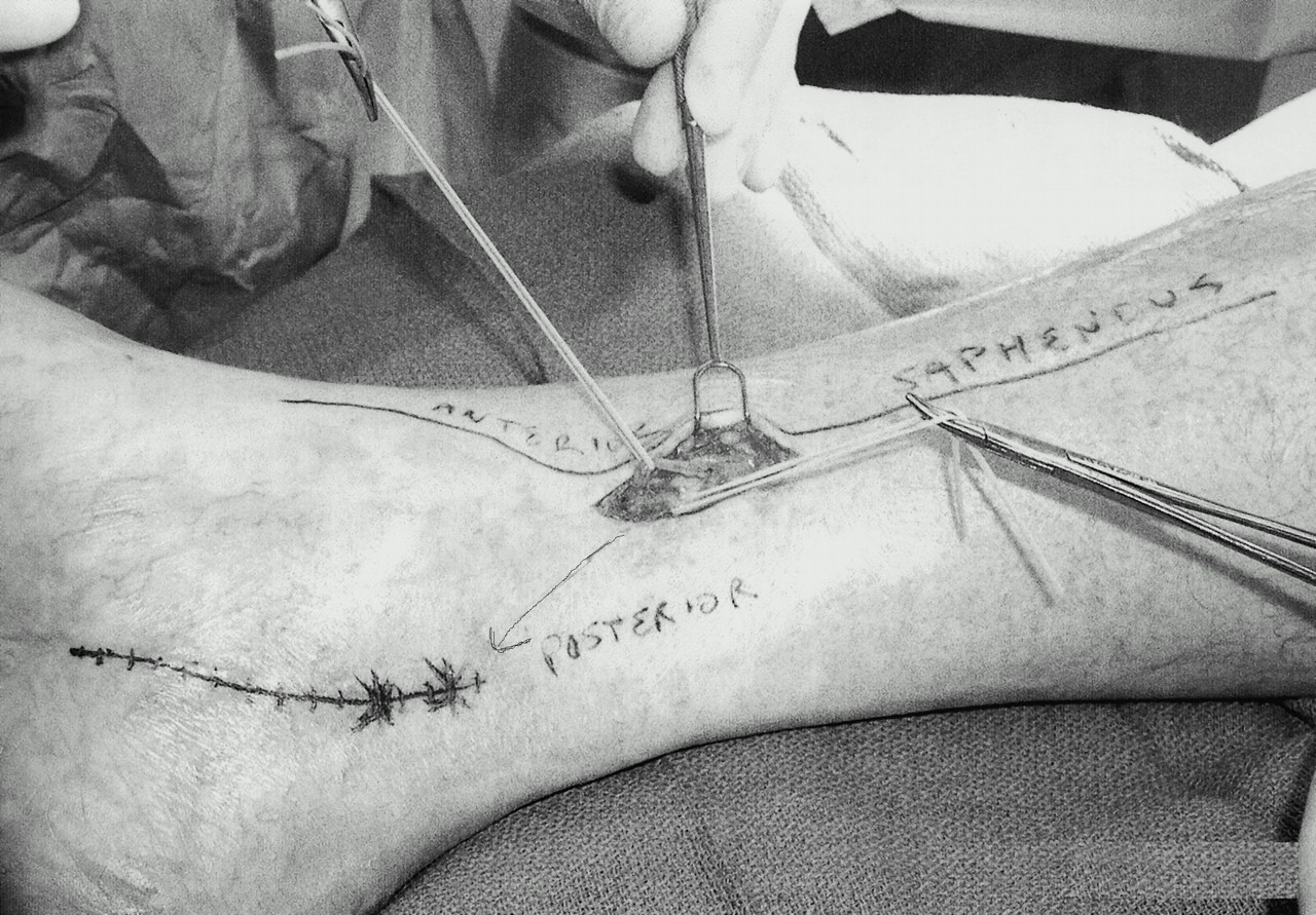
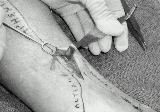
Figure 4.
Both branches of the saphenous nerve are divided. The proximal end is shown. The retractor shows the soleus muscle.
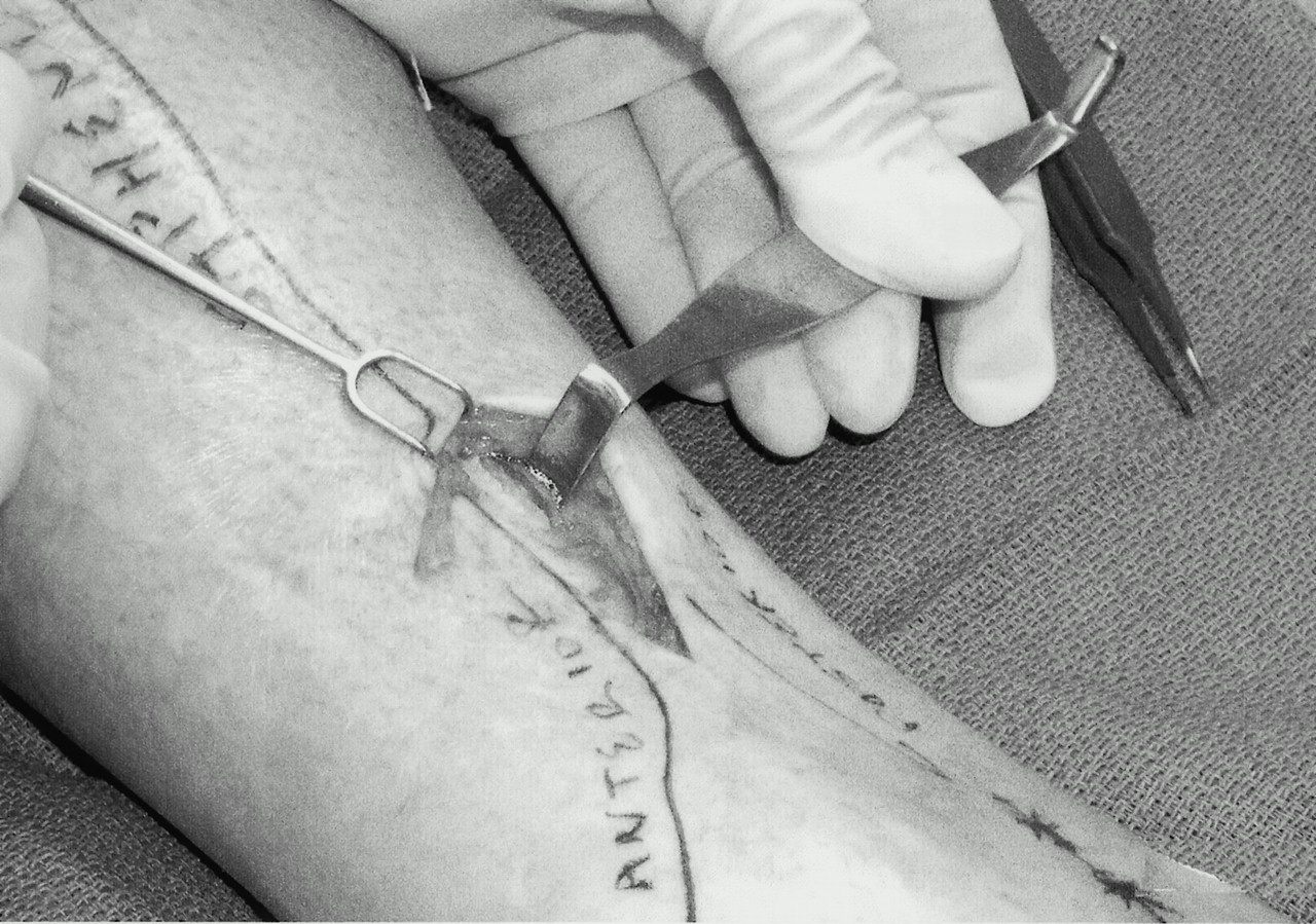
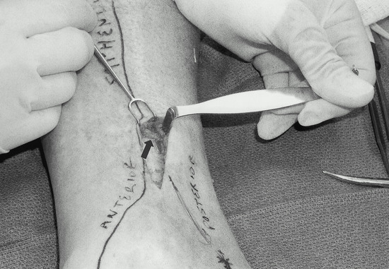
Figure 5.
The saphenous nerve is loosely implanted into muscle. The arrow indicates the saphenous nerve.
Results
Primary wound healing occurred in all patients. The new incision was not painful for any of the patients. The mean patient follow-up time after the neuroma resection and muscle implantation was 18.5 (±9.4) months (range, 6 to 33 months). In 13 cases (76%), patients had complete relief of the pain at the original tarsal tunnel incision site, which was defined as an excellent result. In the other four cases (24%), patients had partial relief of the pain, which was defined as a good result. The one patient who had bilateral neuroma resections had an excellent result on each side.
Discussion
The successful treatment of the painful neuroma depends on the correct identification of the nerve that is the source of the pain, and the correct treatment of that nerve. If there is distal pain at the tarsal tunnel incision site, and if the pain radiates toward the heel, then it is clear that a calcaneal branch is the source of the pain. This observation was made by Skalley et al [28] in their report of clinical results following revision tibial nerve release in 12 patients. If the incision is directed somewhat anteriorly at its distal end, the pain may include a distal branch of the superficial peroneal nerve. This will become clear if this portion of the incision site remains painful after the block of the saphenous nerve. Skalley et al [28] described three patients with pain at the proximal tibial incision site in their report, but they did not attribute the pain to the saphenous nerve. If a block of the distal saphenous nerve at a location proximal and anterior to the medial malleolus relieves the pain at the incision site, then the saphenous nerve is the source of the pain. If the patient is willing to trade the demonstrated area of anesthesia that results from the block for the pain relief that results from the block, then surgical treatment of the painful neuroma is indicated.
The rationale for implanting the proximal end of the nerve into muscle has been described extensively, and demonstrated for both upper- and lower-extremity nerves. [29-35] The present series of patients extends this approach to pain at the tarsal tunnel incision site where the origin of the pain is the distal saphenous nerve. As the present series of patients demonstrates, this problem may coexist with a recurrent tarsal tunnel syndrome. Treating only the recurrent tarsal tunnel syndrome symptoms by neurolysis of the tibial nerve cannot be expected to treat the painful skin incision. If palpating the painful incision site causes pain to radiate into the arch, the heel, or the big toe, then it must be assumed that the pain at the incision site is due in part to adherence of the scar to the tibial nerve itself. This problem may coexist with the neuroma of the distal saphenous nerve. Again, the nerve block is critical in distinguishing these two separate conditions. After a saphenous nerve block, the scar itself will no longer hurt, but there will still be distally radiating pain if the tibial nerve is deeply adherent to the incision.
The treatment described in this article—resection of the distal saphenous nerve—also carries the risk of creating a new site for neuroma pain. A direct injury to the implanted proximal end of the saphenous nerve in its new intramuscular location may initiate recurrent pain. To date, this has not occurred in this series of patients. There is also the risk that the patients will experience some degree of paresthesia over the distal distribution of the saphenous nerve. This is usually due to collateral sprouting from adjacent uninjured nerves. [36] This discomfort usually lasts 3 to 6 weeks, and is treated by gentle massage of the distal region or walking in a heated swimming pool, which are forms of desensitization.
Conclusion
Pain at the incision site following tarsal tunnel decompression may be symptomatic of a neuroma of the distal saphenous nerve. This nerve has a posterior branch that innervates the skin occupied by the proximal portion of the tarsal tunnel decompression incision. This complication of tarsal tunnel decompression may be avoided by making the incision sufficiently posterior to the medial malleolus. The diagnosis is confirmed by local anesthetic block of the distal saphenous nerve. This complication may be treated successfully by resecting the saphenous nerve proximal to the medial malleolus and implanting the nerve into the soleus muscle.
- Lam SJS: A tarsal-tunnel syndrome. Lancet2: 1354, 1962.
- Keck C: The tarsal-tunnel syndrome. J Bone Joint Surg Am44: 180, 1962.
- Dellon AL: Computer-assisted sensibility evaluation and surgical treatment of tarsal tunnel syndrome. Adv Podiatr2: 17, 1996.
- Chaffee H: Treatment of diabetic neuropathy by decompression of the posterior tibial nerve. Plast Reconstr Surg106: 215, 2000.
- Mackinnon SE, Dellon AL: “Tarsal Tunnel Syndrome,” in Surgery of the Peripheral Nerve, ed by SE Mackinnon, AL Dellon, p 350, Thieme, New York, 1988..
- Edwards WG, Lincoln R, Bassett FH et al: Tarsal tunnel syndrome: diagnosis and treatment. JAMA207: 716, 1969.
- Byank RP, Clarke HJ, Bleecker ML: Standardized neurometric evaluation in tarsal tunnel syndrome. Adv Orthop Surg16: 249, 1989.
- Byank RP, Curtis MJ: Diagnosing tarsal tunnel syndrome. Complications in Orthopedics10: 202, 1992.
- Grant JCB: Grant’s Atlas of Anatomy, 7th Ed, ed by JE Anderson, plates 4-7, 4-8, Williams & Wilkins, Baltimore, 1978..
- Aszmann OC, Ebmer JM, Dellon AL: The cutaneous innervation of the medial ankle: an anatomic study of the saphenous, sural and tibial nerve and their clinical significance. Foot Ankle19: 753, 1998.
- Mahan KT, Rock JJ, Hillstrom HJ: Tarsal tunnel syndrome: a retrospective study. JAPMA86: 81, 1996.
- Baba H, Wada M, Annen S, et al: The tarsal tunnel syndrome: evaluation of surgical results using multivariate analysis. Int Orthop21: 67, 1997.
- Carrel JM, Davidson DM, Goldstein KT: Observations on 200 surgical cases of tarsal tunnel syndrome. Clin Podiatr Med Surg11: 609, 1994.
- Ward PJ, Porter ML: Tarsal tunnel syndrome: a study of the clinical and neurophysiological results of decompression. J R Coll Surg Edinb43: 35, 1998.
- Oh SJ, Meyer RD: Entrapment neuropathies of the tibial (posterial tibial) nerve. Neurol Clin17: 593, 1999.
- Ferraresi S, Leidi P, Leidi M: Tarsal tunnel syndrome: report of a case and review of clinical and surgical aspects. Ital J Neurol Sci13: 47, 1992.
- Turan I, Rivero MC, Guntner P, et al: Tarsal tunnel syndrome: outcome of surgery in longstanding cases. Clin Orthop343: 151, 1997.
- Pfeiffer WH, Cracchiolo A III: Clinical results after tarsal tunnel decompression. J Bone Joint Surg Am76: 1222, 1994.
- Takakura Y, Kitada C, Sugimoto K, et al: Tarsal tunnel syndrome: causes and results of operative treatment. J Bone Joint Surg Br73: 125, 1991.
- Stern DS, Joyce MT: Tarsal tunnel syndrome: a review of 15 surgical procedures. J Foot Surg28: 290, 1989.
- Cimino WR: Tarsal tunnel syndrome: review of the literature. Foot Ankle11: 47, 1990.
- Bailie DS, Kelikian AS: Tarsal tunnel syndrome: diagnosis, surgical technique, and functional outcome. Foot Ankle Int19: 65, 1998.
- Lau JT, Daniels TR: Tarsal tunnel syndrome: a review of the literature. Foot Ankle Int20: 201, 1999.
- Novotny DA, Kay DB, Parker MG: Recurrent tarsal tunnel syndrome and the radial forearm free flap. Foot Ankle Int17: 641, 1996.
- Zahari DT, Ly P: Recurrent tarsal tunnel syndrome. J Foot Surg31: 385, 1992.
- LaBan MM, Tamler MS: Clinical results after tarsal tunnel decompression. J Bone Joint Surg Am78: 150, 1996.
- Roosth HP: Clinical results after tarsal tunnel decompression. J Bone Joint Surg Am79: 472, 1997.
- Skalley TC, Schon LC, Hinton RY, et al: Clinical results following revision tibial nerve release. Foot Ankle Int15: 360, 1994.
- Evans GR, Dellon AL: Implantation of the palmar cutaneous branch of the median nerve into the pronator quadratus for treatment of painful neuroma. J Hand Surg19: 203, 1994.
- Dellon AL, Mackinnon SE, Pestronk A: Implantation of sensory nerve into muscle: preliminary clinical and experimental observations on neuroma formation. Ann Plast Surg12: 30, 1984.
- Mackinnon SE, Dellon AL: Results of treatment of recurrent dorsal radial wrist neuromas. Ann Plast Surg19: 54, 1987.
- Dellon AL, Mackinnon SE: Treatment of the painful neuroma by neuroma resection and muscle implantation. Plast Reconstr Surg77: 427, 1986.
- Mackinnon SE, Dellon AL: Algorithm for management of the painful neuroma. Contemp Orthop13: 15, 1986.
- Dellon AL: Treatment of recurrent metatarsalgia by neuroma resection and muscle implantation: case report and algorithm for management of Morton’s “neuroma.”. Microsurgery10: 256, 1989.
- Dellon AL, Aszman OC: Treatment of superficial and deep peroneal neuromas by resection and translocation of the nerves into the anterolateral compartment. Foot Ankle Int19: 300, 1998.
- Dellon AL, Aszmann OC, Muse V: Collateral sprouting documentation using the PSSD. Ann Plast Surg37: 520, 1996.
© 2001 American Podiatric Medical Association