


To the Editor:
Hemangiomas are usually benign (rarely malignant) vascular neoplasms that are seldom found in the foot and ankle. Benign hemangiomas are the most common tumors encountered in children. They are composed of numerous newly formed blood vessels. The majority of hemangiomas are asymptomatic; however, expansive growth may impinge on the surrounding anatomical structures and cause symptoms such as pain and paresthesia.
Compared with hemangiomas of the head and neck, hemangiomas of the lower extremity are notably less common.[1] In a study published in 1995, Kransdorf[2] reported that 8% of 38,484 individuals had benign hemangiomas; 14% of these benign lesions occurred in the lower extremity. Other studies of lower-extremity hemangiomas have reported a distribution in the foot varying from 4.9% to 28.5%.[3–6] In 1970, Berlin[7] reviewed 35 cases of hemangioma of the foot and found that the majority of these cases were of the cavernous type.
There is widespread controversy in the literature as to the precise etiology of hemangiomas; however, most authors believe that hemangiomas are caused by developmental anomalies of endothelial origin that most commonly appear at birth or in early childhood.[2,8–11] Hemangiomas have also been reported to be hereditary.[10,12,13] Beers and Clark,[13] in 1942, noted a positive family history of cavernous hemangioma in a study of three generations. Cavernous hemangiomas can be either congenital (the most common type) or traumatic in origin. In the 1970 study by Berlin[7] previously mentioned, three cases of congenital and one case of traumatically induced cavernous hemangioma were presented. Other etiologies that have been reported include local irritation of newly formed vessels,[3,5,7,14] pregnancy,[8,15] and infection.[16]
Clinically, cavernous hemangiomas occur most often in white children and are two to three times more common in females than in males.[3,12,14] Red, brown, blue, or purple skin discoloration may be present when cavernous hemangiomas develop in close proximity to the overlying epidermis; this discoloration must be differentiated from that of varicose veins.[7] Carnevali[17] described cavernous hemangiomas as being “like spaghetti,” while Margileth and Museles[12] described them as being “worm-like” or like “a bag of worms.” On palpation, cavernous hemangiomas may be either fixed or movable and may be soft and spongy or indurated.[18] Pulsations have been reported and, when present, are palpable distal to the lesion.[14,18] Berlin[7] reported that cavernous hemangiomas may be painful only on movement of the surrounding musculature. Most pedal hemangiomas are also painful as a result of enlargement of the tumorous mass, which causes compression of the surrounding anatomical structures.[15]
Radiographs of benign vascular tumors yield findings that are usually nonspecific and can resemble a malignant process by virtue of their irregular, lytic, or expansive patterns.[2] Phlebolith formation may result from calcification of thrombi within the vessel wall, which is the characteristic radiographic feature of cavernous hemangiomas.[14,19,20] Currently, magnetic resonance imaging (MRI) is considered the most accurate modality for diagnosing and delineating the extent and consistency of deep soft-tissue tumors. The major advantage of MRI over angiography and computed tomography is the explicit difference in contrast between hemangiomas and the surrounding structures.[21] Hemangiomas have slightly higher intensity than skeletal muscle on T1-weighted images and have markedly high intensity on T2-weighted images, with internal septa, lobulations, and irregular borders.[22]
Case Report
A 15-year-old white male presented to the Foot and Ankle Institute at the Temple University School of Podiatric Medicine in Philadelphia with a chief complaint of a lump on the bottom of his left foot. Both he and his mother, who accompanied him, stated that the lump had existed since he was about 7 years old and had become increasingly painful over time. The mother related that it had slightly increased in size. There was no history of trauma, although the patient mentioned that he liked to roller-skate. The patient denied any professional or self-treatment.
The patient’s medical and surgical history were unremarkable. He denied taking any medications or having any food or drug allergies. The results of the vascular and neurologic examinations were within normal limits. The dermatologic examination revealed a slightly raised soft-tissue mass located in the central medial aspect of the left arch (Figs. 1 and 2). Clinically, the lesion measured approximately 5.1 × 6.4 cm. The mass was very soft, tender on palpation, and immobile. The skin overlying the mass showed no increase in temperature and no change in color, and there were no pulsations present. Pain increased with dorsiflexion of the toes; with dorsiflexion of the ankle, the mass disappeared through the plantar fascia as if it were a herniation.
Radiographs revealed an increase of soft-tissue density and volume in the central arch, corresponding clinically to the lump on the patient’s foot. An MRI study was ordered and showed a 5.5 × 3.0?× 2.3-cm mass located on the plantar medial aspect of the left foot. The fast spin-echo image of the mass showed increased signal intensity compared with the surrounding muscle (Fig. 3) and the gradient-echo image showed a bright signal with fine strands of muscle within the mass (Fig. 4). There were also several small areas of low signal intensity, consistent with calcifications within the mass. These MRI findings correlated with the clinical findings and are consistent with a hemangioma, most likely of the cavernous type, located in the left central medial arch.
Surgical excision was discussed with the patient, but he declined this treatment. In place of surgical treatment, conservative treatment consisting of accommodative insoles that off-loaded the lesion site was initiated.
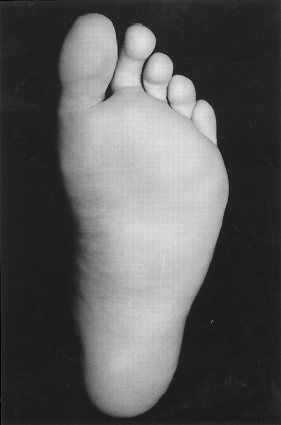
Figure 1 .
Plantar view of the foot, demonstrating the central medial soft-tissue mass.
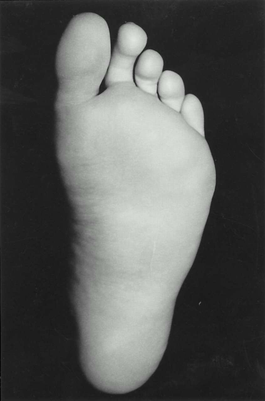
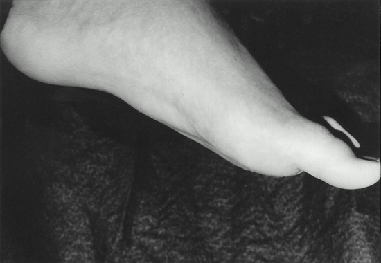
Figure 2 .
Lateral view of the same foot and mass as in Figure 1.
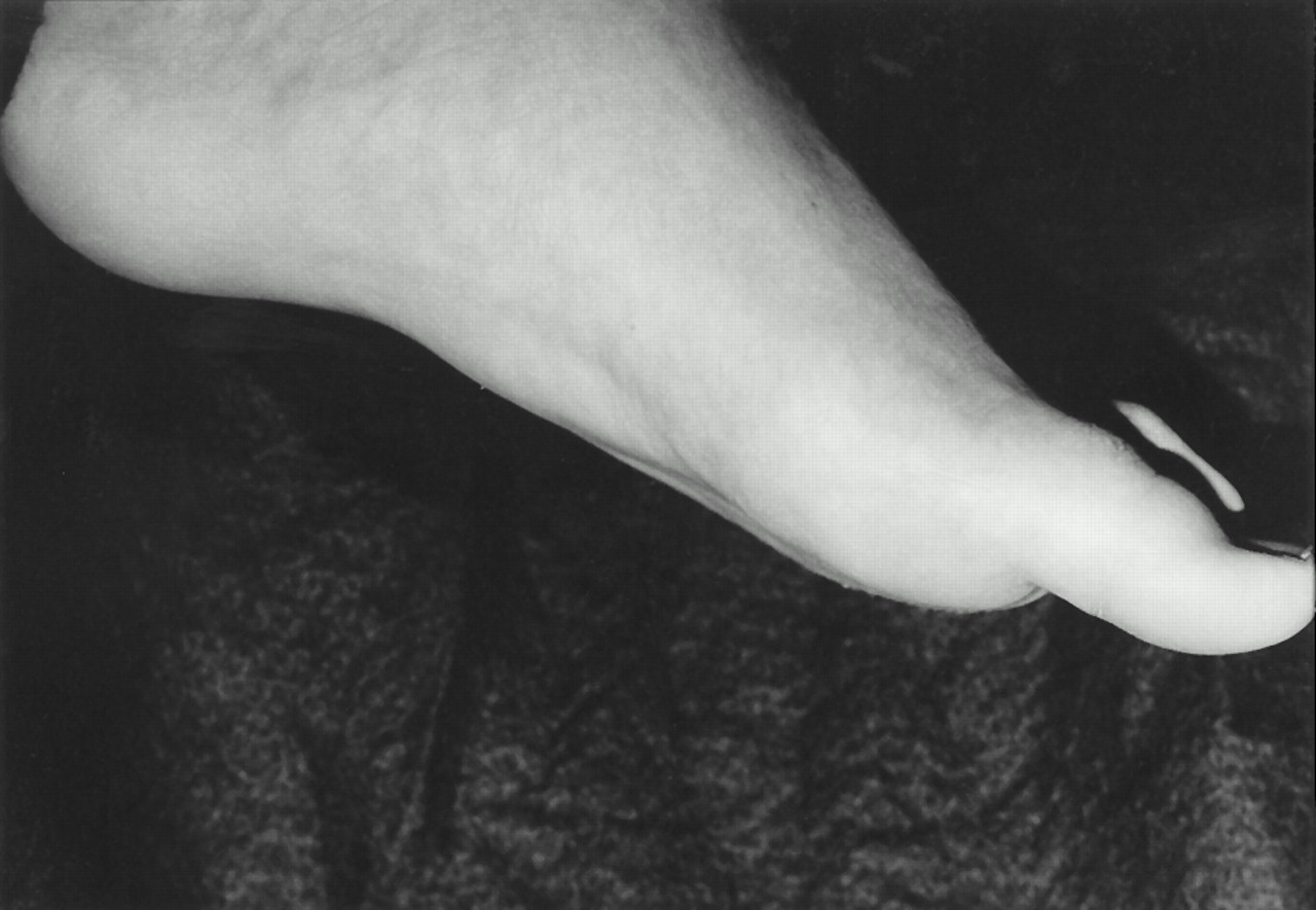
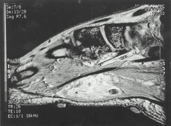
Figure 3 .
A slight increase in signal intensity of the soft-tissue mass compared with the adjacent muscle is evident on this fast spin-echo MR image.
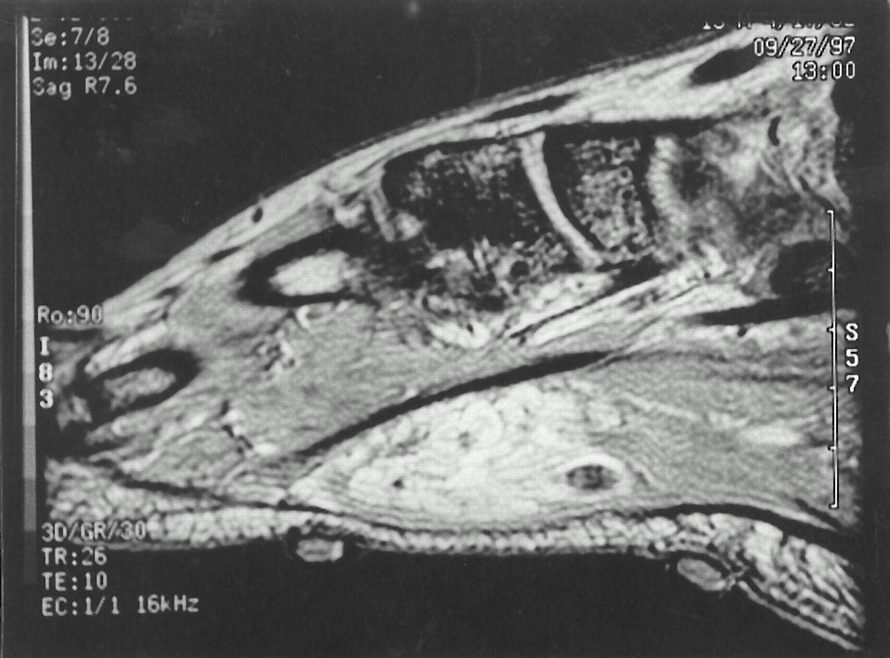
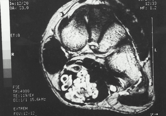
Figure 4 .
An increase in signal intensity of the soft-tissue mass with fine strands within the mass is evident on this gradient-echo MR image. The several areas of low signal intensity within the mass probably represent small calcifications.
Discussion
Soft-tissue tumors, such as hemangiomas, that are located in the foot and ankle are rare and are often difficult to evaluate and treat. The case presented here had the characteristics of a cavernous hemangioma, both clinically and radiographically. This pedal cavernous hemangioma had a cutaneous appearance similar to that of the surrounding skin, and pain was noted on contraction of the surrounding musculature. However, unlike most cavernous hemangiomas, this one occurred in a young white male.[3,12,14]
No soft-tissue calcifications were observed on the radiographs; such calcification is the characteristic radiographic feature of cavernous hemangiomas.[14,19,20] However, the MRI findings were consistent with a cavernous hemangioma, with high intensity on the T2-weighted images.[22]
Many conservative treatment modalities for hemangiomas have been explored, but none have proved very successful. Among these modalities are sclerosing agents, which occlude the vascular supply to the tumor, and radiation therapy, which is often associated with toxic sequelae.[7,18]
Cox,[23] in 1976, concluded that pain, deformity, interference with function, and malignant changes are indications for surgical excision of cavernous hemangiomas. Because cavernous hemangiomas are larger, more diffuse, and located deeper than other types of hemangiomas, surgical intervention can be complex.[3,12,14] Cavernous hemangiomas are difficult to remove when they are located subcutaneously and have poorly defined borders. However, the tumor in the case presented here was located subdermally and had a well-defined border, characteristics that provide for easier surgical excision.[14,24] The tumor may have undergone surgical excision successfully, but the patient preferred conservative treatment. Surgical complications and failures are most often due to inadequate excision of the tumor and consequent excessive bleeding.[7] Therefore, as with most benign tumors, when a hemangioma is incompletely removed, it usually recurs and becomes even more extensive than it was originally.[23]
Conclusion
A case of a benign plantar hemangioma of the foot has been presented. Even though conservative treatment was chosen by the patient in this case, such treatment has shown limited success and, when indicated, surgery is the treatment of choice. However, to achieve optimal results, surgical intervention should not be performed without careful evaluation and adequate diagnostic testing of the tumor.
References
- Giannestras N: Foot Disorders: Medical and Surgical Management, Lea & Febiger, Philadelphia, 1973
- Kransdorf MJ: Benign soft-tissue tumors in a large referral population: distribution of specific diagnoses by age, sex, and location Am J Radiol 164: 395, 1995.
- Johnson EW Jr, Ghormley RK, Dockerty MB: Hemangiomas of the extremities Surg Gynecol Obstet 102: 531, 1956.
- LaSorte AF: Cavernous hemangiomas of striated muscle Am J Surg 100: 593, 1960.
- Weaver JB: Hemangioma of the lower extremities J Bone Joint Surg 20: 731, 1938.
- McNeill TW, Ray RD: Hemangiomas of the extremities: review of 35 cases Clin Orthop 101: 154, 1974.
- Berlin SJ: Hemangioma of the foot JAPA 60: 63, 1970.
- Pack TT, Miller TR: Hemangiomas: classification, diagnosis, and treatment Angiology 1: 405, 1950.
- Waisman M: Common hemangioma: to treat or not to treat Postgrad Med 43: 183, 1968.
- Lampe L, Latourette HB: The management of cavernous hemangiomas in infants Pediatr Clin North Am 6: 511, 1959.
- Cortese CJ: Cavernous hemangioma of the foot J Foot Surg 15: 72, 1976.
- Margileth AM, Museles M: Current concepts in diagnosis and management of congenital hemangiomas Pediatrics 36: 410, 1965.
- Beers CV, Clark LA: Tumors and short toe, a dihybrid pedigree: a family history showing the inheritance of hemangioma and metatarsus atavicus J Hered 33: 366, 1942.
- Midenberg M, Kirschenbaum SE: Benign cavernous hemangioma of the ankle J Foot Surg 22: 294, 1983.
- Castillenti TA: Cavernous hemangioma of the foot: case report and literature review JAPMA 79: 406, 1989.
- Greschickter CF, Keasbey LE: Tumors of blood vessels Am J Cancer 23: 568, 1935.
- Carnevali SL: Sugli angioma dei muscoli striati Arch Orthop 54: 476, 1938.
- Borden JI, Shea TP: Cavernous hemangioma of the foot: a case report and review JAPA 66: 484, 1976.
- Miller SJ, Patton GW, Xenos D, et al: Multiple capillary hemangiomas of the foot with associated phleboliths: a case report JAPA 70: 364, 1980.
- Jones RW, Roberts RE: Calcification and ossification Br J Surg 21: 461, 1933-34.
- Sartoris DJ, Resnick D: Magnetic resonance imaging of pediatric foot and ankle disorders J Foot Surg 29: 489, 1990.
- Wetzel LH, Levine E: Soft-tissue tumors of the foot: value of MR imaging for specific diagnosis Am J Radiol 155: 1025, 1990.
- Cox K: Subcutaneous hemangioma of the foot JAPA 66: 519, 1976.
- Berlin SJ, Members of the Maryland Podiatric Residency Research Committee: “Vascular Tumors of the Foot,” in Soft Somatic Tumors of the Foot: Diagnosis and Surgical Management, Futura Publishing, Mount Kisco, NY, 1976
© 2001 American Podiatric Medical Association