


Abstract
A retrospective study was conducted on the use of the instep plantar fasciotomy for the treatment of recalcitrant plantar fasciitis. A total of 83 patients (94 feet) were analyzed. The average postoperative followup time was 20.9 months. Surgery was deemed successful 93.6% of the time, and in 95.7% of cases, the patient would recommend the procedure to someone with the same condition. The main complications were scarring (9.6%), medial arch or heel pain (7.5%), cramping in the arch (6.4%), lateral column pain (5.3%), aching or pain across the dorsal midfoot (5.3%), and burning or tingling of the ball of the foot (5.3%).
Plantar heel pain is commonly encountered in podiatric medical practice. There is no question that heel pain accounts for a great deal of morbidity. In most instances, plantar heel pain can be attributed to plantar fasciitis or heel spur syndrome. Plantar fasciitis is characterized by pain and inflammation secondary to strain on the intrinsic musculature and plantar fascia at their origin from the medial calcaneal tubercle. The classic physical examination findings are point tenderness at the anterior edge of the fascial attachment to the medial calcaneal tubercle and pain produced by stretching the inflamed plantar fascia with passive hyperextension of the metatarsophalangeal joints. Pain is usually greatest with the first step upon arising in the morning and after periods of rest (poststatic dyskinesia).
The majority of patients respond to conservative treatment. The mainstays of conservative treatment are biomechanical support of the arch, anti-inflammatory medications, physical therapy modalities including stretching, and, in some instances, immobilization.
Surgery is indicated for recalcitrant heel pain. When heel pain is not eradicated by nonsurgical modalities, it is necessary to rule out less common causes of heel pain, including calcaneal stress fracture, tumor, infection, systemic arthritides, and nerve entrapment of the first branch of the lateral plantar nerve. [1]
There have been numerous reports of various approaches to alleviate plantar heel pain caused by plantar fasciitis or heel spur syndrome. [2] It is no surprise that each approach has its own significant complications; as a result, a search is currently under way for a technique that features cost-effectiveness, ease, limited complications, reproducibility, patient acceptance, and an acceptable postoperative outcome.
It is not the intent of this article to review all of the procedures that have historically been advocated to treat heel spur syndrome. The need to resect the heel spur is still controversial. There are significant data to support the efficacy of sectioning the plantar fascia without resection of the osseous spur. A case in point is the popularity, acceptance, and success of the endoscopic plantar fasciotomy. In addition, a recent retrospective analysis of various procedures to treat heel spur syndrome failed to prove the need for spur removal. Postoperative outcomes were similar for all patients, whether or not they had spur resection. [3]
A plantar approach has several advantages. Minimal dissection is required to identify the plantar fascia. There are no significant nerves or blood vessels in the operative site. The procedure can be performed in an expedient manner, and there is no need to use expensive equipment (an endoscope or a monitor). As with the endoscopic technique, most patients return to ambulation rapidly.
Various plantar incisional approaches have been described. Ward and Clippinger [4] advocated a curvilinear incision over the posteromedial longitudinal arch. Boike et al [5] described a transverse plantar incision directly at the site of the heel spur. Self et al [6] and Perelman et al [7] recommended a transverse linear or transverse L incision at various distances from the attachment of the plantar fascia.
The present study was undertaken to determine the efficacy of the instep plantar fasciotomy, to analyze the associated complications, and, finally, to make technical recommendations for future use of this procedure.
Materials and Methods
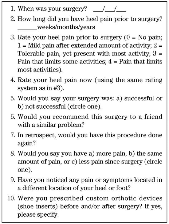
The instep fasciotomy was performed on 83 patients (94 feet) with recalcitrant plantar fasciitis. All of the patients had received extensive conservative treatment; anyone with prior heel surgery was excluded from the study. The surgeries included in the study were performed over a 3-year period. Patients from five surgeons were included in the study. The principal author (W.D.F.) interviewed all patients using a standard questionnaire (Fig. 1). Chart review was undertaken for patients with postoperative complications.
Figure 1.
Patient questionnaire used in the current study.
Surgical Technique
The patient is placed on the operating table in a supine position and prepared for surgery in the standard aseptic fashion. Local anesthetic is administered, consisting of 1% lidocaine with epinephrine and 0.5% bupivacaine hydrochloride without epinephrine. This yields an epinephrine solution of 1:200,000, adequate for hemostasis. A transverse incision is made in the plantar medial arch approximately 2.0 cm in length. The incision placement varied among the five surgeons; the most common placement was approximately 1.5 cm anterior to the fat pad of the heel. The authors believe it is ideal to select a skin crease for placement of the incision, which significantly reduces skin tension during the postoperative healing stages. After the skin incision is made, a hemostat is used to bluntly dissect through the fatty subcutaneous tissue. There are no significant neurovascular structures at this location; there will, however, be some small traversing veins that can be cauterized.
After successful separation of the subcutaneous layer from the deep fascia, the plantar fascia is seen. Palpation reveals the tightest portion of the medial border of the fascia. An incision of the plantar fascia is then made, sectioning the medial one-third of the fascia. Underlying muscle is revealed by dorsiflexing the hallux. Palpation should reveal successful release of the medialmost fibers. Copious lavage is performed, and the subcutaneous tissue and skin are closed as a single layer. Two or three nonabsorbable horizontal sutures are usually adequate.
Postoperatively, patients are placed in a surgical shoe or removable walking cast and are allowed partial weightbearing using crutches for support. Sutures are removed after 2 weeks, and progressive increases in weightbearing and activity are allowed, as tolerated by the patient.
Results
The average duration of heel pain prior to surgery was 14 months, with a range of 2 to 60 months. The average postoperative follow-up time was 20.9 months, with a range of 12 to 43 months. On the pain scale of 1 to 4, the average preoperative heel pain rating was 3.50, and the average postoperative pain rating was 0.59. A paired t-test performed on the pain-level data revealed a statistically significant difference (P < .001).
In six instances (6.4%), surgery was deemed unsuccessful by the patient. However, in only four of these instances (4.3%) did the patient have at least as much pain postoperatively as preoperatively: in two cases (2.1%) the patient had the same amount of pain postoperatively, and in two cases (2.1%) the patient had more pain postoperatively. In four instances (4.3%), the patient would not recommend the instep fasciotomy to someone with a similar problem, and in 90 instances (95.7%), the patient would have the surgery performed again if needed.
Postoperative complications included scarring at the incision site (9.6%), a dull ache or pain in the medial arch or heel (7.5%), cramping in the arch (6.4%), lateral column pain (5.3%), aching or pain across the dorsum of the midfoot (5.3%), burning or numbness under the metatarsal heads (5.3%), pruritus (1.1%), numbness of the hallux (1.1%), and superficial infection (1.1%).
Discussion
These data strongly support the efficacy of the instep plantar fasciotomy. Although this surgical technique has its share of complications, the authors wish to comment on certain technical factors and make recommendations based on these factors. One of the problems inherent in this study is that patients from five different surgeons, with slightly different techniques and postoperative protocols, were analyzed. However, valuable information was gleaned from this. For example, one of the most common postoperative complaints was incisional irritation or scarring. It is theorized that incision placement is the culprit. To illustrate, some surgeons prefer a more distal incision, which makes the dissection easier because there is less fat and the plantar fascia is palpable. After analysis of the data, it was noted that a disproportionately high number of patients with problematic scarring had a more distal fasciotomy. Sixty-seven percent of the scarring resulted from a more distal fasciotomy (six out of nine cases). Very little subcutaneous tissue overlies the fascia at the medial arch region, a fact that would seem to contribute to the incidence of scarring and adhesions.
An explanation for the vague dorsal pain experienced by some patients postoperatively may come from research that supports the existence of greater strain on the medial arch of the foot after the plantar fascia is sectioned. [7,8,9] This increased strain along the medial column has been shown to cause abnormal joint rotation and arch displacement after plantar fasciotomy, especially in the pes planus foot type. However, in the nonpronated foot type, there is significantly less joint displacement or rotation after fasciotomy. [8] Therefore, it would seem logical that in the patient population of the study, those who developed these vague arch fatigue symptoms may have had a more significant flatfoot deformity. Correlating these results with the literature leads one to the conclusion that one should be cautious in using this procedure in patients with severe pes planus. In addition, one should be aware that this patient population may not receive total relief of pain. Moreover, patients with midfoot symptomatology may benefit from stabilizing the hypermobile midfoot with an orthotic device postoperatively.
This theory can be extrapolated to include patients who develop pain after a more aggressive fasciotomy (total plantar fasciotomy, rather than partial fasciotomy). Thordarson et al [9] showed a progressive loss of the arch-supporting function of the plantar fascia as the fascia was sequentially sectioned from medial to lateral. The most significant loss of the windlass mechanism was noted after complete plantar fascial release. [8] It is already widely accepted in the literature that after aggressive fasciotomy, lateral column pain is a common complication secondary to abnormal joint function and subsequent jamming. [10]
Cramping, generalized achiness in the arch, and metatarsalgia-type symptoms are most likely due to stress on the intrinsic musculature. After the plantar fascia is released, the stretch is then transferred to the underlying muscles. This may be more evident after fasciotomy in the hypermobile foot where the intrinsic musculature attempts to stabilize the arch and midfoot.
Most of the previously mentioned postoperative complications, with the exception of scarring, are seen in any type of heel surgery for plantar fasciitis. The loss of support of the plantar fascia results in greater weightbearing forces in the midfoot. Postoperative pain or discomfort is common; however, there is significant improvement. Generalized cramping, achiness, and dorsal midfoot pain tend to resolve in 3 to 6 months. Some patients, however, may need a year or more to resolve these transient symptoms.
Conclusion
The authors believe that instep plantar fasciotomy is an effective surgical treatment for chronic plantar fasciitis. This procedure is well tolerated by patients and is technically nondemanding. More than half of the patients in this study with problematic scarring had distal fasciotomies; to avoid scarring, it is recommended that the incision be placed close to the fat pad of the heel. As with any surgical technique for plantar fasciitis, the clinician will at times need to treat postoperative aches and pain across the dorsum of the foot, the plantar metatarsal region, and the calcaneocuboid joint region. These pains are transient and usually resolve within 6 months.
References
- GROVES, MJ. “Atypical Heel Pain,”. In Reconstructive Surgery of the Foot and Leg: Update ’97; Vickers, NS, Miller, SJ, Mahan, KT, et al., Eds.; The Podiatry Institute: Tucker, GA, 1997; p. 220. [Google Scholar]
- JACOBY, RP; WOLFE, LC. Surgical management of recalcitrant heel pain. Clin Podiatr Med Surg 1991, 8, 187. [Google Scholar] [CrossRef] [Scilit] [PubMed]
- BREKKE, MK; GREEN, DR. A retrospective analysis of minimal-incision, endoscopic, and open procedures for heel spur syndrome. JAPMA 1998, 88, 64. [Google Scholar] [CrossRef] [Scilit]
- WARD, WG; CLIPPINGER, FW. Proximal medial longitudinal arch incision for plantar fascia release. Foot Ankle 1987, 8, 152. [Google Scholar] [CrossRef] [Scilit] [PubMed]
- BOIKE, AM; SNYDER, AJ; ROBERTO, PD; et al. Heel spur surgery: a transverse plantar approach. JAPMA 1993, 83, 39. [Google Scholar] [CrossRef] [Scilit] [PubMed]
- SELF, TC; KUNZ, RE; YOUNG, G. Transverse plantar incision for heel spur surgery: four-year follow-up survey of 35 patients. JAPMA 1993, 83, 259. [Google Scholar] [CrossRef] [Scilit] [PubMed]
- PERELMAN, GK; FIGURA, MA; SANDBERG, NS. The medial instep plantar fasciotomy. J Foot Ankle Surg 1995, 34, 447. [Google Scholar] [CrossRef] [Scilit] [PubMed]
- KITAOKA, HB; LUO, ZP; AN, KN. Mechanical behavior of the foot and ankle after plantar fascia release in the unstable foot. Foot Ankle Int 1997, 18, 8. [Google Scholar] [CrossRef] [Scilit] [PubMed]
- THORDARSON, DB; KUMAR, PJ; HEDMAN, TP; et al. Effect of partial versus complete plantar fasciotomy on the windlass mechanism. Foot Ankle Int 1997, 18, 16. [Google Scholar] [CrossRef] [Scilit] [PubMed]
- BARRETT, SL; DAY, SV. Endoscopic plantar fasciotomy: two portal endoscopic surgical techniques—clinical results of 65 procedures. J Foot Ankle Surg 1993, 32, 248. [Google Scholar] [PubMed]
© 2000 American Podiatric Medical Association