


Abstract
This article discusses the advantages and disadvantages of primary wound healing as compared with primary amputation in individuals with chronic diabetic foot wounds. The authors review the potential benefits of vascular surgical procedures and advanced dressings, including two of the most promising modalities in modern wound care: growth factors and bioengineered skin. In this era of cost-conscious health-care administration, it is incumbent on the practitioner to consider not only the basic science of wound care, but also the economic aspect of treatment rendered. These various interventions, dressings, growth factor delivery systems, and new modalities could significantly reduce healing time, thereby reducing the risk of infection, hospitalization, and amputation while improving quality of life. If so, they may be truly cost-effective.
Foot lesions are a common complication of diabetes mellitus. Occurring in approximately 15% of patients with the disease, [1] they are the primary precipitant of perhaps the most feared complication associated with diabetes: lower-extremity amputation. The impact of major amputation on quality of life is profound. The economic consequences of major amputation are also staggering. The commonly experienced needs for repeat hospitalizations, rehabilitation, social services, and home care add to the cost of major amputation. [2,3]
While there is general consensus among clinical researchers in the United States and Europe regarding the need to reduce the number of lower-extremity amputations, [4,5] there is still great concern among many health-care providers that extensive treatment in high-risk patients may not be cost-effective. However, the data seem to indicate that limb salvage is more cost-effective than amputation in both the intermediate and the long term. [6] Short-term evaluation is often deceptive, as most costs are associated not with the surgical procedure, but with perioperative and immediate postoperative care in nonsurgical units in hospitals and nursing homes.
The total costs of healing of a foot ulcer are closely related to the severity of the lesion. [6,7,8] Complex deep-pedal ulcerations may involve a large area on the foot. Many foot ulcers are further complicated by significant underlying bony architectural abnormalities, peripheral vascular disease, or wound infection. Osseous destruction seen in Charcot’s arthropathy or prior amputation, with its alteration in gait biomechanics, affects wound outcome profoundly. Complex wounds are more economically demanding than most chronic wounds, requiring long-term follow-up care. However, even for the most high-risk patients with profound limb-threatening ischemia, the direct short-term costs of arterial reconstruction are nearly identical to the costs of primary amputation. [9,10] Perler [11] suggests that the rate of revision in revascularization (8%) is significantly lower than the general rate of revision for primary high-level amputation (23%). Singh et al [12] stress the need for appropriate patient selection, as the charges associated with revision of revascularization procedures may increase the overall cost of limb salvage dramatically.
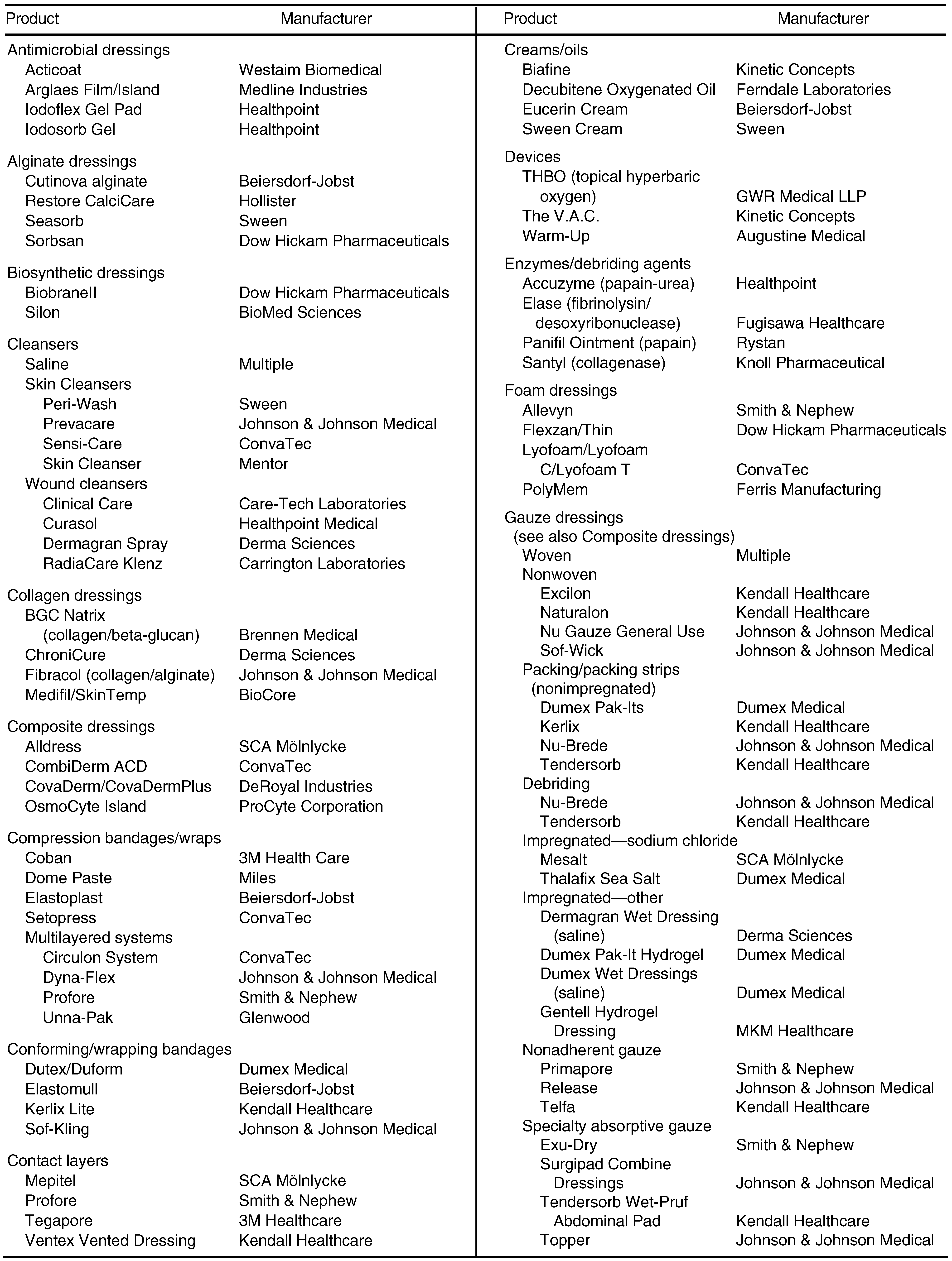
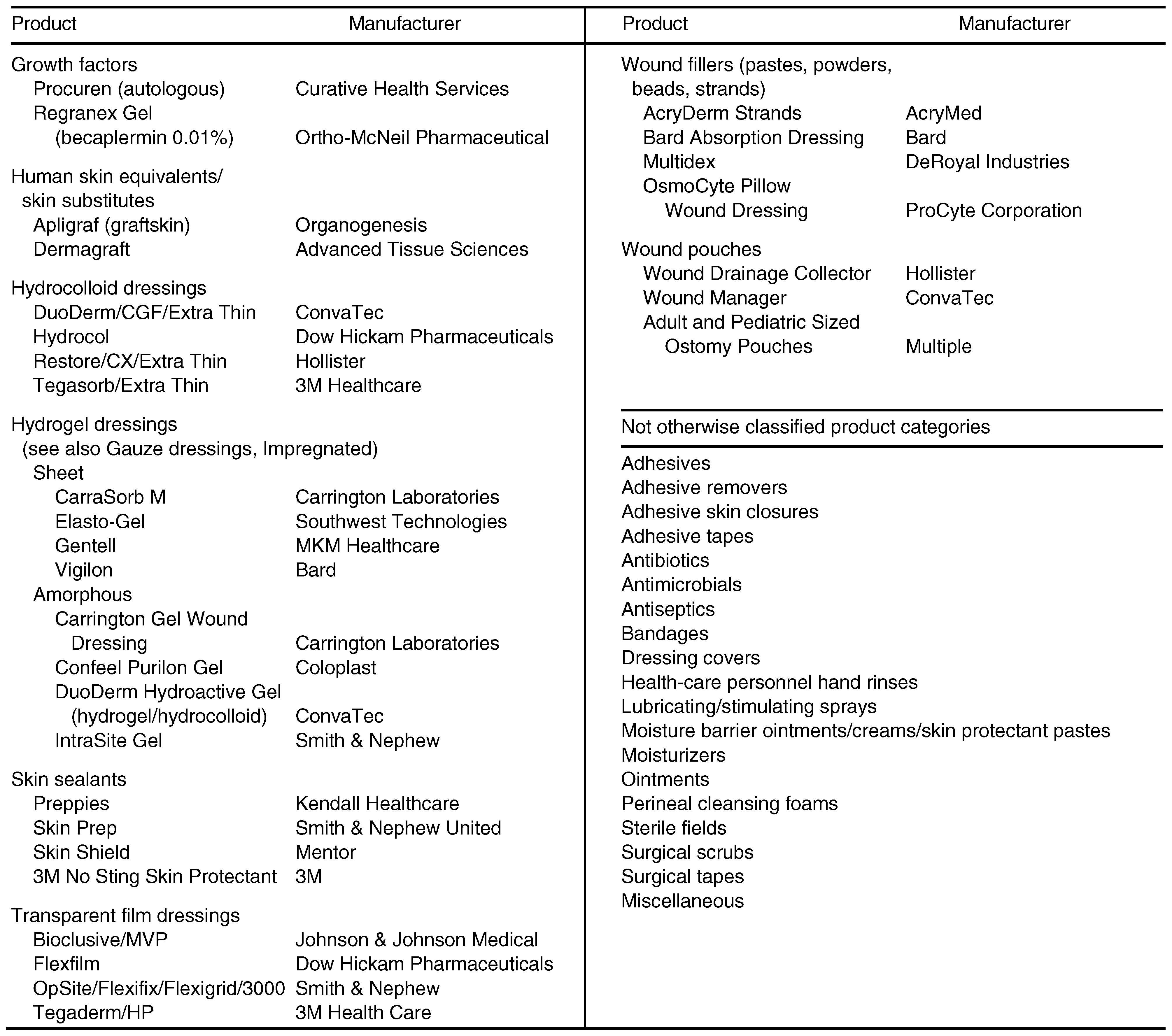
Another seemingly logical argument that has recently been propagated is that antibiotic therapy and the use of advanced dressings may drive up the cost of care, thus obliterating the economic advantages of limb salvage. However, Apelqvist and coworkers6 have argued that even for the healing of complicated wounds, the overall cost of antimicrobial therapy was generally less than 2% of the total cost of hospitalization. Other work from these researchers [13] suggested that advanced dressings, in reducing the frequency of dressing changes, lowered the overall cost of care, mainly because of less need for staff intervention, lower transportation costs, and a smaller quantity of dressing supplies. It should also be noted that this study did not take into account the potential for more rapid healing or a higher proportion of wounds healed in the advanced-dressing group. A categorized list of advanced dressings and woundhealing modalities appears in Table 1. This article will detail the characteristics of some of the more advanced wound-healing modalities: growth factor technologies and bioengineered skin.
Table 1.
Categories of Dressings and Advanced Wound-Healing Modalities
New Modalities
During the past 10 years, significant advances in biomedical technology have made it possible for practitioners to manipulate the wound microenvironment. Molecular techniques have fostered new hope for therapies directed toward treating acute and difficult-to-heal chronic wounds. Investigations into the interaction between the complex molecular processes within the wound space extracellular matrix and its associated microcirculation are increasing in popularity. [14,15,16,17,18,19,20] One significant force driving this research is the growing recognition that care of chronic wounds is time-consuming and extremely expensive and can significantly affect quality of life.
Many of the soluble mediators or growth factors have been isolated from the wound space. Plateletderived growth factor, epidermal growth factor, angiogenesis factor, transforming growth factor, betafibroblastic growth factor, tumor necrosis factor, and interleukin-1 are a few of the better-characterized cytokines. [21] The use of growth factors as possible therapeutic agents for chronic wounds in general, and as adjunctive treatment of diabetic foot wounds in particular, has met with a great deal of enthusiasm among wound-care providers. The traditional passive, watch-and-wait approach to wound care is being rapidly eclipsed by an aggressive, interactive approach favoring active wound therapies.
Growth Factors
The acute wound space contains a variety of growth factors. These local soluble mediators are the primary agents responsible for stimulating cell movement, cell division, and cell–cell interaction. Growth factors are also linked to increases in matrix synthesis and cell mass. Thus these agents are largely responsible for wound maturation and closure. [22,23] Characterization of these factors and their effects, along with the ability to prepare them in large quantities by means of biotechnology, has led to numerous clinical trials of various preparations.
Successful laboratory demonstrations of significant effects in tissue culture preparations have not necessarily led to demonstrable clinical effects. Some of the disappointing clinical outcomes occur because the environment within the chronic wound space differs significantly from that found in the acute wound. This frontier within the chronic wound space environment is largely uncharacterized. However, the importance of its characterization is readily apparent. Clinical studies have demonstrated that exudate extracted from a chronic wound space inhibits the activities of individual cell lines in tissue culture. When this extract was placed in acute wounds, significant inhibition of healing was also demonstrated. [21] Extrinsic factors may have reduced the healing rates reported in some studies. In one large multicenter trial of growth factor technology, [24] pressure off-loading was not standardized from center to center. The results reported in that study might have been even more impressive if appropriate attempts had been made to standardize the method of pressure reduction.
Clinical trials involving growth factors have included platelet-derived wound-healing formula, an autologous preparation consisting of the secretory granules of a patient’s own plasmaphoresed and lysed platelets (also known as platelet releasate). Other cytokines, epidermal growth factor, arginine–glycine– aspartic acid peptide matrix (Argidine Gel, Telios Pharmaceuticals, Inc, San Diego, California), topical beta-fibroblastic growth factor, and recombinant human platelet-derived growth factor (becaplermin) have also been studied. To date, these trials have included only patients with relatively good blood flow who were taking no medications known to inhibit wound healing and had no known comorbid medical conditions (other than diabetes). These studies were also selective with regard to the wound studied and included patients with noninfected full-thickness wounds not extending to bone (below grade 3A on the University of Texas scale). [25]
Knighton and coworkers [26] reported results from the first clinical trial with platelet-derived wound-healing formula. The study was uncontrolled and involved a small study population. This research group [27] subsequently reported on a randomized, single-blind, crossover trial of platelet-derived wound-healing formula for chronic (more than 8 weeks’ duration) cutaneous ulcers of variable etiology and location. The overall results showed a positive response in time to healing in the group treated with platelet-derived wound-healing formula. However, the study did not enroll enough patients to stratify the results according to diagnostic groups. Steed and colleagues [28] reported the results of a randomized, prospectively controlled, double-blind trial of 13 patients (6 controls versus 7 in the treatment group) with neuropathic diabetic foot ulcers. A transcutaneous oximetry level of more than 30 mm Hg was required for entry into the trial. The ulcers were debrided and converted to the acute state prior to randomization. Five of seven ulcers in the treatment group were healed by week 15, as compared with only one of six by week 20 in the control group. The control group was not crossed over in this study. Wound care and patient compliance with the treatment regimen were similar in the two groups.
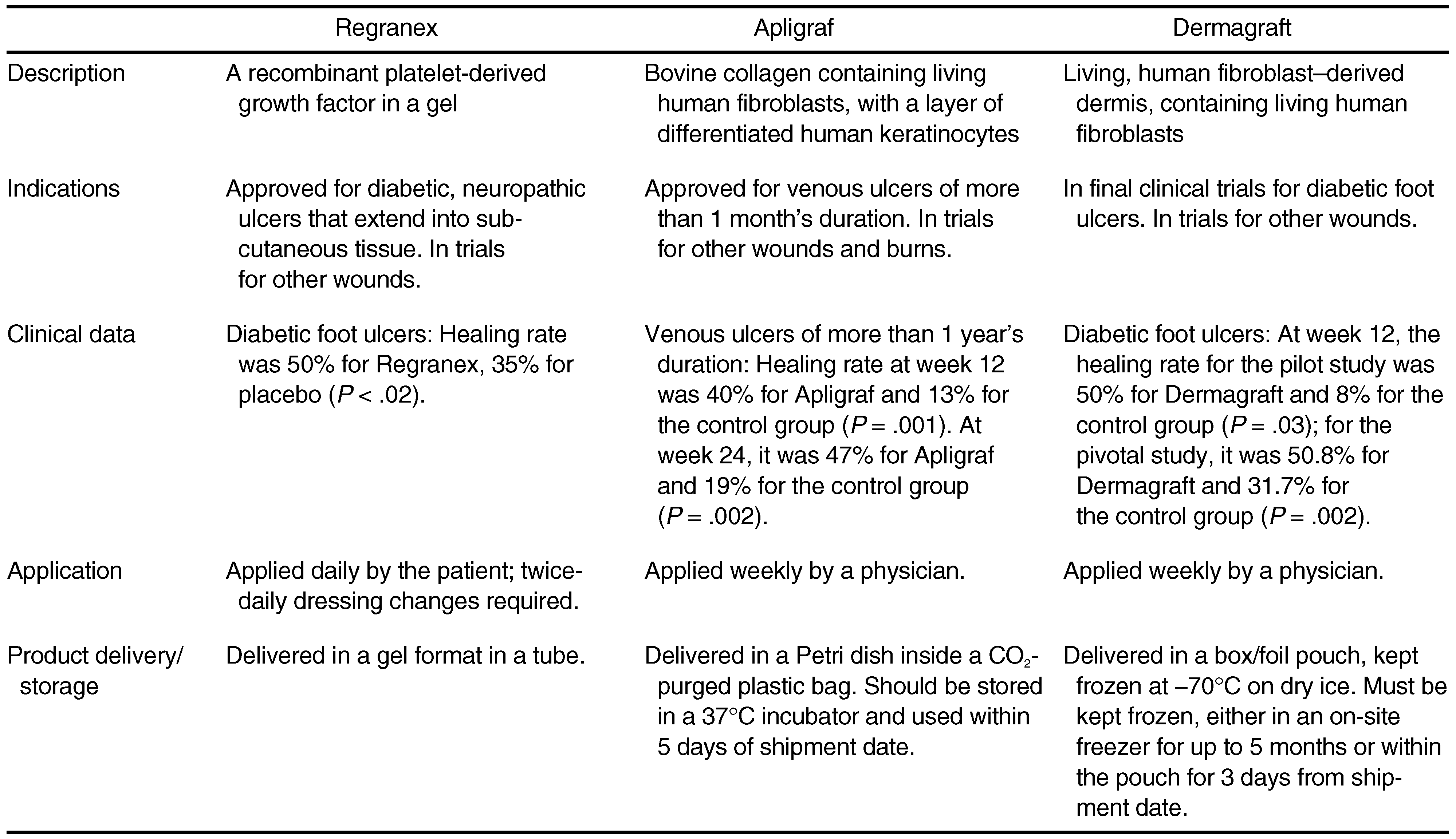
Recombinant growth factor technologies have some advantages over autologous and homologous factors. They avoid the high cost of autologous preparations and the infection hazard of homologous preparations. Additionally, large quantities can be readily produced for research and commercial purposes. The ability to precisely analyze and control the concentration of the engineered growth factor during production and processing can validate a dose-response curve in the chronic wound. Finally, recombinant human platelet-derived growth factor-BB is approved by the US Food and Drug Administration (FDA) as a prescription drug (Regranex Gel, Ortho-McNeil Pharmaceutical, Inc, Raritan, New Jersey) (Table 2).
Table 2.
New Technology Product Descriptions
Steed [24] evaluated recombinant human platelet-derived growth factor-BB (becaplermin) in 118 subjects in a randomized, double-blind, placebo-controlled trial. Becaplermin is produced by recombinant-DNA technology by insertion of the gene for the B chain of platelet-derived growth factor into the yeast Saccharomyces cerevisiae. In the aforementioned study, the preparation was formulated into a gel for the treatment group. The control group received a similar gel without recombinant human platelet-derived growth factor-BB. Using 2.2 μg of recombinant human platelet-derived growth factor-BB per square centimeter of ulcer area, the investigators reported significant differences in complete wound healing (48% for the treatment group versus 25% for the control group) at the end of 20 weeks. Differences in time to complete healing were not evident in the first 3 weeks of the study, but by 68 days, a difference of 30 to 40 days in the time to complete healing was noted between the groups. These results were statistically significant.
A subsequent study of recombinant human plateletderived growth factor enrolled 382 patients in a multicenter, double-blind, placebo-controlled design. Subjects were randomly assigned to one of three groups: becaplermin gel 0.01%, becaplermin gel 0.003%, and placebo gel. In this study, the more concentrated becaplermin gel (0.01%) was associated with a 50% proportion of wounds healed at 20 weeks, the less concentrated gel (0.003%) a 36% proportion, and the placebo gel a 35% proportion. [32] Only becaplermin gel 0.01% differed significantly from placebo in proportion of wounds healed at 20 weeks. A subsequent published report, whose primary aim was to assess the safety of the vehicle gel used in recombinant human platelet-derived growth factor compared with good wound care alone, [33] revealed no significant difference between the vehicle gel (36% proportion of wounds healed; n = 70) and the placebo treatment, termed “good wound care alone” (22% proportion of wounds healed; n = 68). The investigators included a small group (n = 34) of patients treated with becaplermin gel (Regranex Gel) (44% proportion of wounds healed) in this study. The small sample size in the becaplermin gel arm precluded any meaningful comparison with the other two treatment arms. The rationale for inclusion of the becaplermin gel arm in this study is puzzling, as the study was not intended to evaluate wound-healing proportion per se or to judge the clinical efficacy of becaplermin’s gel vehicle but rather to study the safety of becaplermin’s gel vehicle.
Other clinical trials have sought to elucidate the utility of other growth factors in treating chronic wounds. Fibroblast growth factor, epidermal growth factor, and arginine–glycine–aspartic acid have been investigated. The group studying arginine–glycine– aspartic acid [34] reported favorable results of arginine–glycine–aspartic acid gel (Argidine Gel) in the treatment of neuropathic diabetic foot ulcers in the case of full-thickness cutaneous ulcers below grade 3A on the University of Texas scale. The study reported a 35% proportion of wounds completely healed in the treatment group, as compared with 8% in the control group, at the completion of the trial. The description of the study methodology indicated that the study was double-blind, but in fact the treatment complex was in gel form while the control was normal saline. In addition, the hyaluronate carrier was not tested separately as a control. Fibroblast growth factor was used by Richard et al [35] in a pilot study. The researchers randomized patients with diabetic neuropathic ulcers (University of Texas grade 3A or below) in a double-blind fashion to receive fibroblast growth factor or placebo. They were unable to demonstrate any significant difference in wound closure between subjects receiving topical beta-fibroblastic growth factor and those receiving a placebo. Epidermal growth factor, though well characterized, has not yet been used in any trials of diabetic wounds.
Growth factors ultimately require a host cell response to either single or multiple agents added to a wound. Sequential use of topical growth factors may fail because of rapid destruction by proteinases in the wound space, [21] or because wound healing requires cell signaling in an as yet poorly understood cascade. Both despite and as a consequence of this, investigators have sought to enhance the delivery system of growth factors to the wound. [36] One of the most promising forms this delivery system has taken is bioengineered tissue.
Tissue Therapies
Bioengineered tissues may be of dermal origin only or of dermal-epidermal origin. Some investigators now suggest that bioengineered tissues, once considered to be skin substitutes, may be best categorized as potent delivery systems for soluble mediators (such as growth factors). [36] While this may prove to be the case, no conclusive in vivo data currently exist to support this contention. Bioengineered tissues are produced by various proprietary techniques. Both acellular allogeneic dermal grafts and complete dermal-epidermal grafts (human skin equivalents) have been successfully prepared and used in studies involving burn patients. There have been no reports of transmissible disease or immunogenic reactions in studies of either acellular dermal matrix or cellular skin equivalents.
The recent advance in tissue culture technique has made it possible to culture cells from human foreskin donors. Donated newborn foreskins are used to extract epidermal precursor cells and fibroblasts for a master cell bank. Both the cells and the mothers are extensively and repeatedly screened for infectious agents. Very few donors are needed because these cells are transferred to working cell banks, and cells from a few foreskins can produce enough skin substitute to cover the equivalent of four to six football fields. Dermagraft (pending FDA approval; Advanced Tissue Sciences Inc, La Jolla, California) is an in vitro bioengineered dermal skin construct consisting of a bioabsorbable polyglactin mesh (Vicryl, Ethicon Inc, Somerville, New Jersey) and seeded fibroblasts. The seeded fibroblasts proliferate and spread through the mesh, producing cytokines (growth factors), glycosaminoglycans, and other ground substance and matrix proteins, including collagen, fibronectin, and tenascin. [30] After the construct has matured, it is cryopreserved in a bioreactor and then thawed prior to patient application. In the rat aorta ring assay, enhancement of wound tissue expansion and angiogenesis was linked to hepatocyte growth factor/scatter factor produced by the cells. [37]
Gentzkow and coworkers [30] reported the results of a prospective, randomized, single-blind pilot trial of Dermagraft for treatment of neuropathic diabetic foot ulcers. The lesions involved were cutaneous fullthickness ulcers measuring more than 1 cm2 and graded below 3A on the University of Texas scale. Patients were required to have adequate blood flow, manifested by an ankle-brachial index of more than 0.7, and were excluded if infection, medication, or medical conditions known to retard wound healing were present. A standardized wound-care protocol was used in both the treatment and the control groups. In this study, patients who received one piece of Dermagraft per week healed significantly more often (50%; n = 6/12) than patients receiving standard wound care (8%; n = 1/13) during a 12-week period.
A follow-up multicenter study enrolled 129 patients in the control group and 109 in the Dermagraft group. [31] At the interim analysis, it was discovered that the therapeutic range for Dermagraft has been narrower than the original release criteria. Testing is performed before and after cryopreservation to determine a therapeutic range. The assay result was compared with healing rates after application; both very low and very high levels were found to inhibit healing. [30] The 12-week healing rate increased substantially if the product had an appropriate proportion of viable fibroblasts (appropriate therapeutic range), as compared with the entire stock of Dermagraft evaluated. [31] The investigators postulated that the fibroblasts within this living, metabolically active tissue are important in producing matrix components and cytokines interacting physiologically with the host’s tissue response and modulating it. The proportion of wounds completely healed at week 32 for all Dermagraft patients was 57.5%; for therapeuticrange patients only, 57.7%; and for control patients, 42.4%.
The most critical clinical criticism of this trial is similar to the one leveled against the becaplermin trial—that is, the method of off-loading employed. In this trial, subjects were given prescription shoes with molded inlays as the primary off-loading implement. While this method of off-loading was standardized, it was still clearly suboptimal. Lavery and coworkers [38] have reported that therapeutic shoes impart some 900% more pressure to the foot than do total-contact casts or certain removable cast walkers.
Apligraf (graftskin) (Organogenesis, Inc, Canton, Massachusetts) is a skin substitute composed of an epidermis (epidermal cells, stratum corneum) and a dermis (bovine collagen, dermal fibroblasts). [39] In this bilayered construct, bovine collagen is acid-dissociated and added to a transwell with fibroblasts from newborn donors. After 5 to 6 days, epidermal cell precursors are added. When 5 to 15 layers of epithelium are formed, the construct is raised to the air-liquid interface to facilitate the production of a stratum corneum. This skin equivalent is also currently being tested for nonhealing diabetic foot ulcers.
Beyond the clinical issues discussed above, some of the most pressing practical issues surrounding the potential widespread use of tissue-engineered skin concern cost. In addition to clinical effectiveness, cost-effectiveness will be critical to establish in the current environment.
In the clinical setting, it is crucial to determine the optimal application technique to maximize the efficacy of these products. Bacterial burden of the wound, local and regional vascular supply, host factors, and other issues need to be addressed and corrected to achieve optimal results. In the future, many new generations of skin substitutes may become available. The dermis could be manipulated with glycosaminoglycans that can enhance wound healing, and the transplanted cells could be transfected. A viral vector could be used to insert into cells extra copies of genes, such as platelet-derived growth factor, that promote the wound-healing process.
Summary
An emerging approach to the care of chronic wounds is based on an old concept: conversion of a chronic wound into an acute wound. However, it is the maintenance of the wound in this acute state that has called upon the expertise of numerous investigators from various fields. For clinical research in this area, precise control and documentation of wound care is extremely important in order to avoid confounding the effects of a specific modality with those of “good wound care.” Falanga [22] has noted that the neuropathy of diabetes has been given wide support as the primary pathogenic component of diabetic ulcers, while less recognition has been given to the mechanisms that are antagonistic to wound healing. The therapies discussed in this article have taken these mechanisms into account. Furthermore, in the current era of costconscious health-care administration, podiatric physicians must consider the economic component of care in addition to the basic science. The authors believe that only if advanced dressings, growth factor delivery systems, and other procedures are shown to significantly reduce healing time, thereby reducing the risk of potential infection, hospitalization, and amputation while potentially improving quality of life, can they be considered truly cost-effective.
References
- NATIONAL DIABETES DATA GROUP. Diabetes in America; US Department of Health and Human Services: Bethesda, MD, 1995. [Google Scholar]
- Apelqvist J, Ragnarson-Tennvall G, Larsson J; et al. Long-term costs for foot ulcers in diabetic patients in a multidisciplinary setting. Foot Ankle Int 1995, 16, 388. [Google Scholar] [CrossRef] [Scilit]
- LAVERY, LA; VAN HOUTUM, WH; ARMSTRONG, DG. Institutionalization following diabetes-related lower extremity amputation. Am J Med 1997, 103, 383. [Google Scholar] [CrossRef] [Scilit]
- US Department of Health and Human Services. Healthy People 2000, National Health Promotion and Disease Prevention Objectives: Full Report, with Commentary; Public Health Service: Washington, DC, 1991. [Google Scholar]
- Diabetes care and research in Europe: the Saint Vincent declaration. Diabet Med 1990, 7, 360. [CrossRef] [Scilit]
- Apelqvist, J; Ragnarson-Tennvall, G; Persson, U; et al. Diabetic foot ulcers in a multidisciplinary setting: an economic analysis of primary healing and healing with amputation. J Intern Med 1994, 235, 463. [Google Scholar] [CrossRef] [Scilit]
- APELQVIST, J; LARSSON, J; AGARDH, CD. The influence of external precipitating factors and peripheral neuropathy on the development and outcome of diabetic foot ulcers. J Diabetes Complications 1990, 4, 21. [Google Scholar] [CrossRef] [Scilit] [PubMed]
- APELQVIST, J; CASTENFORS, J; LARSSON, J; et al. Wound classification is more important than site of ulceration in the outcome of diabetic foot ulcers. Diabet Med 1989, 6, 526. [Google Scholar] [CrossRef] [Scilit]
- GUPTA, SK; VEITH, FJ; ASCER, E; et al. Cost factors in limbthreatening ischaemia due to infrainguinal arteriosclerosis. Eur J Vasc Surg 1988, 2, 151. [Google Scholar] [CrossRef] [Scilit]
- MACKEY, WC; MCCULLOUGH, JL; CONLON, TP; et al. The costs of surgery for limb-threatening ischemia. Surgery 1986, 99, 26. [Google Scholar] [PubMed]
- PERLER, BA. Cost-efficacy issues in the treatment of peripheral vascular disease: primary amputation or revascularization for limb-threatening ischemia. J Vasc Interv Radiol 1995, 6 (suppl), 111. [Google Scholar] [CrossRef] [Scilit] [PubMed]
- SINGH, S; EVANS, L; DATTA, D; et al. The costs of managing lower limb–threatening ischaemia. Eur J Vasc Endovasc Surg 1996, 12, 359. [Google Scholar] [CrossRef] [Scilit]
- APELQVIST, J. RAGNARSON-TENNVALL G: Cavity foot ulcers in diabetic patients: a comparative study of cadexomer iodine ointment and standard treatment. an economic analysis alongside a clinical trial. Acta Derm Venereol 1996, 76, 231. [Google Scholar] [CrossRef] [Scilit] [PubMed]
- AKBARI, CM; SAOUAF, R; BARNHILL, DF; et al. Endotheliumdependent vasodilatation is impaired in both microcirculation and macrocirculation during acute hyperglycemia. J Vasc Surg 1998, 28, 687. [Google Scholar] [CrossRef] [Scilit] [PubMed]
- VEVES, A; AKBARI, CM; PRIMAVERA, J; et al. Endothelial dysfunction and the expression of endothelial nitric oxide synthetase in diabetic neuropathy, vascular disease, and foot ulceration. Diabetes 1998, 47, 457. [Google Scholar] [CrossRef] [Scilit]
- YOUNG, MJ; BENNETT, JL; LIDERTH, SA; et al. Rheological and microvascular parameters in diabetic peripheral neuropathy. Clin Sci (Colch) 1996, 90, 183. [Google Scholar] [CrossRef] [Scilit]
- YANG, L; QIU, CX; LUDLOW, A; et al. Active transforming growth factor-beta in wound repair: determination using a new assay. Am J Pathol 1999, 154, 105. [Google Scholar] [CrossRef] [Scilit] [PubMed]
- KANZAKI, T; MORISAKI, N; SHIINA, R; et al. Role of transforming growth factor-beta pathway in the mechanism of wound healing by saponin from Ginseng Radix rubra. Br J Pharmacol 1998, 125, 255. [Google Scholar] [CrossRef] [Scilit] [PubMed]
- WATSON, AL; BURTON, GJ. : A microscopical study of wound repair in the human placenta. Microsc Res Tech 1998, 42, 351. [Google Scholar] [CrossRef] [Scilit]
- COLEMAN, C; TUAN, TL; BUCKLEY, S; et al. Contractility, transforming growth factor-beta, and plasmin in fetal skin fibroblasts: role in scarless wound healing. Pediatr Res 1998, 43, 403. [Google Scholar] [CrossRef] [Scilit]
- BRANTIGAN, CO. The history of understanding the role of growth factors in wound healing. Wounds 1996, 8, 78. [Google Scholar]
- FALANGA, V. Growth factors and wound healing. J Dermatol Surg Oncol 1993, 19, 711. [Google Scholar] [CrossRef] [Scilit]
- FALANGA, V. Wound healing: an overview. J Dermatol Surg Oncol 1993, 19, 689. [Google Scholar] [CrossRef] [Scilit]
- STEED, DL. Clinical evaluation of recombinant human platelet-derived growth factor for the treatment of lower extremity diabetic ulcers. Diabetic Ulcer Study Group. J Vasc Surg 1995, 21, 71. [Google Scholar] [CrossRef] [Scilit]
- ARMSTRONG, DG; LAVERY, LA; HARKLESS, LB. Validation of a diabetic wound classification system: the contribution of depth, infection, and ischemia to risk of amputation. Diabetes Care 1998, 21, 855. [Google Scholar] [CrossRef] [Scilit] [PubMed]
- KNIGHTON, DR; CIRESI, KF; FIEGEL, VD. Classification and treatment of chronic nonhealing wounds: successful treatment with autologous platelet-derived wound healing factors (PDWHF). Ann Surg 1986, 204, 322. [Google Scholar] [CrossRef] [Scilit]
- KNIGHTON, DR; CIRESI, K; FIEGEL, VD; et al. Stimulation of repair in chronic, nonhealing, cutaneous ulcers using platelet-derived wound healing formula. Surg Gynecol Obstet 1990, 170, 56. [Google Scholar]
- STEED, DL; GOSLEN, JB; HOLLOWAY, GA; et al. Randomized prospective double-blind trial in healing chronic diabetic foot ulcers: CT-102 activated platelet supernatant, topical versus placebo. Diabetes Care 1992, 15, 1598. [Google Scholar] [CrossRef] [Scilit]
- FALANGA, V; SABOLINSKI, ML. A bilayered living skin construct (Apligraf) accelerates complete closure of hardto-heal venous ulcers. Wound Repair Regen (in press).
- GENTZKOW, GD; IWASAKI, SD; HERSHON, KS. Use of Dermagraft, a cultured human dermis, to treat diabetic foot ulcers. Diabetes Care 1996, 19, 350. [Google Scholar] [CrossRef] [Scilit] [PubMed]
- POLLACK, RA; EDINGTON, H; JENSEN, J. A human dermal replacement for the treatment of diabetic foot ulcers. Wounds 1997, 9, 175. [Google Scholar]
- WIEMAN, TJ; SMIELL, JM; SU, Y. Efficacy and safety of a topical gel formulation of recombinant human plateletderived growth factor-BB (becaplermin) in patients with chronic neuropathic diabetic ulcers: a phase III randomized placebo-controlled double-blind study. Diabetes Care 1998, 21, 822. [Google Scholar] [CrossRef] [Scilit]
- D’HEMECOURT, PA; SMIELL, JM; KARIM, MR. Sodium carboxymethylcellulose aqueous-based gel vs. becaplermin gel in patients with nonhealing lower extremity ulcers. Wounds 1998, 10, 69. [Google Scholar]
- STEED, DL; RICOTTA, JJ; PRENDERGAST, JJ; et al. Promotion and acceleration of diabetic ulcer healing by arginine–glycine–aspartic acid (RGD) peptide matrix. RGD Study Group. Diabetes Care 1995, 18, 39. [Google Scholar] [CrossRef] [Scilit] [PubMed]
- RICHARD, JL; PARER-RICHARD, C; DAURES, JP; et al. Effect of topical basic fibroblast growth factor on the healing of chronic diabetic neuropathic ulcer of the foot: a pilot, randomized, double-blind, placebo-controlled study. Diabetes Care 1995, 18, 64. [Google Scholar] [CrossRef] [Scilit] [PubMed]
- KIRSNER, RS; FALANGA, V; KERDEL, FA; et al. Skin grafts as pharmacological agents: pre-wounding of the donor site. Br J Dermatol 1996, 135, 292. [Google Scholar] [CrossRef] [Scilit] [PubMed]
- JIANG, WG; HARDING, KG. Enhancement of wound tissue expansion and angiogenesis by matrix-embedded fibroblast (Dermagraft), a role of hepatocyte growth factor/scatter factor. Int J Mol Med 1998, 2, 203. [Google Scholar]
- LAVERY, LA; VELA, SA; LAVERY, DC; et al. Reducing dynamic foot pressures in high-risk diabetic subjects with foot ulcerations: a comparison of treatments. Diabetes Care 1996, 19, 818. [Google Scholar] [CrossRef] [Scilit] [PubMed]
- SABOLINSKI, ML; ALVAREZ, O; AULETTA, M; et al. Cultured skin as a “smart material” for healing wounds: experience in venous ulcers. Biomaterials 1996, 17, 311. [Google Scholar] [CrossRef] [Scilit] [PubMed]

© 2000 American Podiatric Medical Association