


To the Editor:
Foreign objects embedded in the foot are commonly seen in podiatric medical practice. Foreign bodies penetrate the foot in a variety of ways, ranging from a person’s stepping on an object, usually while walking barefoot, to a projectile’s entering the foot. The literature indicates that the most common types of foreign bodies encountered are of metal, glass, wood, hair, and thorns [1,2,3]. Except in cases of peripheral neuropathy, the individual is usually well aware that something may have penetrated the skin of the foot because of the associated pain. Occasionally, however, the individual is not aware of the injury. In 1991, Assalita [4] reported on a case of a foreign body that had gone undetected by the patient for 20 years. Obviously, when a patient does not recall an injury on initial presentation, a thorough history and physical examination are essential.
Unless a patient presents with an obvious foreign object or knows that something is lodged in the foot, it is easy to exclude a foreign body as a likely diagnosis. A symptomatic foreign body may present superficially as a mass near the skin, thus mimicking other types of lesions. Dowling [5] discussed the importance of considering the possibility of a ganglion or giant cell tumor. Newman and Hunt [6] emphasized the possibility of a granuloma. Hutchinson and Weir [7] found that foreign bodies may even mimic verrucae.
In certain circumstances, foreign objects embedded in the body’s tissue cause no symptoms and go unnoticed until some of the chronic effects begin to manifest. Scartozzi and Hoffman8 attributed these chronic effects to migration of the foreign body, neurovascular compromise, anemia, lead poisoning, and recurrent infection.
Once the foreign object has been identified and its size and location have been determined, the next step is to consider whether it must be removed. In 1993, Stein and Clark [2] proposed five criteria for determining the necessity of removal of foreign objects: 1) injury to vital structures, 2) evidence of infection or significant contamination, 3) an allergic reaction to the object, 4) pain caused by the object, and 5) the ability to superficially palpate the object.
This article presents a unique case involving a foreign object and discusses the circumstances surrounding its implantation as well as preoperative, intraoperative, and postoperative findings, including pathologic findings.
Case Report
A 34-year-old man presented to the Tower Foot and Ankle Institute in Des Moines, Iowa, with the chief complaint of a mass at the distal, medial aspect of his right fourth digit. The patient stated that this mass had been noticeable for several years, but had become progressively larger over the preceding 6 months. He denied experiencing any sharp pain, but stated that he had experienced some numbness, tingling, and burning toward the tip of his toe. Initially, he denied any trauma to the area. This was the first time he had sought treatment for the problem.
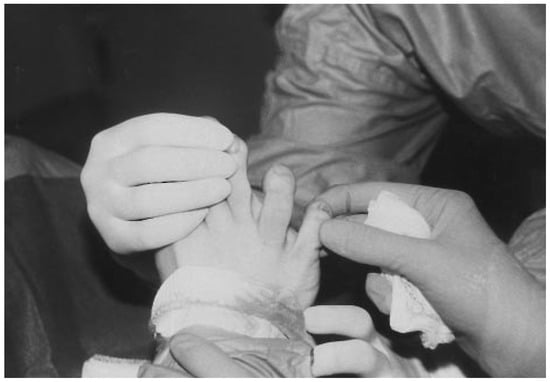
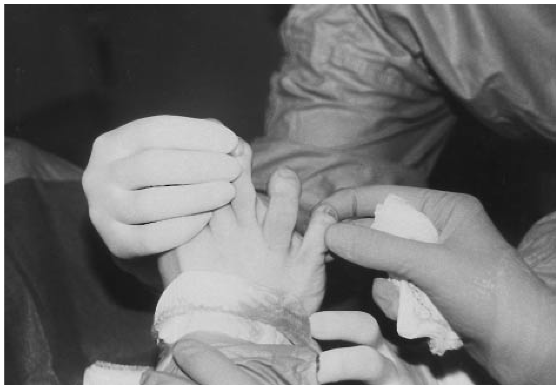
Physical examination revealed a firm, circular, immobile mass over the distal, medial, and slightly plantar aspect of the right fourth digit measuring approximately 1.5 × 1.5 cm (Fig. 1). Mild erythema and edema with a light ecchymosis overlying the taut digit were noted. No pain was elicited with direct or side-to-side compression of the area. With percussion, however, the patient complained of tingling toward the distal aspect of the right fourth digit. The remainder of the physical examination was unremarkable, as were the patient’s medical and surgical histories.
Figure 1.
Prominent mass over the plantar medial aspect of the right fourth digit.
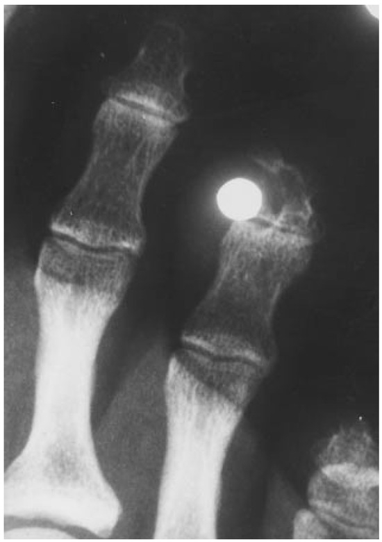
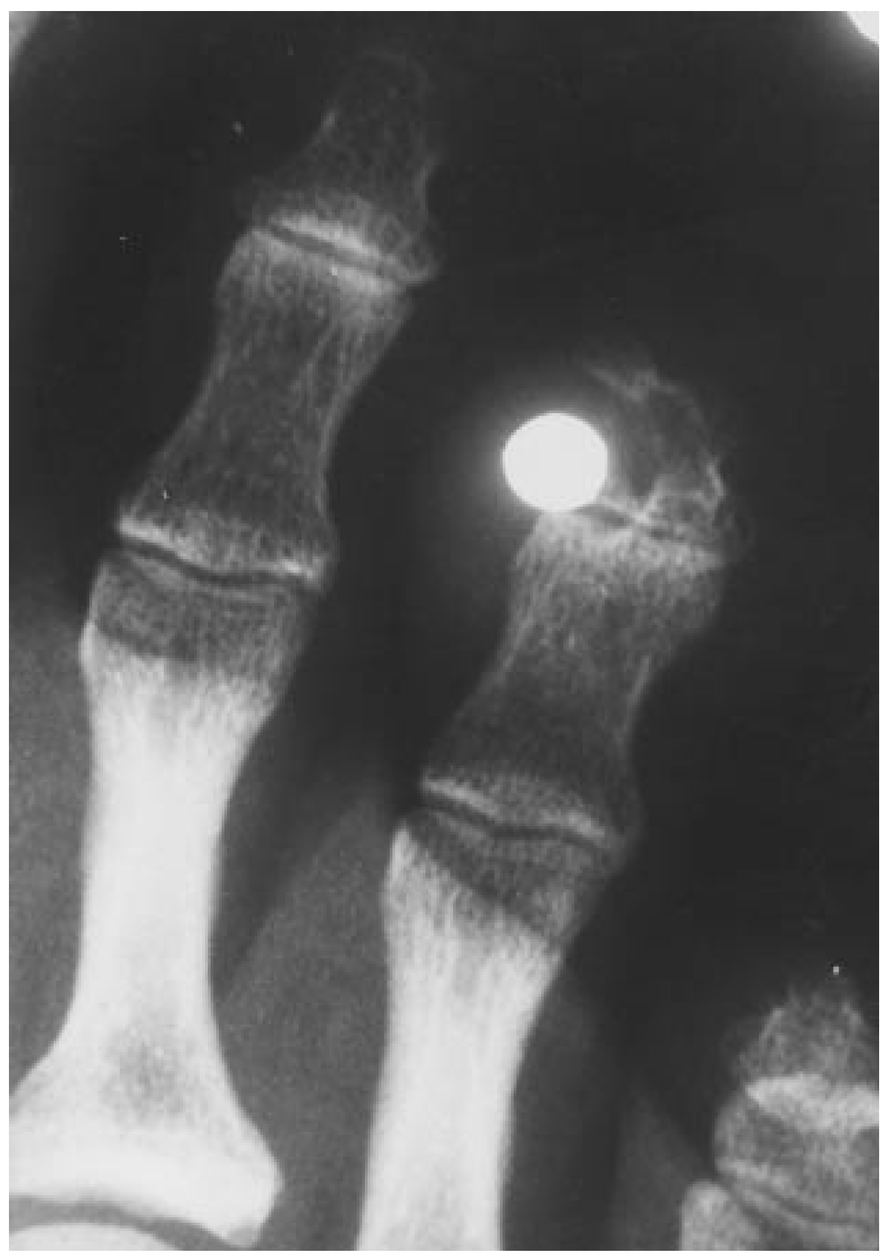
Dorsoplantar and medial oblique radiographs of the patient’s right foot revealed a radiopaque lesion, 4 mm in diameter, located just medial to the distal phalanx of the right fourth digit. Interestingly, the distal phalanx appeared to have grown around the object, with a striking concavity adjacent to the object (Fig. 2).
Figure 2.
Dorsoplantar radiograph demonstrating accommodative growth of the distal phalanx in response to the embedded pellet.
After the radiographs were reviewed with the patient, the patient recalled being shot in the foot with a pellet gun approximately 25 years before. The patient stated that he sought no treatment at the time of the injury, as there was minimal or no bleeding. He had assumed that the pellet caromed off his foot. At this time, the patient was scheduled for surgery to remove the pellet from his right fourth digit.
Surgical Procedure and Findings
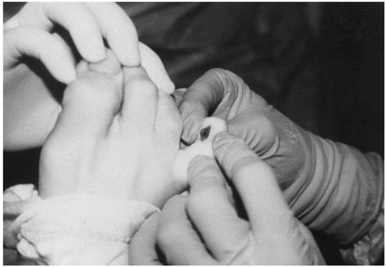
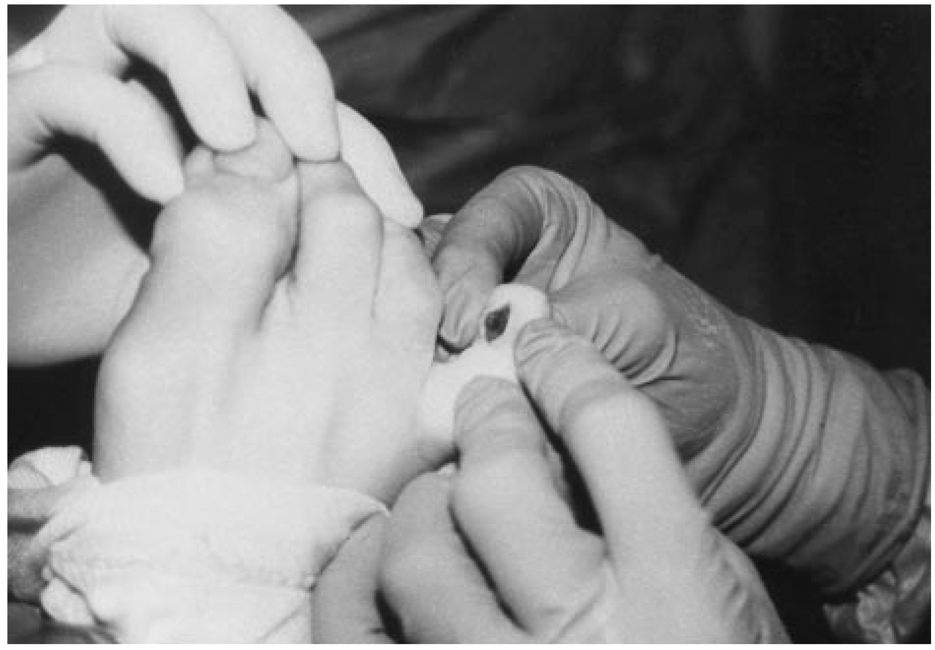
The patient underwent surgical excision of the lesion, which initially revealed a brownish metallic color, similar to that of a penny, staining the subcutaneous tissue (Fig. 3). As dissection continued, the tissue began to appear very darkly stained. Deeper dissection resulted in significant oozing of what appeared to be a tannish purulent liquid with no detectable odor. Owing to the history behind the object, it was suspected that this fluid was the contents of a sterile abscess rather than bacterial in nature. Nonetheless, a culture was obtained and sent to the pathology department for analysis. The pellet was then levered into the surgical site, where it appeared to be in one piece, and was removed in toto. The postoperative course was uneventful.
Figure 3.
Intraoperative photograph of the encapsulated pellet.
Pathologic Findings
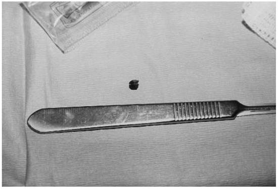
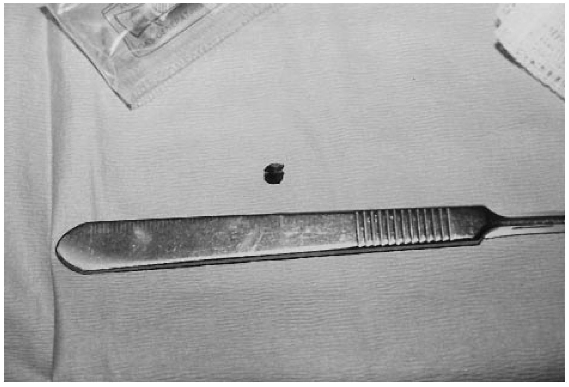
Examination of the foreign body revealed a charcoal gray, spherical object approximately 4 × 4 mm consistent with a lead pellet from a pellet gun (Fig. 4). A Gram’s stain of fibrotic tissue and purulent fluid obtained from the wound demonstrated no white blood cells or other inflammatory cells such as polymorphonuclear leukocytes or macrophages. However, a few gram-positive cocci were observed. A culture demonstrated light growth of coagulase-negative Staphylococcus.
Figure 4.
Excised pellet devoid of alloy coating.
Discussion
This case report is very similar to the one published in 1991 by Assalita [4] in that the patient did not recognize the initial traumatic episode and in the significant amount of time that elapsed before the pellet elicited a symptomatic response by the body’s tissues. In both of these cases, the pellet’s lying dormant for so many years without eliciting a response by the body is inexplicable. Assalita [4] attributes it to the complexities of the human body. The authors concur, as there is no definitive literature to date explaining this phenomenon.
Accidental pellet gun injuries are rather common, especially among school-age children [9] Fortunately, these types of injuries to the foot are rarely life-threatening. Pellet guns produce low-velocity wounds; the projectile has a velocity of less than 2,000 feet per second and typically induces only an entrance wound, with no exit wound and no extreme damage to surrounding tissues [10].
In cases of foreign-body penetration, the human body will attempt to either expel, encapsulate, or dissolve the foreign body [3]. In this case, a capsule was identified intraoperatively; it was composed of darkly stained fibrotic tissue, which, according to Fackler [11], is part of the body’s natural defense mechanism against the foreign body. The authors concur that the darkly stained tissue was attributable to a reaction that occurred between the outer coating of the pellet and the cells within the body. Most ammunition, including pellets, is coated with metal alloys that minimize deformation and tissue damage on contact [10]. In this case, the pellet’s outer covering was completely removed; this further suggests that some type of reaction occurred. To the authors’ knowledge, there is no literature defining the mechanism of the reaction between tissue and the metal alloy that forms the outer covering of pellets. Fackler [11] stated that if the body fails to expel this fibrotic capsule, it will become an abscess. Indeed, in this case, a sterile abscess had formed, although nothing in the patient’s history or physical examination was suggestive of infection.
As seen in the pathology review, no white blood cells, polymorphonuclear leukocytes, macrophages, or other inflammatory cells were present. Assalita [4] used the term “foreign body granuloma” to describe the body’s response to the foreign body. Granuloma is a generic term to describe the aggregation of mononuclear inflammatory cells or a collection of modified macrophages [12]. Given the pathology results in this case, the authors cannot consider the term “granuloma” to describe the response to the foreign body. Furthermore, in the pathology review, it was noted that a few gram-positive cocci were observed and that a culture produced a light growth of coagulase-negative Staphylococcus, which is probably Staphylococcus epidermidis, a component of normal skin flora [13]. These results differ from those obtained by Dowling [5], who observed many coagulase-positive Staphylococcus organisms along with some Escherichia coli organisms in a case where a wooden toothpick was the offending foreign body. Perhaps this difference is due to the two different materials in question, the metal pellet and the wooden toothpick, or perhaps it is due to the situation that led to penetration by the foreign object.
As mentioned previously, embedded foreign objects sometimes remain dormant for years until one of the chronic effects begins to manifest and symptoms appear. One of the reasons this patient sought treatment when he did was that he was experiencing occasional numbness, burning, and tingling toward the distal aspect of his right fourth digit. Clinically speaking, he was describing neurapraxia. Neurapraxia refers to the impairment of the conduction of nerve impulses and is usually completely reversible [14]. This patient suffered impairment of the seventh proper plantar digital nerve. At a 1-month follow-up visit, the patient stated that, for the most part, the feeling had returned to the medial aspect of his toe, and he no longer experienced paresthesia.
Summary
A symptomatic foreign body embedded in the human body can be a frustrating problem for physician and patient alike. A unique case of a retained foreign object resulting from a pellet gun injury has been presented. Although the course of treatment in this case was uncomplicated, it is important to understand the complexities of the human body’s response to foreign bodies.
References
- SCHILLING JJ: Fish fin spine foreign body. JAPMA 84: 196, 1994.
- STEIN RA, CLARK S: Foreign bodies in the foot. JAPMA 83: 284, 1993.
- BYRON TJ: Foreign bodies found in the foot. JAPMA 71: 30, 1981.
- ASSALITA LJ: Delayed foreign body reaction following air gun-pellet trauma. J Foot Surg 30: 187, 1991.
- DOWLING GL: Foreign body: a review of two cases. J Foot Surg 21: 70, 1982.
- NEWMAN JT, HUNT DW: Toothpick foreign body in the foot: a case report. JAPMA 79: 458, 1989.
- HUTCHINSON P, WEIR EC: Retained foreign body in the foot: a case study. Br J Podiatry 50, 1998.
- SCARTOZZI G, HOFFMAN L: Chronic high velocity projectile injury to the foot. JAPMA 79: 236, 1989.
- BRATTON SL, DOWD MD, BROGAR TV, ET AL: Serious and fatal air gun injuries: more than meets the eye. Pediatrics 100: 609, 1997.
- SERGI AR, ACELLO AN: Gunshot wounds to the foot and ankle. Clin Podiatr Med Surg 12: 689, 1995.
- FACKLER ML: Gunshot wound review. Ann Emerg Med 28: 194, 1996.
- Dorland’s Pocket Medical Dictionary, 25th Ed, WB Saunders, Philadelphia, 1995.
- ROBBINS SL, COTRAN RS, KUMAR V: Robbins Pathologic Basis of Disease, 3rd Ed, WB Saunders, Philadelphia, 1984.
- LEPOW GM: “Neurological Considerations,” in Complications in Foot Surgery: Prevention and Management, 2nd Ed, ed by SA Marcus, BH Block, p 456, Williams & Wilkins, Baltimore, 1984.
© 2000 American Podiatric Medical Association