


To the Editor:
Avulsion fractures of the plantar lateral base of the first metatarsal are known to occur with tarsometatarsal joint injuries that involve medial displacement of the first metatarsal. Hardcastle et al[1] documented the appearance of this avulsion fracture in combination with Lisfranc B1 and C injuries. Lisfranc B1 injuries are identified by partial incongruity caused by medial displacement of the first metatarsal alone or in association with medial displacement of one or more of the lesser metatarsals. Medial displacement of the first metatarsal with concomitant lateral displacement of one or more of the lesser metatarsals creates a divergent deformity classified as a Lisfranc C injury.
Hodor et al [2] were the first to describe an avulsion fracture of the plantar lateral base of the first metatarsal not related to a Lisfranc B1 or C injury. As the mechanism of action, the authors proposed a strong, sudden pronatory (abductory) force of the peroneus longus tendon on a planted first ray in a closed kinetic chain. The patients described in their article had difficulty recalling their foot position at the time of injury.
This case presentation will document a similar isolated avulsion fracture of the plantar lateral base of the first metatarsal and propose the mechanism of injury.
The peroneus longus muscle originates from the head and upper two-thirds of the body of the fibula and is the most superficial muscle of the lateral compartment of the lower leg. The tendon passes posterior to the lateral malleolus and runs deep to the superior peroneal retinaculum. As the tendon courses distal to the peroneal trochlea on the lateral aspect of the calcaneus, it travels deep to the inferior peroneal retinaculum. At the cuboid, the peroneus longus tendon continues plantarly and anteriorly along the peroneal groove of the cuboid, deep to the long plantar ligament. It advances medially and anteriorly to insert into the lateral aspect of the first metatarsal base and medial cuneiform.
The base of the first ray is not located in the same transverse plane as the cuboid but is positioned above the plantar surface of the cuboid. Only when this relationship exists can the cuboid function as a pulley for the tendon of the peroneus longus.
The primary functions of the peroneus longus muscle are stabilization of the first ray and stabilization of the head of the first metatarsal against the ground.[3] Contraction of the peroneus longus generates an abductory and pronatory force on the base of the first ray, stabilizing it against the lesser tarsal bones and thereby enabling the first ray to withstand normal ground-reactive forces. The peroneus longus muscle may also exert a strong plantarflexory force upon the first ray when the subtalar joint is in a neutral or supinated position.
Case Report
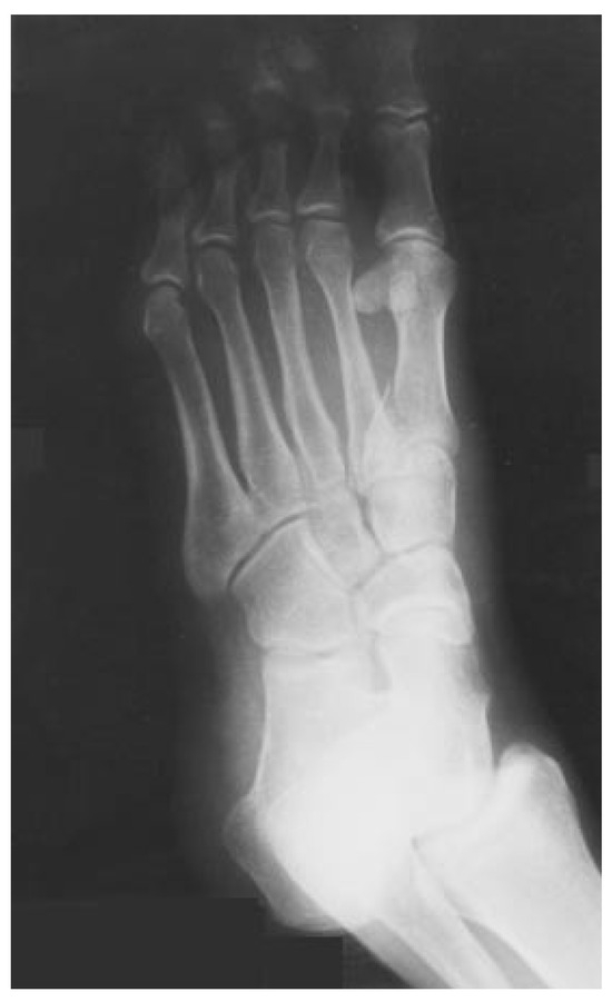
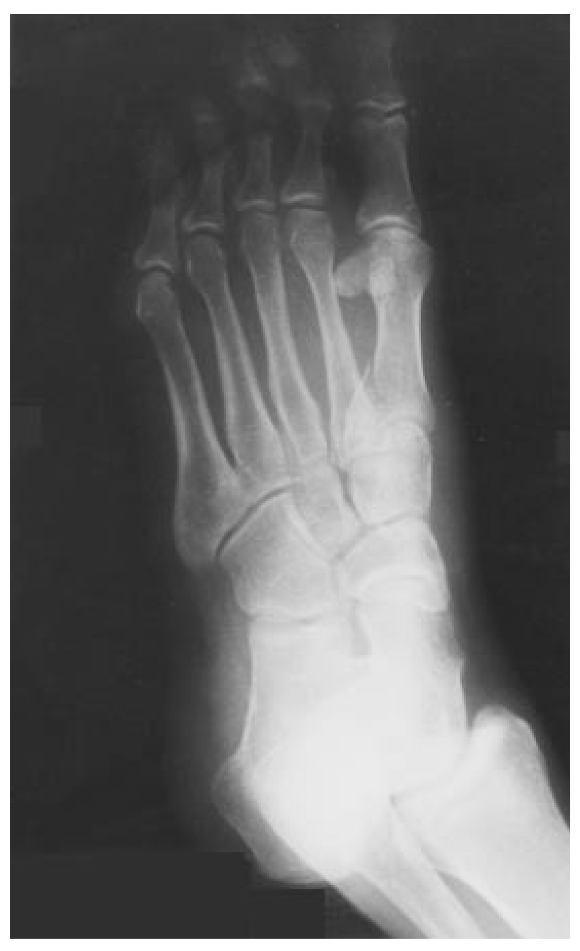
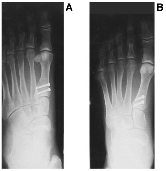
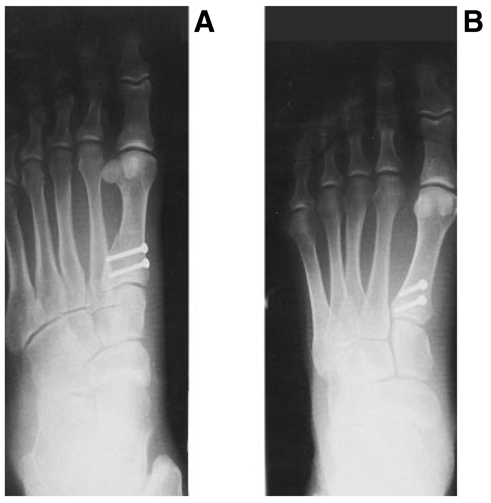
A 30-year-old man presented to the emergency department at Broadlawns Medical Center in Des Moines, Iowa, complaining of a painful left foot and ankle. The patient described an inversion ankle injury occurring the previous evening as he jumped over a fence. Upon landing, he felt a sharp pain in the arch and lateral ankle. Pedal pulses were palpable and light touch sensation was grossly intact. Moderate swelling was noted along the lateral aspect of the left ankle, extending across the plantar aspect of the foot. Maximum pain was elicited on palpation of the first metatarsal base–medial cuneiform articulation. Radiographs of the left foot revealed an oblique intraarticular fracture of the plantar lateral base of the first metatarsal. Fragment displacement measured approximately 2 to 3 mm, with alignment of the remaining first metatarsal considered anatomical (Fig. 1). Radiographs of the left ankle did not reveal osseous pathology. Later that day, the patient was taken to the operating room for open reduction of the fracture. The fracture fragment was reduced with the use of two 2.7-mm cortical screws in lag fashion (Fig. 2). After surgery, the patient was placed in a Jones compression dressing and discharged on crutches with instructions to remain nonweightbearing. The patient returned 5 days later for a dressing change and the application of a synthetic below-theknee cast. He failed to keep subsequent appointments and was lost to follow-up.
Figure 1.
Preoperative medial oblique radiograph of the left foot shows the avulsion fracture of the plantar lateral base of the first metatarsal.
Figure 2.
Medial oblique (A) and anteroposterior (B) radiographs taken immediately after surgery. Note the fixation of the fragment with two 2.7-mm cortical screws. The anteroposterior view demonstrates the normal alignment of the first metatarsal base–medial cuneiform articulation.
Discussion
The first metatarsal base fracture described in this case is similar to the fracture reviewed by Hodor et al. [2] The history, clinical findings, and orientation of the fracture line through the first metatarsal base (perpendicular to the pull of the peroneus longus tendon with lateral displacement) suggest an avulsion fracture associated with an inversion ankle injury.
It is the inversion ankle sprain that provides the basis for the proposed mechanism of injury. Inversion of the ankle at the moment of impact increases tension on the peroneus longus tendon, translating into a traction force applied to the peroneus longus muscle belly. The sudden stretch of the muscle belly elicits a reflex contraction of the muscle known as the myotatic reflex.[4] If the injury is a high-velocity one, these combined actions increase tension at the insertion of the peroneus longus tendon, ultimately resulting in the avulsion fracture of the plantar lateral base of the first metatarsal.
Other avulsion fractures associated with an inversion ankle injury are well documented in the podiatric and orthopedic literature. These include fractures of the tuberosity of the fifth metatarsal, malleoli, and anterior process of the calcaneus. This article has described an additional avulsion fracture occurring in combination with an inversion ankle sprain. Because of the intra-articular nature of the fracture and the likelihood of the development of post-traumatic degenerative joint disease, open reduction with internal fixation was performed.
Conclusion
Lateral ankle sprains are common, accounting for approximately 75% of all ankle injuries.[5] It is therefore particularly important to understand and thoroughly evaluate an injury. In addition to a comprehensive physical examination, appropriate diagnostic tests need to be done to ensure accurate assessment and treatment.
Even though avulsion fractures of the plantar lateral base of the first metatarsal not associated with tarsometatarsal Lisfranc injuries are rare, clinicians must be aware of this injury as an additional pathology related to lateral ankle sprain.
References
- Hardcastle, PH; Reschauer, R; Kutscha-Lissberg, E. AL: Injuries to the tarsometatarsal joint: incidence, classification, and treatment. J Bone Joint Surg Br 1982, 64, 349. [Google Scholar] [CrossRef] [Scilit] [PubMed]
- Hodor, L; Murrhy, RD; Eskandari, NB. Avulsion fracture at the plantar lateral base of the first metatarsal: two case reports. JAPMA 1992, 82, 525. [Google Scholar] [CrossRef] [Scilit] [PubMed]
- Root, ML; Orien, WP. WEED JH: Normal and Abnormal Function of the Foot; Clinical Biomechanics Corp: Los Angeles, 1977. [Google Scholar]
- Mitchell, SJ. “The Motor System,” in Medical Physiology; Rhoades, RA, Tanner, GA, Eds.; Little, Brown & Co: Boston, 1995. [Google Scholar]
- Troop, H; Askling, C; Gillquist, J. Prevention of ankle sprains. Am J Sports Med 1985, 13, 259. [Google Scholar] [CrossRef] [Scilit] [PubMed]
© 2000 American Podiatric Medical Association