


To the Editor:
Morton’s neuralgia is a common cause of foot pain. The etiology is an entrapment of an interdigital nerve between two metatarsal heads, most commonly the third and fourth.
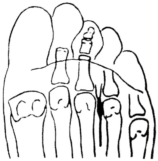
The metatarsal bones articulate proximally with bones of the tarsus, and the metatarsal heads articulate distally with the proximal phalanges. The metatarsophalangeal articulations are enlarged relative to the shaft of the metatarsals, and the interdigital nerves, which run between each two metatarsals, traverse this narrow space. The metatarsal bones are usually of different lengths so that two metatarsal heads are not in the same frontal plane. However, when two metatarsal bones are of the same length, which most frequently occurs for the third and fourth metatarsals, the space for the interdigital nerve is markedly narrowed. If, in addition, the patient wears narrow shoes, the interdigital nerve becomes entrapped between the two metatarsal heads. Continued irritation leads to an inflammatory swelling of the nerve, incorrectly called a “neuroma” (Figure 1). This further increases the force of the entrapment. The pain occurs during walking and may continue when the patient is at rest. It is relieved only when the patient takes off his or her shoes.
Figure 1.
Morton’s “neuroma,” an inflammatory swelling of the interdigital nerve between the third and fourth metatarsals.
Patients with Morton’s neuralgia seen in the author’s office frequently had received prior treatments, such as use of a metatarsal pad, injections, or surgical resection of the “neuroma.” While there may be immediate relief of pain with such methods, recurrences are common. A cortisone injection obviously has only a temporary effect. Surgical resection has a longer effect, but eventually the nerve regrows and pain recurs. The commonly used orthotic correction is a metatarsal pad, but this has only the small effect of spreading the metatarsal heads.
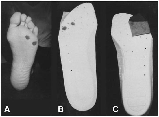
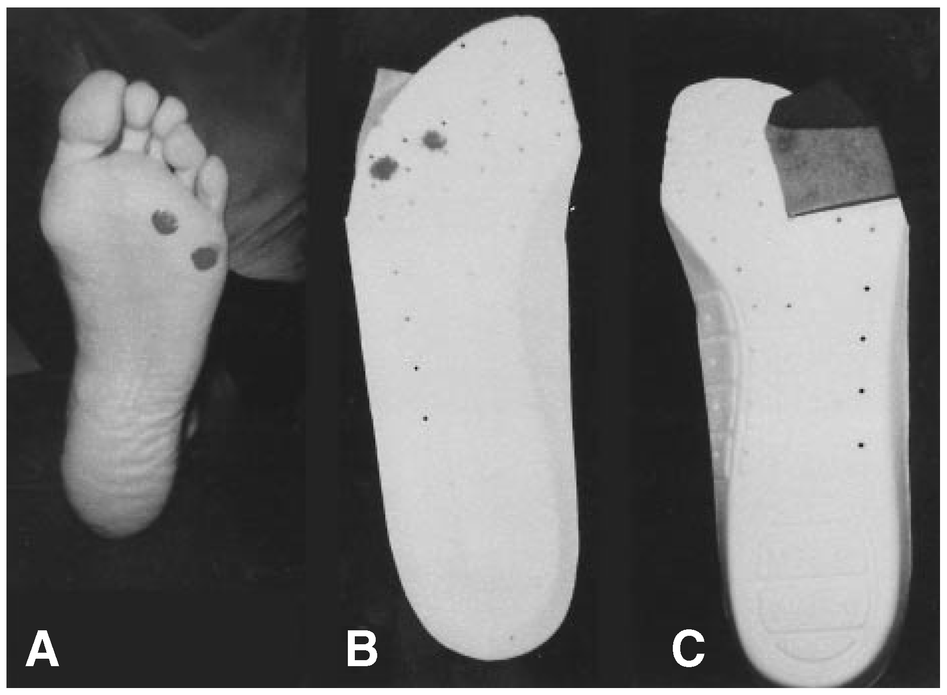
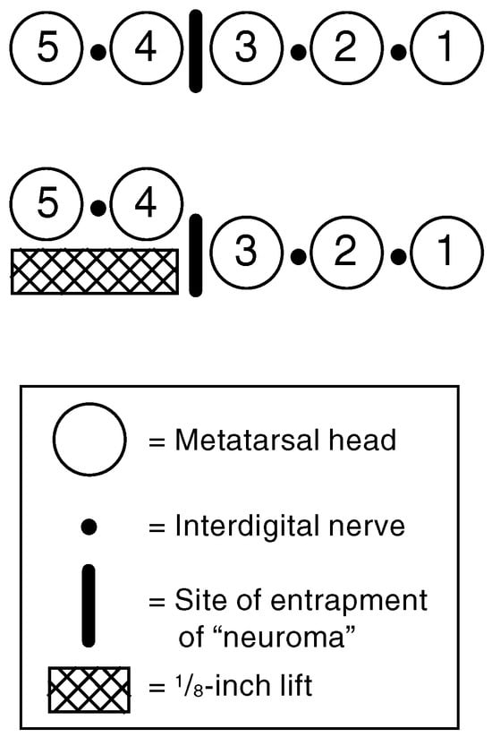
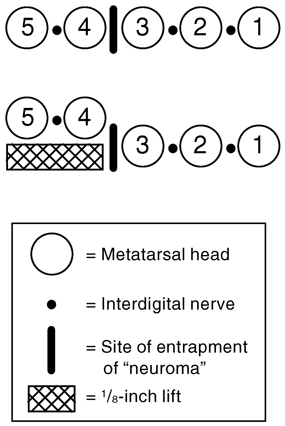
The unconventional treatment method used by the author consists of elevating the fourth and fifth metatarsal heads by approximately 1/8 inch through the application of a 1/8-inch riser to an insole. This is done by marking the patient’s fourth and fifth metatarsal heads with lipstick or ink and having the patient step on the insole, thus marking the exact location for the riser. Then a 1/8-inch pad is glued under the insole (Figure 2). If the patient wears a foot orthosis, the lift can be incorporated. When the patient stands, the weight of the body forces the third metatarsal head down, thus separating the two metatarsal heads vertically and leaving sufficient space to free the “neuroma” (Figure 3). Although this position of the metatarsal heads is not anatomically correct, it is well tolerated by all patients.
Figure 2.
Procedure of placing the 1/8-inch lift under the fourth and fifth metatarsal heads. A, Looking at the sole of the involved foot, mark the fourth and fifth metatarsal heads with lipstick or ink. B, Align the patient’s foot over an insole, and apply pressure to transfer the markings to the insole. C, Glue a 1/8-inch patch of cork, leather, or rubber on the underside of the insole directly under the markings.
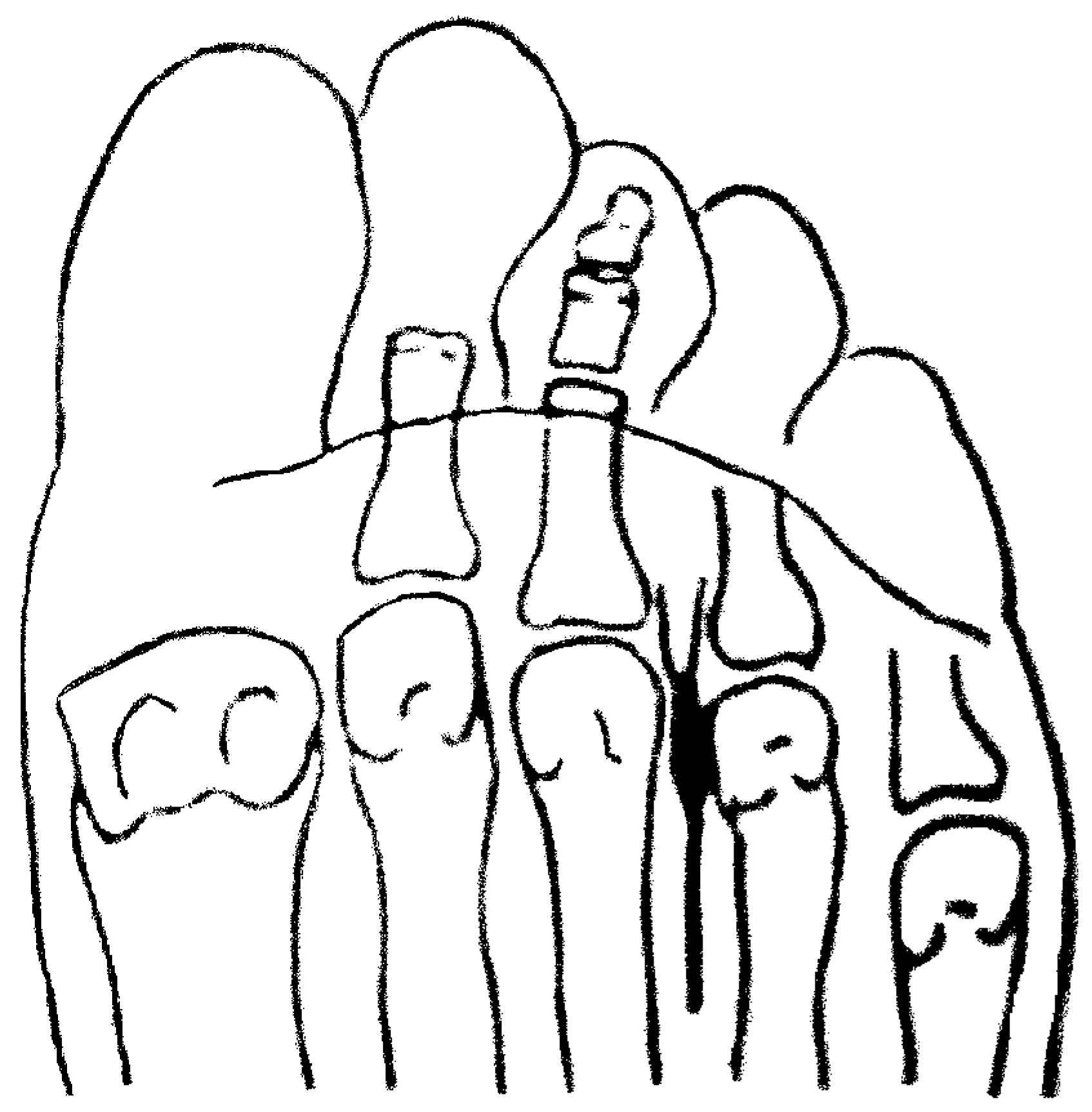
Figure 3.
Diagram showing the effect of a lift under the fourth and fifth metatarsal heads. This lift markedly increases the distance between the third and fourth metatarsal heads, thus freeing the entrapped nerve.
In most cases, the pain with weightbearing is completely and immediately relieved. In the remaining cases, there is immediate improvement, with pain disappearing completely within 2 weeks. There will be no recurrence if the patient wears this orthosis indefinitely.
© 2000 American Podiatric Medical Association