


Abstract
The authors report on a case of disseminated coccidioidomycosis of the medial cuneiform. This is a rare finding, with only a few reports of Coccidioides immitis being cultured from the bones of the foot. A brief overview of coccidioidomycosis, pertinent imaging studies, and histo-pathologic evaluation are presented.
Coccidioidomycosis is a dust-borne fungal infection caused by Coccidioides immitis. Valley fever is the lay term often used for this condition, which is in-digenous to the desert regions of the southwestern United States, notably the San Joaquin Valley of Cali-fornia.[1]
Coccidioidomycosis is contracted by inhaling the fungal spores. The lungs are the primary site of infec-tion; as in tuberculosis, however, extrapulmonary in-volvement may occur with pathologic presentation as a granuloma. Dissemination can occur anywhere in the body. Reports in the literature most commonly describe dissemination to the central nervous sys-tem,[2,3,4,5,6,7] eyes,[8,9] gastrointestinal system,[10,11,12,13,14] genitouri-nary system,[15,16] and musculoskeletal system.[17,18,19,20,21,22,23,24,25,26,27,28,29]
The presentation of coccidioidomycosis can vary greatly. It has been reported that 60% of cases are asymptomatic. The remaining 40% exhibit clinical symptoms of influenza or respiratory infection and pneumonitis.[30] Dissemination may occur in cases of the progressive form of the illness, which is estimat-ed to account for less than 0.1% of cases.[19]
Diagnosis is made by recovering the organism in tissue by means of culture, biopsy, serology, or skin testing. Amphotericin B has been used effectively to treat coccidioidomycosis; however, close monitoring is needed owing to its toxicity. The introduction of azole antifungal agents has shown promising results in treating mild-to-moderate cases. Itraconazole and fluconazole have both been shown to be effective agents.[1] Often, surgical intervention is required to help eradicate the fungal infection. For cases of os-teomyelitis, surgical curettage with bone grafting is recommended with administration of an antifungal agent.[20] According to Bried et al,[26] patients who were treated by medical and surgical means were statisti-cally proven to have less persistent infection or re-currence of infection than those treated with antifun-gal medication alone.
Coccidioidomycosis tenosynovitis has been re-ported in both the hand and the foot,[18,19,29] some-times leading to spontaneous tendon ruptures.[27] Sep-tic arthritis caused by C immitis has been well documented.[22,23,26,28] Coccidioidomycosis osteomy-elitis has been described in the skull,[17] mandible,[21] spine,[25] hand,[20], [24] and foot.[20,29] Stein et al[29] reported on a 29-year-old man with coccidioidomycosis in the tarsal bones of the foot. Bried et al[20] described a 12-month-old boy with coccidioidomycosis of the calca-neus and fifth metacarpal bone.
Case Report
A 40-year-old woman presented to the office of one of the authors (K.S.B.) in Kingman, Arizona, on May 21, 1999. Her main complaints were pain and swelling of the left foot of 3 weeks’ duration. She de-nied any traumatic event and described intermittent pain with gradual worsening. She denied any recent illness, fever, chills, or malaise. Lace-up shoes were irritating to the foot, and prolonged standing was an aggravating factor. She denied ever having this prob-lem in the past. She had not received any prior treat-ment. She was a long-time resident of the Mojave Desert region in Arizona.
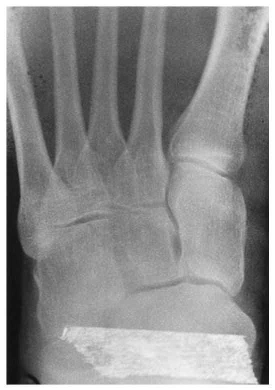
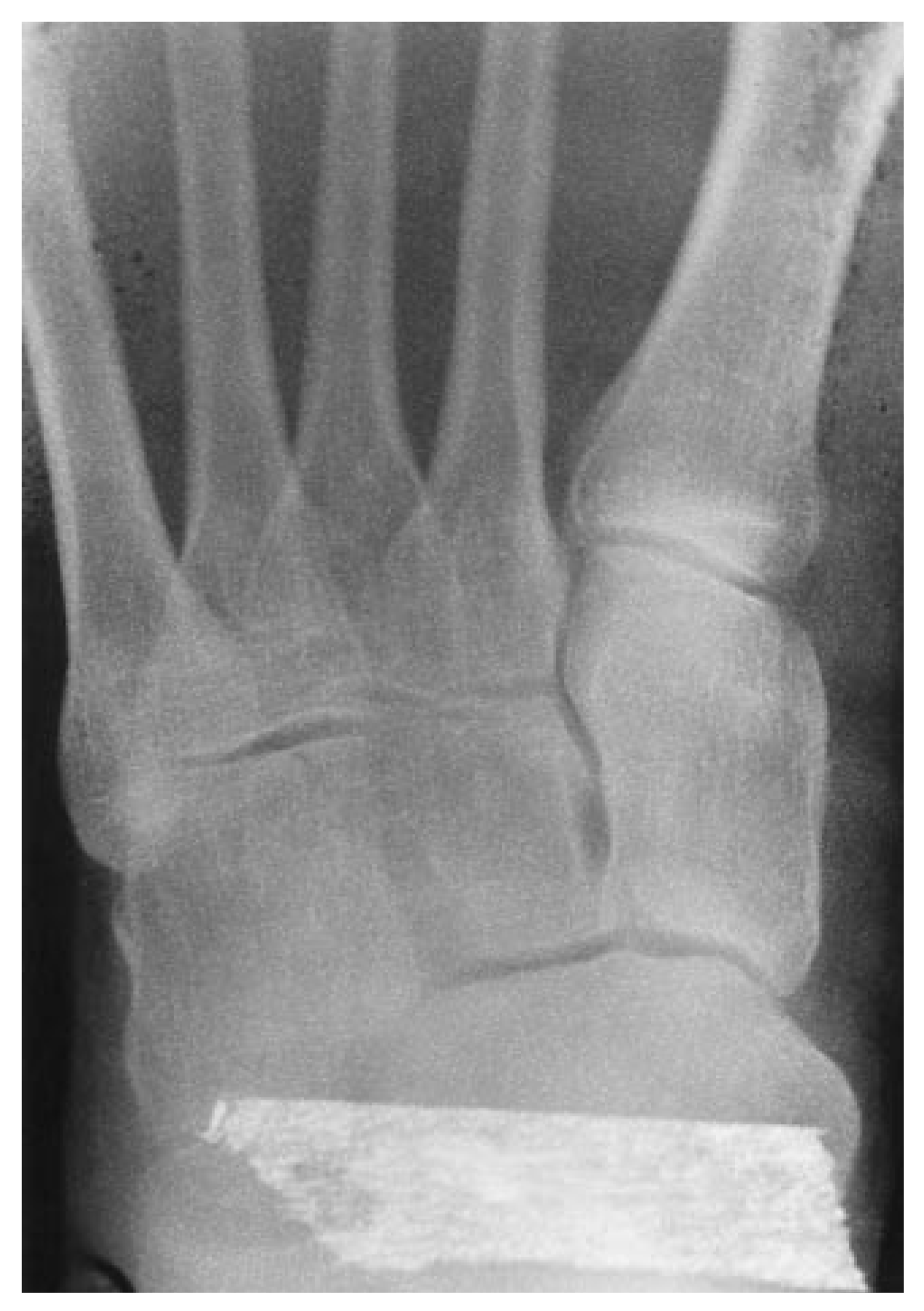
The patient’s medical history was significant for sarcoidosis, mitral valve prolapse, gastric ulcer dis-ease, and skin cancer. She was allergic to penicillin. Her medications included prednisone, 10 mg and 20 mg on alternate days; furosemide, 20 mg daily; lanso-prazole, 15 mg daily; spironolactone, 50 mg daily; and Darvocet (propoxyphene napsylate) as needed for pain. The patient was a nonsmoker and a social drinker. Examination of the left foot failed to reveal any abnormalities of the vascular system. Neurologic examination was unremarkable except for a mild tin-gling sensation with percussion of the medial dorsal cutaneous nerve over the first metatarsocuneiform joint. Dermatologic examination failed to reveal any significant edema, erythema, rash, or changes in tex-ture, temperature, or turgor. Orthopedic examination was significant for pain with palpation along the ante-rior tibial tendon at the region of the first metatarso-cuneiform joint. Plain film radiographs were taken and interpreted as normal (Figure 1). Of note was a mild dorsal prominence at the first metatarsocunei-form joint. The diagnosis at this time was tendinitis of the anterior tibial tendon with neuritis of the dor-sal cutaneous nerve. Treatment included a topical analgesic medication, the avoidance of aggravating shoes, rest, and ice application.
Figure 1.
Anteroposterior radiographic view fails to reveal any obvious pathology of the medial cunei-form.
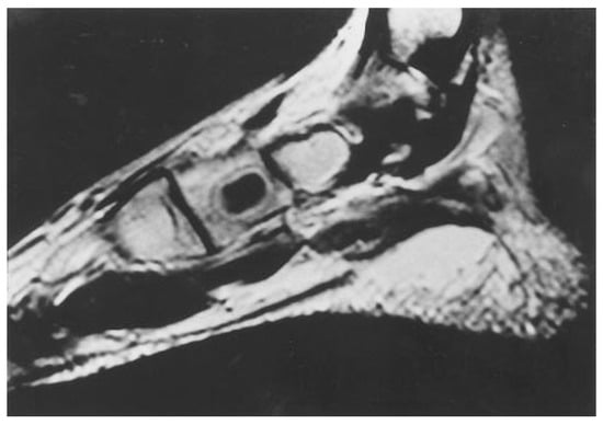
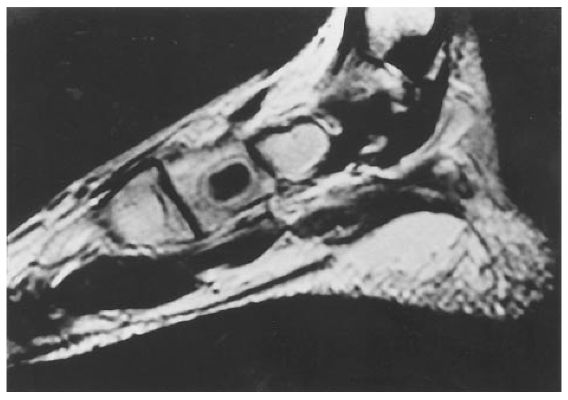
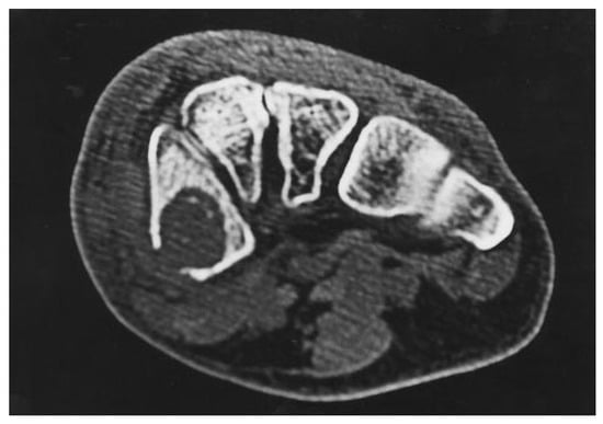
The patient was seen 2 weeks later with worsen-ing pain. Her neuritic pain was improved, but she now had much more pain in the medial arch at the navicular and cuneiform region. Palpation of the na-vicular tuberosity and over the distal portion of the posterior tibial tendon was painful. Muscle power testing showed no loss of strength to either tibial ten-don. The patient was then given a walking cast and told to return to the office in 2 weeks. On her return visit, the patient again showed no clinical improve-ment. Magnetic resonance imaging (MRI) was or-dered to inspect the tibial tendons. In the meantime, the patient was given crutches and told to keep the left foot nonweightbearing. The MR image showed a 2-cm well-defined cystic lesion in the medial cunei-form (Figure 2). At this point, the differential diagnosis included a benign solitary bone cyst, Brodie’s ab-scess, or tumor. A computed tomographic (CT) study was ordered; it revealed a 2.2 × 2.0-cm cystic lesion with breakage of the medial cortex of the medial cu-neiform (Figure 3). This study heightened the authors’ concern that there might be an aggressive inflamma-tory or neoplastic process.
Figure 2.
MR image clearly demonstrates a cystic le-sion in the medial cuneiform.
Figure 3.
CT image illustrating a cortical break in the medial cuneiform.
The patient was then scheduled for an open biopsy of the medial cuneiform. The patient’s preopera-tive laboratory tests were remarkable for a neu-trophil count of 8200/μL (reference range, 1800–7800/μL) and a lymphocyte count of 1000/μL (reference range, 1000–4800/μL). Her remaining hemogram and chemistry study results were within normal limits.
A 4-cm linear incision was placed medially over the medial cuneiform. The cortex of the medial cunei-form was eroded, and soft bone and a gel-like material were encountered. There was complete destruction of the medial cortex. The contents of the cyst were sent for Gram’s stain, culture (aerobic, anaerobic, acid-fast bacilli, and fungal), and histologic evalua-tion. A trephine sample of bone was also taken for the same culture and histologic examination.
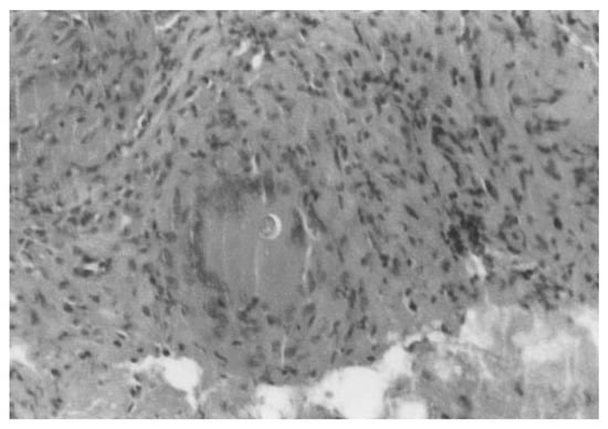
Gram’s stain failed to reveal any bacteria. Aerobic, anaerobic, and acid-fast tests were all negative. After 48 hours, light fungal elements were seen on the fun-gal culture. The specimen was then sent to the state pathology laboratory, which identified C immitis. The histologic examination revealed bone marrow with chronic granulomatous inflammation containing numerous spherules of C immitis (Figure 4).
Figure 4.
Photomicrograph demonstrates a spherule surrounded by a large granuloma (periodic acid–Schiff, ×10).
Diagnosis of disseminated coccidioidomycosis os-teomyelitis was made. Further treatment included oral therapy with fluconazole and surgical curettage of the lesion with bone grafting.
Discussion
This case exemplifies the need to obtain advanced imaging studies when a particular disease process is not responding to typical treatment. Often, initial plain film radiographs are interpreted as normal. When the clinician is looking for subtle changes on plain radiographs, underexposure or overexposure can mask bone changes. Repeating radiographs and using different positions may be of benefit. In this case, both MRI and CT were used to gather information to aid in making the correct diagnosis and for surgical planning.
The hallmark of coccidioidomycosis is tissue demonstrating thick-walled spherules measuring 10 to 80 nm in diameter with endospores clustered in-side.[31] There is very little information in the literature on treatment of disseminated coccidioidomycosis. For the pulmonary form, amphotericin B was the mainstay of treatment until the triazole compounds, which are considerably less toxic, were introduced. Both itraconazole and fluconazole are commonly used. Amphotericin B is still recommended for ex-tremely ill patients with coccidioidomycosis.
This patient has never been diagnosed with pul-monary coccidioidomycosis. She does, interestingly, have pulmonary disease caused by sarcoidosis. Sar-coidosis is a similar disease process that causes gran-ulomatous inflammation. Pulmonary symptoms may have been attributed to sarcoidosis, and as a result, the patient never received a workup for coccidioido-mycosis. This patient was probably predisposed to disseminated coccidioidomycosis owing to her im-munocompromised state from taking long-term oral prednisone.
Conclusion
The authors presented a rare case of disseminated coccidioidomycosis with localization to the medial cuneiform of the foot. Imaging studies helped un-ravel a case in which an infectious inflammatory pro-cess was mistaken for tendinitis. Tissue diagnosis was promptly made, and the patient underwent treat-ment, which included fluconazole administration and surgical curettage and bone grafting.
References
- KIRKLAND, TN; FIERER, J. Coccidioidomycosis: a reemerg-ing infectious disease. Emerg Infect Dis 1996, 2, 198. [Google Scholar]
- SCANARINI, M; ROTILIO, A; RIGOBELLO, L. ET AL: Primary intrasellar coccidioidomycosis simulating a pituitary ade-noma. Neurosurgery 1991, 28, 748. [Google Scholar] [CrossRef] [Scilit]
- HADLEY, MN; MARTIN, NA; SPETZLER, RF; et al. Multiple intracranial aneurysms due to Coccidioides immitis in-fection: case report. J Neurosurg 1987, 66, 453. [Google Scholar] [CrossRef] [Scilit]
- MEYER, PR; HUI, AN; BIDDLE, M. Coccidioides immitis meningitis with arthroconidia in cerebrospinal fluid: re-port of the first case and review of the arthroconidia literature. Hum Pathol 1982, 13, 1136. [Google Scholar] [CrossRef] [Scilit] [PubMed]
- WILLIAMS, PL; JOHNSON, R; PAPPAGIANIS, D; et al. Vasculitic and encephalitic complications associated with Coc-cidioides immitis infection of the central nervous sys-tem in humans: report of 10 cases and review. Clin Infect Dis 1992, 14, 673. [Google Scholar] [CrossRef] [Scilit]
- NINO OBERTO, S; PONCE DE LEON, A; SIERRA MADERO, J. Nino Oberto S, Ponce de Leon A, Sierra Madero. Rev Invest Clin 1999, 51, 43. [Google Scholar]
- DEWSNUP, DH; GALGIANI, JN; GRAYBILL, JR; et al. Is it ever safe to stop azole therapy for. Coccidioides immitis.
- meningitis? Ann Intern Med 1996, 124, 305.
- MAGUIRE, LJ; CAMPBELL, RJ; EDSON, RS. Coccidioidomycosis with necrotizing granulomatous conjunctivitis. Cornea 1994, 13, 539. [Google Scholar] [CrossRef] [Scilit]
- CUTLER, JE; BINDER, PS; PAUL, TO. ET AL: Metastatic coccidioidal endophthalmitis. Arch Ophthalmol 1978, 96, 689. [Google Scholar] [CrossRef] [Scilit] [PubMed]
- RAMIREZ, FC; WALKER, GJ; SANOWSKI, RA. ET AL: Obstructive jaundice due to Coccidioides immitis. Gastroin-test Endosc 1997, 43, 505. [Google Scholar] [CrossRef]
- WEISMAN, IM; MORENO, AJ; PARKER, AL; et al. Gastrointestinal dissemination of coccidioidomycosis. Am J Gastroenterol 1986, 81, 589. [Google Scholar]
- DODD, LG; NELSON, SD. Disseminated coccidioidomyco-sis detected by percutaneous liver biopsy in a liver transplant recipient. Am J Clin Pathol 1990, 93, 141. [Google Scholar] [CrossRef] [Scilit] [PubMed]
- 14. PLATT MA: Laryngeal coccidioidomycosis. JAMA 1977, 237, 1234. [CrossRef] [Scilit]
- ZANGERL, B; EDEL, G; VON MANITIUS, J. ET AL: Coccidioidomycosis as the cause of granulomatous hepatitis [in German]. Med Klin 1998, 93, 170. [Google Scholar] [CrossRef] [Scilit] [PubMed]
- KUNTZE, JR; HERMAN, MH; EVANS, SG. Genitourinary coc-cidioidomycosis. J Urol 1988, 140, 370. [Google Scholar] [CrossRef] [Scilit]
- BELLIN, HJ; BHAGAVAN, BS. Coccidioidomycosis of the prostate gland: report of a case and review of the lit-erature. Arch Pathol 1973, 96, 114. [Google Scholar] [PubMed]
- LOW, WS; SEID, AB; PRANSKY, SM. ET AL: Coccidioides immitis subperiosteal abscess of the temporal bone in a child. Arch Otolaryngol Head Neck Surg 1996, 18 122, 189. [Google Scholar] [CrossRef] [Scilit]
- REID, GD; KLINKHOFF, A; BOZEK, C. ET AL: Coccidioidomycosis tenosynovitis: case report and review of the lit-erature. J Rheumatol 1984, 11, 392. [Google Scholar]
- GROPPER, PT; PISESKY, WA; BOWEN, V. ET AL: Flexor tenosynovitis caused by Coccidioides immitis. J Hand Surg [Am] 1983, 8, 344. [Google Scholar] [CrossRef] [Scilit]
- BRIED, JM; SPEER, DP; SHEHAB, ZM. Coccidioides immi-tis osteomyelitis in a 12-month-old child. J Pediatr Or-thop 1987, 7, 328. [Google Scholar] [CrossRef] [Scilit]
- SIEBER, OF; LARTER, W; Smith, PJ. Coccidioidomycosis of the knee diagnosed by fine-needle aspiration: a case report. J Oral Surg 1977, 35, 721. [Google Scholar]
- ACREE, T; ABREO, F; BAGBY, J. Coccidioidomycosis of the knee diagnosed by fine-needle aspiration: a case report. Diagn Cytopathol 1998, 19, 110. [Google Scholar] [CrossRef] [Scilit]
- PANKOVICH, AM; JEVTIC, MM. Coccidioidal infection of the hip: a case report. J Bone Joint Surg Am 1973, 55, 1525. [Google Scholar] [CrossRef] [Scilit]
- THORPE, CD. SPJUT HJ: Coccidioidal osteomyelitis in a child’s finger: a case report. J Bone Joint Surg Am 1985, 67, 330. [Google Scholar] [CrossRef] [Scilit]
- WESSELIUS, LF; BROOKS, RJ; GALL, EP. Vertebral coccidi-oidomycosis presenting as Pott’s disease. JAMA 1977, 238, 1397. [Google Scholar] [CrossRef] [Scilit] [PubMed]
- BRIED, JM; BENJAMIN, JB; GALGIANI, JN. Coccidioides immitis: an unusual presentation. Orthopedics 1990, 13, 345. [Google Scholar]
- SZABO, RM; LANZER, WL; GELBERMAN, RH; et al. Extensor tendon rupture due to Coccidioides immitis: report of a case. Clin Orthop 1985, 194, 176. [Google Scholar] [CrossRef] [Scilit]
- LANTZ, B; SELAKOVICH, WG; COLLINS, DN. ET AL: Coccidioidomycosis of the knee with a 26-year follow-up evalu-ation: a case report. Clin Orthop 1988, 234, 183. [Google Scholar] [CrossRef] [Scilit]
- STEIN, SR; LEUKENS, CA; BAGG, RJ. Treatment of coccidi-oidomycosis infection of bone with local amphotericin B suction-irrigation: report of a case. Clin Orthop 1975, 108, 161. [Google Scholar] [CrossRef] [Scilit]
- GALES M, MCCLAIN PHILLIPS C: Coccidioidomycosis: a mycotic infection on the rise. Clinician Reviews 1997, 7, 71.
- ROBBINS, SL. “Infectious Diseases,” in Pathologic Basis of Disease,; Robbins, SL, Ed.; WB Saunders: Philadelphia, 1974; p. 446. [Google Scholar]
Disclaimer/Publisher’s Note: The statements, opinions and data contained in all publications are solely those of the individual author(s) and contributor(s) and not of MDPI and/or the editor(s). MDPI and/or the editor(s) disclaim responsibility for any injury to people or property resulting from any ideas, methods, instructions or products referred to in the content. |
© 2000 American Podiatric Medical Association