


Abstract
Background: Models using a double-twist tension band, two partially threaded cancellous screws, a single-twist tension band, and a single-screw tension band were biomechanically compared for fixation of medial malleolus fractures. Methods: All 72 composite cortical tibial bones used in the study were osteotomy suitable for medial malleolus fracture. They were divided into four groups: double-twist tension band (group 1), two partially threaded cancellous screws (group 2), single-twist tension band (group 3), and single-screw tension band (group 4), with 18 composite tibial bones in each group. Bones in each group were divided into three subgroups and subjected to biomechanical tests by applying force as tension, transverse, and axial loading. Results: In the tension and transverse loading tests, the highest mean stiffness was in groups 1 and 4. Group 4 had statistically significantly higher stiffness values than groups 2 and 3. All of the other groups had statistically significantly higher stiffness values than group 2. A statistically significant difference was found comparing group 4 (with the highest value in the axial loading test) with the other groups. No significant difference was found between groups 1 and 3 in any of the biomechanical tests. Conclusions: Tension band method did not make a difference biomechanically when applied as a double or single twist. Single-screw tension band was found to be the most stable fixation method in tension, transverse, and axial loading tests. We think that the single-screw tension band method is a stable and suitable fixation method and should find more place in current surgical practices.
The ankle is a joint in which the talus body is bounded by the medial malleolus, tibial plafond, and distal fibula, and stability is provided by the very strong ligaments between these bones. The upper part of the body of the talus is in full harmony with the tibial plafond, and as a result of this harmony, which is especially impaired in fractures around the ankle, it reduces the joint contact surface by 42%, even with a 1-mm displacement of the talus. [1] Therefore, medial malleolar fractures, whether isolated or not, are very important for the integrity and function of ankle joint stability and should be treated appropriately.
Many methods have been described in the literature for the fixation of medial malleolus fractures. In deciding by which method the medial malleolar fracture will be detected, the orientation of the fracture line, the size of the fractured fragment, and the age of the patient are effective. [2] In 1945, Müller recommended surgical fixation of all medial malleolar fractures, and today, surgical treatment is recommended for medial malleolar fractures, except for isolated fractures where the fracture line does not reach the tibial plafond. [3] Unstable ankle circumference fractures, including medial malleolus fractures, generally require surgical fixation. [4–9] Many methods, including bioabsorbable implants, have been used for surgical fixation in the treatment of medial malleolar fractures. [10] However, the fixation method with two 4-mm partially threaded cancellous screws placed perpendicular to the fracture line, recommended by the AO ASIF, is the most widely used technique. In general, according to the recommendation by the AO, standard conventional techniques for medial malleolar fracture fixation are two 4.0-mm partially threaded cancellous screws for malleolus fractures with large fragments, and a tension band with Kirschner wire and stainless steel wire for fractures with smaller fragments. [8]
Almost all of the studies in the literature on tension band application used in current treatment have used the tension band method with a double twist. In the literature, generally double twist and tension band are recommended, but we see that there is no study showing that there is a difference in terms of biomechanical stability between single twists. On the other hand, we aim to biomechanically compare the effect of tension band application with cancellous screws instead of Kirschner wires used in standard application on fracture stability.
Materials and Methods
Study Design
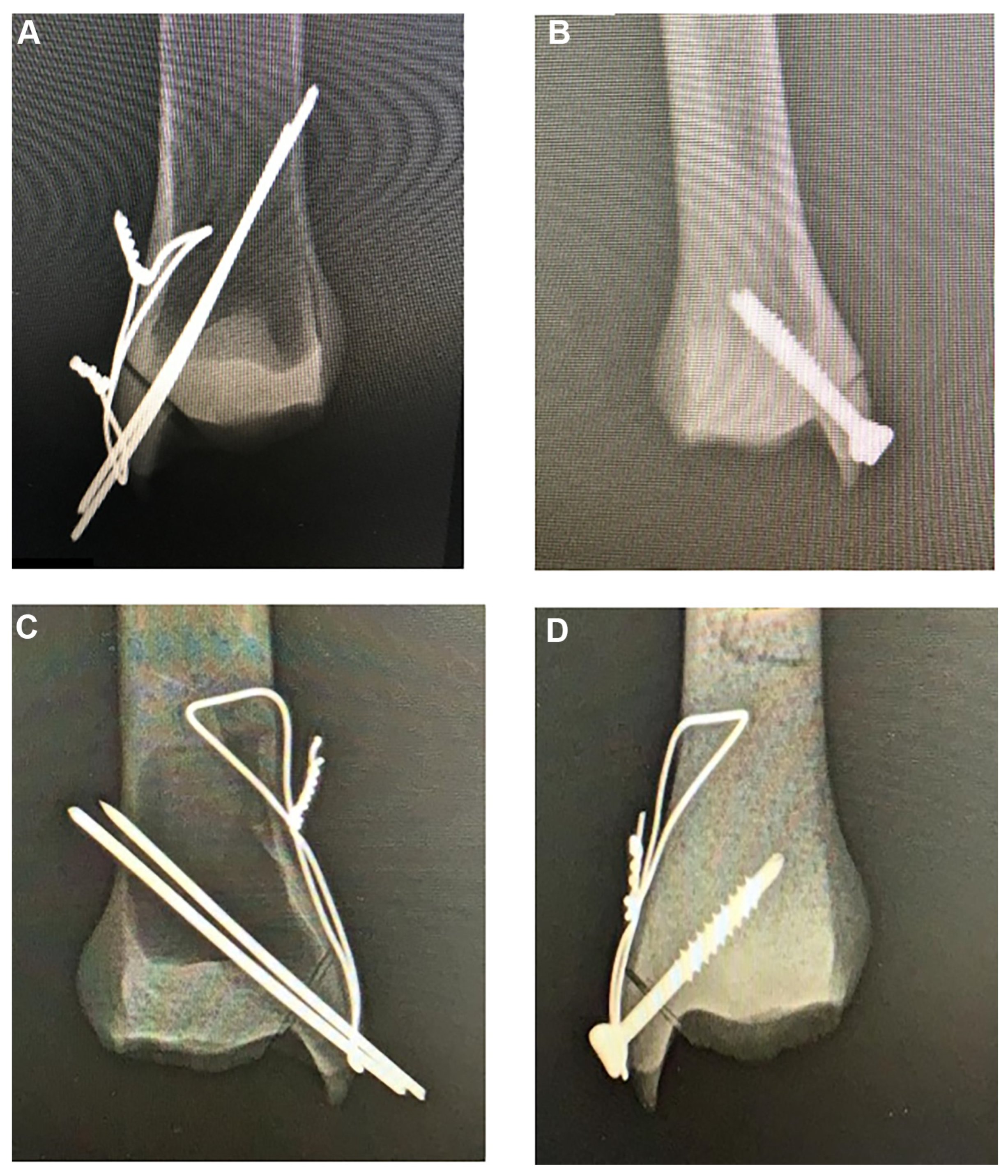
The 72 fourth-generation composite tibial bones (Selbones; Sel Technique, Kayseri, Turkey) prepared for this study were divided into four equal groups. A fracture model was created by performing oblique osteotomy–like AO type 44-B2.2 fractures on all tibial composite bones at the medial malleolus zone. [11] The osteotomies were performed by a single surgeon (G.N.) using a micro sagittal saw. Fixation was provided to the fracture line in four different constructs according to the groups. In group 1, double-twist tension band, the fracture line was fixed with a tension band by double twisting wire using 18-gauge stainless steel wire and 2-mm Kirschner wires (Fig. 1A). In group 2, two cancellous screws, the fracture line was fixed with two 4.0-mm partially threaded cancellous screws (AO recommendation) (Fig. 1B). In group 3, single-twist tension band, the fracture line was fixed with a tension band by single twisting wire using 18-gauge stainless steel wire and 2-mm Kirschner wires (Fig. 1C). In group 4, single-screw tension band, the fracture line was fixed with a tension band by using a 4.0-mm partially threaded cancellous screw and a 2.0-mm stainless steel wire (Fig. 1D).
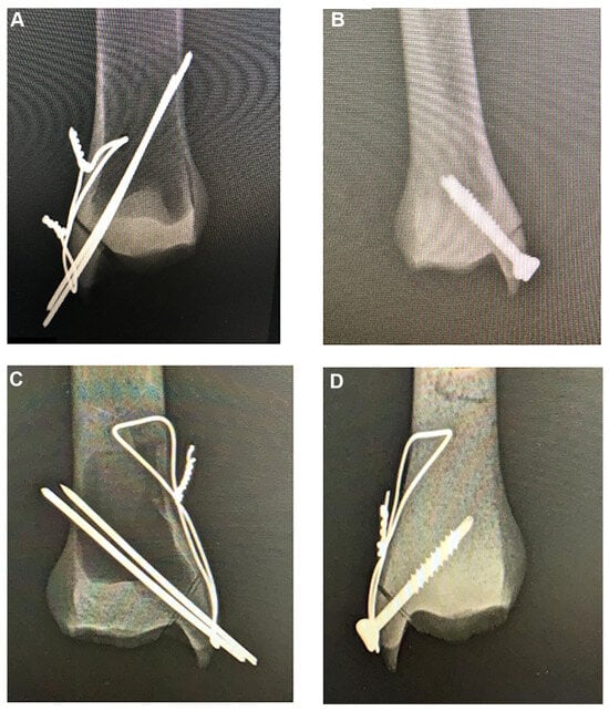
Figure 1.
Anteroposterior radiographs of composite tibial bones demonstrating fixation with a double-twist tension band (A), two cancellous screws (B), a single-twist tension band (C), and a single-screw tension band (D).
Anteroposterior and lateral radiographs were obtained in all of the fixation models. To facilitate fixing of the bones during the experiments, the proximal sides of the bones were embedded in a solid block of thermoset material.
All of the groups were tested in three scenarios, and all of the scenarios were performed using an electromechanical universal testing device (TCS Machinery, Ankara, Turkey) with a mean ± SD 20.0 ± 0.25 kN of tension/compression load cell and a displacement-controlled rate of 20 mm/min. In the tensile, offset axial, and offset transfer loading scenarios, the test was terminated when the test failed, 150 N of force was reached, and 110 N of force was reached, respectively. The stiffness of each construct was calculated from the curve slope of the force-displacement graph. The maximum displacement or force was determined according to the specified criteria.
First, the tensile test was performed based on the study by Fowler et al,[11] similar to the study by Uygur et al. [12] While tensioning with a metal cable, attention was given to the use of pins to ensure equal stress distribution of the force on the bone. The artificial tibia bone was placed upside down in the universal test device by fixing it medially with custom-made fixation plates. Considering the direction of the deltoid ligament, the bones were positioned so that they did not overlap with the loading axis. The experiments were continued until the samples failed.
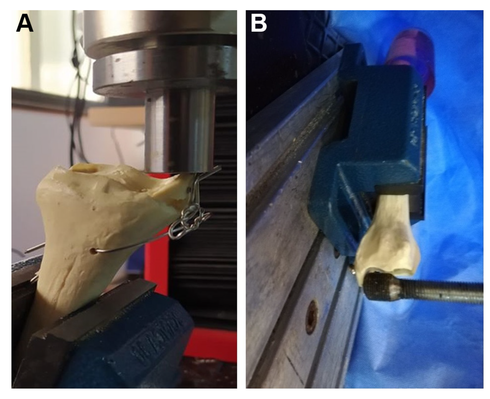
Offset axial and transverse loading was performed according to the study by Dumigan et al. [13] A clamp was used to hold the specimens. For both offset axial and cantilever loading on the fractured small bone part, a clamp was mounted to the testing machine, and bones were tilted approximately 17° from the anatomical axis of bone for offset axial loading. In this scenario, the tests were terminated by reaching a force of 150 N. For the cantilever bending test, the samples were positioned in a vice so that the long axis of the bones overlapped with the horizontal axis and then rotated 30° outward along the anatomical axis. The loading head of the device for both loading conditions was positioned over the broken small bone fragment (Fig. 2).
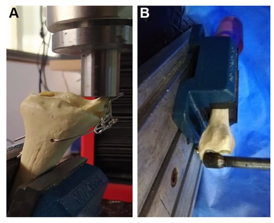
Figure 2.
Photographs showing the axial loading (A) and transverse loading (B) mechanisms.
Statistical Analyses
The data obtained in this study were analyzed with IBM SPSS Statistics for Windows, Version 22.0 (IBM Corp, Armonk, New York). The normality of the distribution for the variables was tested with the Shapiro-Wilk normality test. Nonparametric tests were used for variables without normal distribution. Continuous data without normal distribution were analyzed by Mann-Whitney U and Kruskal-Wallis tests. Quantitative data are expressed as mean ± SD, minimum and maximum values. The confidence interval was 95%, and P < .05 was considered statistically significant.
Results
In the tension testing, there were significant differences between the groups in terms of stiffness (P = .002) and maximum load (P = .001) according to the Kruskal-Wallis test. Analysis of the data from the offset tension testing model demonstrated that groups 1 and 4 exhibited superior stiffness to all of the other constructs tested. Group 4 specimens displayed mean stiffness of 70.1 N/mm. The remaining specimens in descending order from most to least stiffness were group 1 (60.7 N/mm), group 3 (47.2 N/mm), and group 2 (37.5 N/mm) (Table 1 and Fig. 3A). There was a statistically significant difference between group 4 and groups 2 and 3 in terms of stiffness, but no statistically significant difference was found between groups 1 and 4 (Table 2). No statistically significant difference was found between groups 1 and 3. Analysis of the data from the offset tension testing model demonstrated that group 4 exhibited superior maximum load to all of the other constructs tested. Group 4 specimens displayed mean stiffness of 720.2 N/mm. The remaining specimens in descending order from most to least maximum load were group 1 (530.8 N/mm), group 3 (497.2 N/mm), and group 2 (349.2 N/mm) (Table 1 and Fig. 3B). Group 4 had a statistically significantly higher mean maximum load than all of the other fixation methods. Although it was seen that group 1 had a statistically significantly higher maximum load value than group 2, no significant difference was found between groups 1 and 3 (Table 2).

Table 1.
Forced to Displacement Failure of the Groups in Tension Loading Conditions, and Stiffness in Different Loading Conditions

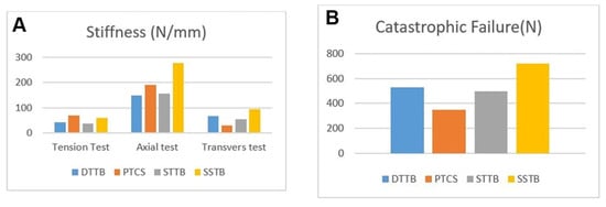
Figure 3.
A, Stiffness values of the fixation methods in tension, axial, and transverse loading. B, Maximum load values of the fixation methods in tension loading. DTTB indicates double-twist tension band; PTCS, partial thread compression screw; SSTB, single-screw tension band; STTB, single-twist tension band.
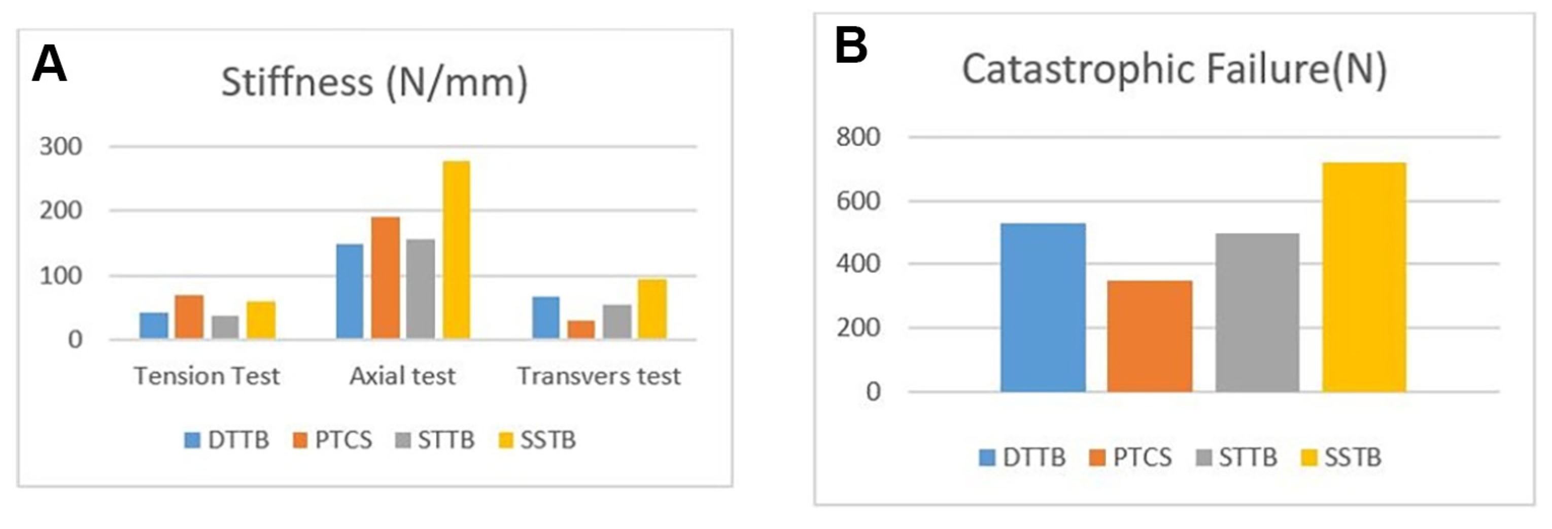
Table 2.
P Values for Groups
In the transverse testing, there were significant differences between the groups in terms of stiffness (P < .0001) according to the Kruskal-Wallis test. The offset transverse loading model produced a slightly different picture, with group 4 being the stiffest construct (93.52 N/mm). The remaining specimens in descending order from most to least stiff were group 1 (64.5 N/mm), group 3 (56.9 N/mm), and group 2 (37.3 N/mm) (Table 1 and Fig. 3A). In transverse loading, group 4 showed significantly higher overall stiffness than the other groups (P < .05) (Table 2). Again, the originally published technique using two 4-mm partially threaded cancellous screws ranked lowest in stiffness. All of the groups had statistically significantly higher stiffness than group 2.
In the axial testing, there were significant differences between the groups in terms of stiffness (P = .003) according to the Kruskal-Wallis test. Analysis of the data from the offset axial testing model demonstrated that group 4 exhibited superior stiffness to all other constructs tested. Group 4 specimens displayed mean stiffness of 278.1 N/mm. The remaining specimens in descending order from most to least stiff were group 2 (191.56 N/mm), group 1 (152.58 N/mm), and group 3 (149.17 N/mm) (Table 1 and Fig. 3A). In axial loading, group 4 showed significantly higher overall stiffness than the other groups (P < .05) (Table 2).
Discussion
In the surgery of medial malleolar fractures, the stability of the fixation is very important for the compatibility of the tibiotalar joint. For this reason, it is important that the fixation method applied is biomechanically rigid and stable in terms of allowing movement and weightbearing to the ankle in the earlier period after surgery.
Two parallel 3.5- or 4.5-mm screws in medial malleolus fractures are used for the treatment. However, in some cases, medial malleolar fractures may not be suitable for the placement of two screws. If the fragment to be fixed is not large enough, the use of a screw and a Kirschner wire (rotational stability) is also a method that can be applied. [14] The use of Kirschner wires causes problems such as soreness over the implant and scar, reduced movement of the ankle joint, and activity-related pain and requires implant removal. [15],[16] In the present study, instead of using Kirschner wires in traditional treatment, we used stainless steel wire over one cancellous screw sent perpendicular to the fracture line. We thought that this application is a method that can be used in cases where at least one screw can be applied in medial malleolar fractures, and it will provide less implant irritation and more stability. On the other hand, DeMill et al[17] reported that fixation screws applied from the middle and posterior regions of the medial malleolus (zones 2 and 3) increase the risk of posterior tibial tendon irritation and rupture. We think that the use of a single screw instead of two screws and the use of zone 1 will reduce this risk with our fixation method. Considering the studies in the literature, we have seen that there is no biomechanical and clinical study similar to the fixation technique we performed. The present study showed that the single-screw tension method is suitable and stable.
During screw fixation, the Weber cerclage technique over two Kirschner wires has been the preferred fixation method in cases where the fractured fragment is thought to be small for screw fixation and may pose a risk of fragmentation in the fractured fragment. This method, which is known as tension tape and is used frequently, has many different techniques in terms of application. In the tension band technique, the stainless steel wire is applied over the Kirschner wires as standard. [18] As a standard, literature and treatment guidelines recommend using the tension band method as a double twist. [19–21] There is no comparison made about the effect of applying the tension band method as a double twist or a single twist on the stability and safety of the method in medial malleolar fractures. In a biomechanical study of tension band application in patella fractures, the authors showed that contrary to what is known, the application of tension tape as a single twist does not differ from the application of double twists. [22] The authors also showed that the tension band method applied as a single twist did not cause any deterioration in fixation strength. In another study on this subject, although it is widely accepted that the double-twist technique helps the surgeon better control both wire lengths, previous studies have shown that it leads to a power loss of approximately 25%. [23] In another study, it was reported that compression between fracture fragments increased in the double-twist technique. [24] In the present study, we found that there is no biomechanical difference between the single-twist or double-twist application of the tension band method, similar to the study by Ali et al. [22] For this reason, we think that applying the tension band method as a double or single twist does not have many advantages over each other.
In the transverse and axial loading tests performed under a constant load, group 4 had the highest stiffness values and the least implant failure. These results suggest that the single-screw tension band fixation method is the most appropriate for resisting transverse loading. In the evaluation made in terms of stiffness in the tension loading test, it was concluded that groups 4 and 2 were biomechanically superior to groups 1 and 3. When the catastrophic failure values were compared, group 4 was found to be superior. Although there was no difference between groups 4 and 2 in terms of stiffness, the statistically significantly higher catastrophic failure value made us think that the single-screw tension band method is a more stable fixation method when higher tension forces are involved. We think that this fixation method is an alternative method that can be used in cases where the fracture fragment in the medial malleolus allows single-screw fixation. On the other hand, considering that medial malleolar fractures are subjected to tension loading, especially under the pull and effect of the deltoid ligament, we suggest using the single-screw tension band method, which is not defined as a standard treatment method in the literature but as an alternative method.
This study has several limitations. First, this study was performed on synthetic bone models. Therefore, the results of this study cannot be generalized to in vivo situations and should be supported by clinical studies. Second, no comparison could be made with bicortical screw application, which is considered more stable biomechanically than standard treatments in the study. Third, irritation from stainless steel wire use may be greater in larger gauges. This study can be performed again in different gauge wires. Finally, we were not able to directly compare the results because there are no similar studies with the single-screw tension band. On the other hand, being the first biomechanical study on this subject in the literature is the strength of the study.
Conclusions
It has been observed that the tension band method, which is one of the classical fixation methods used in current surgical practice, does not make a biomechanical difference when applied as a double auger or a single auger. For this reason, we think that these applications do not have superiority over each other in application of the tension band method. We think that the single-screw tension band method, which is the other subject of this study and is not among the classic methods in the current treatment guidelines, is a stable and appropriate fixation method; therefore, it should find more place in current surgical practices.
Financial Disclosure: This work was supported by the Scientific Research Coordination Unit of Çanakkale Onsekiz Mart University (project TSA-2020-3173).
Conflict of Interest: None reported.
References
- Ramsey PL, Hamilton W: Changes in tibiotalar area of contact caused by lateral talar shift. J Bone Joint Surg Am 58: 356, 1976.
- King CM, Cobb M, Collman DR. et al: Bicortical fixation of medial malleolar fractures: a review of 23 cases at risk for complicated bone healing. J Foot Ankle Surg 51: 39, 2012.
- Muller GM: Fractures of internal malleoulus. BMJ 2: 320, 1945.
- Femino JE, Gruber BF, Karunakar MA: Safe zone for the placement of medial malleolar screws. J Bone Joint Surg Am 89: 133, 2007.
- Georgiadis GM, White DB: Modified tension band wiring of medial malleolar ankle fractures. Foot Ankle Int 16: 64, 1995.
- Johnson BA, Fallat LM: Comparison of tension band wire and cancellous bone screw fixation for medial malleolar fractures. J Foot Ankle Surg 36: 284, 1997.
- Kanakis TE, Papadakis E, Orfanos A, et al: Figure eight tension band in the treatment of fractures and pseudarthroses of the medial malleolus. Injury 21: 393, 1990.
- Weber BG, Colton C: “Malleolar Fractures,” in Manual of Internal Fixation: Techniques Recommended by the AO-ASIF Group, 3rd Ed, ed by ME Mueller, M Allgower, R Schneider, et al, p 595, Springer-Verlag, New York, 1991.
- Perren SM, Frigg R, Hehli M, et al: “Lag Screw,” in AO Principles of Fracture Management, ed by CL Colton, A Fernandez Dell’Oca, U Holz, p 157, Thieme, Stuttgart, Germany, 2000.
- Bucholz RW, Henry S, Henley MB: Fixation with bioabsorbable screws for the treatment of fractures of the ankle. J Bone Joint Surg Am 76: 319, 1994.
- Fowler TT, Pugh KJ, Litsky AS, et al: Medial malleolar fractures: a biomechanical study of fixation techniques. Orthopedics 34: e349, 2011.
- Uygur E, Poyanli O, Mutlu İ, et al: Medial malleolus fractures: a biomechanical comparison of tension band wiring fixation methods. Orthop Traumatol Surg Res 104: 1259, 2018.
- Dumigan RM, Bronson DG, Early JS: Analysis of fixation methods for vertical shear fractures of the medial malleolus. J Orthop Trauma 20: 687, 2006.
- White TO, Bugler KE: “Ankle Fractures,” in Rockwood and Green’s Fractures in Adults, 8th Ed, ed by CM Court-Brown, JD Heckman, MM Mc Queen, et al, p 2542, Wolters Kluwer, New York, 2015.
- Wild M, Fischer K, Hilsenbeck F, et al: Treating patella fractures with a fixed-angle patella plate: a prospective observational study. Injury 47: 1737, 2016.
- Jacobsen S, Lichtenberg MHD, Jensen CM, et al: Removal of internal fixation: the effect on patients’ complaints: a study of 66 cases of removal of internal fixation after malleolar fractures. Foot Ankle Int 15: 170, 1994.
- DeMill SL, Bussewitz BW, Philbin TM: Injury to the posterior tibial tendon after open reduction internal fixation of the medial malleolus. Foot Ankle Specialist 8: 360, 2015.
- Hahn DM, Colton CL: “Malleolar Fractures,” in AO Principles of Fracture Management, edited by TP Rüedi, WM Murphy, CL Colton, et al, p 575, Thieme, Stuttgart, Germany, 2000.
- Shayaa S: Comparative study of operative fixation of fracture of medial malleolus by malleolar screw versus tension band wiring. Int J Surg 4: 157, 2020.
- Georgiadis GM, White DB: Modified tension band wiring of medial malleolar ankle fractures. Foot Ankle Int 16: 64, 1995.
- Clyde J, Kosmopoulos V, Carpenter B: A biomechanical investigation of a knotless tension band in medial malleolar fracture models in composite Sawbones®. J Foot Ankle Surg 52: 192, 2013.
- Ali M, Kuiper J, John J: Biomechanical analysis of tension band wiring (TBW) of transverse fractures of patella. Chinese J Traumatol 19: 255, 2016.
- Marsh JL, Slongo TF, Agel J, et al: Fracture and dislocation classification compendium-2007: Orthopaedic Trauma Association Classification, Database and Outcomes Committee. J Orthop Trauma 21(suppl): S1, 2007.
- Loveday DT, Arthur A, Tytherleigh-Strong GM: Technical tip: fixation of medial malleolar fractures using a suture anchor. Foot Ankle Int 30: 68, 2009.
© 2025 American Podiatric Medical Association