


Abstract
We present a case report of revision talonavicular joint arthrodesis following painful nonunion. Following the removal of the previously united subtalar joint arthrodesis hardware, the posterior screw tunnel was used to access the internal aspect of the calcaneus for bone and marrow harvesting avoiding a second graft site incision. A suction curettage system was then used to acquire adequate autologous cancellous bone graft and nondiluted bone marrow. The talonavicular joint was then prepared for revision arthrodesis, followed by augmentation with the autologous cancellous bone graft and rigid internal fixation placed across the joint for compression. There were no complications noted at the donor site or the arthrodesis site immediately following surgery or during follow-up. Radiographs obtained 4 months postoperatively demonstrate osseous union at the talonavicular joint and near complete consolidation at the calcaneal donor site. The patient is currently walking pain-free in a walking boot and will be continually followed to allow for long-term outcome measures.
The talonavicular joint (TNJ) is one of the three joints that comprise the subtalar complex, and thus, it plays an important role in the foot’s overall function. Loss of this joint severely restricts subtalar motion as well as forefoot to rearfoot alignment [1]. Fusion of this joint may be necessary and is a suitable treatment option in cases of post-traumatic arthrosis, rheumatoid arthritis, degenerative joint disease, severe deformity, or idiopathic etiologies.
With any arthrodesis procedure, the surgeon must consider the possibility of nonunion. Although there are a multitude of factors that may lead to a clinical or radiographic nonunion, such as joint preparation technique, joint alignment, and surgical fixation, this concern remains relevant with nonunion rates of up to 37% reported in the literature with regard to medial column arthrodesis [2]. Nonunion can also be related to infection, tobacco usage, medical comorbidities, metabolic disorders, chronic immunosuppression, and patient noncompliance.
In many cases of revisional arthrodesis, the surgeon may use either autologous or nonautologous bone grafts to aid in facilitating osseous union. Nonautologous options may include but are not limited to orthobiologics, allografts, bone morphogenic protein, and demineralized bone matrix. For lower-extremity surgery, the surgeon may harvest autologous bone from sites such as the calcaneus, distal or proximal tibia, or iliac crest [3]. Compared to traditional harvest techniques, the novel vacuum-assisted suction curettage system may allow acquisition of both autologous cancellous bone/bone marrow in a timely manner through a minimal incisional approach.
Case Presentation
A 63-year-old male with a past medical history of well-controlled type 2 diabetes mellitus, hyperlipidemia, hypertension, and deep venous thrombosis presented with a complaint of continued stiffness and pain to his right foot after being treated for a workers’ compensation injury. He previously had a right foot subtalar joint (STJ) and TNJ arthrodesis with a peroneus brevis tendon debridement and partial excision in 2020 by an outside provider. He was placed in a tall controlled ankle movement (CAM) boot postoperatively and completed physical therapy. He used an external bone stimulator for approximately 5 months and wore custom orthotics. He stated that his pain was rated at a 9 out of 10 and was recurrent on a daily basis. He had pain with walking short distances and had issues with walking up and down stairs. He had both hips replaced many years ago and was starting to have recurrent pain in his right hip.
Physical examination revealed restricted right metatarsophalangeal joint, ankle, and STJ motion. There was a well-healed incision along the medial and lateral column of the right foot. There was severe loss of the medial and lateral longitudinal arches, residual skew foot deformity on the right, and pain directly on the palpation along the medial TNJ and lateral STJ of the right foot. There was medial translocation of the forefoot, in comparison to the midfoot and hindfoot on the right, along with pain on midfoot range of motion. He walked with a slightly propulsive gait and had a weakened single-stance toe raise on the right foot. The remaining vascular, neurologic, and dermatologic exams were within normal limits.
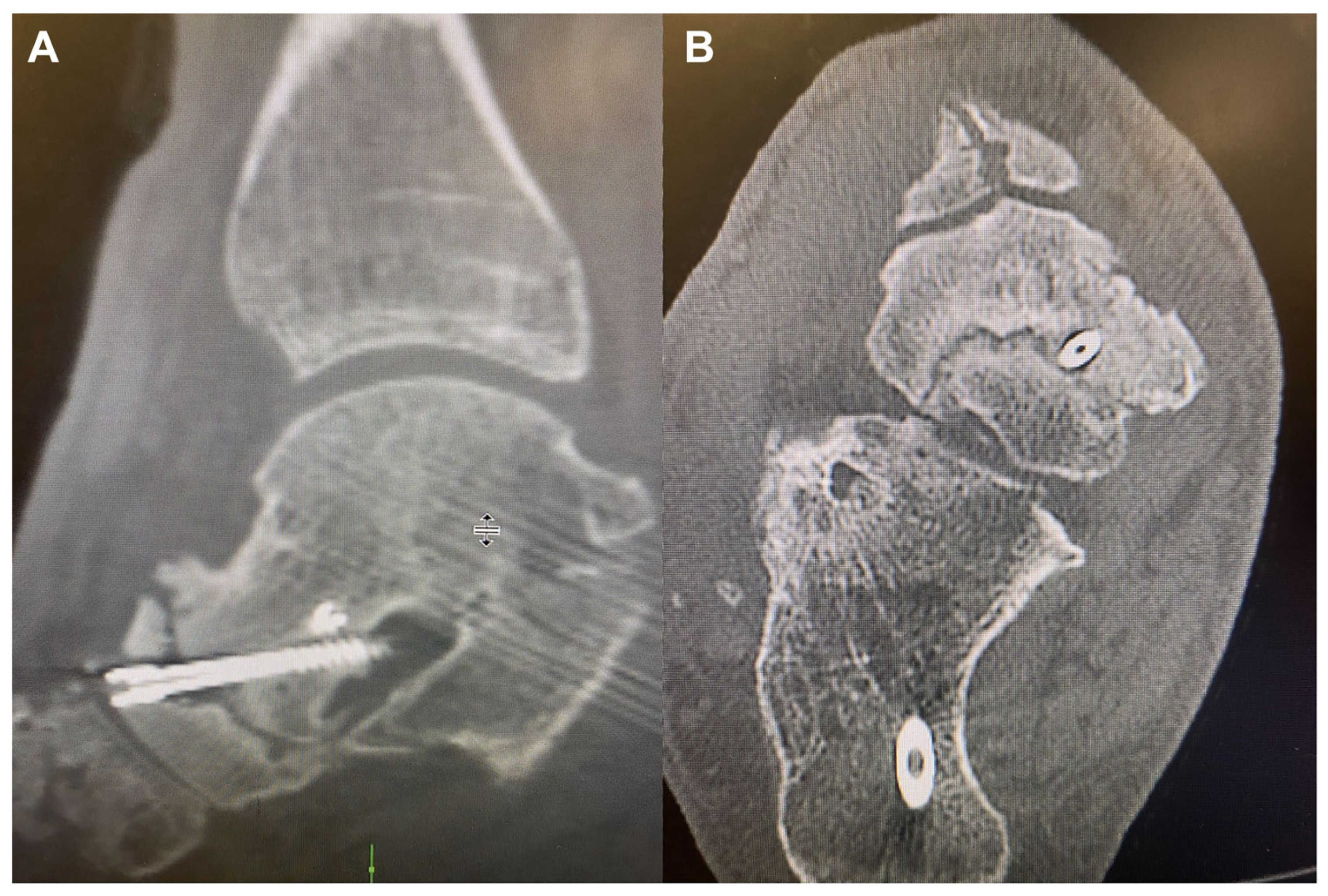
Radiographs of the right foot noted a previous arthrodesis of the STJ and TNJ with a two-screw fixation. There was a slightly persistent lucency noted in the STJ and transarticular trabeculation across the TNJ. A computed tomography (CT) scan was then obtained of the right hindfoot and ankle, noting irregularity and thinning of the TNJ without adequate bone bridging present (Figure 1). Two threaded pins were visualized crossing the TNJ with lucency measuring up to 1.5 mm adjacent to the dorsal aspect of the lateral pin, which extended into the sinus tarsi at its most dorsal aspect. The CT also noted solid fusion at the posterior STJ with two pins crossing the posterior STJ.
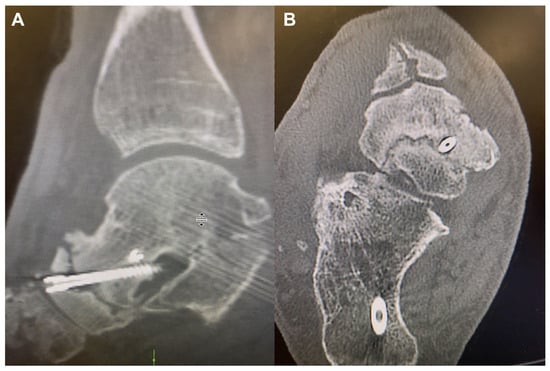
Figure 1.
Preoperative sagittal (A) and coronal (B) computed tomographic views showing complete nonunion at the talonavicular joint.
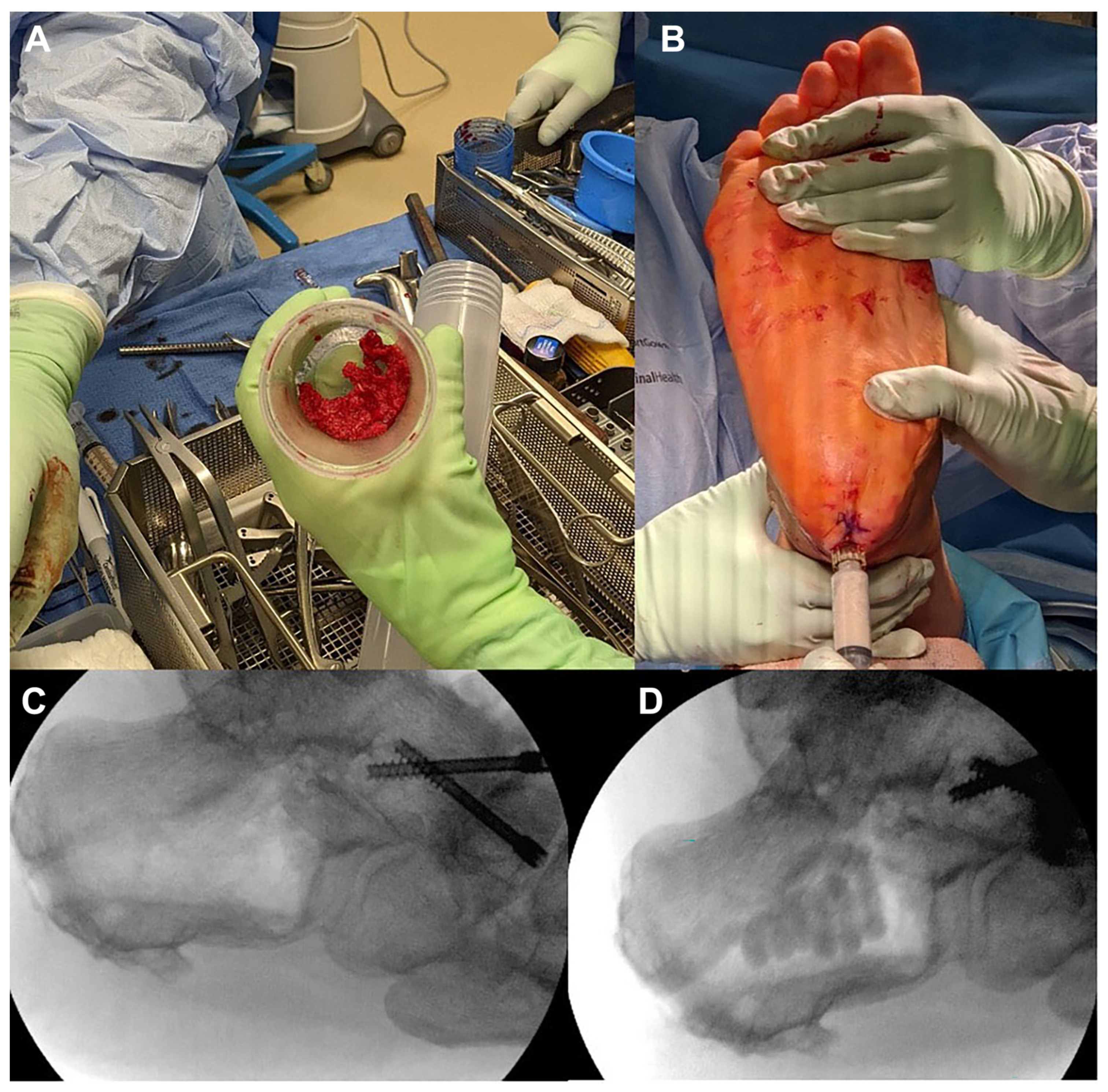
Due to the extent of the patient’s pain and degenerative joint disease, the decision was made to perform a revisional TNJ arthrodesis with autologous bone grafting and fixation with peroneal tendon excision of the right foot. In the operating room, attention was first directed toward the posterior calcaneus under fluoroscopic guidance to locate and remove two 7.0-mm headless cannulated partially threaded screws traversing the STJ. Once both screws were removed, an Avitus Pilot Hole Creator (Avitus Orthopaedics, Shelton, Connecticut) was inserted into the most proximal screw tunnel under fluoroscopic visualization, and angled distal centrally (Figure 2). The Avitus Pilot Hole Creator (8 mm) was used to bore out a slightly larger circular cortical hole than the diameter of the original screw tunnel to allow insertion of the Avitus Bone Harvester (6 mm). The vacuum-assisted bone harvester was then used in the standard manufacturer technique, taking care to acquire adequate cancellous bone and marrow, while maintaining cortical integrity. The device enables scraping of the cancellous bone while simultaneously aspirating the bone fragments and requisite marrow from the harvest site. While scraping, the cancellous bone fragments and marrow collect inside the receptacle located inside the handle of the device, and the volume of graft can be approximated. Approximately 12 mL of calcaneal autograft was harvested along with 10 mL of bone marrow aspirate in less than 5 minutes. The collected bone and marrow were kept on the back table for later use (Figure 3). The calcaneal defect was backfilled with approximately 5 mL of injectable demineralized bone matrix.
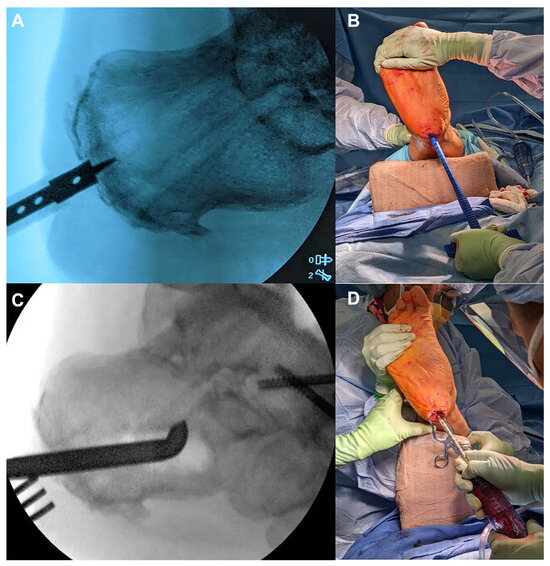
Figure 2.
A, Radiograph showing Avitus Pilot Hole Creator inserted into most proximal STJ screw tunnel. B, Clinical photo of Avitus Pilot Hole Creator insertion into STJ screw tunnel. C, Radiograph of Avitus Bone Harvester obtaining cancellous graft in 360° within the calcaneus. D, Clinical photo of Avitus Bone Harvester suction curettage capturing autologous bone into the receptacle of the handle.
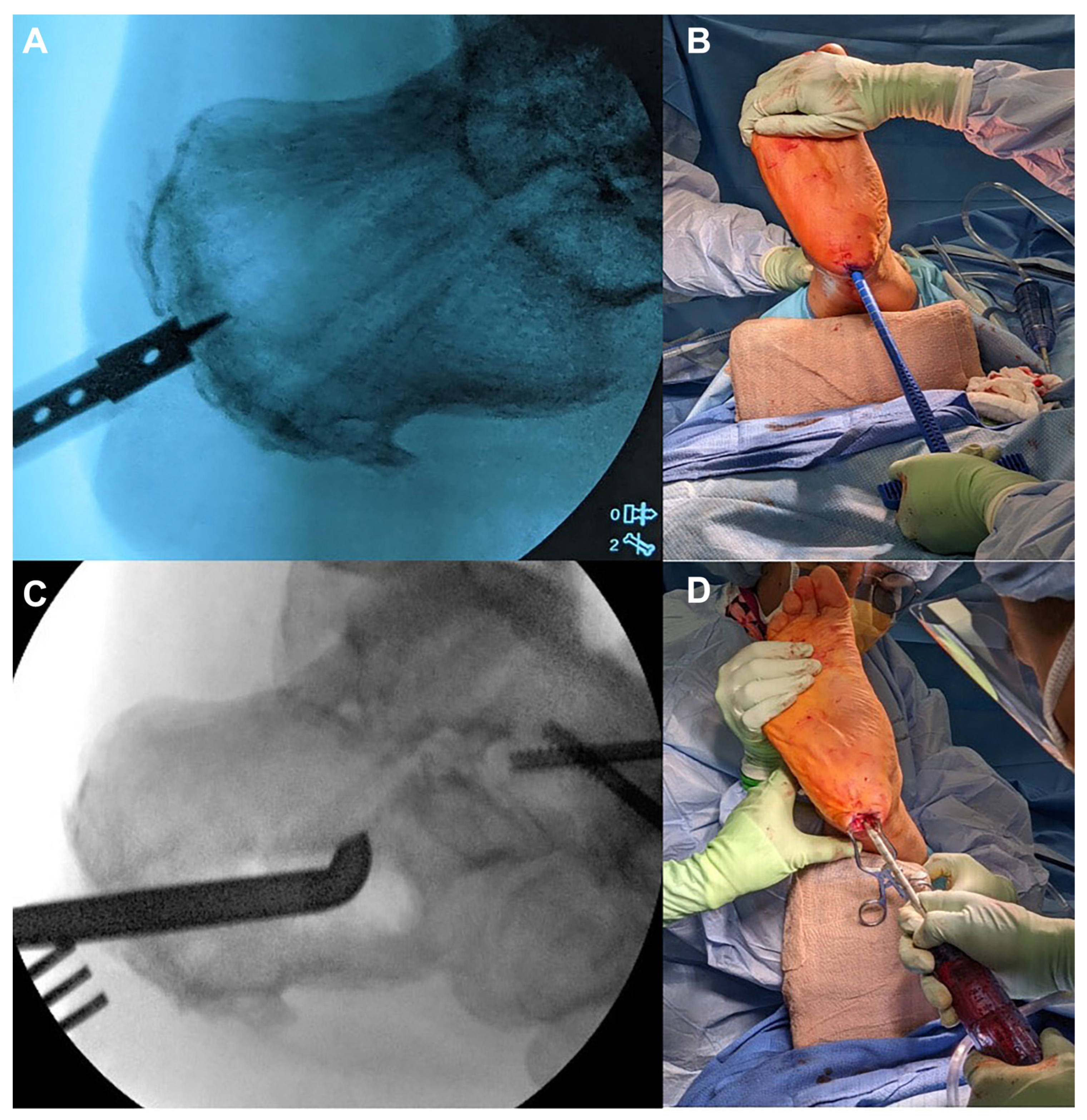
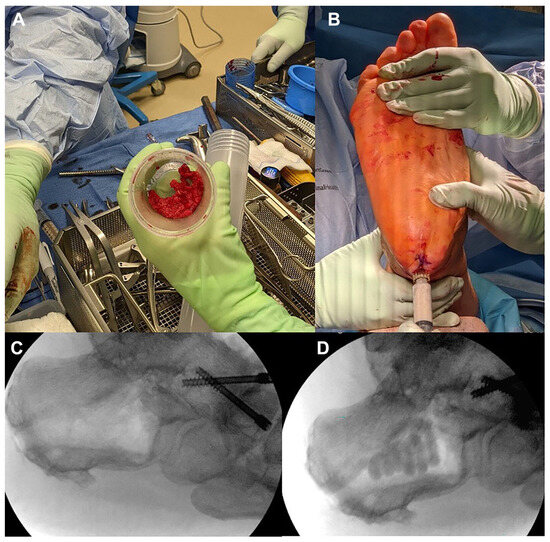
Figure 3.
A, 12 mL of chunky cancellous bone was harvested through the posterior screw hole approach in under 5 minutes with the Avitus Bone Harvester. B, Backfill of calcaneal graft defect with injectable demineralized bone matrix. C, Radiograph of calcaneal body after autograft harvest. D, Radiograph of calcaneal body after backfill.
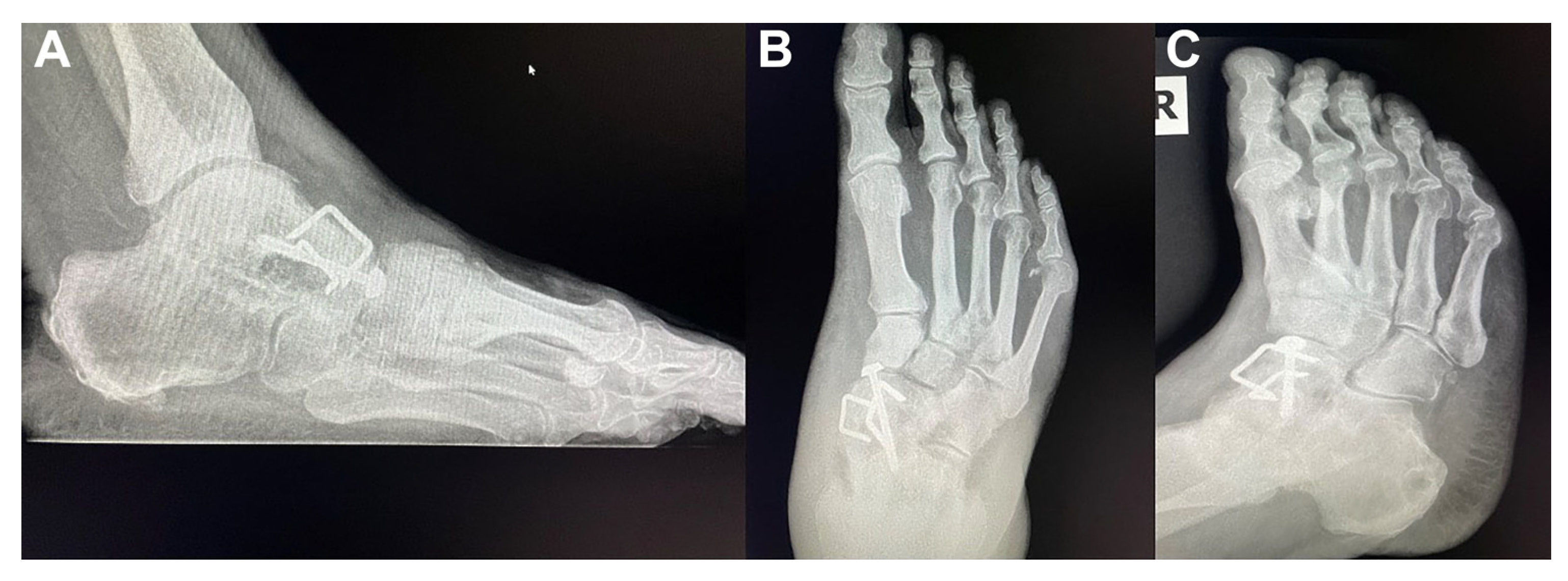
Attention was then directed to the TNJ, using a curvilinear skin incision and anatomical dissection to obtain full visualization of the nonunited TNJ. Under fluoroscopic visualization, residual retained hardware at the TNJ was removed. Joint preparation was performed with power instrumentation and smooth/curved osteotomes. Complete fenestration was performed at the navicular and distal talus to allow osseous channels to facilitate healing. The harvested calcaneal autograft was placed into the TNJ helping facilitate complete osseous union (Figure 4). The graft had a chunky consistency that was easy to pack and held under compression of the joint. Fixation was completed with a 5.0-mm cannulated screw and washer placed from the central navicular into the central talus, an 18-mm barbed staple into the medial TNJ, and a 20-mm barbed staple placed into the lateral TNJ. The incision at the TNJ was closed in routine layered fashion and the posterior calcaneal stab incisions with simple interrupted sutures. At the 3-month follow-up, the patient denied pain and was full weightbearing with the use of a CAM walker boot and crutches. Follow-up plain film three-view radiographs (Figure 5) at 4 months postoperative demonstrate complete osseous union at the TNJ arthrodesis site, and no evidence of complications were noted to the calcaneal donor harvest site with a near complete trabeculation present. There were no further complications during the recovery period.
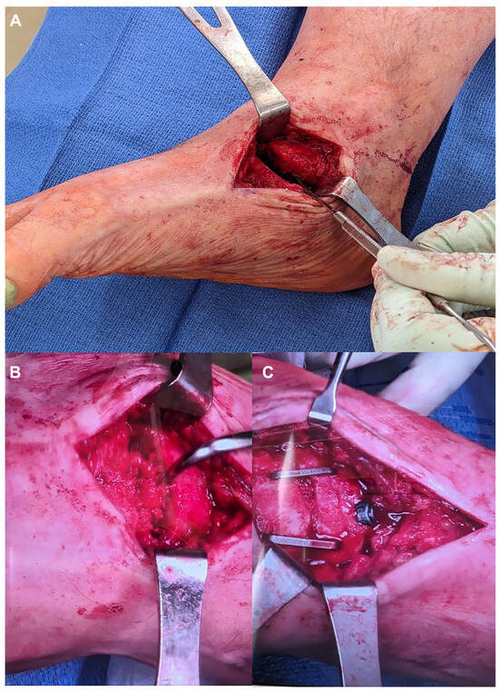
Figure 4.
A, Exposure and joint preparation of talonavicular joint nonunion. B, Insertion of calcaneal autograft into talonavicular joint. C, Final fixation of revision talonavicular joint fusion.
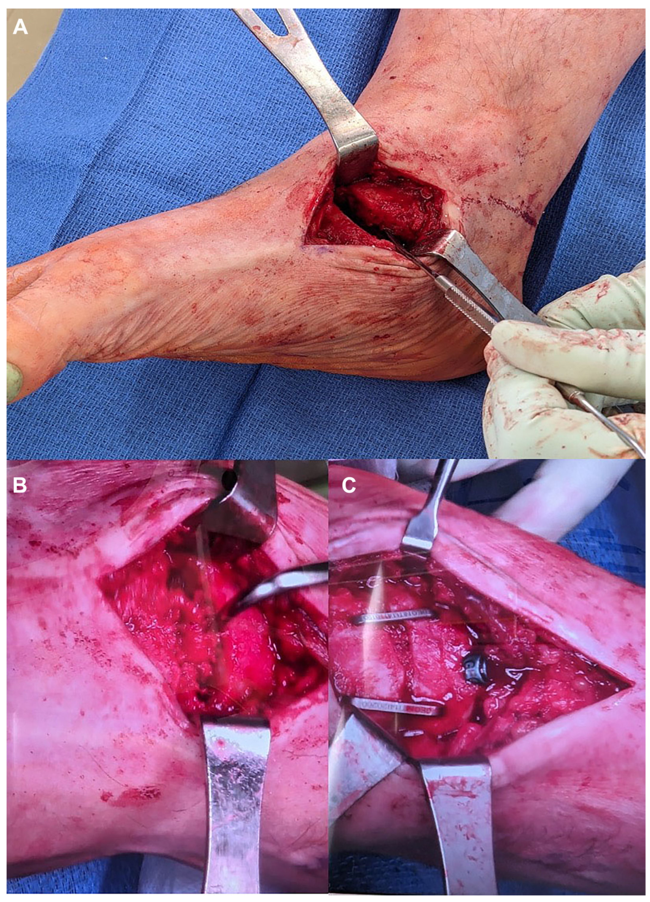
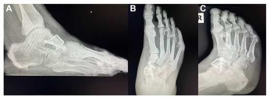
Figure 5.
A, 4-month postoperative lateral radiograph showing complete osseous union at the talonavicular joint. B, anteroposterior radiograph showing complete osseous union at the talonavicular joint. C, Medial oblique radiograph showing complete osseous union at the talonavicular joint. Additionally, note the calcaneal graft harvest site is nearly fully consolidated on all three views.
Discussion
Revision surgery can be difficult, and it poses a challenge to the operating surgeon. Achieving solid osseous union with functionally pain-free ambulation and a braceable or shoeable foot is ideal. The surgeon must consider potential donor site complications, medical comorbidities, underlying metabolic disease, tobacco usage, patient expectations, and a multitude of other considerations. Due to the complex nature of revisional surgery, the operative time and the duration of general anesthesia are worth considering. In 2020, Gowd et al [4] found that longer operating room time is associated with surgical-site infection and wound dehiscence after open reduction/internal fixation for ankle fractures. The surgeon should strive to maximize the quality of bone graft, reduction of deformity, and placement of hardware, while minimizing complications and the length of the operation to minimize risk of nonunion.
The primary reason an autogenous bone is considered superior to a nonautologous bone substitute relates to the presence of osteogenic, osteoinductive, and osteoconductive properties, along with numerous growth factors, like mesenchymal stem cells, bone morphogenic proteins, and platelet-derived growth factors found in autologous cancellous bones. There are many locations readily available to the foot and ankle surgeon for the harvest of autogenous bone graft/marrow in the lower extremity, including the proximal tibial, distal tibia, and the calcaneus. There are many benefits to these locations over Iliac crest harvest sites, including less reported minor/major donor-site complications, adequate graft volume available, short harvest time, similar fusion rates and the adjacent nature to primary procedure site in the context of lower-extremity procedures. In comparison, major and minor complication rates associated with Iliac crest bone graft harvests have been reported at 19.3% and 13.7%, respectively [5]. These complications constitute a wide spectrum, including but not limited to: hematoma, seroma, superficial versus deep infection, incisional hernia, paresthesia, chronic pain, gait disturbances, and even donor-site fracture [6,7,8].
The calcaneus was selected in our case study due to its proximity to the primary revision site, ease of access to cancellous bone following the removal of retained hardware, and low reported complication rates. The decision to use the screw tunnels for the autograft harvest was to avoid the need for another ancillary incision, which the more traditional lateral calcaneal harvest approach requires. Also, leaving the screws in place would have complicated the traditional lateral calcaneal harvest approach as well. Hence, the decision was made to remove them and use that screw tunnel for novel autograft harvest access. Attia et al [9] reviewed eight studies with 1,557 patients undergoing calcaneal autograft bone harvest, noting only five donor site fractures (0.32%), no infections, and 20 cases of transient sural neuritis (1.28%) that were resolved. All of the reported calcaneal fractures healed nonoperatively with immobilization and casting, except one that required an additional operative intervention. Of the calcaneal fractures that occurred, nearly all were due to a pathologic fracture or insufficiency at the donor site, with one being related to a fall. The traditional access for calcaneal graft is via a lateral approach, followed by a subsequent corticotomy and cancellous bone harvest, as described by many authors including Khademi et al in 2021 [10]. Numerous others have also described a percutaneous calcaneal approach, with Roukis et al [11] reporting a technique for bone marrow aspirate and bone graft harvest. After removing the screws from the posterior aspect of the calcaneus, we used this same percutaneous incision as an access point for the Avitus Bone Harvester. This obviated the need for an additional incision. We were able to obtain adequate autogenous graft volume while minimizing additional operative time and maintaining cortical integrity of the calcaneus. The suction curettage bone harvester system enabled capture of both autologous cancellous bone and bone marrow simultaneously while reducing the time associated with traditional bone harvest techniques. Since our postoperative weightbearing protocol is based on the primary TNJ revision arthrodesis site, we were confident that the calcaneus would be appropriately healed for weightbearing when initiated. We are not aware of this calcaneal bone graft harvest technique being reported in the literature previously.
Conclusions
This case study presents a patient undergoing revision TNJ arthrodesis for painful nonunion. Our new approach to harvesting autogenous calcaneal bone graft and bone marrow through an existing posterior calcaneal screw track allowed successful hardware removal. With a novel vacuum-assisted suction curettage system, we were able to quickly harvest autologous bone and marrow with an adequate volume for TNJ arthrodesis augmentation and eliminate the need for an ancillary incision.
At the initial 3-month short-term follow-up, our patient had a solid osseous union demonstrated on the postoperative plain film radiographs. There were no issues related to the posterior calcaneal harvest site to date. The patient can fully weightbear and walk without pain with the assistance of a CAM walker boot. He is advancing physical therapy and beginning to ween off the CAM walker boot. We plan mid- and long-term follow-up and updates (6–9 months and 12–24 months, respectively) for this case study.
Acknowledging that this scenario of posterior calcaneal access due to the removal of retained hardware, and the revision fusion of a nearby joint being uncommon, we still plan to continue this technique when at all possible. There are clear limitations to this study based on its single patient size, short-term follow-up duration, and the patient’s continued use of a CAM walker boot. However, we are encouraged by the current results of our type 2 diabetes mellitus patient with multiple medical comorbidities achieving solid osseous union of a previously failed TNJ arthrodesis, and the ease at which this anatomic approach allowed harvesting of autogenous bone graft and marrow. The novel vacuum-assisted suction curettage bone harvesting system was able to obtain adequate volumes of autologous cancellous bone and bone marrow in a quick seamless manner, thus reducing overall anesthesia time and operative time associated with conventional harvest techniques. Rancy et al [12] and Owoola et al [13] both reported average harvest times of 28 minutes and 18.7 minutes, respectively, by using traditional methods. On the other hand, Lukasiewicz et al [14] reported an average of 5 minutes for harvesting by using our same technique of a novel vacuum-assisted suction curettage technique with the Avitus Bone Harvester system. We will continue to use this method for harvesting autologous cancellous bone and bone marrow for reconstructive lower-extremity procedures.
Financial Disclosure
None reported.
Conflict of Interest
None reported.
References
- Webster J, Murphy D: Atlas of Orthoses and Assistive Devices 5th Edition, Elsevier Health Sciences, Philadelphia, 2017.
- de Moraes Barros Fucs PM, Svartman C, de Assumpção RM, et al: Surgical technique: Medial column arthrodesis in rigid spastic planovalgus feet. Clin Orthop Relat Res 470: 1334, 2012.
- Klassen LJ, Shi E, Weinraub GM, et al: Comparative Nonunion Rates in Triple Arthrodesis. J Foot Ankle Surg 57: 1154, 2018.
- Gowd AK, Bohl DD, Hamid KS, et al: Longer operative time is independently associated with surgical site infection and wound dehiscence following open reduction and internal fixation of the ankle. Foot Ankle Spec 13: 104, 2020.
- Dimitriou R, Mataliotakis GI, Angoules AG, et al: Complications following autologous bone graft harvesting from the iliac crest and using the RIA: a systematic review. Injury 42: S3, 2011.
- Kim DH, Rhim R, Li L, et al: Prospective study of iliac crest bone graft harvest site pain and morbidity. Spine J 9: 886, 2009.
- Sudhakar KNV, Mohanty R, Singh V: Evaluation of donor site morbidity associated with iliac crest bone harvest in oral and maxillofacial, reconstructive surgery. J Clin Diagn Res 11: ZC28, 2017.
- Ahlmann E, Patzakis M, Roidis N, et al: Comparison of anterior and posterior iliac crest bone grafts in terms of harvest-site morbidity and functional outcomes. J Bone Joint Surg Am 84: 716, 2002.
- Attia AK, Mahmoud K, ElSweify K, et al: Donor site morbidity of calcaneal, distal tibial, and proximal tibial cancellous bone autografts in foot and ankle surgery. A systematic review and meta-analysis of 2296 bone grafts. Foot Ankle Surg 7: 2473011421S00094, 2022.
- Khademi F, Erfani A, Erfani MA, et al: Bone graft harvesting from the calcaneus using lateral wall corticotomy technique by an osteotome. Foot Ankle Spec 14: 298, 2014.
- Schade VL, Roukis TS: Percutaneous bone marrow aspirate and bone graft harvesting techniques in the lower extremity. Clin Podiatr Med Surg 25: 733, 2008.
- Rancy SK, Malliaris SD, Wolfe SW: Tibial bone grafting for wrist reconstruction. J Wrist Surg 6: 158, 2017.
- Owoola AM, Odunubi OO, Yinusa Y, et al: Proximal tibial metaphysis: its reliability as a donor site for grafting. West Afr J Med 29: 403, 2010.
- Lukasiewicz AM, Bagi PS, Yu KE, et al: Novel vacuum-assisted method for harvesting autologous cancellous bone graft and bone marrow from the proximal tibial metaphysis. Foot Ankle Orthop 6: 2473011420981901, 2021.
© 2025 American Podiatric Medical Association