


Abstract
Background: The primary aim of this study is to examine the effect of the stiffness of rehabilitation material on muscle activation in individuals with low medial longitudinal arch. The secondary aim is to compare the effects of single-leg balancing and single-leg squat exercises on the activation of the muscles around the ankle in the same individuals. Methods: Twelve individuals with low medial longitudinal arch and 18 healthy control participants were included. Participants were asked to perform the single-leg balance and single-leg squat exercises on Theraband stability trainers of three different stiffness levels. Activation of the peroneus longus, peroneus brevis, tibialis anterior, medial and lateral gastrocnemius muscles during exercises was evaluated by surface electromyography. Results: The single-leg balancing exercise performed on soft and very soft ground was more effective than single-leg squat in increasing the activation of the lateral gastrocnemius muscle in participants with a low medial longitudinal arch. It was determined that performing single-leg squat exercise on a very soft surface instead of hard ground caused more muscular activation in the peroneus brevis, tibialis anterior, and medial gastrocnemius in the same individuals. Conclusions: The exercise progression to be applied on different surfaces to increase the activation of the muscles around the ankle in individuals with a low medial longitudinal arch and in healthy individuals without a low medial longitudinal arch was different.
Pes planus is defined as structural change in the medial longitudinal arch structure. The incidence of pes planus, which can develop due to congenital or acquired reasons, varies between 2% and 23% [1]. In this case, the talus pronates more than normal, and the forefoot goes into more abduction and supination [2]. These changes in foot biomechanics are an important risk factor for the development of over-use injuries [3,4]. For this reason, it is very important to strengthen the muscles around the ankle in patients with a low medial longitudinal arch (LMLA) and to improve the proprioceptive sense.
It is known that the risk of ankle injury may increase in individuals with a LMLA [4]. The strength of the peroneus longus (PL) and peroneus brevis (PB) muscles, in particular, is very important in the prevention and treatment of ankle injuries, as they provide support to the lateral ligament [5,6]. In addition, tibialis anterior (TA), lateral gastrocnemius (LG), and medial gastrocnemius (MG)muscles play a key role during single-leg activities [7]. Therefore, we think that neuromuscular training of the muscles around the ankle will be important in the physiotherapy and rehabilitation program in these individuals. Rehabilitation materials with different stability properties are frequently used in the treatment of proprioceptive sensory deficit and muscular imbalance [8]. Unstable mats, balance board, stability trainers, and oscillation devices can be given as examples of rehabilitation materials [9].
To our knowledge, there is no study in the literature on the type of exercise required to activate the muscles around the ankle in individuals with an LMLA and how the exercise will progress. The primary aim of our study is to examine the effect of the stiffness of the rehabilitation material on muscle activation in people with an LMLA and in healthy individuals without an LMLA. The secondary aim of this study is to compare the effects of single-leg balancing (SLB) and single-leg squat (SLS) exercises on the activation of the muscles around the ankle in those with LMLA.
Materials and Methods
Participants
This prospective study was carried out at Gazi University, Faculty of Health Sciences, Department of Physiotherapy and Rehabilitation (Ankara, Turkey) and approved by the Gazi University Ethics Committee. A total of 30 university students (25 women and 5 men) between 18 and 35 years old were included in the study. Eighteen healthy individuals comprised the control group, and 12 individuals with LMLA comprised the study group. All participants were right-side dominant. Participants with soft-tissue injuries in the lower extremity, bone fractures, those who had surgery due to a pathology in the lower extremity, and those with pain during exercise were excluded.
Procedures of the Study
Each participant signed an informed consent form. The age, gender, height, weight, whether they had any injury to the dominant leg, and personal and family history of all participants who agreed to participate in the study were recorded.
Participants were divided into two groups according to the presence or absence of LMLA. The navicular drop test was used to evaluate the arch structure. If the amount of navicular drop was 10 mm or more, it was accepted that there was an LMLA. On the test day, the participants were first asked to balance on one leg with their hands at the waist on four different hard floors, and then they were asked to do an SLS exercise. For the SLB and SLS exercise on different surfaces, they were asked to perform three successful repetitions for 30 seconds and a 30-second rest break was given between each repetition.
Evaluation of Muscular Activation
All muscular activation measurements were taken from the right extremity. An 8-channel electromyography (EMG) device (Noraxon MiniDTS system; Noraxon, Inc, Scottsdale, Arizona) was used to measure muscular activation. The common noise reduction ratio of this device is over 100 dB, the differential input impedance is over 100 ohms, and the sampling rate is between 1500 Hz and 3000 Hz per channel. To record the EMG signals, disposable, self-adhesive Ag/AgCl electrodes were used for superficial EMG applications only [10]. The distance between the electrodes was 20 mm, the diameter of two circular adhesives was 1 cm, and the size of the 8-shaped adhesive was 4 cm × 2.2 cm. Before the electrodes were attached, the area was first shaved in order to reduce the skin impedance below 5 kX, then sandpapered and cleaned with cotton dipped in alcohol until the skin was slightly red. The electrodes were placed parallel to the identified muscle fibers as recommended by the European Recommendations for Surface Electromyography [11]. EMG signals were analyzed with 500 Hz low-pass and 10 Hz high-pass filters. Then, root mean square values were calculated from the raw EMG data in successive time windows (time windows: 0.1 sec) to evaluate the EMG signals. The values obtained after each trial were normalized to the mean value on the hard floor and expressed as a percentage value.
Characteristics of the Theraband Stability Trainers
The characteristics of the Theraand Stability Trainers (Theraband, Akron, Ohio) of different hardness that we used in our study during exercise practices are shown in Table 1 [12].
Table 1.
Comparison of the Features of Theraband Stability Trainers in Different Colors.
Exercise Description
Single-Leg Balancing. Balance exercises are often performed on both legs or on one leg. The purpose of these exercises is to reduce sensorimotor deficits and restore neuromuscular activation. These exercises are frequently used in rehabilitation programs after an ankle injury [13]. Participants were asked to balance on their right foot for 30 seconds while performing this movement with their hands on their waists and their left knees at 90 degrees flexion on floors of different hardness. Then a 30-second rest break was given. In this way, they were asked to balance on one leg 3 times in total.
Single-Leg Squat. The single-leg squat exercise is frequently used in the clinic to strengthen the lower extremity and hip muscles. During this exercise, the knee should not go to varus/valgus and the hip must be prevented from falling/raising [14]. During the study, the participants were asked to bend their right knee 30 degrees and extend their left leg forward while their hands were on their waists in a standing position. Participants were asked to wait 30 seconds at the end of the movement. They were asked to repeat this movement 3 times and have a rest for 30 seconds between repetitions. Knee flexion angle was checked with the goniometer application. Studies have shown that this application is a valid and reliable method for measuring range of motion [15].
Statistical Analysis
Statistical analyses were performed using SPSS version 24 software (IBM, Armonk, New York). The conformity of the variables to the normal distribution was examined using visual (histogram and probability graphs) and analytical methods (Shapiro-Wilk Tests). Because the data showed normal distribution according to descriptive analyses, the variables were presented using the mean and standard deviation. Demographic information and muscle activation values of individuals with and without low medial longitudinal arch were compared using the independent sample t test. Changes caused by surface differences in muscle activation were analyzed separately for each group with repeated measures analysis of variance (ANOVA). The Greenhouse-Geisser correction was used when the sphericity assumption was not met. The surface condition caused the difference between muscle activations was determined using Bonferroni adjusted post hoc pairwise comparisons. For statistical significance, the total type-1 error level was determined as 5%. Post-power analysis of the study was conducted using G*Power 3.0.10 program (HeinrichHeine-Universita¨t Du¨ sseldorf, Du¨ sseldorf, Germany). In this analysis, bidirectional a 5 0.05 was accepted. In participants with a LMLA, the statistical power (1 - β) was determined to be between 0.90 and 0.98 in general, according to the muscle group selected and the type of exercise.
Results
Demographic Characteristic of Participants
Twenty-five of the participants were women and five were men. The dominant extremity was the right side in all participants. All muscle activation measurements were taken from the right extremity. Low medial longitudinal arch was seen in 12 of the participants; the remaining 18 did not have LMLA. Demographic data and arch height of the participants included in the study are shown in
Table 2. Age, height, weight and body mass index of individuals in both groups were similar (P . 0.05).
Table 2.
Demographic and Clinical Data of the Participants.
Comparison of Muscular Activation Between Groups During Single-Leg Balancing Exercise Between Floors of Different Hardness
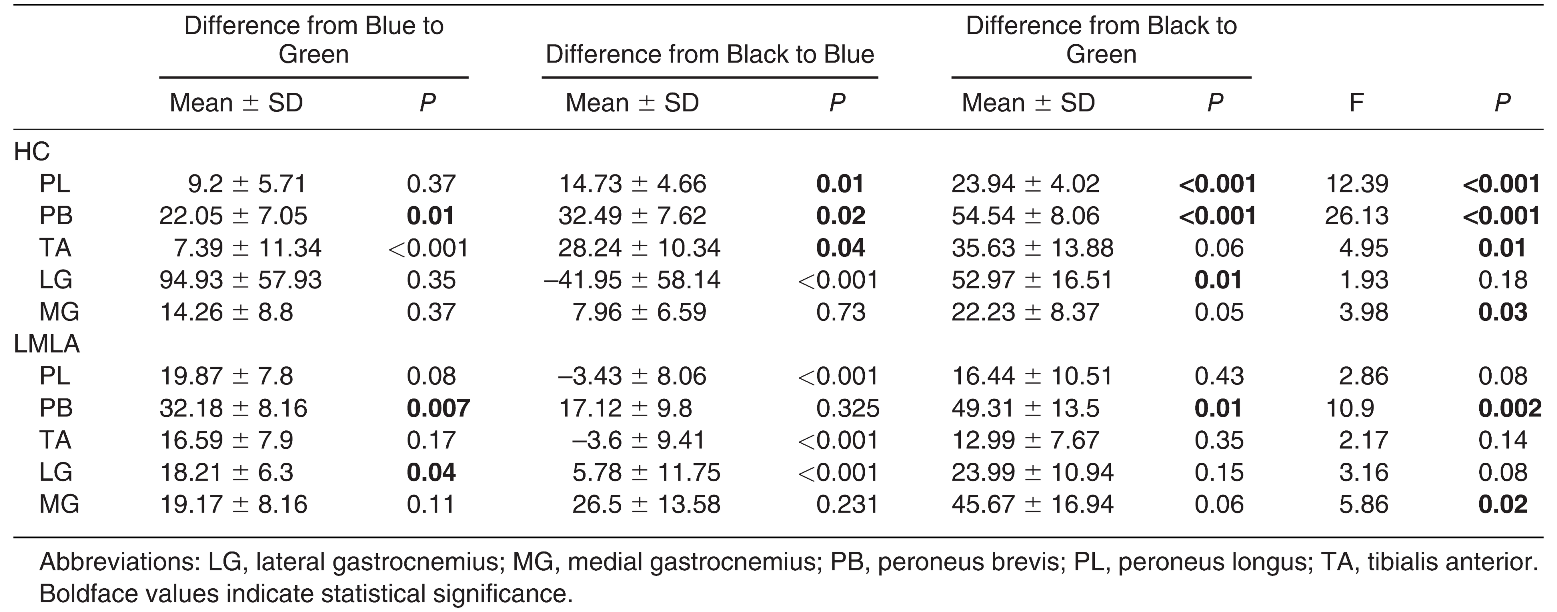
The transition from green surface to blue during SLB exercise of the individuals in the control group caused a significant increase in the activation of the PB muscle (P 5 0.018). During the transition from blue to black, an increase in muscular activation is observed in the PL, PB, and TA (P < 0.05). In addition, with the transition from green to black, there was a statistically significant increase in PL, PB, and LG muscle activation (P < 0.05). In individuals with LMLA, increased activation of the PB and LG muscles was observed during the transition from green surface to blue surface SLB exercise. In addition, an increase in activation in the PB muscle is observed during the transition from green to black (Table 3).
Table 3.
Comparison of Muscular Activations of Healthy Controls (HC) and Individuals with Low Medial Longitudinal Arch (LMLA) During Single-Leg Balancing Exercise.
Comparison of Muscular Activation During Single-Leg Squat Between Groups on Surfaces of Different Hardness
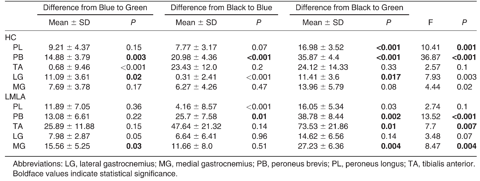
Individuals in the control group showed an increase in activation in the PB and LG muscles with the transition from green to blue during the SLB exercise (P < 0.05). During the transition from blue surface to black, in the PB muscle (P < 0.001); during the transition from green surface to black an increase in activation is observed in the PL, PB, and LG muscles in healthy individuals (P < 0.05). During the SLS exercise of individuals with LMLA, there was an increase in MG muscle activation (P = 0.039) when transitioning from the green surface to the blue surface. In addition, during the transition from the green surface to the black surface, there was an increase in activation in the PB, TA, and MG muscles (P < 0.05) (Table 4).
Table 4.
Comparison of Muscle Activations of Healthy Controls (HC) and Individuals with Low Medial Longitudinal Arch (LMLA) During Single-Leg Squat Exercise on Different Floors.
Differences in Muscular Activation During Two Different Exercises Performed on Different Surfaces
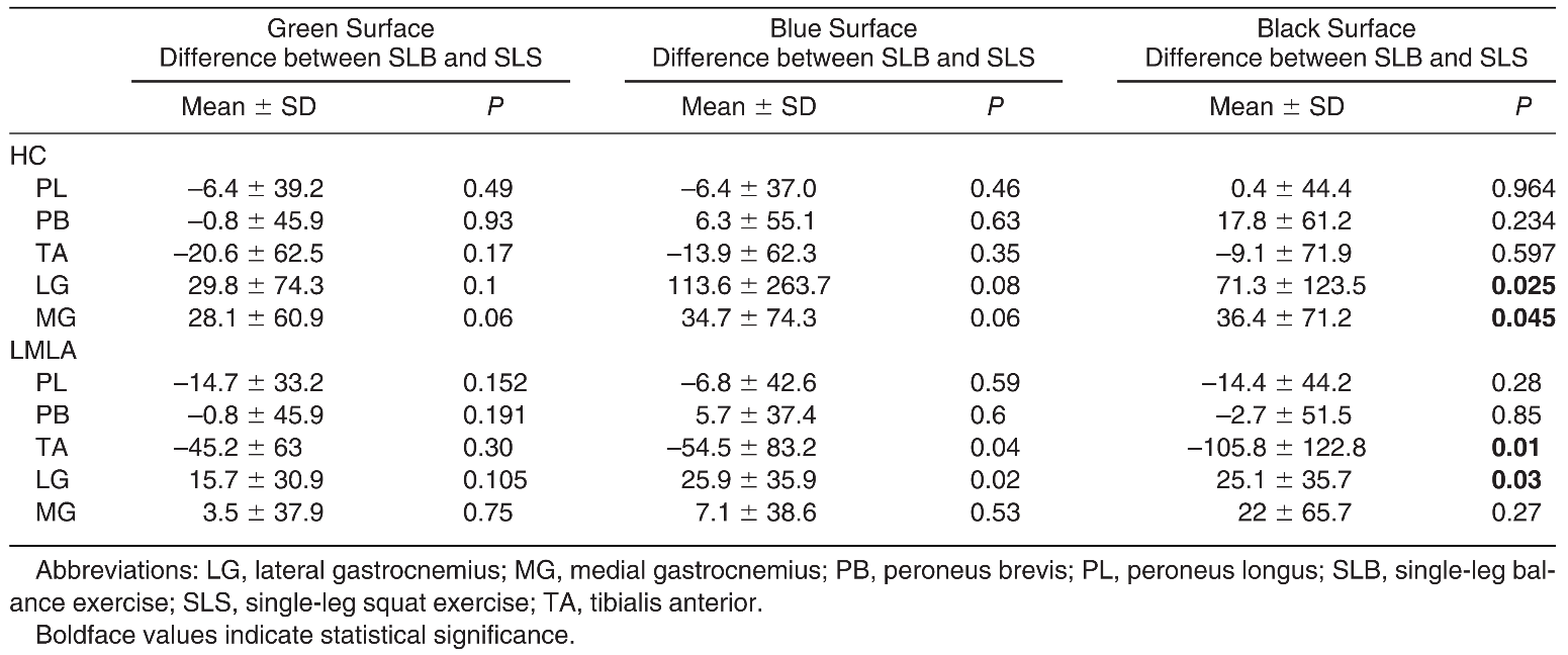
The muscle activation observed in the SLS and SLB on the three stability trainers was generally similar in individuals without LMLA. However, the SLB exercise on the black stability trainer was more effective than SLS exercise in increasing activation of both parts of the gastrocnemius (P < 0.05). Single-leg balance exercise performed on the blue and black stability trainers was more effective than SLS in activating the LG muscle (P < 0.05). Additionally, the SLS exercise performed on the blue and black stability trainers was more effective than SLB exercise in increasing activation of the TA muscle ((P < 0.05) (Table 5).
Table 5.
Muscular Activation During Two Different Exercises Performed on Different Surfaces.
Discussion
The main aim of our study was to examine the effect of the stiffness of the rehabilitation material on muscle activation in individuals with LMLA. The secondary aim of this study is to compare the effects of SLB and SLS exercises on the activation of the muscles around the ankle in patients with LMLA.
The progression of exercise using stability trainers of three different stiffness levels to increase activation of the muscles around the ankle was different between the group with LMLA and the control group. SLS exercise was observed to be more effective than SLB exercise to strengthen the TA muscle, while SLB exercise was observed to be more effective than SLS to strengthen the LG muscle in individuals with LMLA. In addition, it was determined that SLB and SLS exercises were not superior to each other in increasing the activation of PL and PB muscles in either the LMLA or the control group.
Despite the clinical significance of LMLA, there is limited information regarding the difference in biomechanical factors between individuals with and without LMLA [16]. This situation makes it difficult to interpret and discuss the results of our study.
We observed a difference between the type and progression of exercise that should be performed to activate the muscles around the ankle in both individuals with and without LMLA. The reason for this difference may be the medial shift of the ground reaction force due to excessive pronation in the stance phase of gait in people with LMLA [17]. The shift of the ground reaction force toward the medial causes abnormalities in the pressure distribution on the plantar surface of the foot. It has been reported that while pressure increases in the big toe, central forefoot, and medial midfoot were observed, pressure decreases in the medial and lateral forefoot in individuals with LMLA [18]. These pressure differences observed between those with and without LMLA may cause differences in muscle activation during different exercises performed on floors of different hardness.
In a study by Angin et al [19], it was observed that the cross-sectional area and thickness of the abductor hallucis, flexor hallucis brevis, peroneus longus, and brevis muscles were lower in people with LMLA, while the cross-sectional area and thickness of the flexor digitorum longus and flexor hallucis longus muscles were found to be greater. In addition, it was observed that the anterior part of the plantar fascia was 21.7% thinner and the middle part was 10.6% thinner in those with LMLA compared to those without LMLA. It has been shown that the plantar fascia can tolerate less weight as a result of being thinner in certain areas [19].
We observed that the changes in cross-sectional area and thickness of the extrinsic and intrinsic muscles of the foot and plantar fascia may contribute to differences in muscle activation during exercise in people with LMLA and healthy controls.
In general, it is thought that the decrease in the stiffness of the rehabilitation materials will increase the muscular activation as it causes a more challenging effect on the neuromuscular system [20]. Wolburg et al [12] included 25 healthy individuals in their study to determine how rehabilitation materials of different stiffnesses affect activation in lowerextremity muscles. The researchers evaluated the muscular activation of MG, LG, TA, and PL with superficial EMG while participants performed SLB exercises on five rehabilitation materials of varying stiffness. In that study, as the stiffness of the rehabilitation material decreased in the muscles other than the PL, the muscular activation increased [12]. In our study, we revealed that the gradual decrease in surface stiffness during SLB exercise in people with LMLA did not cause a statistically significant change in the activation of PL and MG muscles, while it did cause a significant increase in the TA, LG, and PB muscles. This finding shows that the decrease in the stiffness of the rehabilitation material used does not always result in an increase in muscular activation.
In our study, SLB and SLS exercises on three different surfaces in the healthy control group did not cause a significant difference in the muscular activation of PL and TA. There are studies with results similar to ours. Harput et al [21] asked 24 healthy participants to perform the SLB and SLS exercise on a hard surface (Theraband wobble board) and a soft surface (Both Sides Up-BOSU). While the participants were doing these exercises on different surfaces, PL and TA muscle activation was evaluated with superficial EMG. As a result of the study, they revealed that the surface hardness and exercise type did not cause a statistically significant change in the muscular activation level of the PL and TA. Braun Ferreirae et al [22] determined that there was no difference in the muscular activation of TA during SLB exercise performed on the trampoline and balance board. Unlike these results, Blackburn et al [23] reported that the EMG signal amplitude of the PL and TA muscles was greater during SLB exercise on a hard surface compared to a soft surface. As can be seen, there is no consensus in the literature on how the change in the rehabilitation material used during exercise affects the activation of the TA muscle. Future studies should focus on how the rehabilitation material should be selected to increase the progression of exercises focused on strengthening the TA muscle.
Squat exercises are very important in maintaining the correct biomechanical alignment of foot posture. Therefore, squat exercises are used to prevent chronic ankle injuries [24]. Previous studies have shown that single-leg squats provide greater neuromuscular activation than double-leg squats due to reduced mediolateral support. Increased neuromuscular activity has been reported to have positive effects on proprioception and functional activities [25,26]. Although squat exercises are a frequently used exercise type in the clinic, no study has been found on how the use of surfaces of different hardness during SLS exercise affects muscular activation in individuals with LMLA. Our study is the first to examine this subject. As a result of our study, it was concluded that SLS exercise performed on a black background was more effective than SLS exercise on a green ground in increasing the activation of PB, TA and MG muscles in cases with LMLA. In these cases, it was observed that the decrease in the hardness of the surface used during SLS exercise did not change the activation of the PL and LG muscles.
To the best of our knowledge, this is the first study to compare the effectiveness of SLB and SLS exercises on people with LMLA. As a result of our study, it was seen that SLS exercise was more effective than SLB exercise to strengthen the TA muscle on blue and black backgrounds in those with LMLA. This may be due to the fact that maintaining the anterior-posterior stability of the ankle during squats is particularly dependent on the activation of the TA [27,28]. It would be beneficial to choose SLS exercise instead of SLB exercise when TA muscle weakness occurs in individuals with LMLA.
Rehabilitation materials used in physiotherapy and rehabilitation clinics can be divided into stable and unstable materials. While BOSU and stability trainer are shown as examples of stable rehabilitation materials, balance board, wobble boards, and rocker boards can be given as examples of unstable rehabilitation materials. In their study, Dohm-Acker et al [29] concluded that after standing on one leg for 15 seconds on unstable surfaces, muscular activation in the lower extremities decreased significantly. Therefore, when examining the effect of unstable rehabilitation materials on muscular activation during SLB, especially the first 15 seconds of the exercise should be considered. However, when the literature was examined, it was not possible to obtain information about the time from which muscle activation decreased during SLB on the stable surface. In future studies, it should be investigated that the lower-extremity muscular activation starts to decrease from the second during SLB and SLS exercise on a stable surface in those with and without LMLA.
In superficial EMG (sEMG), after the raw data are processed with the MATLAB program (MathWorks, Natick, Massachusetts) the maximum and average EMG amplitudes for each muscle are formed. In this study, we used the mean EMG amplitudes for each muscle. However, the EMG amplitudes used in studies on sEMG vary. In addition, the low-pass and highpass values selected during the processing of sEMG signals also show great differences between studies. This situation makes it difficult to compare and interpret our study results with other study results in the literature.
As a result of this study, it is seen that while different exercises performed on different surfaces cause an increase in activation in some muscles, it does not cause a change in activation in other muscles. However, the effect of using SLB and SLS exercises on Theraband stability trainers for a longer period instead of a few seconds is unknown. In addition, it may be necessary to use kinematic analysis to determine what type of postural control strategies individuals use when trying to balance on stable or unstable rehabilitation materials. In our study, we evaluated the muscular activation during the whole balancing activity. Sudden movements of individuals to maintain their balance may have caused an increase in extra muscle activation by reflex mechanisms [30]. The possibility that foot deformities may also increase extra muscle activation should be taken into consideration in future studies [31].
The type of exercise and rehabilitation material to be used to activate the muscles around the ankle in people with LMLA and in healthy individuals vary according to the muscles in our target group. In addition, contrary to general belief, it is seen that the decrease in the stiffness of the rehabilitation material does not cause an increase in activation of all muscles in the lower extremity. For this reason, when determining the exercise program, the weak muscle should be identified. Also, the appropriate exercise type and appropriate rehabilitation material should be selected. In addition, it should be considered whether individuals who apply to physiotherapy clinics do indeed have an LMLA, and it should not be forgotten that this situation will cause a change in the rehabilitation program.
Our study has limitations. Because only young individuals were included in this study, and because the majority of the participants were women, the results cannot be generalized to the larger population.
Funding
None reported.
Acknowledgments
Murat Esmer, PhD, would like to thank TUBITAK (Scientific and Technological Research Council of Turkey) for their support through the TUBITAK 2211-A Domestic Graduate Scholarship Program during his doctoral studies.
Conflicts of Interest
None reported.
References
- SHIBUYA N, JUPITER DC, CILIBERTI LJ ET AL: Characteristics of adult flatfoot in the United States. J Foot Ankle Surg 49: 363, 2010.
- MYERSON M: Adult Acquired Flat Foot Deformity, WB Saunders, San Diego, 2003.
- LEVY JC, MIZEL MS, WILSON LS, ET AL: Incidence of foot and ankle injuries in West Point cadets with pes planus compared to the general cadet population. Foot Ankle Int 27: 1060, 2006.
- KAUFMAN KR, BRODINE SK, SHAFFER RA, ET AL: The effect of foot structure and range of motion on musculoskeletal overuse injuries. Am J Sports Med 27: 585, 1999.
- WILLEMS T, WITVROUW E, VERSTUYFT J, ET AL: Proprioception and muscle strength in subjects with a history of ankle sprains and chronic instability. J Athlet Train 37: 487, 2002.
- GLICK JM, GORDON RB, NISHIMOTO D: The prevention and treatment of ankle injuries. Am J Sports Med 4: 136, 1976.
- HOUGLUM PA, BERTOTI DB: Brunnstrom’s Clinical Kinesiology, 6th Edition, FA Davis Co, Philadelphia, 2011.
- HUPPERETS MD, VERHAGEN EA, VAN MECHELEN W: Effect of sensorimotor training on morphological, neurophysiological and functional characteristics of the ankle. Sports Med 39: 591, 2009.
- HUPPERETS MD, VERHAGEN EA, VAN MECHELEN W: The 2BFit study: is an unsupervised proprioceptive balance board training programme, given in addition to usual care, effective in preventing ankle sprain recurrences? Design of a randomized controlled trial. BMC Musculoskel Dis 9: 1, 2008.
- Hermens HJ, Freriks B, Disselhorst-Klug C, et al: Development of recommendations for SEMG sensors and sensor placement procedures. J Electromyography Kinesiol 10: 361, 2000.
- HERMENS HJ, FRERIKS B, MERLETTI R, ET AL: European recommendations for surface electromyography. Roessingh Res Develop 8: 13, 1999.
- WOLBURG T, RAPP W, RIEGER J, ET AL: Muscle activity of leg muscles during unipedal stance on therapy devices with different stability properties. Phys Therap Sport 17: 58, 2016.
- EILS E, ROSENBAUM D: A multi-station proprioceptive exercise program in patients with ankle instability. Med Sci Sport Exerc 33: 1991, 2001.
- WILLY RW, DAVIS IS: The effect of a hip-strengthening program on mechanics during running and during a single-leg squat. J Orthop Sports Phys Ther 41: 625, 2011.
- >Dos Santos RA, Derhon V, Brandalize M, et al: Evaluation of knee range of motion: Correlation between measurements using a universal goniometer and a smartphone goniometric application. J Bodywork Movement Thera 21: 699, 2017.
- Panichawit C, Bovonsunthonchai S, Vachalathiti R, et al: Effects of foot muscles training on plantar pressure distribution during gait, foot muscle strength, and foot function in persons with flexible flatfoot. J Med Assoc Thai 98: S12, 2015.
- BENNETT JE, REINKING MF, PLUEMER B, ET AL: Factors contributing to the development of medial tibial stress syndrome in high school runners. J Orthop Sports Phys Ther 31: 504, 2001.
- LOUDON JK, JENKINS W, LOUDON KL: The relationship between static posture and ACL injury in female athletes. J Orthop Sports Phys Ther 24: 91, 1996.
- ANGIN S, CROFTS G, MICKLE KJ, ET AL: Ultrasound evaluation of foot muscles and plantar fascia in pes planus. Gait Posture 40: 48, 2014.
- CRISWELL E. Cram’s Introduction to Surface Electromyography, Jones & Bartlett Publishers, Burlington, MA, 2010.
- HARPUT G, SOYLU AR, ERTAN H, ET AL: Activation of selected ankle muscles during exercises performed on rigid and compliant balance platforms. J Orthop Sports Phys Ther 43: 555, 2013.
- FERREIRA LAB, PEREIRA WM, ROSSI LP, ET AL: Analysis of electromyographic activity of ankle muscles on stable and unstable surfaces with eyes open and closed. J Bodywork Movement Thera 15: 496, 2011.
- LEHMAN GJ, MCGILL SM: The importance of normalization in the interpretation of surface electromyography: a proof of principle. J Manipulative Physiol Therapeutics 22: 444, 1999.
- MCPOIL TG, CORNWALL MW, MEDOFF L, ET AL: Arch height change during sit-to-stand: an alternative for the navicular drop test. J Foot Ankle Res 1: 1, 2008.
- SONG J, HILLSTROM H, SECORD D, ET AL: Foot type biomechanics. comparison of planus and rectus foot types. JAPMA 86: 16, 1996.
- BULDT AK, ALLAN JJ, LANDORF KB, ET AL: The relationship between foot posture and plantar pressure during walking in adults: A systematic review. Gait Posture 62: 56, 2018.
- SEVERIN AC, BURKETT BJ, MCKEAN MR, ET AL: Quantifying kinematic differences between land and water during squats, split squats, and single-leg squats in a healthy population. PloS one 12: e0182320, 2017.
- GRACI V, VAN DILLEN LR, SALSICH GB: Gender differences in trunk, pelvis and lower limb kinematics during a single-leg squat. Gait Posture 36: 461, 2012.
- DOHM-ACKER M, SPITZENPFEIL P, HARTMANN U: Effect of proprioceptive trainings tools for the muscles in stance stability. Sportverletzung Sportschaden: Organ der Gesellschaft fur Orthopadisch-Traumatologische Sportmedizin 2: 52, 2008.
- SOLOMONOW M, KROGSGAARD M: Sensorimotor control of knee stability. A review. Scandinavian J Med Sci Sport 1: 64, 2001.
- C INAR-MEDENI Ö , ATALAY GUZEL N, BASAR S: Mild hallux valgus angle affects single-limb postural stability in asymptomatic subjects. J Back Musculoskel Rehabil 29: 117, 2016.



© 2024 American Podiatric Medical Association