Next Article in Journal
Pedal Vessel Calcification and Risk of Major Adverse Foot Events in the Diabetic Neuropathic, Nephropathic Foot
Previous Article in Journal
Muscular Activation Levels of Subjects with Low Medial Longitudinal Arch During Single-Leg Squat and Single-Leg Balancing Exercises on Surfaces of Different Hardness
 
 
Journal of the American Podiatric Medical Association is published by MDPI from Volume 116 Issue 1 (2026). Previous articles were published by another publisher in Open Access under a CC-BY (or CC-BY-NC-ND) licence, and they are hosted by MDPI on mdpi.com as a courtesy and upon agreement with American Podiatric Medical Association.
Article

Hoop Stress Elicited at Medial Tibial Crural Fascia Attachment During Passive Dorsiflexion: A Proof-of-Concept Study for Medial Tibial Stress Syndrome Causation

by
Amy H. Amabile
1,*,
Thomas A. Hulcher
2,
Mariano Figueroa-Perez
2 and
Madeline R. Reich
2
1
Temple University Lewis Katz School of Medicine and School of Podiatric Medicine, MERB 457, 3500 North Broad Street, Philadelphia, PA
2
Department of Physical Therapy, Thomas Jefferson University, Philadelphia, PA
*
Author to whom correspondence should be addressed.
J. Am. Podiatr. Med. Assoc. 2024, 114(5), 23169; https://doi.org/10.7547/23-169
Published: 1 September 2024

Abstract

Background: Identification of a specific causal mechanism for medial tibial stress syndrome has been elusive, although there is a consensus that it may be caused by traction on the tib-ial periosteum elicited by soft tissues. The crural fascia (CF) attaches directly to the tibia throughout the length of the leg, encircling it in a grossly cylindrical fashion, and the leg may thus be viewed as a type of fluid-filled cylinder, subject to both longitudinal and hoop stresses. Prior researchers have not considered the possibility that strain on the medial tibia could be produced by the CF during gait and passive stretching, secondary to fluid pressure increases in the fascial compartments of the leg. The purpose of the present research was to verify the existence of measurable hoop strain in the CF of a cadaver donor at the medial tibial border during a heel cord stretch. Methods: Strain gauges were affixed to the CF of a cadaver donor to measure hoop and longitudinal strain during repeated heel cord stretches applied manually, and with measure-ments taken from each strain gauge separately. Results: Passive heel cord stretches produced 182.96 × 10²³ mV/V and 138.00 × 10²³ mV/V hoop strain in the CF, in the distal third and middle third of the leg, respectively. A maximum longitudinal strain in the CF of the superficial posterior compartment of 75.00 × 10²³ mV/V was also produced. Conclusions: A heel cord stretch applied to a cadaver donor can elicit a measurable hoop strain within the CF attachment to the medial border of the tibia, in a grossly 2-to-1 manner consistent with the ratio of hoop to longitudinal strain seen with gases and liquids in a closed cylinder. Further research is indicated to replicate these results in multiple subjects, with var-iation in cadaver fixative and experimental set-up.

Medial tibial stress syndrome (MTSS) denotes a pain syndrome of the leg that presents commonly in runners and other types of athletes [1,2,3],most often in the distal two-thirds of the medial tibia [4,5,6,7]. Multiple names have been given to this condition, including “shin splints” and “tibial periosteitis.” [6,8]. The etiology is, however, unproven in spite of a consensus that traction on the tibial periosteum by muscles or other soft tissues is a likely underlying cause [3,6,8,9]. Heel cord stretching is a passive stretch into dorsi-flexion, and is one intervention that is used as both a preventive and treatment intervention for MTSS [10,11,12,13,14]. The crural fascia (CF), also known as the tibial fascia or deep fascia of the leg, envelopes the entire leg, running from both malleoli inferiorly to the fascia lata superiorly. It is also thought to be continuous with the plantar fascia via the calcaneal periosteum [15,16]. The CF has attachments directly along the length of the tibia at its anterior and medial borders, and indirectly to the fibula through the anterior and posterior intermuscular septa [15].
Because the CF encircles the leg from the ankle to the knee in an irregular cylindrical fashion, it may be subject to the same forces as can be found in thin-walled fluid or gas-filled cylinders under pressure. These forces include: longitudinal, radial, and circumferential, or hoop, stress (Figure 1), which is known to be twice the magnitude of longitudinal stress [17]. Pressure in the four compart-ments of the leg has been shown to increase with passive dorsiflexion of the ankle [18,19], which creates a scenario whereby hoop stresses within the CF could be produced.
Multiple studies [1,8,9] from the orthopedic and podiatric literature have hypothesized that a type of tibial fasciitis arising from the CF is the source of MTSS pain, via traction on the medial tibial border, secondary to contraction of superficial and deep posterior compartment muscles. These researchers have focused largely on the role of muscle contractions in production of stress in the CF. This is problematic because none of the muscles studied actually attaches to the CF or to the distal tibia where MTSS pain is most com-monly sensed. We have found no studies showing that strain could be produced within the CF dur-ing passive or active dorsiflexion of the ankle sec-ondary to fluid pressure increases.
The purpose of the present research was to mea-sure hoop strain in the CF of a cadaver donor at the medial tibial border during a heel cord stretch.
Our hypothesis was that a passive heel cord stretch would impart a hoop stress, and measura-ble hoop strain, within the CF at its attachment to the distal medial border of the tibia, secondary to increased intracompartmental pressure in the su-perficial and deep posterior compartments of the leg.

Methods

Authorization for the present research was received from the body donor program that provided our cadaver donor. Our subject was a 78-year-old female, postmortem, formalin-fixed cadaver donor, with a cause of death of breast cancer.
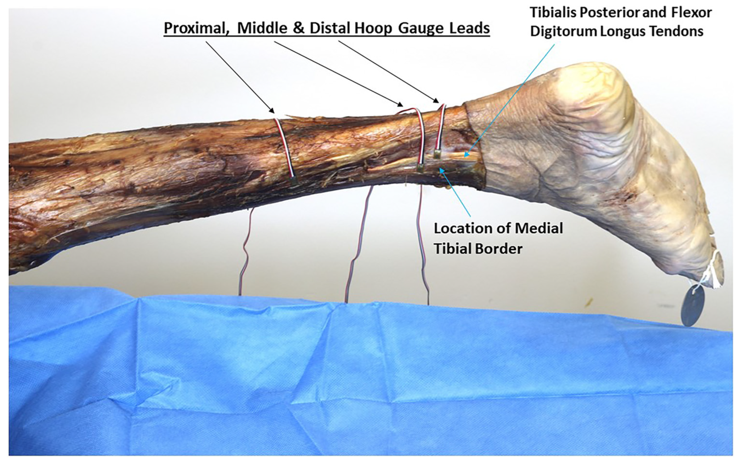
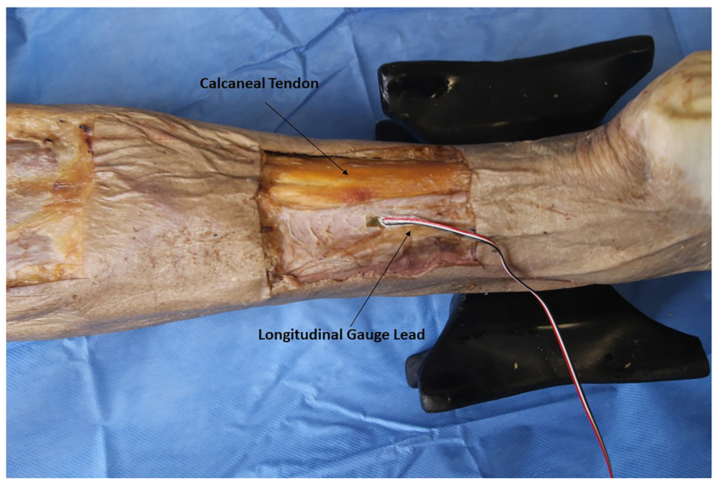
The subject was placed in prone and the skin and subcutaneous fat were removed to expose the CF in the posterior compartments of both legs. The test-ing area of the CF was cleaned with isopropyl alco-hol pads and abraded with fine sandpaper before placement of the strain gauges (SG). Microstrain gauges 3.18 mm in length with pre-attached leads (VPG Micro-Measurements, Raleigh, NC), were used. M-Bond Adhesive (VPG Micro-Measurements, Raleigh, NC) was applied to each SG and manual pressure was maintained for 2 minutes after applying adhesive, and allowed to dry for 30 hours before data collection to ensure maximal adhesion to the CF. Three horizontally oriented SGs were placed on the distal right leg of the cadaver donor (6, 7, and 15.5 cm vertical distance from the medial malleo-lus), with the most proximal and distal gauges located 10 mm posterior to the medial tibial bor-der; and the middle gauge overlapping the medial border of the tibia by 5 mm (Figure 2). A vertically oriented SG was placed in the posterior compart-ment of the donor’s left leg, 9.5 cm superior to the medial malleolus, and 6 mm lateral to the cal-caneal tendon, to measure longitudinal strain (Figure 3). Gauges were placed in the middle and distal thirds of the leg to coincide with the seg-ments of the tibia where MTSS symptoms are most commonly felt [4,5,6,7].
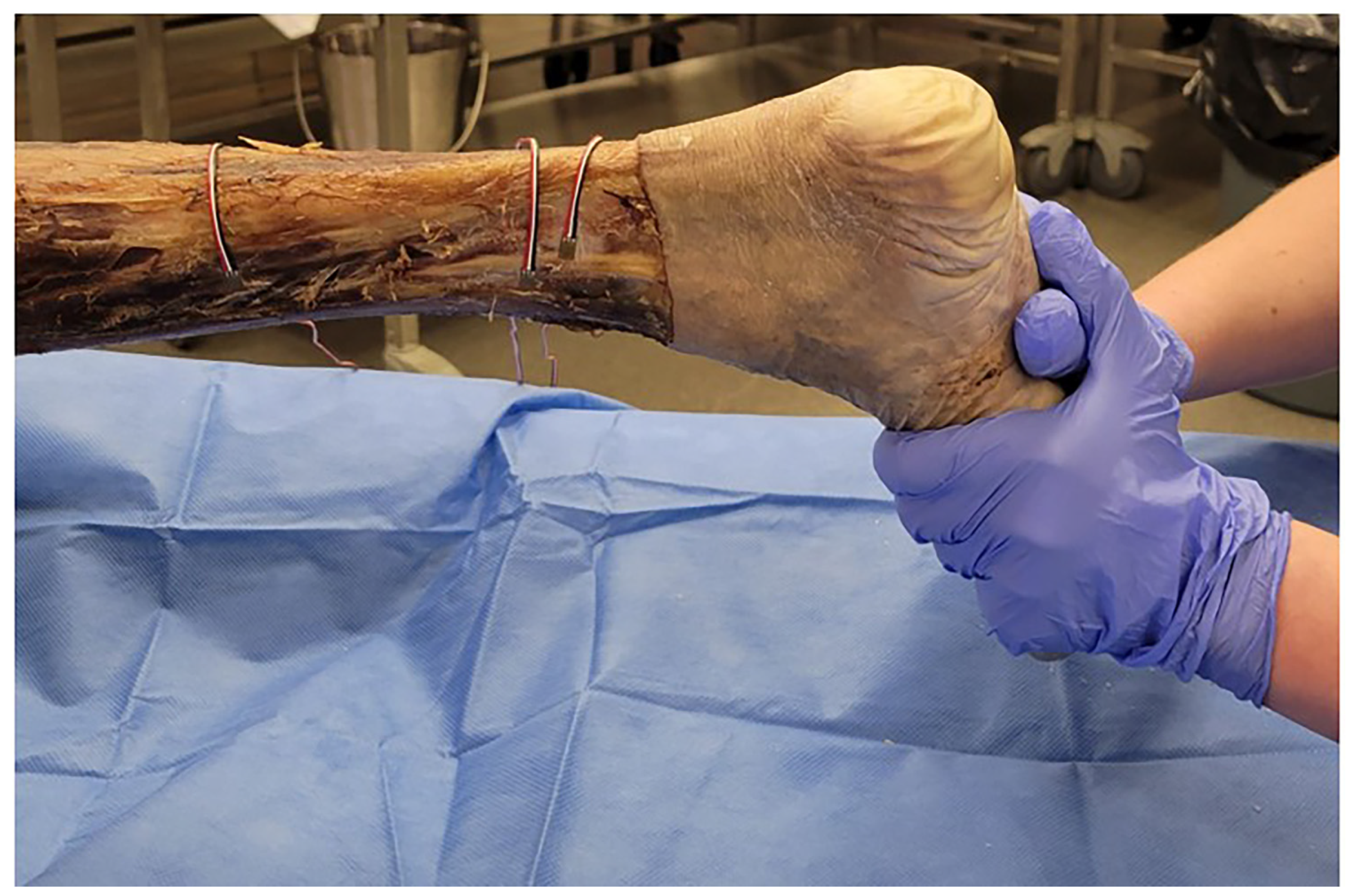
Measurements were taken from each of the SGs independently. Leads from each SG were crimped to an RJ45 Connector (Glark, Shenzhen, China) and plugged into a Student Data Acquisition Unit (VPG Micro-Measurements, Malvern, PA) and connected to a Dell laptop. Data was analyzed with MM01 MultiDAQ software (V. 2.0.1.3 Vishay Measurements Group, Raleigh, NC) A passive stretch into dorsi-flexion was applied manually to the metatarsal heads by one investigator (M.R.R.) and held for approximately 5 seconds, and then repeated up to five times with increasing force in order to obtain greater excursion into dorsiflexion with successive stretches (Figure 4). A maximal force of 341 N was applied during the heel cord stretch as measured by a MicroFet2 Digital Muscle Tester (Hoggan Scientific, Salt Lake City, UT) prior to the start of strain gauge data collection.

Results

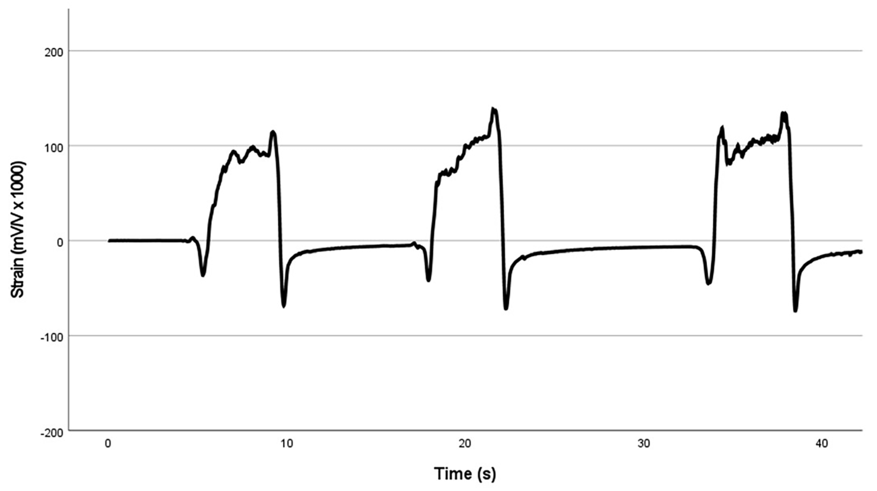
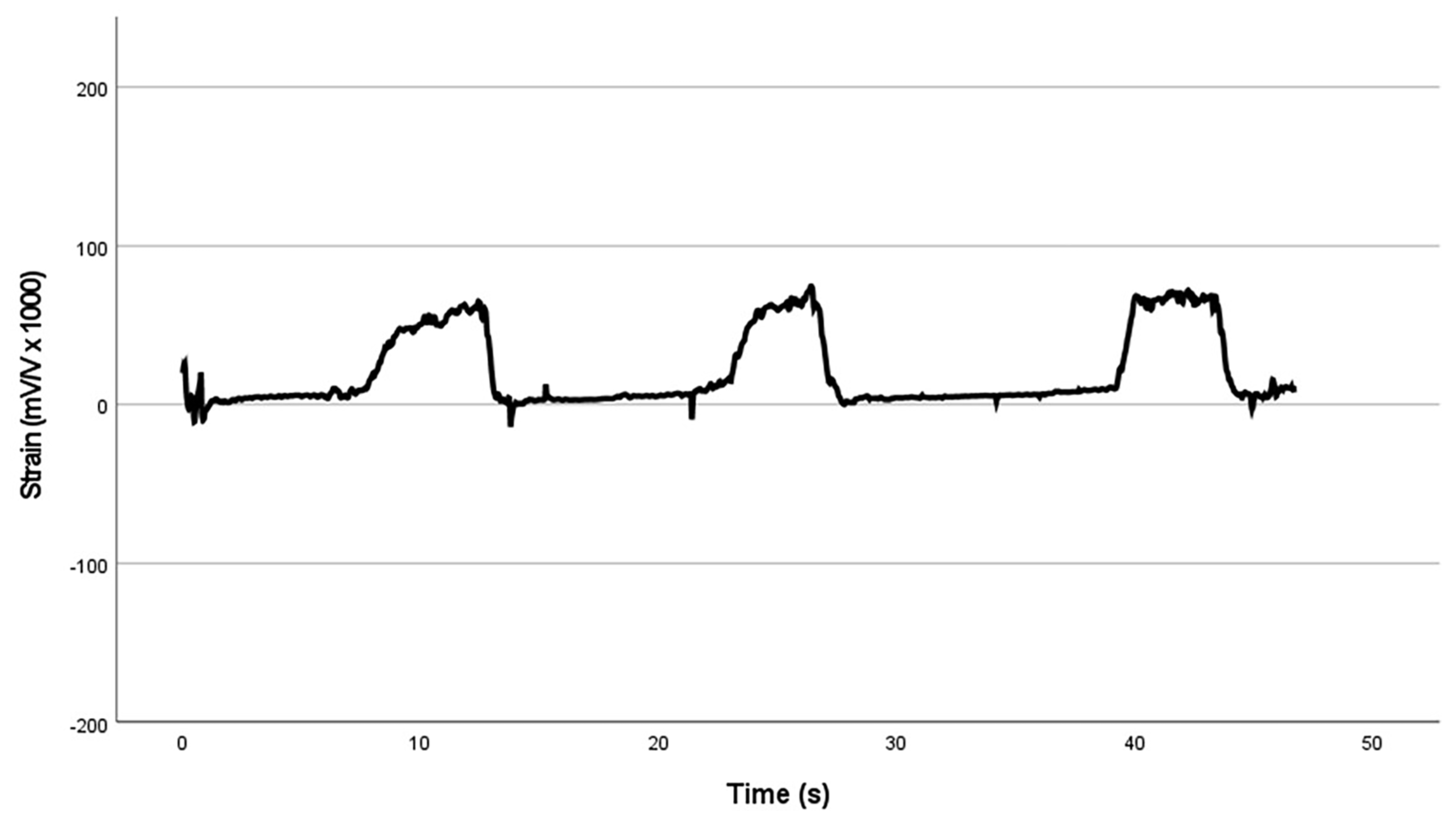
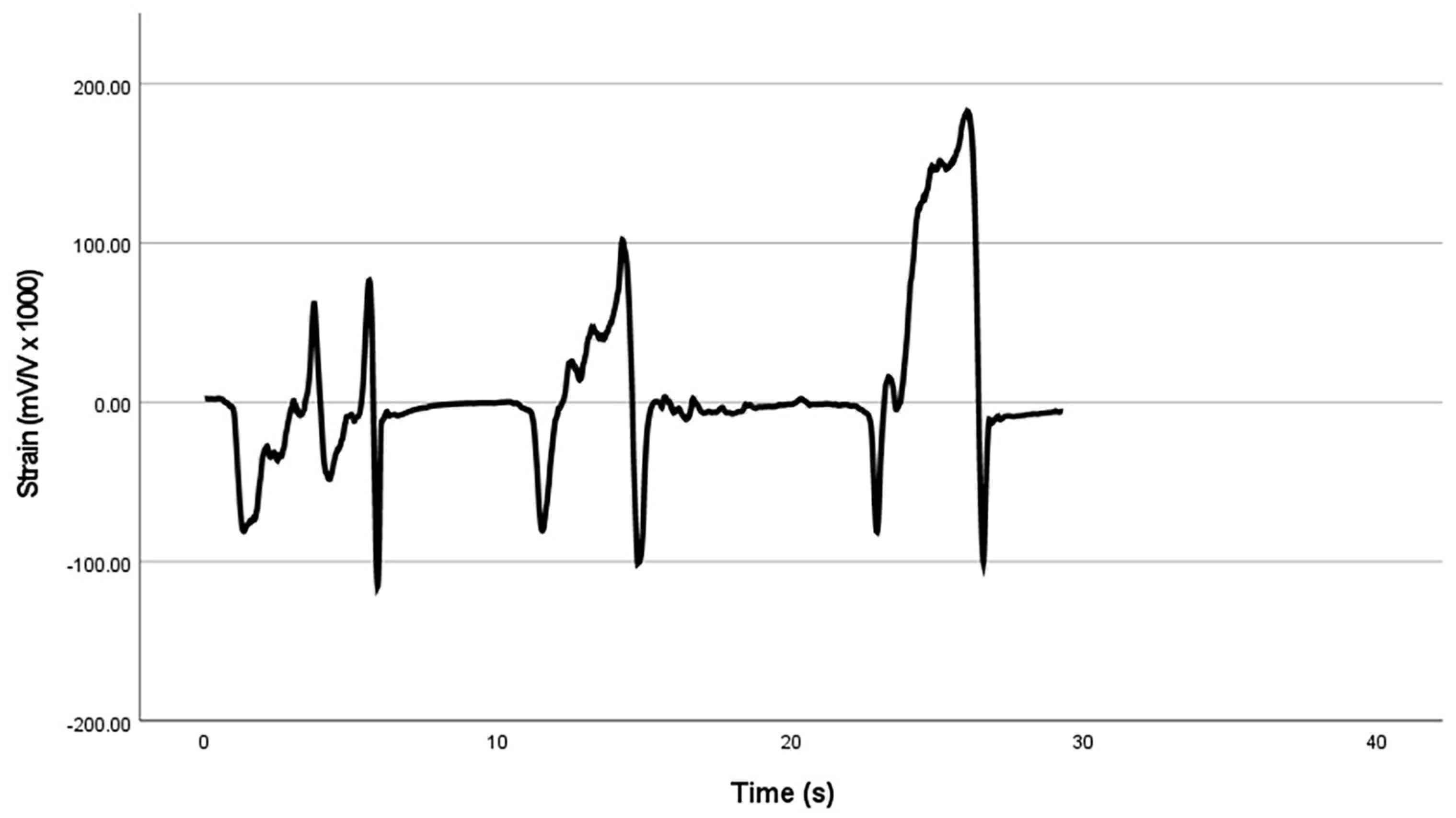
Repeated heel cord stretches resulted in production of a maximum of 182.96 × 10²³ mV/V hoop strain at the CF attachment to the medial tibial border in the distal SG (Figure 5); and a maximum of 138.00 × 10²³ mV/V hoop strain at the CF attachment to the medial tibial border in the proximal SG (Figure 6). The strain data for the middle gauge, which overlapped the tibia, produced error data with a negative sign and so was thrown out. Passive dorsiflexion generated a maximum longitudinal strain in the CF of the superficial posterior compartment of 75.00 × 10²³ mV/V (Figure 7). Maximum strain values for the above SGs are contained in Table 1.

Discussion

Our results show that a heel cord stretch applied to a cadaver donor can produce a measurable hoop strain within the CF attachment to the medial border of the tibia. This happened in a grossly 2-to-1 manner consistent with the ratio of hoop to longitudinal strain seen with gases and liquids in a closed cylinder. It is an accepted principle of physics that a tensile force cannot produce stress in a vector that is perpendicular to the application of the force [20]. It has previously been, therefore, considered counter-intuitive that a heel cord stretch causing longitudinal traction forces would be able to create a horizontal stress on the CF attachment to the medial tibia. To our knowledge, this is the first research hypothesizing an MTSS etiology based on thin-walled cylinder stress behaviors resulting from increased intercompartmental pressure in the leg with passive stretch.
Our study builds on the work of Bouche´ and Johnson [8], who found measurable strain in the CF in horizontally placed microstrain gauges when tension was applied, via cables and pneumatic actuators, to the flexor digitorum longus and soleus muscles of three disarticulated cadaver limbs. Based on these experiments, they proposed a “tibial fascial traction theory,” whereby MTSS is caused by a “tenting” effect in the CF at the medial tibial border, secondary to eccentric con-traction of these muscles during stance phase of gait. They did not, however, explain the specific mechanism whereby contraction of these muscles could produce this strain, given that neither has an attachment to the CF.
The deep fascia is a connective tissue that has been classified as both dense, irregular [21] and dense regular [22] connective tissue, depending on the source. Composed mostly of Type I collagen and elastin, it is richly innervated by both myelinated and unmyeli-nated neurons, and has been shown to serve both a proprioceptive and nociceptive function [22,23,24].The CF contains nociceptors [25] that may be implicated in MTSS pain. It also tractions the periosteum at its attachments to the tibia, potentially causing pain production arising from periosteal nociceptors [26]. Transmission of forces through the deep fascia has been shown in multiple body regions. Sheets of fascia cross joints and are able to elicit mechanical force transmission when joints are moved actively or passively. This happens through either direct fascial connections, such as CF to fascia lata force transmission, or via intervening muscles as seen with gluteus maximus to thoracolumbar fascia, to latissimus dorsi force transmission [16,27,28,29].
Sustained heel cord stretching is known to increase ankle range of motion over time [30]. Stretching before a workout is thought to increase connective tissue compliance in an unloaded or low-loaded condition prior to initiation of vigorous high-load activities such as running and jumping [31]. Runners with MTSS have been found to have significantly decreased dorsiflex-ion range of motion [32], and heel cord stretching has been widely used in prevention and treatment of MTSS [10,11,12,13,14].Yet none of these studies have conclusively demonstrated a significant effect of this type of intervention. Sample size limits, as well as a lack of control of covariates, may have been a factor in the lack of significance.
Our study has several limitations. Due to rigor mortis, side effects of the formalin fixative, and pos-sible premorbid tissue shortening, our subject’s ankle initially presented with a fairly rigid plantar-flexion contracture. This limited our ability to precisely replicate a heel cord stretch comparable to that seen in a healthy, living athlete. The contralateral leg was used for longitudinal strain measurements due to damage to the CF during dissection, which prevented adequate adherence of the longitudinal strain gauge to the ipsilateral hoop strain test leg. Dynamometer readings of the force of the man-ual stretch were taken separately from the gauge readings, due to limitations in the experimental set up. Finally, although hoop strain was produced, it is unclear whether this level of strain would be considered clinically significant in a living human subject.

Conclusions

Hoop strain at the most common site of MTSS pain was generated with passive heel cord stretch in a single cadaver donor. Further research is required to replicate these results in a larger sample, with biomechanical instrumentation that allows concur-rent data collection from multiple sensors, and real-time linking of strain measurements with stretching force. Use of a softer embalming solution, as seen with Thiel fixation[33,34], in future research would also yield more biofidelic results.

Funding

None reported.

Acknowledgments

We gratefully acknowledge the support of our cadaver donor and her family, and the Willed Body Program of the University of Kansas Medical Center, without whom this research would not be possible.

Conflicts of Interest

None reported.

References

  1. STICKLEY CD, HETZLER RK, KIMURA IF, ET AL: Crural fascia and muscle origins related to medial tibial stress syn-drome symptom location. Med Sci Sports Exerc 41: 1991, 2009.
  2. THACKER SB, GILCHRIST J, STROUP DF, ET AL: The prevention of shin splints in sports: a systematic review of litera-ture. Med Sci Sports Exerc 34: 32, 2002.
  3. DETMER DE: Chronic shin splints. Classification and management of medial tibial stress syndrome. Sports Med 3: 436, 1986.
  4. BATT ME, UGALDE V, ANDERSON MW, ET AL: A prospective controlled study of diagnostic imaging for acute shin splints. Med Sci Sports Exerc 30: 1564, 1998.
  5. FREDERICSON M, BERGMAN AG, HOFFMAN KL, ET AL: Tibial stress reaction in runners. Correlation of clinical symptoms and scintigraphy with a new magnetic reso-nance imaging grading system. Am J Sports Med 23: 472, 1995.
  6. RESHEF N, GUELICH DR: Medial tibial stress syndrome. Clin Sports Med 31: 273, 2012.
  7. ORAVA S, PURANEN J: Athletes’ leg pains. Br J Sports Med 13: 92, 1979.
  8. BOUCHE´ RT, JOHNSON CH: Medial tibial stress syndrome (tibial fasciitis): a proposed pathomechanical model involving fascial traction. JAPMA 97: 31, 2007.
  9. BROWN AA: Medial tibial stress syndrome: muscles located at the site of pain. Scientifica (Cairo) 2016: 7097489, 2016.
  10. KORTEBEIN PM, KAUFMAN KR, BASFORD JR, ET AL: Medial tibial stress syndrome. Med Sci Sports Exerc 32: S27, 2000.
  11. ANDRISH JT, BERGFELD JA, WALHEIM J: A prospective study on the management of shin splints. J Bone Joint Surg Am 56: 1697, 1974.
  12. CRAIG DI: Medial tibial stress syndrome: evidence-based prevention. J Athl Train 43: 316, 2008.
  13. CORTE´ S GONZA´ LEZ RE: Successful treatment of medial tib-ial stress syndrome in a collegiate athlete focusing on clinical findings and kinesiological factors contributing to pain. Physiother Theory Pract: 38: 1, 2020.
  14. MOEN MH, HOLTSLAG L, BAKKER E, ET AL: The treatment of medial tibial stress syndrome in athletes; a randomized clinical trial. Sports Med Arthrosc Rehabil Ther Technol 4: 12, 2012.
  15. MOORE KL, DALLEY AF, AGUR AMR: Clinically Oriented Anatomy, illustrated edition, Lippincott Williams & Wilkins, New York, 2013.
  16. MYERS TW: Anatomy Trains: Myofascial Meridians for Manual and Movement Therapists, Elsevier Health Sciences, Amsterdam, 2009.
  17. IBRAHIM A, RYU Y, SAIDPOUR M: Stress analysis of thin-walled pressure vessels. MME 5: 1, 2015.
  18. TSINTZAS D, GHOSH S, MAFFULLI N, ET AL: The effect of ankle position on intracompartmental pressures of the leg. Acta Orthop Traumatol Turc 43: 42, 2009.
  19. GERSHUNI DH, YARU NC, HARGENS AR, ET AL: Ankle and knee position as a factor modifying intracompartmental pressure in the human leg. J Bone Joint Surg Am 66: 1415, 1984.
  20. BENENSON W, HARRIS JW, STOCKER H, ET AL eds: Handbook of Physics, Springer New York, New York, NY, 2006.
  21. LANGEVIN HM, HUIJING PA: Communicating about fas-cia: history, pitfalls, and recommendations. Int J Ther Massage Bodywork 2: 3, 2009.
  22. STECCO C, PORZIONATO A, LANCEROTTO L, ET AL: Histological study of the deep fasciae of the limbs. J Bodyw Mov Ther 12: 225, 2008.
  23. SCHLEIP R, KLINGLER W, LEHMANN-HORN F: Active fascial contractility: Fascia may be able to contract in a smooth muscle-like manner and thereby influence musculoskel-etal dynamics. Med Hypotheses 65: 273, 2005.
  24. KLINGLER W, VELDERS M, HOPPE K, ET AL: Clinical relevance of fascial tissue and dysfunctions. Curr Pain Headache Rep 18: 439, 2014.
  25. FEDE C, PETRELLI L, GUIDOLIN D, ET AL: Evidence of a new hidden neural network into deep fasciae. Sci Rep 11: 12623, 2021.
  26. NENCINI S, IVANUSIC JJ: The physiology of bone pain. how much do we really know? Front Physiol 7: 157, 2016.
  27. BARKER PJ, BRIGGS CA, BOGESKI G: Tensile transmission across the lumbar fasciae in unembalmed cadavers: effects of tension to various muscular attachments. Spine 29: 129, 2004.
  28. CARVALHAIS VO DO C, OCARINO J DE M, ARAU´ JO VL, ET AL: Myofascial force transmission between the latissimus dorsi and gluteus maximus muscles: an in vivo experiment. J Biomech 46: 1003, 2013.
  29. KRAUSE F, WILKE J, VOGT L, ET AL: Intermuscular force transmission along myofascial chains: a systematic review. J Anat 228: 910, 2016.
  30. GAJDOSIK RL, LEVEAU BF, BOHANNON RW: Effects of ankle dorsiflexion on active and passive unilateral straight leg raising. Phys Ther 65: 1478, 1985.
  31. WITVROUW E, MAHIEU N, DANNEELS L, ET AL: Stretching and injury prevention: an obscure relationship. Sports Med 34: 443, 2004.
  32. TWEED JL, CAMPBELL JA, AVIL SJ: Biomechanical risk fac-tors in the development of medial tibial stress syndrome in distance runners. JAPMA 98: 436, 2008.
  33. REDDY R, IYER S, PILLAY M, ET AL: Soft embalming of cadav-ers for training purposes: Optimising for long-term use in tropical weather. Indian J Plast Surg 50: 29, 2017.
  34. LIAO P-Y, WANG Z-G: Thiel-embalming technique: investi-gation of possible modification in embalming tissue as evaluation model for radiofrequency ablation. J Biomed Res 33: 280, 2019.
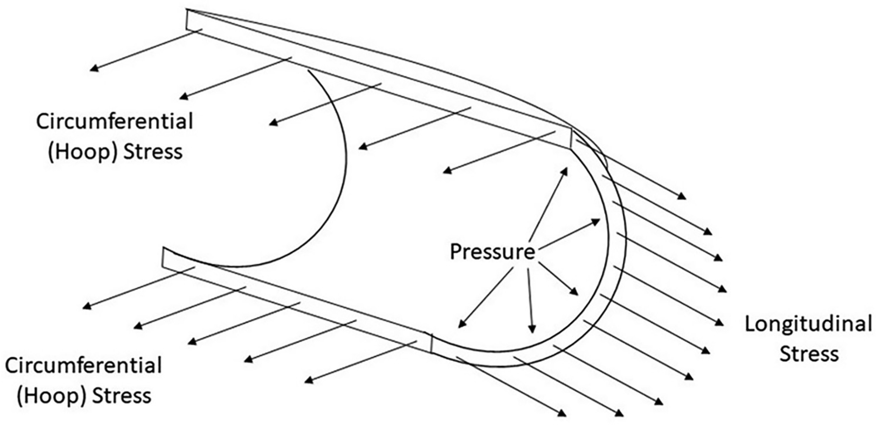
Figure 1. Longitudinal and hoop stress in a cylinder.
Japma 114 23169 g001
Figure 2. Placement of gauges to measure hoop strain.
Japma 114 23169 g002
Figure 3. Placement of gauge to measure longitudinal strain.
Japma 114 23169 g003
Figure 4. Heel cord stretch technique.
Japma 114 23169 g004
Figure 5. Hoop strain measurements from distal gauge.
Japma 114 23169 g005
Figure 6. Hoop strain measurements from proximal gauge.
Japma 114 23169 g006
Figure 7. Longitudinal strain measurements.
Japma 114 23169 g007
Table 1. Maximum Hoop and Longitudinal Strain Measurement.
Japma 114 23169 i001