


Abstract
Background: Isolated medial malleolar fractures are observed less often than are lateral malleolar fractures, bimalleolar fractures, and trimalleolar fractures. The aim of treatment is to provide anatomical reduction, protect this reduction until the fracture heals, and to regain normal ankle function. The aim of our study was to evaluate the clinical and radiological out-comes of patients with isolated medial malleolus fracture treated with cross-screw fixation. Methods: The study included 13 patients: 9 males with a mean age of 37.1 years (range, 25–64 years) and 4 females with a mean age of 40.2 years (range, 24–62 years) who were treated with the cross-screw fixation method for an isolated medial malleolar fracture. The fracture union status was evaluated on the 3-month postoperative radiographs, and the functional outcomes with the American Orthopaedic Foot and Ankle Scale (AOFAS) ankle score. Results: The mean AOFAS score was 82 at 3 months postoperatively, evaluated as an excellent outcome in 2 patients, good in 9, and fair in 2. At the 24th month postoperatively, the mean AOFAS score was determined to be 92.2, evaluated as excellent in 11 patients and good in 2. On palpation of the medial malleolus, the mean VAS pain score was 2 (range, 1–3.8) at 3 months postoperatively, and a mean of 0.6 (range, 0.2–3) at 24 months. Conclusions: Cross-screw fixation can be preferred for the fixation of isolated medial malleolar fractures as it causes less soft-tissue damage because it can be performed percutaneously, and the screw head has a low profile, thereby eliminating the need for a Kirschner wire to be applied together with the screw to prevent rotational deformity.
Of all fractures of the skeletal system, ankle fractures are the second most frequently seen after hip fractures [1]. Isolated medial malleolar fractures are observed less often than are lateral malleolar fractures, bimalleolar fractures, and trimalleolar fractures [2]. Together with the better understanding of ankle biomechanics, the strategies used in treat-ment have shown differences over time. The aim of treatment is to provide anatomic reduction, protect this reduction until the fracture heals, and to regain normal ankle function. Previous studies have shown that prognosis is more positive when full anatomic reduction is obtained [3,4].
A reduction that is not fully anatomic can cause post-traumatic arthritis in the joint, restricted joint range of movement, and pain [5]. However, no consensus has yet been reached on the subject of the optimal treatment protocol. Nondisplaced isolated medial malleolar fractures can be treated conservatively [6]. Anatomic reduction and fixation is required for a displaced medial malleolus fracture. When deter-mining which fixation method to use in a medial mal-leolus fracture, one must take into account the orientation of the fracture line, the size of the fracture fragments, and the patient’s age [7].
Partially threaded screws, fully threaded screws, anti-slip plates and Kirschner wires, and the tension band method can be used in the fixation of these fractures. If the fracture fragment is big enough, 2 screws can be used parallel to each other. If the fragment is not big enough, the use of a screw and Kirschner wire is recommended to provide rotational stability. The tension band method can be used in fragmented fractures or small avulsion fractures [8].
Cross-screw fixation is the surgical method applied together with the plate used in osteoporotic proximal humerus fractures [9]. We applied the cross screws alone for the medial malleolar fixation.
The aim of this study was to evaluate the clinical and radiological outcomes of patients with isolated medial malleolus fractures treated with cross-screw fixation. Our report seems to be the first in the literature for cross-fixation screw application for isolated medial malleolar fractures.
Methods
The Clinical Research Ethics Committee of Giresun University Faculty of Medicine, Giresun, Turkey, approved the present study. The patients were informed that medical records could be used for scientific purposes only, and a written informed con-sent was obtained from every patient.
The records of patients who underwent surgery for a diagnosis of an isolated medial malleolar fracture were screened in the orthopaedics and traumatology clinic. The patients included in the study were those 18 to 90 years old who were applied with cross-screw fixation because of an isolated medial malleolus fracture. The age and gender of the patients, and mechanism of injury were recorded. Patients were excluded from the study if they had any concomitant injury or syndesmotic injury.
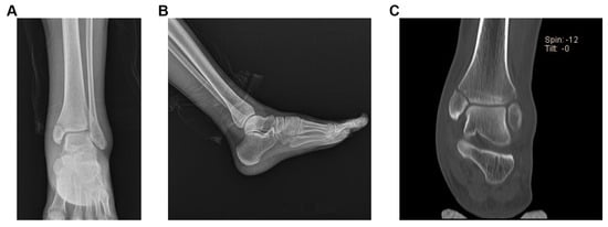
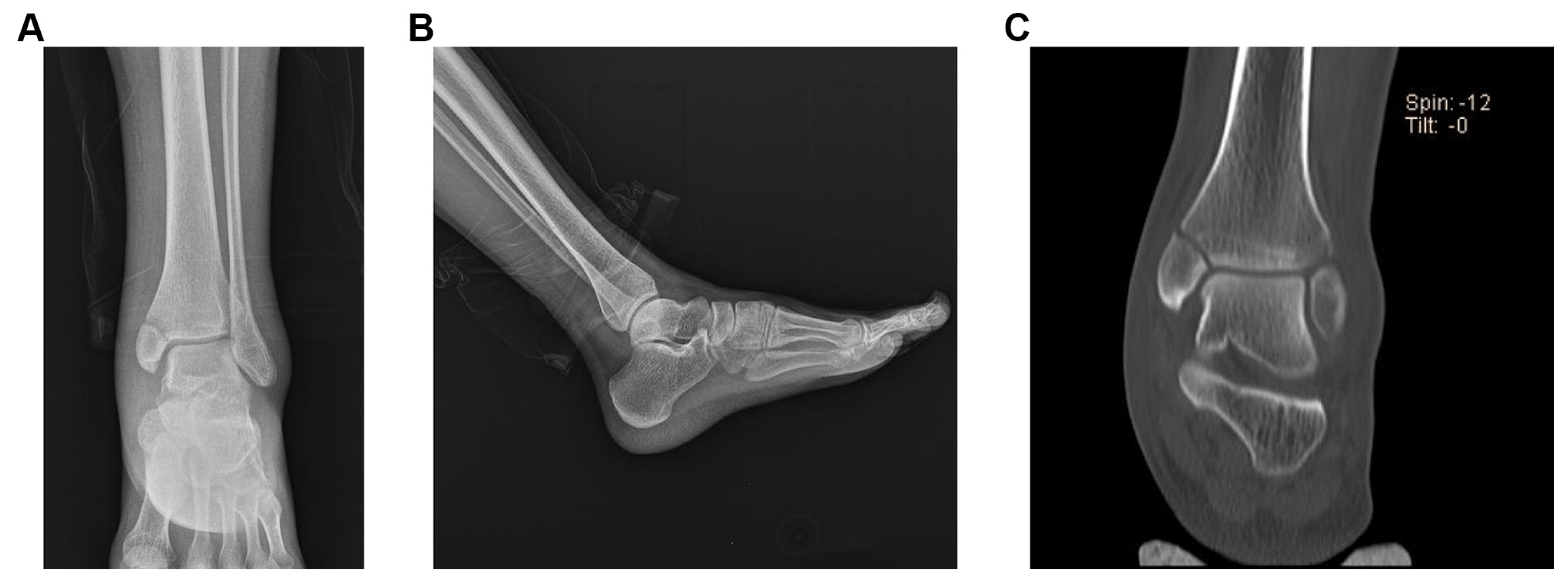
Radiographs of the anteroposterior, lateral, and mortise view of the involved ankle were obtained preoperatively (Figure 1), postoperatively, and at every follow-up time point. The fracture pattern and treatment method were evaluated with the radiographs. Patients with an implant other than cross-screws were excluded from the study.
Figure 1.
Preoperative anteroposterior (A) and lateral (B) radiographs of an isolated medial malleolar frac-ture, and preoperative computed tomographic scan (C) of the patient shows an isolated medial malleolar fracture.
The evaluation of the fracture pattern was based on the fracture level as described in the Herscovici et al [10] classification system. Medial malleolar fractures are classified as Type A: avulsion of the malleolus end distal of the ankle joint line, Type B: fracture between the plafond level and the end of the malleolus, Type C: fracture at the level of the plafond, and Type D: fracture extending vertically above the level of the tibial plafond.
Radiological union was accepted as the fracture line not visualized on radiographs and/or bony callus tissue formed in the fracture line. The absence of signs of progressive fracture healing in a period of 3 months was accepted as representing delayed union, and if this period was longer than 6 months, it was accepted as nonunion.
The clinical outcomes were evaluated using the American Orthopaedic Foot and Ankle Society (AOFAS) ankle-hindfoot score. In the 100-point AOFAS scoring system, the scores are evaluated as $90 points: excellent; 80–89 points: good; 70–79 points: fair; and #69 points: poor outcome [11].
The study included 13 patients who were treated with the cross-screw fixation method for an isolated medial malleolar fracture diagnosed in our hospital during the study period. Anteroposterior, lateral, and mortise radiographs were obtained 4 weeks postoperatively and then at 6, 8, and 12 weeks until radiographic union was obtained.
Functional outcomes were evaluated with the AOFAS ankle score. At the end of the 24th month postoperatively, the patients were contacted by telephone and called for a follow-up examination.
Radiographs were taken for the evaluation of the fracture union, and the functional outcomes were evaluated with the AOFAS scores. The severity of pain over the medial malleolus was evaluated using a visual analog scale (VAS), 10-cm horizontal line in length, marked from 0 (no pain) to 10 (severe pain). The patients were instructed to mark the point rep-resenting the severity of pain felt and the points were rounded to the nearest whole number (eg, 2.3 cm 5 2, 0.8 cm 5 1).
After appropriate sterile draping, antibiotic pro-phylaxis was administered. All the operations were performed under spinal anaesthesia with the patient positioned supine and a pneumatic tourniquet was used in all cases. The lower extremity was washed with 10% povidone iodine soap and draped under sterile conditions. Using a clamp, closed reduction was performed on the medial malleolus under fluoroscopy guidance. When the reduction was seen to be appropriate, a Kirschner wire was advanced from the midline of the tip of the medial malleolus to be parallel to the medial cortex. After the drilling procedure, the fracture was fixed by advancing a cancellous screw over the Kirschner wire.
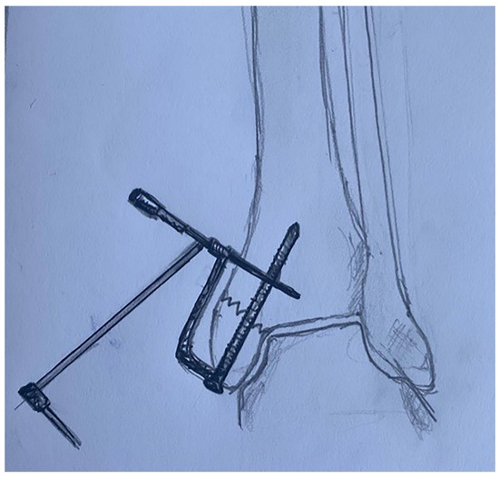
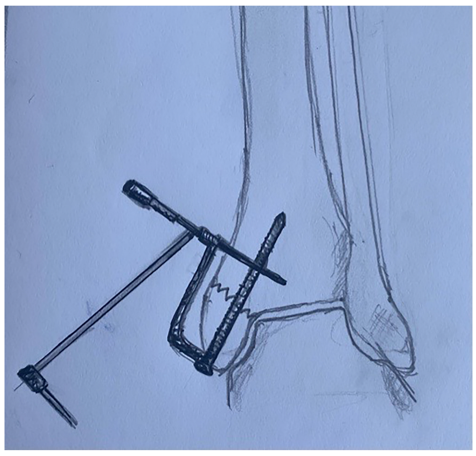
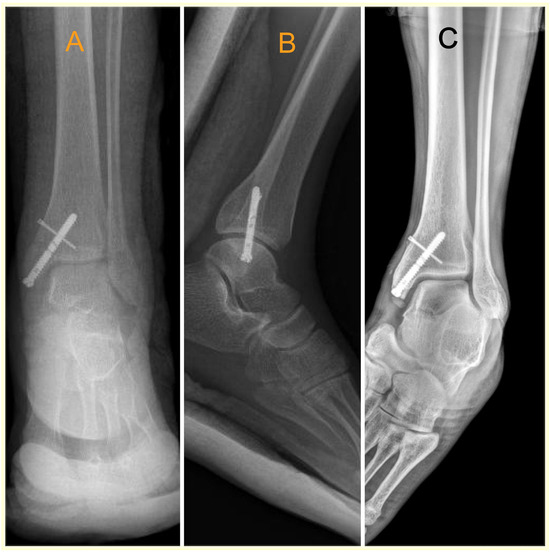
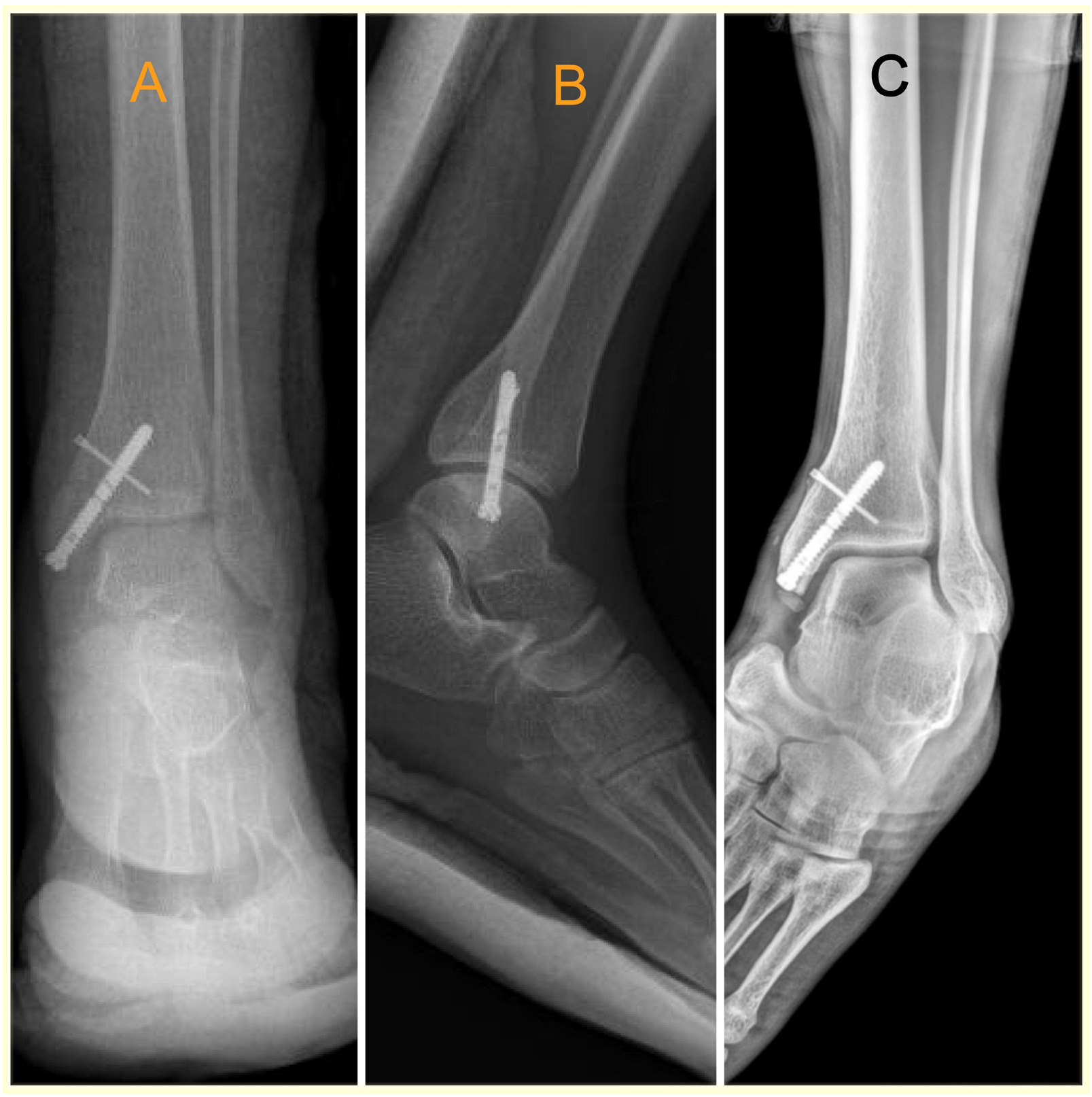
The cross-screw guide is threaded onto the post with a retainer screw. After applying the screw, the cross-screw guide was installed. Then, a Kirschner wire was passed through the cross-screw guide sleeves into the bone (Figure 2). Both anteroposterior and lateral views were captured to assess the placement of the Kirschner wire using fluoroscopy. The wire was then removed, and an anti-rotational screw was convergently advanced to pass distal to the screw. Final reduction and placement were evaluated using fluoroscopy, and the subcutaneous tissues and skin were sutured. A short leg splint was applied to all the patients. Postoperative radiographs of all patients’ ankles were taken (Figure 3).
Figure 2.
Schematic view of the application of the cross-screw guide and the cross screw.
Figure 3.
Postoperative anteroposterior (A), lateral (B), and (C) mortise radiographs of the patient treated with 1 screw that was fixed with an anti-rotational screw.
All of the patients were followed up according to the standard postoperative follow-up protocol established in our clinic. All of the patients were immobilized for the first 1 to 2 weeks postoperatively with a short-leg splint. After removal of the splint, active and passive ankle joint range of motion exercises were started. Weightbearing was permitted when there was radiographic evidence of healing together with clinical examination findings.
RESULTS
We evaluated 13 patients: 9 (69.2%) males with a mean age of 37.1 years (range, 25–64 years) and 4 (30.7%) females with a mean age of 40.2 years (range, 24–62 years). Right-side medial malleolus fracture was present in 8 patients and left-side medial malleolus fracture in 5. The fracture resulted from an out-of-vehicle traffic accident in 2 patients, a motorcycle accident in 1, direct trauma in 4, twisting of the ankle in 1, a fall in 2, and an in-vehicle traffic accident in 3. According to the Herscovici classification, Type B in 5, Type C in 5, and Type D in 3.
There was no malunion, loosening of the implant, wound site problems, or loss of reduction in any patient. When the postoperative radiographs were examined, anatomic reduction was observed in all the patients. Radiological bone union was achieved in all 13 patients. There was no requirement for implant removal in any patient.
The mean AOFAS score was 82 at 3 months post-operatively, evaluated as an excellent outcome in 2 patients, good in 9, and fair in 2. At the 24th month postoperatively, the mean AOFAS score was deter-mined to be 92.2, evaluated as excellent in 11 patients and good in 2.
On palpation of the medial malleolus, the VAS pain score was mean 2 (13.8) at postoperative 3 months and mean 0.6 (0.2–3) at 24 months.
Discussion
Together with the anterior talofibular ligament, the medial malleolus prevents medial translation of the talus. Therefore, maintaining congruity within the ankle mortise of the medial malleolus is necessary for normal tibiotalar contact surface and normal tibiotalar pressure distribution [12]. Even millimetric talar migration can reduce the contact area in the tibiotalar joint by approximately 40%, and this can cause post-traumatic osteoarthritis and poor healing outcomes [13]. With surgical treatment of intra-articular fractures providing full reduction and early joint movement, restricted movement, the risk of increased degeneration, and loss of workforce can be reduced [14]. Therefore, isolated medial malleolus fractures are generally treated surgically [5,7,8].
Various fixation methods have been described in the surgical treatment of medial malleolus fractures. The application of 1 or 2 spongious screws, Kirschner wires, and the tension band method are frequently used methods. However, there is no clear evidence about which technique is clinically superior.
Considering the inadequate soft-tissue support around the ankle, particularly in the ankle area, there has been a notable increase in the frequency of implant-related complications associated with traditional fixation methods, especially the tension band method [15]. Despite full bone union in operations performed using cancellous screws, tension band and Kirschner wire may cause discomfort. Additionally, medial malleolar screws may result in irritation of the posterior tibialis tendon, other soft-tissue irritations, or bony impingement [16,17]. The most common causes of discomfort are pain, prominent hardware, difficulty in shoe wear and functional dissatisfaction [18]. These reasons are usually due to soft-tissue destruction associated with the implant, and the pain felt by the patient may be at a level that will require removal of the implant [19]. Removing the implant may not be a harmless procedure as expected since it may cause several complications such as skin necrosis, infection, and several iatrogenic injuries [20,21].The screws used in the cur-rent study have a low profile, less protrusion on the bone surface, and fewer skin complications, which was confirmed by the fact that chronic pain was not observed in the long term in the current study patients. There was no requirement for implant removal in any patient during the mean 2-year follow-up and this seems to be an advantage.
In this study, the clinical and radiological out-comes were evaluated of patients for whom percutaneous cross-screw fixation was applied following closed reduction of an isolated medial malleolus fracture. Cross-screw fixation provides additional stability and minimizes the risk of reduction loss and protrusion into the articular surface of the humeral head. Minimally invasive approaches that reduce soft-tissue stripping have gained popularity, including a mini-arthrotomy technique involving a 3-cm incision at the superomedial aspect of the mortise, medial to the tendon of the tibialis anterior [22]. This approach allows for articular inspection, irrigation, and fracture reduction while fixation is performed through a separate stab incision. A purely percutaneous approach, used in high-risk multi-comorbid patients through a 1-cm wound distal to the tip of the malleolus, has shown promising results [23]. Weinraub et al [24] reported a higher rate of nonunion in patients who underwent pure percutaneous medial fixation compared to those who underwent open reduction. The authors attributed this difference to the interposed periosteal flap, which can become trapped during the rotational aspect of the injury and is not retrieved during per-cutaneous fixation. It’s important to note that this study may have had selection bias due to its retrospective design and the disparity in group sizes. Additionally, radiological follow-up was not con-ducted beyond eight weeks, making it impossible to compare nonunion rates. In our study, none of the cases showed evidence of non-union. The results obtained according to the AOFAS ankle-hindfoot scale for the 13 patients included in the study were excellent in 11 (84.6%) patients and good in 2 (15.4%) patients. The mean AOFAS ankle-hindfoot score was determined to be 92.2 (range, 80–95). The mean VAS score was 2 (range, 1–3.8) at 3 months postoperatively and 0.6 (range, 0.2–3) at 24 months. Our results are similar for both radiologic and functional outcomes that have been reported in the literature.
Traditionally, 2 4.0-mm screws were used for medial malleolar fixation instead of 1 to ensure rotational control. When only 1 screw is used, significant torsional forces can lead to medial malleolus failure in tension or compression [17]. Consequently, stable fix-ation may necessitate the use of 2 screws. It’s worth noting that the incidence of implantrelated complications might potentially be reduced through single-screw fixation. Femino et al [19] reported that in sur-geries performed with screws applied to the region greatly posterior of the medial malleolus or fracture fixation with 2 screws, the screws applied in zone 3 were in direct contact with the tibialis posterior ten-don, which was found to cause irritation in the tendon Using 2 screws for fixation in a small medial malleolar fragment can result in comminution of this small piece of bone [18]. In the current study, fracture fixation in all the patients was achieved with 1 screw and because of the divergent locking there was no need for a second screw.
Full bone union was obtained in all the patients in this study. There were no complaints associated with malunion, wound site problems, screw dis-placement, loss of reduction, or tibialis posterior tendon damage. These results demonstrate that isolated medial malleolar fractures can be successfully treated with cross-screw fixation.
Limitations of this study include the retrospective design, the absence of a control group, the low number of patients, and lack of biomechanical comparison.
In conclusion, cross-screw fixation can be preferred for the fixation of isolated medial malleolar fractures as it causes less soft-tissue damage because it can be performed percutaneously, and the screw head has a low profile, thereby eliminating the need for a Kirschner wire to be applied together with the screw to prevent rotational deformity.
Funding
None reported.
Conflicts of Interest
None reported.
References
- COURT-BROWN CM, CAESAR B: Epidemiology of adult fractures: a review. Injury 37: 691, 2006; doi: https://doi.org/10.1016/j.injury.2006.04.130.
- JAFFE D, CHRISTIAN MW, WEBER A, ET AL: Incarceration of the posterior tibial tendon in an isolated comminuted medial malleolus fracture. J Foot Ankle Surg 56: 1312, 2017; doi: https://doi.org/10.1053/j.jfas.2017.05.016.
- GEHR J, FRIEDL W: Intramedullary locked fixation and compression nail (IP-XS-Nail): treatment of ankle joint fractures. Operative Orthopadie und Traumatologie 18: 155, 2006; doi: https://doi.org/10.1007/s00064-006-1168-0.
- BREEDERVELD R, VAN STRAATEN J, PATKA P, ET AL: Immediate or delayed operative treatment of fractures of the ankle. Injury 19: 436, 1988; doi: https://doi.org/10.1016/00201383(88)90142-8.
- CARTER TH, DUCKWORTH AD, WHITE T: Medial malleolar fractures: current treatment concepts. Bone Joint J 101: 512, 2019; doi: https://doi.org/10.1302/0301-620x.101B5.BJJ-2019-0070.
- Hoelsbrekken SE, Kaul-Jensen K, Mørch T, et al: Nonoperative treatment of the medial malleolus in bimal-leolar and trimalleolar ankle fractures: a randomized con-trolled trial. J Orthop Trauma 27: 633, 2013; doi: https://doi.org/10.1097/BOT.0b013e31828e1bb7.
- KING CM, COBB M, COLLMAN DR, ET AL: Bicortical fixation of medial malleolar fractures: a review of 23 cases at risk for complicated bone healing. J Foot Ankle Surg 51: 39, 2012; doi: https://doi.org/10.1053/j.jfas.2011.09.007.
- OSTRUM RF, LITSKY AS: Tension band fixation of medial malleolus fractures. J Orthop Trauma 6: 464, 1992; doi: https://doi.org/10.1097/00005131-199212000-00013.
- GONZALEZ-HERNANDEZ E, PIETRZAK WS, JAIN S, ET AL: Cross elements to enhance fixation in osteoporotic bone with application to proximal humeral locking plates: a bio-mechanical study. J Shoulder Elbow Surg 29: 1606, 2020; doi: https://doi.org/10.1016/j.jse.2019.12.017.
- HERSCOVICI D JR, SCADUTO JM, INFANTE A: Conservative treatment of isolated fractures of the medial malleolus. J Bone Joint Surg Br 89: 89, 2007; doi: https://doi.org/10.1302/0301-620X.89B1.18349.
- IBRAHIM T, BEIRI A, AZZABI M, ET AL: Reliability and validity of the subjective component of the American Orthopaedic Foot and Ankle Society clinical rating scales. J Foot Ankle Surg 46: 65, 2007; doi: https://doi.org/10.1053/j.jfas.2006.12.002.
- LAREAU CR, BARITEAU JT, PALLER DJ, ET AL: Contribution of the medial malleolus to tibiotalar joint contact charac-teristics. Foot Ankle Spec 8: 23, 2015; https://doi.org/10.1177/1938640014546862.
- LLOYD J, ELSAYED S, HARIHARAN K, ET AL: Revisiting the concept of talar shift in ankle fractures. Foot Ankle Int 27: 793, 2006; doi: https://doi.org/10.1177/107110070602701006.
- ARAZI M: Bimalleoler Ayak Bilegi Kırıkları. Turkiye Klinikleri J Surg Med Sci 3: 72, 2007.
- GEORGIADIS GM, WHITE DB: Modified tension band wiring of medial malleolar ankle fractures. Foot Ankle Int 16: 64, 1995; doi: https://doi.org/10.1177/107110079501600202.
- BARNES H, CANNADA LK, WATSON JT: A clinical evaluation of alternative fixation techniques for medial malleolus fractures. Injury 45: 1365, 2014; doi: https://doi.org/10.1016/j.injury.2014.05.031.
- BUCKLEY R, KWEK E, DUFFY P, ET AL: Single-screw fixation compared with double screw fixation for treatment of medial malleolar fractures: a prospective randomized trial. J Orthop Trauma 32: 548, 2018; doi: https://doi.org/ 10.1097/BOT.0000000000001311.
- FEMINO JE, GRUBER BF, KARUNAKAR MA: Safe zone for the placement of medial malleolar screws. J Bone Joint Surg Am 89: 133, 2007; doi: https://doi.org/10.2106/JBJS.F.00689.
- MAY H, ALPER KATI Y, GUMUSSUYU G, ET AL: Bioabsorbable magnesium screw versus conventional titanium screw fixation for medial malleolar fractures. J Orthop Trauma 21: 1, 2020; doi: https://doi.org/10.1186/s10195-020-00547-7.
- KASAI T, MATSUMOTO T, IGA T, ET AL: Complications of implant removal in ankle fractures. J Orthop 16: 191, 2019; doi: https://doi.org/10.1016/j.jor.2019.02.017.
- KAISER PB, WATKINS I, RIEDEL MD, ET AL: Implant removal matrix for the foot and ankle orthopaedic surgeon. Foot Ankle Spec 12: 79, 2019; doi: https://doi.org/10.1177/1938640018791015.
- SAINI P, AGGRAWAL A, MEENA S, ET AL: Mini arthrotomy assisted percutaneous screw fixation for displaced medial malleolus fractures - a novel technique. J Clin Orthop Trauma 5: 252, 2014; doi: https://doi.org/10.1016/ j.jcot.2014.07.003.
- SMITH M, MEDLOCK G, JOHNSTONE AJ: Percutaneous screw fixation of unstable ankle fractures in patients with poor soft tissues and significant co-morbidities. Foot Ankle Surg 23: 16, 2017; doi: https://doi.org/10.1016/j.fas.2015.11.008.
- WEINRAUB GM, LEVINE P, SHI E, ET AL: Comparison of medial malleolar fracture healing at 8 weeks after open reduction internal fixation versus percutaneous fixation: a retrospective cohort study. J Foot Ankle Surg 56: 277, 2017; doi: https://doi.org/10.1053/j.jfas.2016.11.003.
© 2024 American Podiatric Medical Association