


Abstract
Background: Syndesmosis injuries occur in approximately 10% of all ankle fractures. The integrity of the deltoid ligament is important in the decision of surgical treatment of lateral malleolus fractures. Methods: Patients who were operated on for Weber B ankle fracture were evaluated retrospectively, and the relationship between tenderness around the medial malleolus and syndesmosis injury was investigated. Patients with visual analog scale, Foot and Ankle Ability Measure daily living, and sports activity scores in their files were included. This study enrolled 38 patients. The patients were divided into two groups. Group 1 consisted of patients with a medial space greater than 4 mm on preoperative radiographs and a positive intraoperative Cotton test, in which a syndesmosis screw was used. Group 2 consisted of patients with a medial space less than 4 mm on preoperative radiographs and negative intraoperative Cotton test, for whom no syndesmosis screw was used. Results: In 17 of 38 patients, syndesmosis screws were used because of intraopeative positive Cotton test. In 21 patients, the Cotton test was negative and the syndesmosis screw was not used. Comparing the groups statistically revealed no statistically significant difference in all scores. Tenderness around the medial malleolus was detected in two patients in group 2 and nine patients in group 1. A statistically significant difference was detected in terms of medial clear space values and tenderness around the medial malleolus between both groups. Conclusions: The absence of tenderness around the medial malleolus in Weber B ankle fractures indicates no syndesmosis injury, whereas the presence of tenderness around the medial malleolus does not mean that there is a syndesmosis injury.
In hospitals, ankle fractures are the most often diagnosed fractures, and are responsible for approximately 10% of all fractures. Weber B ankle fractures are the most common type [1]. Ankle fractures are the most often treated injury by orthopedic surgeons [2].
Approximately 10% of ankle fractures result in syndesmosis injuries. Syndesmosis injuries usually occur with pronation external rotation, pronation abduction and, more rarely, supination external rotation mechanisms [3]. Tibiofibular syndesmosis provides a soft-tissue connection between the distal tibia and the fibula. It includes the anteroinferior tibiofibular ligament, the posteroinferior tibiofibular ligament, the inferior transverse ligament, and the interosseous ligament [4].
The integrity of the deltoid ligament is important in the decision of surgical treatment of lateral malleolus fractures. Surgical treatment for the lateral malleolus is performed because it causes instability of the ankle in total deltoid ligament ruptures [5]. The deltoid ligament has superficial and deep layers. The deep layer originates from the intercollicular groove and posterior colliculus, adhering to the talus. The superficial layer originates from the anterior colliculus and clings to the navicular, calcaneal, talus, and spring ligaments [6]. The purpose of this study was to evaluate patients who had undergone surgery for Weber B ankle fractures and to determine the relationship between tenderness around the medial malleolus and syndesmosis injury.
Methods
This study was conducted with approval of the institutional ethics committee (2020-11/02). Patients who underwent surgical treatment for malleolus fracture were evaluated retrospectively. Patients whose files included visual analog scale (VAS), Foot and Ankle Ability Measure (FAMM) daily living, and sports activity scores at the time of their last visit were included [7]. Patients with incomplete radiographs or no evaluation score in their files; those with Weber A and C ankle fractures, open fractures, ankle fractures, or a history of surgery; and those with a follow-up of less than 1 year were excluded. The patients were divided into two groups. Group 1 consisted of patients with a medial space greater than 4 mm on preoperative radiographs and a positive intraoperative Cotton test, in which a syndesmosis screw was used. Group 2 consisted of patients with a medial space less than 4 mm on preoperative radiographs and negative intraoperative Cotton test, for whom no syndesmosis screw was used. According to the preoperative analyses of the patients’ files, those with and without tenderness around the medial malleolus were identified.
Surgical Procedure
All operations were performed under a tourniquet after cefazolin sodium prophylaxis. The lateral malleolus was reached using a classic lateral incision. After fracture reduction in the lateral malleolus, it was fixed by locking anatomical distal fibula plates. The Cotton test was performed following fixation under fluoroscopy control with a bone hook [8]. Separation greater than 2 mm was considered syndesmosis injury. The ankle was placed in 10° of dorsiflexion, fixations were made with a screw and three cortices, and syndesmosis fixation was completed. All patients were not permitted to load for 3 weeks following surgery. Movements of the ankle were allowed. In some patients, a short leg splint application was performed not exceeding 2 weeks with the recommendation of the surgeon. In group 1, patients were not loaded for 6 weeks and then the syndesmosis screws were removed and the patients were fully loaded. In group 2, partial body weight was loaded at the end of the third week and full body weight at the end of the sixth week.
Medial Space Measurement Method and Radiologic Evaluation
Radiographs of the ankles were taken using the gravity stress method [9]. The medial space was measured as the distance between the most medial cortical level of the talus and the most lateral cortical part of the medial malleolus from 5 mm distal to the talus dome. Medial space greater than 4 mm was evaluated as instability [10].
Radiographic evaluation includes recent follow-up radiographs from posttraumatic radiographs. Displacement fixation deficiencies in the fracture line and the findings of fracture union were evaluated in the radiographs taken at postsurgical follow-up. Callus tissue seen in three of four cortices on anteroposterior and lateral radiographs was considered radiographically as union.
Statistical Analysis
The compliance of the data to normal distribution was tested, and because they were not normally distributed, the Mann-Whitney U test, which is a nonparametric method, was used to compare numerical variables, and the χ2-Fisher exact test was used for categorical data. Receiver operating characteristic curve analysis was performed in terms of sensitivity and specificity between the patient’s new test. The value of P < .05 was considered statistically significant in the 95% confidence interval.
Results
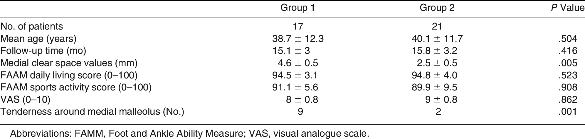
The study enrolled 38 patients (29 male patients and nine female patients). Group 1 included 17 patients, and group 2 included 21 patients. The mean age of the patients was 39.5 ± 11.8 years. The mean follow-up duration was 15.5 ± 3.1 months. The mean medial widening value of the patients measured before surgical treatment was 3.4 ± 1.1 mm. The mean age was 38.7 ± 12.3 years and the mean follow-up duration was 15.1 ± 3 months in group 1. In this group, the mean FAAM daily living score was 94.5 ± 3.1, mean FAAM sports activity score was 91.1 ± 5.6, and mean VAS score was 8 ± 0.8. The medial clear space measured before surgery in this group was 4.6 ± 0.5 mm. The mean age of patients in group 2 was 40.1 ± 11.7 years, and the mean follow-up duration was 15.8 ± 3.2 months. The mean FAAM daily living score was 94.8 ± 4.0, the mean FAAM sports activity score was 89.9 ± 9.5, and the mean VAS score was 9 ± 0.8. In this group, the preoperative medial clear space was 2.5 ± 0.5 mm. Demographic, radiographic, and clinical values of patients are shown in Table 1.

Table 1.
Demographic, Radiographic, and Clinical Values of Patients
Tenderness around the medial malleolus was noted in 11 of 38 patients during preoperative examinations. In group 2, tenderness around the medial malleolus was not detected in 19 patients before surgery, but it was detected in two patients. In group 1, tenderness around the medial malleolus was detected in nine patients but not in eight patients. Comparisons of the tenderness around the medial malleolus with the Cotton test are shown in a 2 × 2 table (Table 2 and Fig. 1). According to 2 × 2 table diagnostic analysis, tenderness in the medial malleolus has a sensitivity of 52.9%, a specificity of 90.5%, a positive predictive value of 81.8%, and a negative predictive value of 70.4%.
Table 2.
2 × 2 Table Showing Diagnostic Analysis of Tenderness Around Medial Malleolus
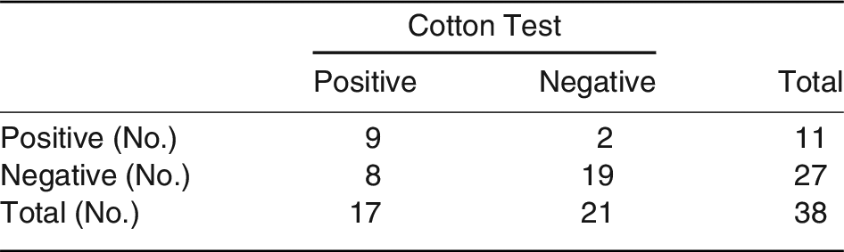
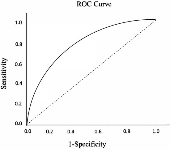
Figure 1.
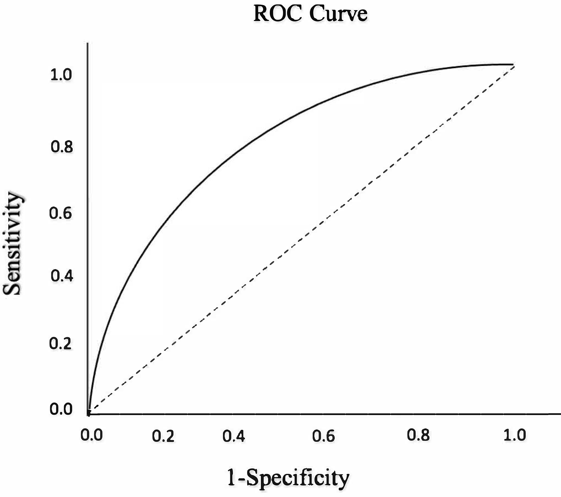
The receiver operating characteristic curve illustrates the relationship/tradeoff between clinical sensitivity and specificity of tenderness around the medial malleolus. ROC, receiver operating characteristic.
Early superficial skin infection was observed in two patients in group 1 and in one patient in group 2, and they were treated with antibiotic therapy. No patients needed revision surgery during the follow-up period. Radiographic union was detected in all patients at their last visit.
Statistical comparisons of the groups did not reveal a statistically significant difference between sex distribution, age, follow-up duration, FAAM daily activity score, FAAM sports activity score, and VAS scores (P > .05). A statistically significant difference was detected in terms of medial clear space widening (P = .05). In terms of the tenderness around the medial malleolus, there was a statistically significant difference between both groups (P = .001). The tenderness around the medial malleolus was 52.9% in the group where the syndesmosis screw was used, whereas the tenderness around the medial malleolus was determined to be 0.5% in the group without the syndesmosis screw.
Discussion
To achieve good clinical results in ankle fractures, the main goals are to ensure the stability of the ankle and the alignment of the fracture with surgical or nonsurgical treatment methods [11]. Osteoarthritis and poor functional consequences are more common in patients with mortise enlargement [12].
According to the literature, surgical treatment of Weber B ankle fractures is not very effective. It has been reported that syndesmosis rupture has no effect on clinical radiographs or ankle dorsiflexion in Weber B ankle fractures [13]. The relationship between increasing obliquity, fracture length, and mortise instability has not been determined in Weber B ankle fractures [14]. After 5.3 years of follow-up, it was established that surgical treatment for Weber B ankle fractures is not superior to conservative treatment in terms of pain, ankle function, and quality of life [15].
Current treatment protocols are based on ankle mortise stability. Lateral malleolus fractures without lateral subluxation of the talus are considered stable and are less likely to be displaced, and good results can be achieved with nonsurgical treatment [11]. In contrast, lateral malleolus fractures with incompatible ankle mortise are considered unstable and require surgical fixation [16]. The length of the fibula is important for the stability of the ankle joint. Changes in the length or sequence of the lateral malleolus cause significant biomechanical instability and lead to poor clinical consequences [17]. Fixation with locking plates is an important surgical treatment technique. Displaced ankle lateral malleolus fractures should be anatomically reduced to ensure absolute stability [18]. In our study, surgical treatment was performed to provide absolute stability in patients who were considered stable and had nonsurgical treatment capability, and as a result of our follow-up, no statistically significant difference was found between the clinical results of both groups, and the clinical results of both groups were evaluated as good. For all Weber B ankle fractures, we recommend surgical treatment to ensure absolute stability to ensure early range of joint motion. As a result of the study based on the visualization of the arthroscopic deltoid ligament, Schuberth et al [19] determined the false-positive rates of deltoid ligament rupture as 88.5, 53.6, 26.9, and 7.7%, respectively, when there is a 3-, 4-, 5-, and 6-mm opening in the medial clear space and have concluded that the medial clear space is not a good indicator of the deltoid ligament integrity. In the radiographic examination of 49 ankles without any pathology, Murphy et al [20] found that the medial clear space was 3.8 ± 0.7 mm in men and 2.9 ± 0.5 mm in women. They indicate that the increased medial clear space is associated with deltoid ligament rupture and ankle instability. In this study, the medial clear space was measured as 4.6 ± 0.5 mm in group 1, and it was determined as 2.5 ± 0.5 in group 2. We conclude that the medial space is a good indicator of ankle instability based on the results of this study, but our findings are limited in terms of indicating deltoid ligament rupture.
Magnetic resonance imaging and ultrasonographic imaging are not used in evaluating ankle fractures but can be used to evaluate ligamentous structures. Ultrasonography defines medial deltoid ruptures as 100% sensitivity and 100% specificity.[21,22] Magnetic resonance imaging and ultrasonographic imaging, which are recommended for better evaluation of soft tissues, were not performed in this study, and this limits the study.
Tenderness and swelling in the medial ankle are not signs of instability, as previously thought [10]. Medial pain and ecchymosis can occur as a result of injury to the superficial components of the deltoid ligament, and the deep deltoid ligament is intact and ensures ankle stability.[23] Most orthopedic surgeons find clinical findings of ecchymosis, swelling, and tenderness to be reliable in evaluating the integrity of medial structures [24]. Stenquist et al [25] have shown that medial tenderness is associated with instability. A review supports that medial tenderness in supination external rotation type ankle fractures is an important finding of deep deltoid ligament rupture [26]. DeAngelis et al [10] evaluated 55 patients with Weber B ankle fractures and normal medial joint space and determined the sensitivity of medial side tenderness on deltoid ligament injury as 57% and specificity as 59%. According to the results of our study, there was a statistically significant difference between both groups in terms of tenderness around the medial malleolus (P ≤ .05). The tenderness around the medial malleolus was 52.9% in the group where the sindesmosis screw was used, whereas the tenderness around the medial malleolus was determined to be 0.5% in the group without the syndesmosis screw. The absence of sensitivity tenderness around the medial malleolus in Weber B ankle fractures indicates no syndesmosis injury, whereas the presence of tenderness around the medial malleolus does not mean that there is a syndesmosis injury.
In their cadaver study, Davis et al [9] examined the biomechanical properties of locking and unlocking plates, and they did not detect a significant difference between the two groups in terms of torsion, amount of loading for insufficiency, and axial stiffness. In addition, Davis et al [27] compared the contoured distal fibular plates with one-third tubular plates in a 2-year follow-up retrospective study, and no difference was found in terms of complications and revision surgery needs. Early superficial skin infection was observed in two patients in group 1 and one patient in group 2, and they were treated with antibiotherapy. During the follow-up period, no patient required revision surgery because of implant failure, and our postoperative complications were in line with the literature.
After the fixation of fractures, the evaluation of syndesmosis is carried out by conducting a hook test during surgery. There is no consensus on an optimal method of syndesmosis fixation. Although some studies have concluded that neither method is superior than the other in comparisons [19,28], some studies concluded that flexible fixation showed better outcomes [29,30]. Small fragment screws (3.5-mm) or large fragment screws (4.5-mm), fixation of three or four cortices, fixation on a plate, and flexible suture tools are used. In group 1, three cortices of fixation were done with 4.5-mm screws, and the syndesmosis screws were removed after 6 weeks.
Conclusions
In Weber B ankle fractures, the absence of tenderness around the medial malleolus indicates no syndesmosis injury, whereas the presence of tenderness around the medial malleolus does not mean that there is a syndesmosis injury.
Financial Disclosure
None reported.
Conflict of Interest
None reported.
References
- Donken CCMA, Al-Khateeb H, Verhofstad MHJ, et al: Surgical versus conservative interventions for treating ankle fractures in adults. Cochrane Database Syst Rev 8: CD008470, 2012.
- Court-Brown CM, Caesar B: Epidemiology of adult fractures: a review. Injury 37: 691, 2006.
- Wuest TK: Injuries to the distal lower extremity syndesmosis. J Am Acad Orthop Surg 5: 172, 1997.
- Norkus SA, Floyd RT: The anatomy and mechanisms of syndesmotic ankle sprains. J Athl Train 36: 68, 2001.
- Aiyer AA, Zachwieja EC, Lawrie CM, et al: Management of isolated lateral malleolus fractures. J Am Acad Orthop Surg 27: 50, 2019.
- Close JR: Some applications of the functional anatomy of the ankle joint. J Bone Joint Surg Am 38: 761, 1956.
- Çelik D, Malkoç M, Martin R: Evidence for reliability, validity and responsiveness of Turkish Foot and Ankle Ability Measure (FAAM). Rheumatol Int 36: 1469, 2016.
- Cotton FJ: Fractures and Joint Dislocations, WB Saunders, Philadelphia, 1910.
- Michelson JD, Varner KE, Checcone M: Diagnosing deltoid injury in ankle fractures: the gravity stress view. Clin Orthop Relat Res 387: 178, 2001.
- DeAngelis NA, Eskander MS, French BG: Does medial tenderness predict deep deltoid ligament incompetence in supination-external rotation type ankle fractures? J Orthop Trauma 21: 244, 2007.
- Gougoulias N, Khanna A, Sakellariou A, et al: Supination-external rotation ankle fractures: stability a key issue. Clin Orthop Relat Res 468: 243, 2010.
- Weening B, Bhandari M: Predictors of functional outcome following transsyndesmotic screw fixation of ankle fractures. J Orthop Trauma 19: 102, 2005.
- Kohake MB, Wiebking U, O’Loughlin PF, et al: Mid- to long-term outcomes after Weber B-type ankle fractures with and without syndesmotic rupture. In Vivo 33: 255, 2019.
- Velasco BT, Miller CP, Michael YY, et al: Does fracture obliquity in Weber B fibula fractures correlate with mortise instability? Injury 50: 2113, 2019.
- Van Leeuwen CA, Hoffman RP, Hoogendoorn JM: Long-term outcome in operatively and non-operatively treated isolated type B fibula fractures. Injury 50: 2318, 2019.
- Makwana NK, Bhowal B, Harper WM, et al: Conservative versus operative treatment for displaced ankle fractures in patients over 55 years of age: a prospective, randomised study. J Bone Joint Surg Br 83: 525, 2001.
- Uchiyama E, Suzuki D, Kura H, et al: Distal fibular length needed for ankle stability. Foot Ankle Int 27: 185, 2006.
- Smith WR, Ziran BH, Anglen JO, et al: Locking plates: tips and tricks. J Bone Joint Surg Am 89: 2298, 2007.
- Schuberth JM, Collman DR, Rush SM, et al: Deltoid ligament integrity in lateral malleolar fractures: a comparative analysis of arthroscopic and radiographic assessments. J Foot Ankle Surg 43: 20, 2004.
- Murphy JM, Kadakia AR, Schilling PL, et al: Relationship among radiographic ankle medial clear space, sex, and height. Orthopedics 37: e449, 2014.
- Nortunen S, Lepojärvi S, Savola O, et al: Stability assessment of the ankle mortise in supination-external rotation-type ankle fractures: lack of additional diagnostic value of MRI. J Bone Joint Surg Am 96: 1855, 2014.
- Henari S, Banks LN, Radiovanovic I, et al: Ultrasonography as a diagnostic tool in assessing deltoid ligament injury in supination external rotation fractures of the ankle. Orthopedics 34: e639, 2011.
- Gougoulias N, Sakellariou A: When is a simple fracture of the lateral malleolus not so simple? How to assess stability, which ones to fix and the role of the deltoid ligament. Bone Joint J 99: 851, 2017.
- Ebraheim NA, Elgafy H, Padanilam T: Syndesmotic disruption in low fibular fractures associated with deltoid ligament injury. Clin Orthop Relat Res 409: 260, 2003.
- Stenquist DS, Miller C, Velasco B, et al: Medial tenderness revisited: is medial ankle tenderness predictive of instability in isolated lateral malleolus fractures? Injury 51: 1392, 2020.
- Baumhauer JF, Geppert MJ, Michelson JD: “Ankle and Foot: Trauma,” in K Koval, ed, Orthopaedic Knowledge Update, 7th Ed, Rosemont, IL, AAOS, 2002.
- Davis AT, Israel H, Cannada LK, et al: A biomechanical comparison of one-third tubular plates versus periarticular plates for fixation of osteoporotic distal fibula fractures. J Orthop Trauma 27: e201, 2013.
- Moore JA Jr, Shank JR, Morgan SJ, et al: Syndesmosis fixation: a comparison of three and four cortices of screw fixation without hardware removal. Foot Ankle Int 27: 567, 2006.
- Elabd A, Abdullah S, Kandel W, et al: Syndesmotic stabilization: syndesmotic screw versus flexible fixation: a systematic review. J Foot Ankle Surg 60: 998, 2021.
- Xu Y, Kang R, Li M, et al: The clinical efficacy of suture-button fixation and trans-syndesmotic screw fixation in the treatment of ankle fracture combined with distal tibiofibular syndesmosis injury: a retrospective study. J Foot Ankle Surg 61: 143, 2022.
© 2023 American Podiatric Medical Association