


Abstract
Variations of the lumbricals of the foot are not common, unlike the lumbricals of the hand. Few cases of unilateral absence of lumbricals of the foot have been reported. There have been far fewer cases of bilateral asymmetric absence of the lumbricals of the foot reported. The study reports the findings observed during the routine dissection for undergraduate medical students in the Department of Anatomy. We observed that the third lumbrical in both the feet was absent in an elderly male cadaver. The other features of both feet were usual. Externally, there was no apparent deformity, operation mark, or evidence of injury in any of the feet. Considering the functional role of the lumbricals in particular and other small intrinsic muscles in general, on the mechanics of foot movement, it is of much significance to have the knowledge of the possible variations and their clinical implications, which should be studied using different diagnostic tools such as imaging techniques, dynamometry, and other modalities.
The foot hosts 18 intrinsic muscles arranged in four layers. All these intrinsic muscles are important for proper locomotion and weightbearing when the heel is lifted off the ground, which strongly activates them. The intertarsal joints become closely packed, accompanied by the extension of the metatarsophalangeal and interphalangeal joints. The foot loses all its pliancy in this position and becomes an effective lever [1]. The weight is shifted to the forefoot as the intrinsic muscles contribute greatly to maintaining the muscular support of the arch. They exert considerable flexion force in the forepart of the foot, as their line of pull lies in the long arch of the foot. They are the principal stabilizers of the transverse arch, as the pull is perpendicular to the latter. The pronated or flat foot demands greater activity of the intrinsic muscles to stabilize the midtarsal and subtalar joints than does the normal foot. It can be observed in walking [1]. The study has been approved by the Bioethics Cell, All India Institute of Medical Sciences, Raebareli, Uttar Pradesh, India.
According to Mann and Inman [2], the plantar intrinsic foot muscles function as a unit to resist subtalar joint pronation, observed as calcaneal eversion (frontal plane), combined with medial deviation (transverse plane) and reduced vertical height (sagittal plane) of the navicular bone [3]. As foot posture and function are known to impact single-leg balance, activation of the plantar intrinsic foot muscles may be used to help stabilize the foot, thereby improving balance. Mickle et al. [4] hypothesized that weakness in the intrinsic foot muscles is associated with poor balance and increased risk of falls in the elderly.
Kelly et al. [5] have observed that plantar intrinsic foot muscle activation was strongly correlated with mediolateral postural sway in a single-leg stance, with increasing activity observed during sway to the medial border of the foot. In addition, these muscles display highly correlated intermuscular activation patterns during standing.
Among these 18 intrinsic muscles, the lumbricals of the foot are one of the important sets of muscles (Fig. 1). There are four of them, and they are numbered from medial to lateral [1]. They increase the grip of the metatarsal heads by producing plantarflexion at the metatarsophalangeal joints and dorsiflexion at the interphalangeal joints of the lateral four toes. They also prevent toes from buckling under during walking and running [6]. Ishizawa and Yamasaki [7] in their study observed multiple and asymmetric absence of lumbricals in both feet. In their case report, they reported that the second and third lumbricals in the right foot and the second, third, and fourth lumbricals in the left foot were absent. Absence or dysfunction (Charcot-Marie-Tooth disease) of lumbrical muscles contributes to the clawing of the toes [8].
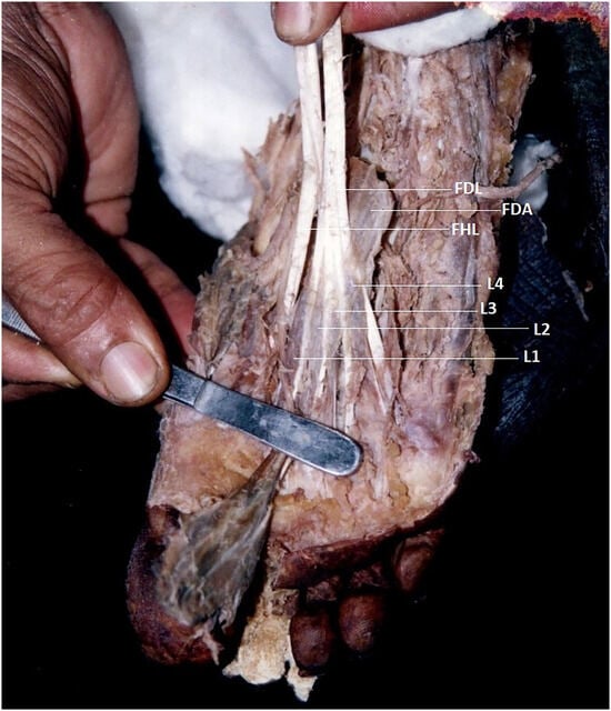
Figure 1.
Image showing lumbricals in a normal foot. FDL, flexor digitorum longus; FDA, flexor digitorum accessorius; FHL, flexor hallucis longus; L1, first lumbrical; L2, second lumbrical; L3, third lumbrical; L4, fourth lumbrical.
Hur et al. [9] have stated some variations in the origin of the lumbricals of the foot. In their study, 66 lower limb specimens from 19 male and 14 female cadavers were studied for the origin of lumbricals of the foot. They observed that lumbricals of the foot had origins from the tendons of flexor digitorum longus and tendinous slips of flexor hallucis longus. They did not report the absence of lumbricals of the foot, either unilateral or bilateral.
Kara et al. [10] conducted a study in 30 human fetuses and observed that there were no variations in the number of lumbricals in all 60 feet: 55 feet showed normal innervation pattern of lumbricals and five feet showed variations in innervation of the lumbricals. Chaney et al. [11] examined 257 formalin-fixed feet and observed that absence of one or more lumbricals occurred in 117 of the limbs (45.9%) dissected. Of the 257 limbs studied, there were no lumbricals present in 3.1%, one lumbrical present in 3.5%, two lumbricals present in 12.1%, and three lumbricals present in 27.2%. Kura et al. [12] studied 11 fresh frozen cadaveric feet and observed that one foot showed the absence of the second and third lumbrical muscles. Kura et al referred to an old study of Schmidt et al. [13] on 100 formalin fixed feet. They observed that the fourth lumbrical was most commonly absent (3%; 12 of 100), followed by the third lumbrical (2.5%; 10 of 100), the second lumbrical (2.25%; nine of 100), and the first lumbrical (0.25%; one of 100). In total, an absence of one or more lumbricals occurred in 8% (32 of 100) [13].
From different studies as discussed above, lesser degrees of variation with respect to the origin, number, and nerve supply of lumbricals of the foot have been observed, unlike the high incidence of their counterparts in the hand. Much difficulty has been observed while measuring the strength of the individual intrinsic muscles of the foot by indirect and direct methods, as the intrinsic muscles are closely arranged and they more or less work as a unit [14]. The presence of any alteration of that unit would have an impact on the distal joints of the foot, which could not be stiffened sufficiently to provide normal push-off against the ground during late stance [15]. Oukouchi et al. [16], in their study on 25 cadaveric feet, reported a rare atavistic anomaly in a few specimens that showed division of lumbricals into two tails before inserting into the proximal phalanges of the contiguous toes, even as they had constant and thick insertions into the dorsal aponeuroses (dorsal digital expansion) of the toes.
Case Report
During routine dissection performed by undergraduate medical students in the Department of Anatomy, we observed that the third lumbrical was absent in an elderly male cadaver in both feet (Fig. 2). Apart from that, the other features of both feet were usual. Externally, there was no apparent deformity, marks indicating surgery, or evidence of injury in any of the feet. However, the existing lumbrical muscles of both feet retained their normal morphology and nerve supply. The first one received the nerve supply from the medial planter nerve and the second and fourth received their nerve supply from the deep branches of the lateral planter nerve. This rare observation of symmetric absence of lumbricals of the foot has enthused us to bring this information to the notice of all through publication (Figs. 3 and 4).
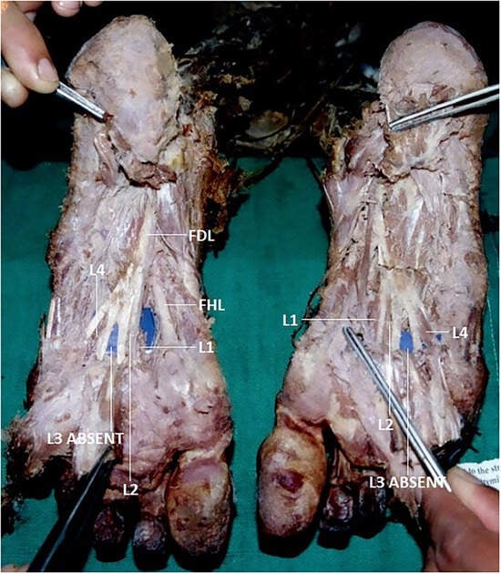
Figure 2.
Image showing bilateral absence of third lumbricals. FDL, flexor digitorum longus; FHL, flexor hallucis longus; L1, first lumbrical; L2, second lumbrical; L4, fourth lumbrical (blue paper pieces were used to make the background prominent).
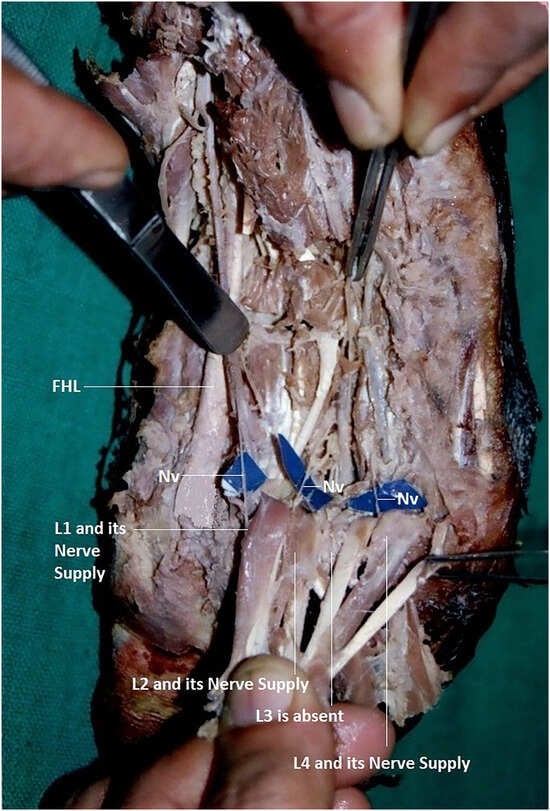
Figure 3.
Image showing lumbricals with their nerves of the left sole with flexor digitorum longus being pulled distally to expose the muscles from deep surface. L1, first lumbrical; L2, second lumbrical; L4, fourth lumbrical; Nv, nerve.
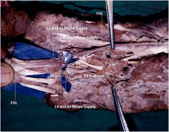
Figure 4.
Image showing lumbricals with their nerves of the right sole with flexor digitorum longus being pulled distally to expose the muscles from deep surface. FHL, flexor hallucis longus; L1, first lumbrical; L2, second lumbrical; L4, fourth lumbrical; Nv, nerve.
Discussion
In our case, we noted bilateral symmetric absence of the third lumbrical in both feet. However, the origin, morphology, and nerve supply of the remaining lumbricals were normal. The first one received its nerve supply from the medial planter nerve, whereas the second and fourth lumbricals received their nerve supply from the deep branches of the lateral planter nerve (Figs. 2–4). The comparison of the percentage of absence of first, second, third, and fourth lumbricals of the foot in different studies and the presence of the total number of lumbricals of the foot in different studies are shown in Tables 1 and 2.

Table 1.
Percentage of Absence of First, Second, Third, and Fourth Lumbricals of the Foot

Table 2.
Presence of Total Number of Lumbricals of the Foot

In this case, any functional deficiency whatsoever attributable to the symmetric absence of one lumbrical in each foot might have been compensated for by the dorsal expansion through its attachment to the other lumbricals, being further facilitated by the symmetric presence of the lumbricals, which created a balancing act. These factors may explain the absence of any external manifestation on the concerned toes, such as clawing-related issues. Perhaps this might be the reason for the absence of any deformity in the distal joints of the involved toes as otherwise envisaged in a report [15].
In our case, we observed only the absence of the lumbrical muscle, but no presence of any variation in the insertion pattern of the other lumbrical muscles in either of the feet, unlike that reported by Oukouchi et al. [16]. This may be suggestive of the absence of any compensatory morphologic and functional modifications despite the absence of one lumbrical, and the effect of that deficiency might have been compensated for by the factors as explained above. This compensatory mechanism is further facilitated by the recruitment of the other functional planter intrinsic muscles of the foot with increasing postural demand [5]. In this rare case, the presence of an effective mechanism of the unit formed by the plantar intrinsic muscles and bones facilitated by the symmetric and bilateral absence of only one lumbrical might have ensured the smooth gripping function of the toes being maintained by the concerned leverage factors.
Conclusions
In conclusion, it can be appreciated well that, considering the functional role of the lumbricals in particular and other small intrinsic muscles in general on the mechanics of the foot movement, it is of much significance to have the knowledge of the possible variations and their clinical implication, which should be studied using different diagnostic tools such as imaging techniques, dynamometry, and other modalities. The information and knowledge would definitely provide reasonable help to podiatric surgeons in the management of patients with foot problems.
Acknowledgment
The All India Institute of Medical Sciences, Raebareli, Uttar Pradesh, India, for providing the logistics to support this work.
Financial Disclosure
None disclosed.
Conflict of Interest
None disclosed.
References
- Salmons S. “Muscle,” in Gray’s Anatomy: The Anatomical Basis of Medicine and Surgery, 38th Ed, edited by Lawrence B, Martin MB, Collins P, et al, p 895, Churchill Livingstone, Edinburgh, 2000.
- Mann RA, Inman VT: Phasic activity of intrinsic muscles of the foot. J Bone Joint Surg 46: 469, 1964.
- Razeghi M, Batt ME: Foot type classification: a critical review of current methods. Gait Posture 15: 282, 2002.
- Mickle KJ, Munro BJ, Lord SR, et al: ISB Clinical Biomechanics Award 2009: toe weakness and deformity increase the risk of falls in older people. Clin Biomech (Bristol, Avon) 24: 787, 2009.
- Kelly LA, Kuitunen S, Racinais S, et al: Recruitment of the plantar intrinsic foot muscles with increasing postural demand. Clin Biomech (Bristol, Avon) 27: 46, 2012.
- Moore KL, Dailey AF, Augur AMR: Clinically Oriented Anatomy, SAE (Indian Adaptation), Vol 2, p 771, Wolters Kluwer, New Delhi, 2018.
- Ishizawa A, Yamasaki M: Plural absence of the lumbrical muscles observed in bilateral feet of a body [in Japanese]. Kaibogaku Zasshi 77: 31, 2002.
- Davies MS: “Foot and Ankle,” in Gray’s Anatomy: The Anatomical Basis of Clinical Practice, 39th Ed, edited by Standring S, p 1539, Elsevier, Philadelphia, 2005.
- Hur MS, Kim JH, Gil YC, et al: New insights into the origin of the lumbrical muscles of the foot: tendinous slip of the flexor hallucis longus muscle. Surg Radiol Anat 37: 1161, 2015.
- Kara BA, Uzmansel D, Beger O: Innervation patterns of the lumbrical muscles of the foot in human fetuses. JAPMA 110: Article 8, 2020.
- Chaney DM, Lee MS, Khan MA, et al: Study of ten anatomy variants of the foot and ankle. JAPMA 86: 532, 1996.
- Kura H, Luo ZP, Kitaoka HB, et al: Quantitative analysis of the intrinsic muscles of the foot. Anat Rec 249: 141, 1997.
- Schmidt R, Reissig D, Heinrichs HJ: The musculi lumbricales of the human foot [in German]. Anat Anz 113: 450, 1963.
- Soysa A, Hiller C, Refshauge K, et al: Importance and challenges of measuring intrinsic foot muscle strength. J Foot Ankle Res 5: 29, 2012.
- Farris DJ, Kelly LA, Cresswell AG, et al: The functional importance of human foot muscles for bipedal locomotion. Proc Natl Acad Sci U S A 116: 1645, 2019.
- Oukouchi H, Murakami T, Kikuta A: Insertions of the lumbrical and interosseous muscles in the human foot. Okajimas Folia Anat Jpn 69: 77, 1992.
© 2023 American Podiatric Medical Association