


Abstract
Background: The aim of this study was to share the clinical results of open calcaneus fractures, which are extremely rare and have become the fearful nightmare of orthopedic surgeons. Methods: Between June of 2014 and June of 2020, of 196 patients with the diagnosis of calcaneus fracture, Gustilo-Anderson type IIIB, 17 patients (11 men and six women; mean age, 36.2 ± 4.6 years; range, 22–56 years) under prospective follow-up were retrospectively screened and included in the study. Patients were recorded in terms of classification of fracture, mechanism of injury, comorbidities, surgical progress, complications, and associated orthopedic and nonorthopedic injuries. Functional clinical results and quality of life of the patients were evaluated at the 6-month follow-up with the American Orthopedic Foot and Ankle Society score and the 36-Item Short Form Health Survey questionnaire. Results: The most common etiologic factor was work-related injuries, with 29.4%. Although triple arthrodesis surgery was performed in three cases because of pseudoarthrosis, amputation was performed in one case because of deep infection. The most common soft-tissue defect was located at the medial aspect of the foot, which was commonly reconstructed with the used of reverse flow island sural flap. The average total American Orthopedic Foot and Ankle Society hindfoot score was 51.6 (range, 48–74) and similarly, physical component, pain, and social parameters on the 36-Item Short Form Health Survey were statistically significant compared to Turkish population normal scores. Conclusions: A long and laborious process is required for both the patient and the surgeon in the treatment of open calcaneal fractures. However, it is possible to obtain good results with soft-tissue reconstructions.
Calcaneus fractures are a relatively common injury, making up approximately 1% of all fractures [1]. Management of closed calcaneus fractures requires knowledge of the osseous anatomy, careful decision-making, and meticulous surgical technique [2]. In contrast, open calcaneus fractures are considerably rarer compared with closed fractures, representing from less than 5% [3,4] to 11% [5] of overall calcaneal fractures. Within the subcategory of open calcaneus fractures, there is a wide range in severity of injury, with a multitude of both osseous and soft-tissue considerations and a correspondingly large number of treatment options. These injuries represent a rare and challenging problem for the treating orthopedic surgeon and a potential limb-altering and/or limb-threatening injury for the patient. As with any open fracture, there is an increased risk of infection, wound healing problems, and the possibility for soft-tissue coverage needs to complement fracture management. Open calcaneus fractures impart significant cost to both the individual and society in the form of lost wages, decreased productivity, short and possible long-term disability, and often prolonged treatment courses [6–8]. Despite the myriad articles dedicated to the classification and treatment of closed calcaneal fractures, controversy continues to surround the question of the open versus closed surgical treatment of this injury.
Until more recently, the progress of open calcaneal fractures largely was unknown. To that end, by reporting the results of our relatively large series of open calcaneal fractures to bring further attention to this uncommon injury pattern, which is crucial for understanding fully the appropriate treatment and realistic outcome for this complex injury. The present study aims to report on the associated injuries, demographic distribution, and the results of treatment of patients sustaining open calcaneus fractures requiring soft-tissue reconstruction, presenting at a single, tertiary level trauma center.
Methods
After obtaining institutional ethical board approval, a prospectively followed up, retrospectively compared cohort-type study was conducted by orthopedic surgeons in a tertiary trauma center. Medical records were retrieved using International Classification of Diseases, 10th Revision codes. Closed and open calcaneus fractures were identified by International Classification of Diseases, 10th Revision codes S92.0, S92.00, and S92.01.
Between June of 2014 and June of 2020, 196 patients who had calcaneus fractures were treated. Twenty-nine of these patients had open fractures. Seventeen of these open fractures were treated and followed up with the diagnosis of Gustilo-Anderson (GA) type IIIB open fractures, which were determined to be the sample of the present study. Allpatients included in the current study had undergone a full trauma evaluation and initial assessment performed by traumatologists. Standard radiographs and computed tomographic scans of the affected extremities were obtained according to our institution’s trauma protocol, including, but not limited to, computed tomography of the head, chest, abdomen, pelvis, and spine. The remaining data were obtained from the medical records of 17 open calcaneus fractures by reviewing the physical examination and radiographic findings, clinic progress, hospital progress, and operative notes, and other ancillary studies to collect associated injuries. The questionnaires were administered during patient check-ups in the outpatient policlinic. The functional outcomes of the patients were evaluated by using the American Orthopedic Foot and Ankle Society (AOFAS) scores conducted with a Turkish-validated form, at the end of each follow-up [9]. Fracture classification was performed by two orthopedic surgeons on an intraobserver basis and one radiologist on an interobserver basis using the AO/OTA and Sanders classifications. The 36-Item Short-Form Health Survey (SF-36) questionnaire, a Turkish-validated form, was also administered to measure the general health status of all sample participants at month 6 after the follow-up, face to face [10].
All data were analyzed by SPSS for Windows version 22.0 (SPSS Inc, Armonk, New York). In the statistical evaluation of the data, the independent t test was used for determination of the mean/median differences. The Mann-Whitney U test was used for those that did not fit the normal distribution. The χ2 test was used to compare the categorical variables. Mean, standard deviation, and median (minimum to maximum) were used as descriptive statistics and the Tukey test was used for post hoc analysis. In the comparisons, a value of P < .05 was considered statistically significant.
Results
The mean age of the patients was 36.2 ± 4.3 years (range, 22–56 years), with a male-to-female ratio of 1.8:1 (11 men and six women), a mean hospitalization interval of 14.3 ± 2.7 days (range, 8–35 days) including intensive care unit stay, and a mean follow-up period of 21.6 ± 7.2 months (range, 16–38 months). The most common etiologic factor was work-related injuries (n = 5 cases [29.4%]) (Table 1). Three cases did not return to work and thus were disabled and retired. Of the remaining 14 cases, the average time to return to work was 15.2 ± 3.6 months (range, 11–19 months). Average number of surgical interventions, including nonorthopedic, was 13.4 ± 2.2 (range, 9–21). In contrast, the average number of surgical interventions just for GA type IIIB calcaneus fracture was 8.7 ± 1.3 (range, 7–11) including all debridement under sedation anesthesia. In all cases, after radical debridement, wound care was performed by means of vacuum-assisted compress, with an average of 4.2 sessions per case in an average 3.4 ± 0.7 intervals (range, 3–5 intervals).

Table 1.
Etiology and Patient Demographics.
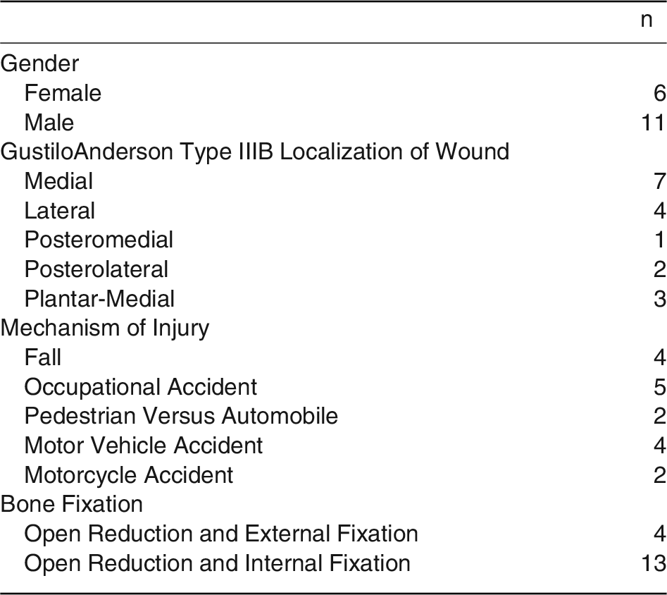
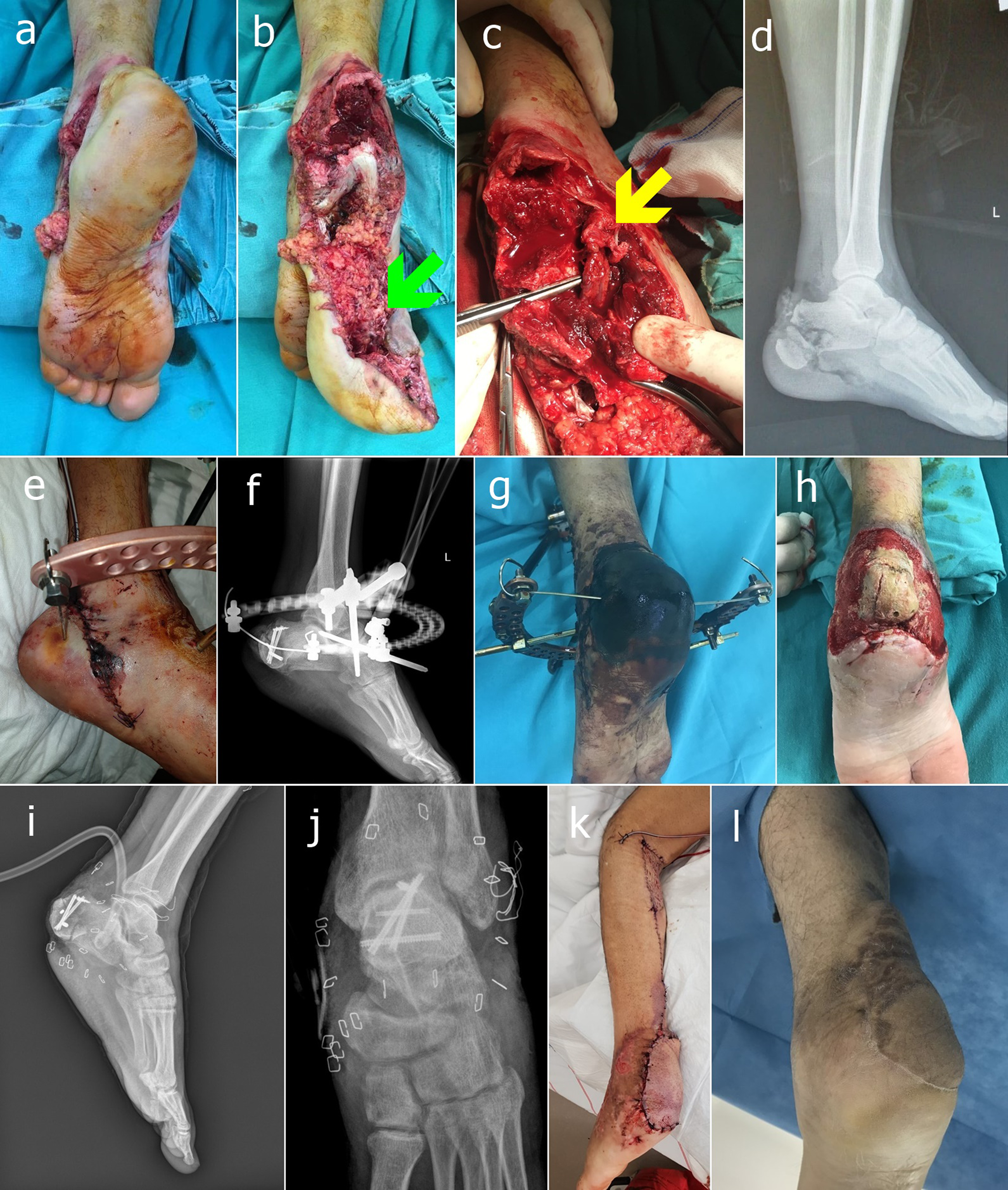
A total of 14 patients (82.3%) had more than two additional orthopedic injuries. Associated orthopedic and nonorthopedic injuries were listed at Tables 2 and 3, respectively. Soft-tissue restoration was performed with free and pedicle flap options in all 17 cases in the current study. Medially located defects in seven cases were reconstructed with reverse-flow island sural flap like that shown in Figure 1. Laterally located defects in four cases were reconstructed with adductor digiti minimi pedicle flaps with full-thickness skin grafts in three cases and a lateral supramalleolar propeller pedicle flap in one case. Posteromedially located defects in four cases were all reconstructed with medial plantar pedicle flaps. Posterolaterally located defects in two cases were all reconstructed with a reverse-flow island sural flap in one case and other with lateral a supramalleolar perforator pedicle flap. Lastly, in one case, a plantar-medial–located defect was reconstructed with a free anterolateral thigh flap.
Table 2.
Associated Orthopedic Injuries.
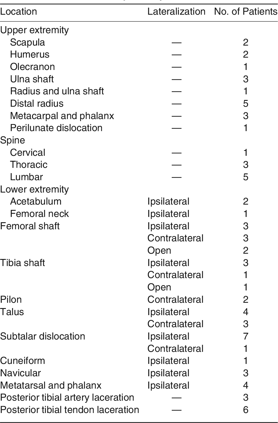
Table 3.
Associated Nonorthopedic Injuries*.
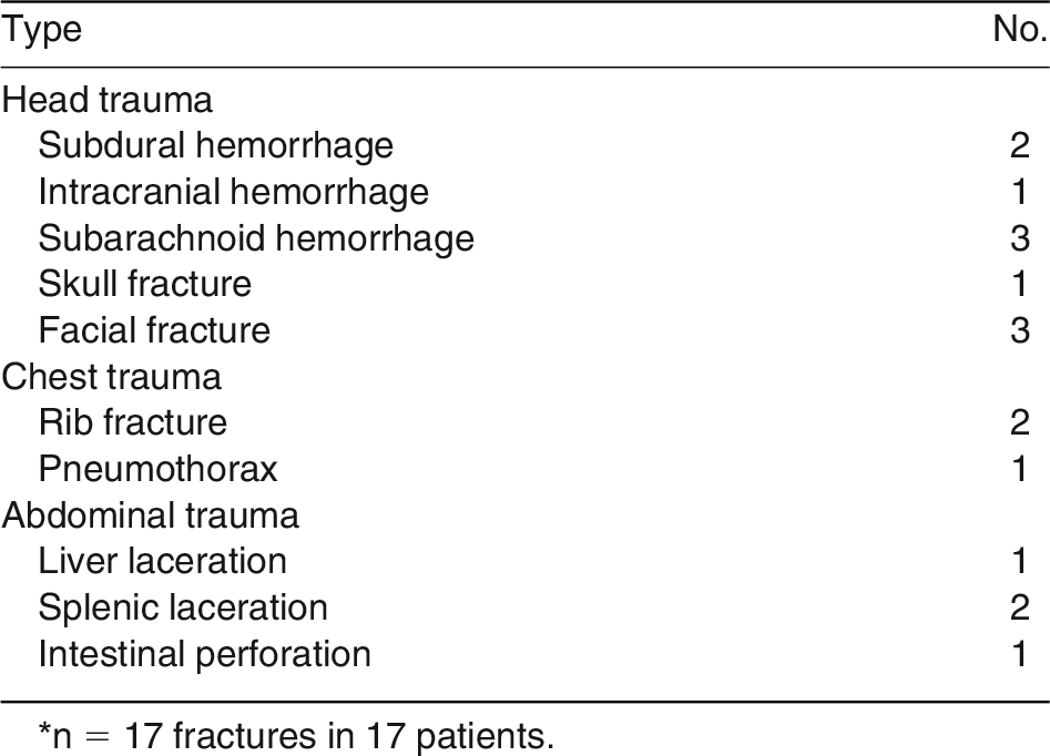
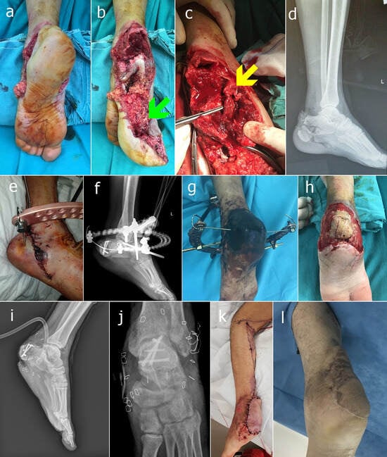
Figure 1.
Chronologic views of a representative case (patient 10). A, Degloving injury of the hindfoot; clinical view of the case; B, explorative view of the trauma (green arrow refers to degloving); C, intraoperative view of the microvascular and neural end-to-end anastomosis labeled with yellow arrow; D, preoperative radiographic lateral view of the case; E, early clinical postoperative view of the case; F, early postoperative radiographic view of the case; G, necrosis of the hindfoot at week 3; H, clinical view of the foot after radical debridement of the necrotic tissue; I and J, radiographic views of the case after external fixation was removed with the vacuum-assistant care of the wound; K, early postoperative view of the foot reconstructed with reversed flow island sural flap; L, final clinical view of the case.
Because the health institution where the authors are located is a reference trauma center, various types of trauma cases underwent primary surgical intervention with bony and/or soft-tissue complications. Four of the cases were referred after first bone fixation was performed and soft-tissue defects without infections occurred. In three of those four cases, one had deep infection that was eventually concluded with amputation and the rest had superficial infection overcome with intravenous antibiotics with the consultation of the Infectious Disease Department of our institution. The pathogen responsible for deep infection was nosocomial type, Pseudomonas aeruginosa; superficial infections were caused by Staphylococcus aureus.
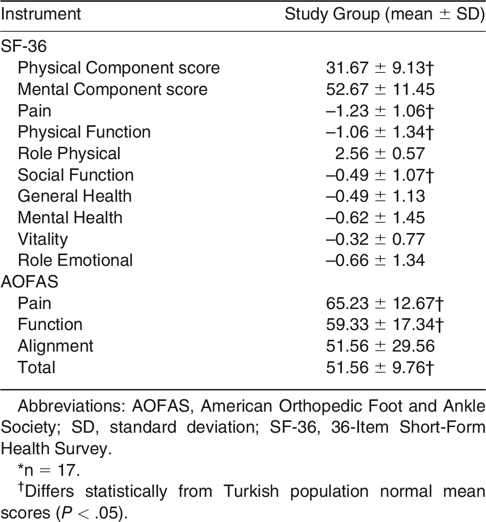
The AOFAS hindfoot clinical rating system was used to evaluate subjective and objective findings at follow-up. The average total AOFAS hindfoot score was 51.6 of a possible 100 (range, 48–74), which was statistically significant compared to Turkish population normal scores. Similarly, three SF-36 scores, Physical component, Pain, and Social parameters were statistically significant compared to Turkish population normal scores (Table 4).
Table 4.
Functional Outcome Score Results*.
Discussion
Every single open calcaneus fracture remains a challenging clinical problem for even the best-equipped clinics and the most skilled orthopedic surgery teams. Immediate initiation of appropriate antibiotic treatment together with urgent surgical irrigation and radical debridement is the mainstay of early treatment. Various treatment methodologies are available and should be selected on a patient-by-patient basis, taking into account bone and soft-tissue injuries, the experience and technical comfort of the treating orthopedic surgeon, and ultimately patient preference.
Open fractures of the calcaneus are relatively rare injuries, representing only 0.8% to 10% of all calcaneus fractures [11]. The data in the current study were in the upper portion of that range at 14.7%. Previous studies have reviewed the demographic data associated with calcaneus fractures and concomitant injuries, but none has specifically focused on GA type IIIB open calcaneus fractures [4–11].
Spierings et al [5] have recently reported a systematic review of open calcaneus fractures, including 616 injuries in 598 patients, that represents the most comprehensive evaluation of open calcaneus fractures in the literature. In that review, 8.8% of injuries were GA type I, 31.1% were GA type II, and 50.1% were GA type III, similar to current findings, which are 58.6%.
Limited data are available in the literature regarding the effect of posterior tibial artery injuries associated with open calcaneus fractures. Ricci et al [12] reported on eight patients with nine transcalcaneal talonavicular dislocations. Five of these injuries presented with open wounds and three had posterior tibial artery lacerations. None of the injuries with a concomitant posterior tibial artery laceration required amputation. Overall, five B/K amputations were performed on patients with complications secondary to open calcaneus fractures. Of the four patients with posterior tibial vessel injuries, two subsequently required a B/K amputation. In the current study, only one case had the posterior tibial artery injury in which, luckily, the amputation was not performed.
Not too much, but sufficient studies were conducted in the literature regarding the associated orthopedic injuries in calcaneus fracture. Myerson and Quill [13] discussed the associated orthopedic injuries and late complications of calcaneus fractures. The mechanism of injury was a fall from a height in 33 patients (77%) and a motor vehicle accident in six (14%). The calcaneus fracture was an isolated injury in 36 patients (86%) [1]. In addition, Worsham et al [14] found that a total of 57 patients (91.9%) in a series of 69 open calcaneus fractures had more than one additional orthopedic injury. All data presented did not match the data in the current study. A possible reason could be the mechanism of injury. Myerson and Quill [13] found that greater than 70% of fractures had resulted from falls from a height. In contrast, in our study, traffic accidents predominated. This was also shown in patients presenting after a traffic accident with several coinjuries that were both orthopedic and nonorthopedic in open calcaneus fractures. Also, Worsham et al [14] report on any associated orthopedic injuries higher than our findings numerically. However, the current finding is more than two additional orthopedic injuries.
Associated nonorthopedic injuries occurred in 15 patients (88.2%), with the most common areas being the head and thorax (Table 3). These included 10 head injuries (58.8%) with subdural, intracranial, subarachnoid hemorrhages and skull fractures. In the 69 open calcaneus fracture patients reported by Worsham et al [14], 17 (24.6%) had head trauma. The higher head trauma ratios of our study should be the effect of our institution position, which is the reference trauma center for four provinces with a population of approximately 3 million. Three patients (17.6%) sustained an injury to the chest wall, with one being a pneumothorax. Only one study has noted the association between calcaneus fractures and pneumothoraces [15], and our current study would be the second. This emphasizes the need to be careful in evaluating all other body systems, especially the chest and head, in patients presenting with open calcaneus fractures. Historically, 10% of patients presenting with a calcaneus fracture have abdominal trauma [16]. Clinical evaluation with general surgery consultation of the patient has been recommended because of the high incidence. In our study, similar results were found with abdominal trauma in three patients (17.6%).
The skin layers over the calcaneus are thicker than those of any other parts of the body. The subcutaneous fat tissue in this area is affixed to the periosteum. This unique structure of the sole enables it not only to withstand shearing forces but also to support body weight during walking or standing [17]. Therefore, under these considerations, the modality of reconstruction for soft-tissue defects of calcaneus should be chosen as a local flap of the tissue from nonweightbearing parts of the lower extremity or a free flap that uses distant fasiocutaneous tissue or muscle tissue with a skin graft. In our study, all the soft-tissue defect reconstructions were performed with the above conditions. That is probably the reason why the current study did not encounter any flap failure.
This study is probably the first to specifically evaluate the functional outcomes of GA type IIB open calcaneal fractures using validated foot and ankle scores, AOFAS, and a global health outcome measure (SF-36). According to a metanalysis conducted by Spierings et al [5], AOFAS questionnaire scores ranged from 59.4 to 84, with a median of 73.7. When we compared the scores of this study with our own, we found the scores of our study unusually low. The main reason for this expected decrease is that the sample of our study consisted of only GA type IIIB fractures.
Inability to standardize all factors those are etiologic factors, surgical treatment modalities, and soft-tissue coverage reconstruction techniques are the most important limitations of the study. Another limitation is that the associated orthopedic and nonorthopedic injuries were not nominally distributed among the patients, which can easily and probably procure the functional outcomes of the patients in both AOFAS and SF-36 aspects.
Conclusions
Meticulous soft-tissue care and guarded prognosis should be taken with lateral and plantar wounds regarding the extreme comminution of the bone, and poor host. Early decision-making with the tissue defect and determination of surgical techniques for coverage considerations is highly recommended when primary closure is not possible. Patients should be counseled that there is a significant likelihood of treatment requiring multiple operations and that amputation could result from this injury.
Acknowledgments
To freelance native speaker English editor, Russell Fraser, russafraser@gmail.com, for editing; and Seyfi Tuncay for tireless assistance at photographs taken.
Financial Disclosure
None reported.
Conflict of Interest
None reported.
References
- Court-Brown CM, Caesar B: Epidemiology of adult fractures: a review. Injury 37: 691, 2006.
- Zhang X, Liu Y, Peng A, et al: Clinical efficacy and prognosis factors of open calcaneal fracture: a retrospective study. Int J Clin Exp Med 8: 3841, 2015.
- Lawrence SJ, Singhal M: Open hindfoot injuries. J Am Acad Orthop Surg 15: 367, 2007.
- Wiersema B, Brokaw D, Weber T, et al: Complications associated with open calcaneus fractures. Foot Ankle Int 32: 1052, 2011.
- Spierings KE, Min M, Nooijen LE, et al: Managing the open calcaneal fracture: a systematic review. Foot Ankle Surg 25: 707, 2019.
- Coughlin M: Calcaneal fractures in the industrial patient. Foot Ankle Int 21: 896, 2000.
- Batıbay SG, Bayram S: Comparing open reduction and internal fixation versus closed reduction using dual-point distraction and percutaneous fixation for treating calcaneal fractures. Jt Dis Relat Surg 31: 193, 2020.
- Aslan A, Sargın S, Gülcü A, et al: Clinical, radiological and patient-reported outcomes in intra-articular calcaneal fractures: comparison of conservative and surgical treatment. Eklem Hastalik Cerrahisi 30: 143, 2019.
- Akbaba YA, Celik D, Ogut RT: Translation, cross-cultural adaptation, reliability, and validity of Turkish version of the American Orthopaedic Foot and Ankle Society Ankle-Hindfoot Scale. J Foot Ankle Surg 55: 1139, 2016.
- Demiral Y, Ergor G, Unal B, et al: Normative data and discriminative properties of short form 36 (SF-36) in Turkish urban population. BMC Public Health 6: 247, 2006.
- Berry GK, Stevens DG, Kreder HJ, et al: Open fractures of the calcaneus: a review of treatment and outcome. J Orthop Trauma 18: 202, 2004.
- Ricci W, Bellabarba C, Sanders R: Transcalcaneal talonavicular dislocation. J Bone Joint Surg Am 84: 557, 2002.
- Myerson M, Quill GE: Late complications of fractures of the calcaneus. J Bone Joint Surg Am 75: 331, 1993.
- Worsham JR, Elliott MR, Harris AM: Open calcaneus fractures and associated injuries. J Foot Ankle Surg 55: 68, 2016.
- Sillesen NH, Nielsen LT, Bonde C: Complex injuries associated with somnambulism [in Danish]. Ugeskr Laeger 172: 3489, 2010.
- Rowe CR, Sakellarides HT, Freeman PA, et al: Fractures of the os calcis: a long term follow-up study of 146 patients. JAMA 184: 920, 1963.
- Uslu AB, Ünlü RE: Heel reconstruction with bipedicled flap: a salvage procedure. Int J Low Extrem Wounds 19: 377, 2020.
© 2023 American Podiatric Medical Association