


Abstract
The presence of an os sustentaculum bone is extremely rare. Given the scarcity of clinical literature reporting such a finding, the condition may be misdiagnosed as a fracture of the sustentaculum tali. We describe such an incident in a 16-year-old male athlete presenting for a recent ankle sprain with no other history of trauma or pain on the medial aspect of the ankle. The original computed tomographic scan reported a possible nonunion of a sustentaculum fracture, or peripherally corticated ossification in association with a possible talocalcaneal fibrocartilaginous coalition. Given no history of high-level trauma to the area, the clinical presentation, and the radiographic findings, the more likely diagnosis was confirmed to be an os sustentaculum. The goal of the authors of this article is to report such findings to raise awareness of a rare clinical presentation to avoid misdiagnosis.
The os sustentaculum (OS) is an accessory bone that can be found along the medial aspect of the ankle [1]. This rare skeletal variant attaches to the talus at the sustentaculum tali, and can be bridged by means of the fibrocartilaginous or fibrous tissue. As currently reported, the prevalence of this condition is 0.3% of the population [2,3]. Typically, the presence of an OS remains asymptomatic; however, symptoms may arise in cases where an osseous, cartilaginous, or fibrous coalition is present [4,5].
The presence of an OS is scarcely reported in the literature. In this case report, we document not only this rare finding, but also its presence in an asymptomatic young athletic with suspicion of a fibrocartilaginous coalition. It is the goal of this study to present such a finding to increase awareness of its presence, and thereby decrease the chance of an inaccurate diagnosis in atraumatic cases.
Case Report
A 16-year-old boy presented to our office with a chief complaint of pain in the left ankle from a sprain that occurred approximately 1 week previously. The patient’s father was present at the time of the initial office encounter, noting that his son twisted his ankle while pivoting on the basketball court during a game the previous week. The pain was mild to moderate initially and improved to the point of only an occasional dull pain when jumping. The patient had attempted to wear a supportive sock that was purchased online but denied the use of any brace or wrap in the management of his condition. He indicated that the pain was only on the “outside” of his ankle, to which he had applied ice intermittently for approximately 48 hours.
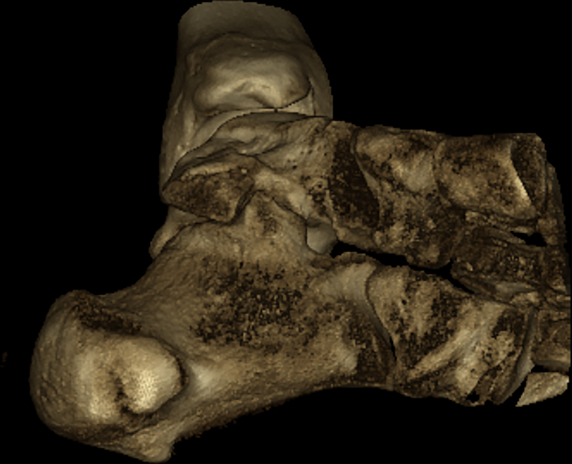
On clinical examination, the patient had full range of motion for dorsiflexion, plantarflexion, inversion, and eversion without pain or crepitation. There was pain reported on the anterior lateral aspect of the left ankle at the level of the anterior talofibular ligament on palpation. All other areas of the left foot and ankle were unremarkable. Radiographs of the left ankle were unremarkable for fracture or dislocation; however, irregularities within the medial aspect of the ankle prompted further investigation with high-resolution imaging. Using the Verity system (Planmed, Helsinki, Finland), a high-resolution, low-dose pediatric computed tomographic (CT) scan was obtained on the left ankle to rule out the presence of an occult fracture (Figs. 1 and 2). The formal read from the CT scan suggested a “chronic ununited fracture fragment” of the sustentaculum tali or “peripherally corticated ossification” and a possible fibrocartilaginous coalition between the “fracture fragment” and the sustentaculum tali. Given the absence of any history of trauma to the left ankle other than the sprain 1 week previously, and no pain appreciated or reported on the left medial ankle, a chronic fracture was not likely. The alternative, and more likely diagnosis of an OS of the left foot was discussed with the patient and his father. Given the absence of symptoms in the left medial ankle, the collective decision was made to monitor the condition and follow-up with a magnetic resonance imaging scan should symptoms arise.
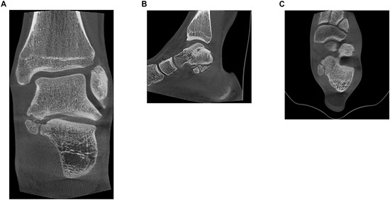
Figure 1.
Images of high-resolution, low-dose computed tomographic scan of the left ankle. A, Coronal view of the subtalar joint reveals the presence of an os sustentaculum on the medial aspect of the ankle. B, Sagittal view displays the os sustentaculum just inferior to the talus. C, Axial view exhibits the smooth margins of the ossicle.
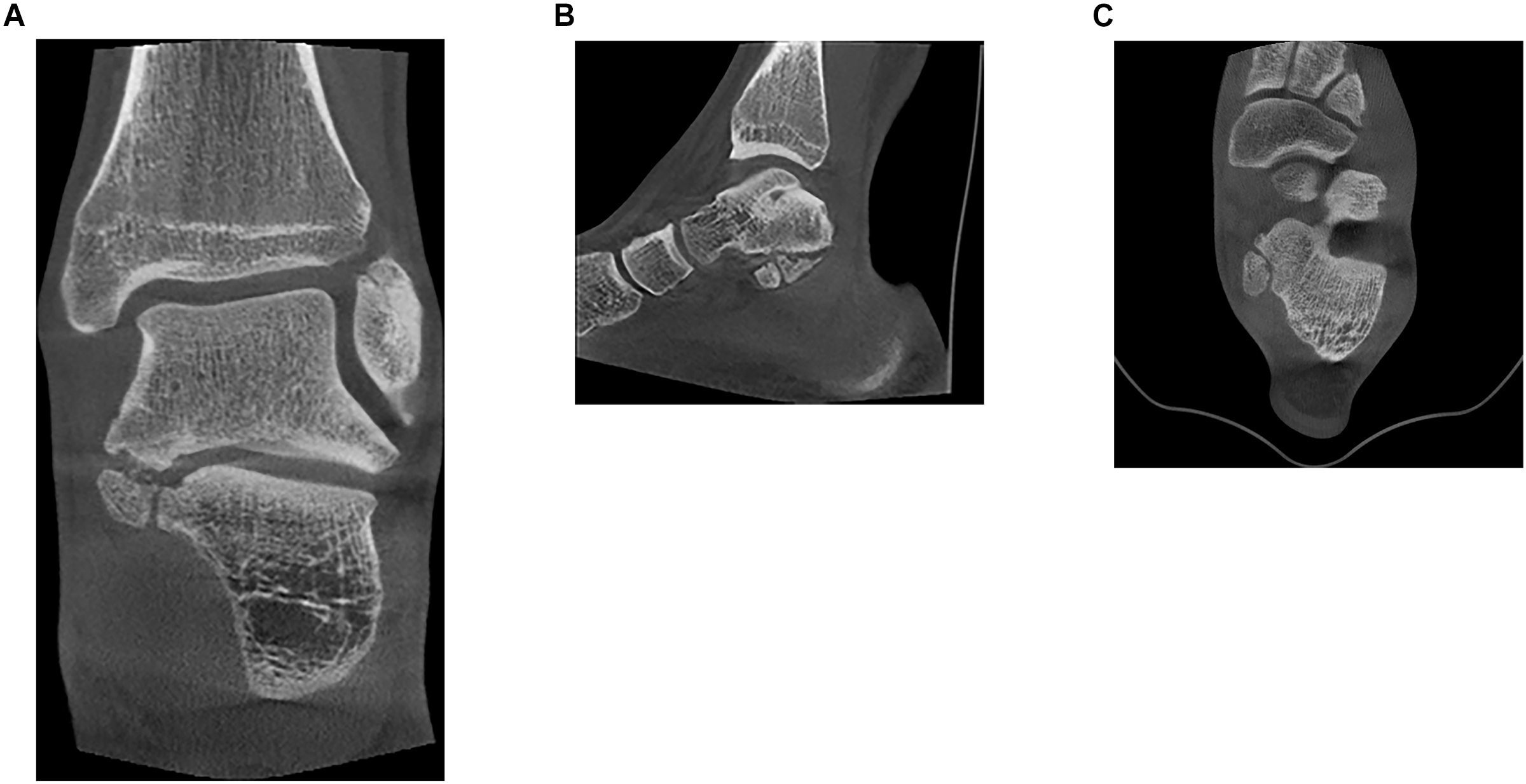
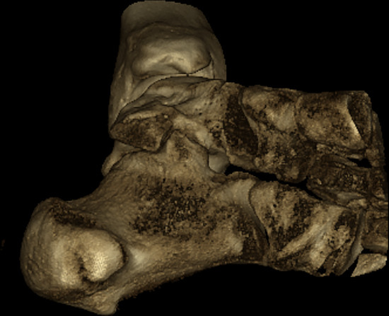
Figure 2.
Image of the left ankle. A three-dimensional rendering of the left ankle in which the os sustentaculum is visible near the medial aspect of the subtalar joint again denotes the smooth margins of the ossicle.
Discussion
The presence of an OS is rarely reported in the literature. One study by Yun et al [5] did report the condition in the setting of a medial facet coalition; however, the patient’s symptoms prompted this discovery. Another study, by Hong et al [6], also reported the presence of an OS, but this was again in the setting of symptoms from a mass effect of the lesion in the tarsal tunnel. In our presented case study, the patient had no symptoms on the medial aspect of the left ankle. To the best of our knowledge, such a condition has not been reported in an asymptomatic patient. In addition, the patient presented was an extremely active young athlete nearing complete osseous maturity. This unusual presentation suggests that an OS, like many other accessory bones, may not always pose a threat to normal activity or functioning.
Conclusions
In this case report, we present the rare finding of an OS in a 16-year-old athlete. Given the rare occurrence of this accessory bone in the literature, the initial CT scan report suggested a fractured sustentaculum. The presence of smooth edges and cortical bone surrounding the entire osseous entity is more suggestive of an accessory bone than a nonunion of a previous fracture. In addition, the absence of any history of trauma other than the left ankle sprain 1 week previously, further supports this unusual and rare finding. It is the hope of the authors to further increase awareness of a rare clinical finding and present an asymptomatic case of an OS.
Financial Disclosure
None reported.
Conflict of Interest
None reported.
References
- Bloom R, Libson E, Lax E, et al: The assimilated os sustentaculi. Skeletal Radiol 15: 455, 1987.
- Tsuruta T, Shiokawa Y: Radiological study of the accessory skeletal elements in the foot and ankle. J Jpn Orthop Assoc 55: 357, 1981.
- Bencardino J, Rosenberg ZS, Beltran J, et al: Os sustentaculi: depiction on MR images. Skeletal Radiol 26: 505, 1997.
- Mellado J, Salvado E, Camins A, et al: Painful os sustentaculi: imaging findings of another skeletal variant. Skeletal Radiol 31: 53, 2002.
- Yun S, Wook J, Kim G, et al: A different type of talocalcaneal coalition with os sustentaculum: the continued necessity of revision of classification. AJR Am J Roentgenol 205: W612, 2015.
- Hong C, Lee Y, Won S, et al: Tarsal tunnel syndrome caused by an uncommon ossicle of the talus. Medicine (Baltimore) 97: e11008, 2018.
© 2023 American Podiatric Medical Association