


Abstract
Background: Herein, we present a case series of tibiotalocalcaneal arthrodesis (TTCA) with a distal femoral locking plate and describe the short-term clinical and radiographic results of the procedure. Methods: We performed a case review of four patients with hindfoot and ankle pathology who had been treated with a TTCA using a distal femoral locking plate (Depuy Synthes, Zimmer Biomet) by a single surgeon (H.A.H.) from January 2018 to December 2020. Results: At final follow-up, there was a 75% complication rate (3/4) with two developing a nonunion of the ankle joint and one developing a postoperative wound and surgical site infection. Conclusions: Lateral locking plate fixation with a distal femoral locking plate system may be an alternative fixation option for tibiotalocalcaneal fusions in complex patients. However, a larger sample size with longer term follow-up is needed to make any significant recommendations on the use of this plating system.
The tibiotalocalcaneal arthrodesis (TTCA) is a powerful procedure whose primary goals include pain relief, correction of deformity, stability, and maintenance and restoration of a stable hindfoot alignment. There are a variety of indications, including total ankle arthroplasty failure, talus fractures, talar avascular necrosis, Charcot neuroarthropathy, severe ankle and subtalar arthritis, and varus and valgus deformity of the ankle joint (AJ) and the subtalar joint (STJ) [1–3]. Many times, the TTCA is used as an end-stage or salvage procedure, and most patients have unfavorable medical comorbidities and have had previous surgical procedures performed, all increasing the overall complexity of the case. There have been many described surgical techniques with different approaches and fixation methods, each with their own pros and cons. Because of the complexity and salvage nature of the procedure, reducing the overall complication rate is of benefit. Tibiotalocalcaneal arthrodesis complications include perioperative or postoperative fractures, malunion, nonunion, and infection [1–3]. Proper patient selection with surgical planning can help to minimize postoperative complications; however, improving the fixation constructs and limiting the amount of soft-tissue damage have been essential.
In patients with poor bone stock, crossed lag screw fixation can cause instability and implant loosening, low fusion rates, and issues with correcting varus and valgus deformity. Other more robust fixation options offer different advantages. Retrograde intramedullary nail (IMN) fixation has a biomechanical advantage; however, it may be more technically demanding in terms of placement of the nail [1]. Looking at different IMNs, Griffin and Coughlin [7] and Dujela et al [5] found that the combined fusion rates of the ankle and subtalar joints were only 65% and 68.2%, respectively. Ankle union rates with IMNs have ranged from 80% to 94%, with subtalar union rates ranging from 74% to 88% [3–7]. There are, however, reports of increased primary stability and reduced soft-tissue damage with use of a nail that may allow earlier return to weightbearing and activities for patients compared to traditional constructs [3–7]. The posterior blade plate had been reported to have high fusion rates and avoids previous incisions and areas of tenuous soft tissue with a posterior incision. This allows for direct visualization of both joints and provides a viable alternative for those who have had issues with an anterior or lateral soft-tissue envelope [8]. However, in more complex patient groups, especially those who have had revisions, there may be an increase in delayed union and nonunion with this approach [9]. Peterson et al [8] looked at nine patients undergoing TTCA using a posterior locking plate and reported a 78% union rate, including one at both the AJ and STJ and one at the ankle only. Gorman et al [9] found a union rate of 82% in 40 patients.
In terms of primarily lateral approaches, locking plate options have been described. There has been literature using proximal humeral plates that augmented fixation with a medial lag screw to allow for compression of the medial aspect of the AJ and STJ [10,11]. There have been a couple of studies that discuss some advantages of lateral plating that include faster surgical times, ease of use, and more stability [10–12]. DiDomenico and Wargo-Dorsey [13] published a technique guide using the lateral condylar locking plate (LCP) (VA-LCP Condylar Plate 4.5/5.0; DePuy Synthes, Raynham, Massachusetts) with adequate correction; however, there was a lack of long-term follow-up. The hardware purported is robust, designed in mind for distal femur fractures. The plate has a limited contact design to reduce plate-to-bone contact and preserve periosteum. It has combination holes that allow us to create a fixed-angle construct to prevent collapse of bone in those with significant deformity and osteopenic bone [13]. Research has shown that lateral plating may be biomechanically more stable compared to IMN, especially in those with osteopenic bone [10,14]. In addition, the use of hybrid fixation in lateral plates with nonlocked and locked screws have shown to be 7% stronger in bending and 42% in torsion with only a 7% loss in axial compression compared to locked plate constructs. Constructs are noted to fail by screw breakage in torsion, and the use of hybrid plating may be an alternative for those with osteoporosis [15]. A study by Zhang et al [16] showed similar fusion rates for IMN and locked plate fixation, 90.6% compared to 95.4%, respectively; however, the locking plate fixation showed significantly shorter operation times, less blood loss, and fewer intraoperative fluoroscopy sessions than the IMN group.
The purpose of our case series was to follow a group of patients who underwent limb salvage with a TTCA using a distal femoral lateral locking plate to see whether the outcomes were comparable to other reported techniques, including intramedullary nailing and posterior plating. In addition, we wanted to review whether this nonanatomical locking plate in a lateral approach could provide another option for limb salvage for a subset of patients with diabetic peripheral neuropathy. This comorbidity has many deleterious effects on the immune system that may affect the fusion of bone, the healing of incisions, and overall proclivity to infections, making the TTCA significantly more difficult. Because of the increased rate of nonunion and malunion at the fusion sites, we also wanted to see whether this construct could also provide relative stability for ambulation for those that do not go on to complete union. We hypothesized that this construct may provide an alternative that may be less technically demanding for surgeons and more biomechanically stable than intramedullary nailing and posterior plating for a complicated patient population.
Methods
A retrospective review was performed of patients who had undergone TTCA. Indications for hindfoot arthrodesis were Charcot neuroarthropathy of the midfoot or ankle, and posttraumatic arthritis of the AJ and STJ. Patients were to have preoperative assessment and preoperative and postoperative radiographs. The TTCA was performed with a distal femoral locking plate from 2018 to 2020 by a single surgeon (H.A.H.). Four total patients were identified and included in this case review. There were three male patients and one female patient. Radiographs were used to determine union of the joints.
Surgical Technique
The patient was placed in the supine position on the operating table with a bump under the ipsilateral hip. Following general anesthesia, the extremity was then scrubbed, prepared, and draped in aseptic fashion. A linear incision was made over the distal fibula extending approximately 10 cm proximal to 2 cm below the distal tip of the fibula. The incision was carried down to the fibula and the soft-tissue attachments were freed up. The fibula was resected approximately 7 to 8 cm proximally using a sagittal saw and removed to use as autologous bone graft. With the fibula removed, we gained exposure of the AJ and STJ and the lateral wall of the calcaneus. At this time, the AJ and STJ were prepared using a combination of curettes, osteotomes, and drills. Cartilage was denuded and the subchondral plate was fenestrated. The fibula was then milled and used as autogenous graft in the AJ and STJ.
At this time, the AJ and STJ were placed with the foot in plantigrade position and with 0° to 5° of valgus. If necessary, a percutaneous tenotomy of the Achilles tendon was performed to allow for reduction. If possible, a home run screw from the tibia to talus to calcaneus was placed to hold the reduction, but in other cases, a screw across the ankle joint from tibia to talus or from the calcaneus to the talus was placed. The LCP condylar plate (4.5/5.0) of the LCP periarticular plating system or the NCB distal femur lateral plating system (Zimmer-Biomet, Warsaw, Indiana) was placed laterally over the distal tibia, talus, and calcaneus. The plate position was then confirmed using anteroposterior and lateral fluoroscopic images. A combination of nonlocking and locking screws were then placed within the plate using the manufacturer’s guidelines. At least three points of fixation were placed to the tibia and the calcaneus.
Closure was obtained deep with absorbable suture, and skin was closed with nonabsorbable suture. A well-padded posterior splint was applied postoperatively. Patients followed up within 1 week and were placed in a well-padded, short leg cast. Cast changes were performed every 2 weeks thereafter, with a total time of nonweightbearing being 8 weeks. They were transitioned to a B/K controlled ankle movement (CAM) walker to begin transitioning to partial weightbearing depending on radiographic findings of early consolidation. Patients were then transitioned to full weightbearing without the boot.
Case Reports
Case 1
A 66-year-old man with a medical history of diabetes mellitus (DM) type 2 (5 years in duration; reported hemoglobin [Hb] A1c level, 6.1%) presented to the office with complaints of right foot and ankle pain. He had fractured his right ankle 7 months previously and had a subsequent open reduction and internal fixation of the right ankle performed by an outside doctor. He recently had been developing pain to the same ankle, with more severe pain in the past month.
On physical examination, the patient had no breakdown of skin. Previous incisions to the lateral and medial ankle were healed. There were palpable posterior tibial (PT) and dorsalis pedis (DP) pulses, and he had notable nonpitting edema of the right ankle. He had diminished protective sensation determined by means of Semmes-Weinstein monofilament and diminished vibratory sensation determined by means of tuning fork. Radiographs showed retained plate and screws within the fibula. Magnetic resonance imaging of the right ankle showed partial fusion of the fibula and significant degeneration of cartilage within the ankle joint. There was anterior and medial subluxation of the tibia relative to the talar dome.
At this time, we had discussed reconstructive surgery of his foot and ankle because of the Charcot breakdown of the ankle with TTCA. The patient had consented to the procedures with one of the authors (H.A.H.) after hearing the risks, benefits, alternatives, and postoperative course. The patient underwent surgery and was placed in a sugar-tong splint and was later discharged to home the next morning (Fig. 1). The patient had short leg casts applied for a total of 12 weeks. Sutures were removed at week 4 with no issues with skin healing. He began weightbearing with a CAM boot at 12 weeks with physical therapy. Radiographs were taken at that time (Fig. 2). The patient noted tightness and pain localized to the Achilles tendon with weightbearing. At 32 weeks, the pain persisted, and we discussed tenotomy of the Achilles tendon. At 40 weeks postoperatively, the patient had the Achilles tenotomy performed. There were some improvements in pain; however, pain persisted, and a computed tomographic scan revealed significant cystic changes and nonunion of the right ankle. Currently, the patient has been using a bone stimulator and has been weightbearing with a CAM boot with slow improvement.
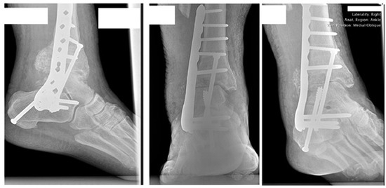
Figure 1.
Two-week postoperative ankle radiographs for patient 1. There are two screws, one from the tibia traversing into the talus and one from the calcaneus to the talus.
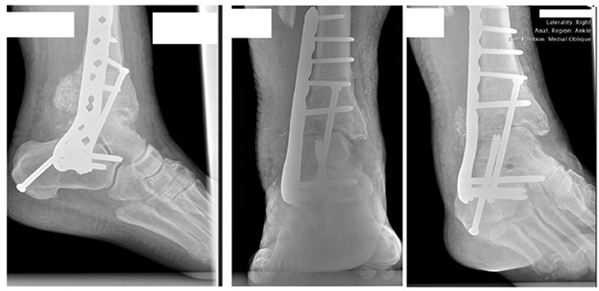
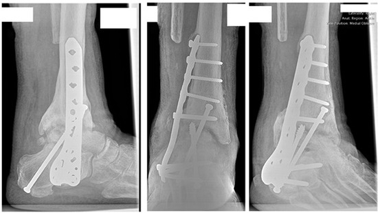
Figure 2.
Three-month postoperative ankle radiographs for patient 1. There is resorption of bone at the talus and tibia, which is concerning for nonunion or further Charcot breakdown. No hardware failure was noted at this time.
Case 2
A 53-year-old man with a medical history of DM type 2, on insulin (10 years in duration), initially had presented to the hospital with worsening infection of his right foot. He was noted to have osteomyelitis of the right third toe. In addition, there was a wound to the medial aspect of the right foot from previous Charcot neuroarthropathy. He underwent a debridement of the right foot wound and amputation of the third digit. The patient underwent various offloading/bracing and debridement of the right lower extremity that helped to close the medial foot wound; however, he continued to have breakdown of skin in that area. He returned 4 years later with a worsening medial foot wound. His HbA1c level at that time was 7.6%.
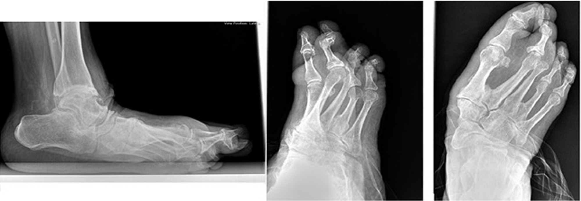
On physical examination, the patient had a wound to the plantar medial arch measuring 6.0 × 4.0 × 0.2 cm with subcutaneous tissue exposed. There was a negative probe-to-bone test. He had palpable pulses to both the PT and DP with nonpitting edema to the right foot. Neurologically, he had absent sensation determined by mean of Semmes-Weinstein monofilament and absent vibratory sensation determined by means of tuning fork. He had significant collapse of the midfoot at the talonavicular joint with abduction of the forefoot. There was significant equinus deformity. Radiographs had shown significant collapse of the midfoot and magnetic resonance imaging of the foot showed no significant changes on T1-weighted imaging of the medial cuneiform (Fig. 3).
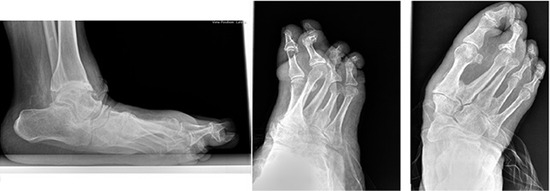
Figure 3.
Preoperative foot radiographs for patient 2. There is significant Charcot collapse of the midfoot with forefoot abduction. There is a significant decrease in calcaneal inclination angle and increase in talar declination angle.
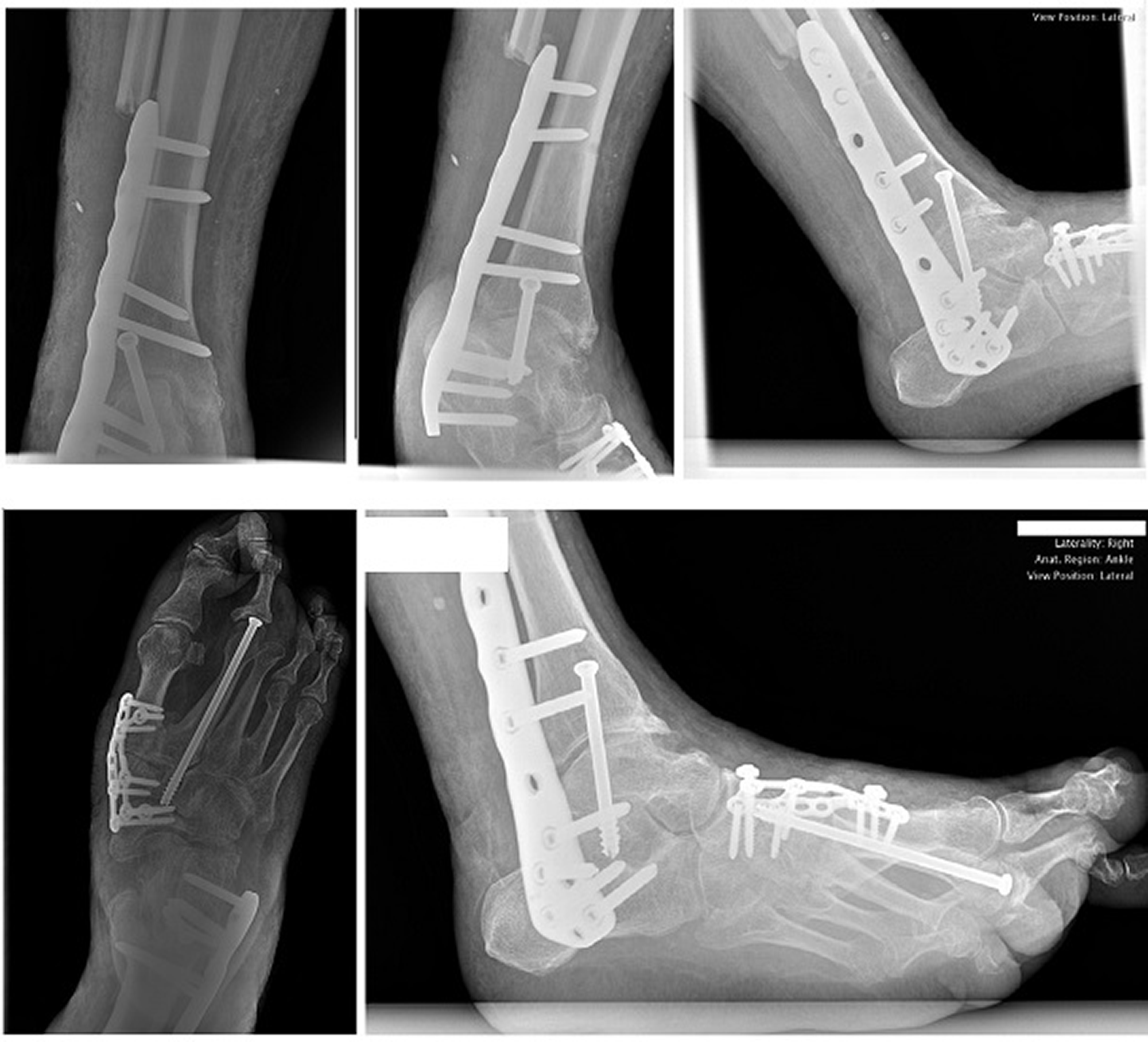
At this time, we discussed reconstructive surgery of his foot and ankle because of the recalcitrant wound (including a TTCA), Achilles tenotomy, medial column fusion, and debridement of the wound. The patient had consented to the procedures (with H.A.H.) after hearing the risks, benefits, alternatives, and postoperative course. The patient underwent his procedure at the hospital, and was placed in a sugar-tong splint (Fig. 4). He was discharged to a skilled nursing facility. Postoperatively, there was marginal skin necrosis of the distal lateral incision that later formed an eschar. The plantar medial foot wound would go on to heal along with the incisions of the foot. The eschar laterally broke down, exposing hardware distally. Four weeks postoperatively, he was started on a course of intravenous antibiotics for 2 weeks by infectious disease. Ten weeks postoperatively, the patient was undergoing vacuum-assisted wound closure to the lateral wound with improvement in wound size weekly with no exposed hardware. He is currently not on intravenous antibiotics and is nonweightbearing in a CAM boot.
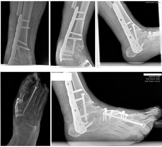
Figure 4.
Postoperative foot and ankle radiographs for patient 2. There is one screw from proximal to distal across the ankle and subtalar joint for positioning. There is a medial column plate along with beam across the second metatarsal to the intermediate cuneiform.
Case 3
A 53-year-old man with a medical history of end-stage renal disease on hemodialysis, DM type 2 on insulin, and Charcot to the left foot initially had presented to the office with blisters on the left foot. He continued to have issues with nonhealing wounds to his left foot.
On physical examination, the patient had a wound to the left plantar medial arch measuring 2.0 × 1.5 × 0.5 cm with subcutaneous tissue exposed. There was a negative probe-to-bone test. He had palpable pulses to both the PT and DP and had edema of the foot. Neurologically, he had absent sensation determined by means of Semmes-Weinstein monofilament and diminished vibratory sensation determined by means of tuning fork. Structurally, he had significant collapse of the midfoot at the talonavicular joint with abduction of the forefoot and a significant equinus deformity. Radiographs had shown significant collapse of the midfoot (Fig. 5) and magnetic resonance imaging of the foot showed no significant changes on T1-weighted images of the medial cuneiform. His HbA1c level at that time was 6.7%.
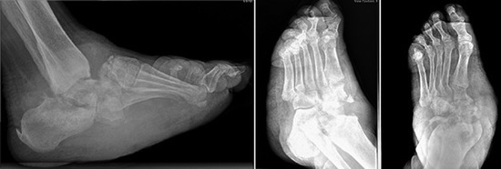
Figure 5.
Preoperative foot radiographs for patient 3. There is Charcot neuroarthropathy with significant destruction of bone at the midtarsal joints with bayonet deformity. There is a significant decrease in calcaneal inclination angle and increase in talar declination angle.
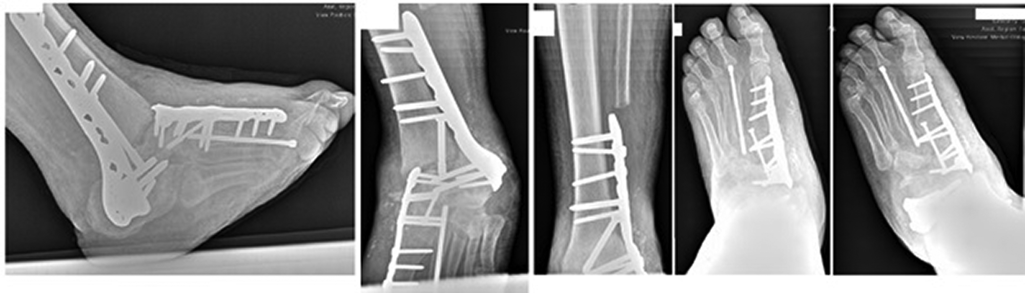
At this time, we discussed reconstructive surgery of his foot and ankle because of the recalcitrant wound, including a TTCA, Achilles tenotomy, medial column fusion, and excision of the wound with primary closure. The patient had consented to the procedures with one of the authors (H.A.H.) after hearing the risks, benefits, alternatives, and postoperative course. The patient underwent his procedure, and was placed in a sugar-tong splint (Fig. 6). He was discharged to a skilled nursing facility. He was unable to make his initial postoperative appointments because of coronavirus disease of 2019. He had sutures removed at 6 weeks postoperatively, new radiographs taken, and was placed in a short leg cast (Fig. 7). At 9 weeks postoperatively, he was allowed to bear weight with the CAM boot and started physical therapy. At 12 weeks, he was back to weightbearing in diabetic shoes.
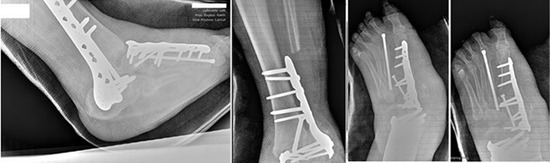
Figure 6.
Two-week postoperative foot and ankle radiographs for patient 3.
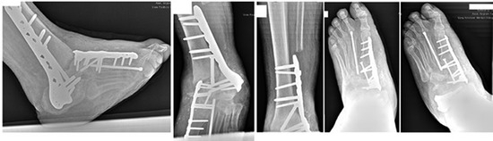
Figure 7.
Six-week postoperative foot and ankle radiographs for patient 3.
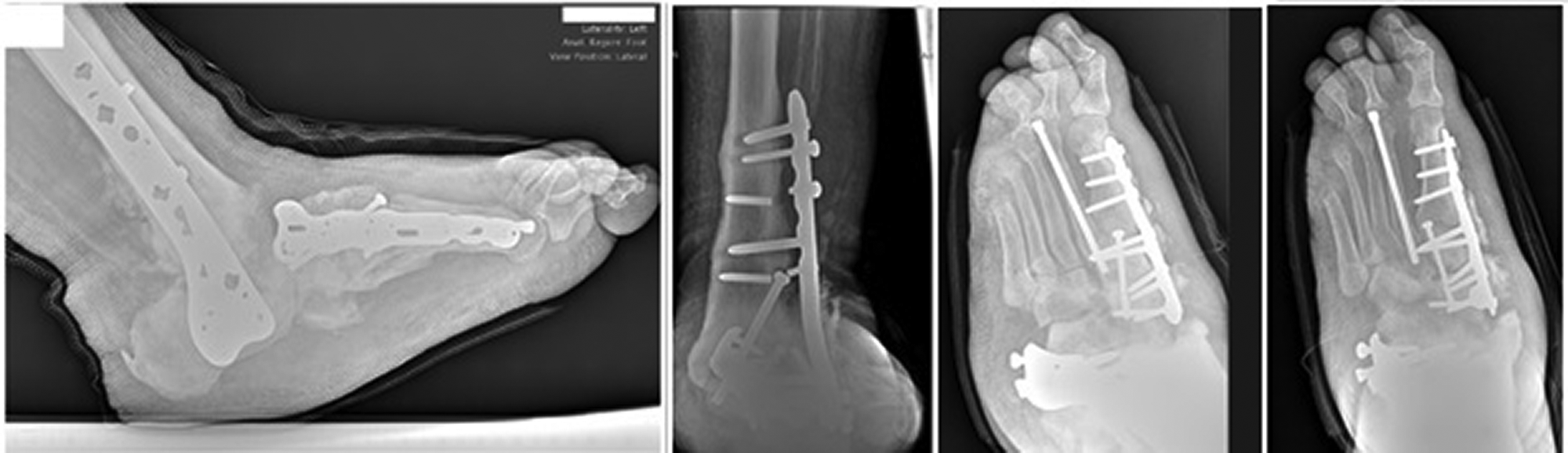
At 13 months postoperatively, the patient had developed a blister to the plantar foot along with a wound to the fourth digit of the left foot. New radiographs showed failure of hardware (Fig. 8). The plantar blister healed without issue, but the patient had a fourth- and fifth-digit amputation performed because of infection. At 14 months postoperatively, the patient had hardware removed from the TTCA site and had an external fixator applied.
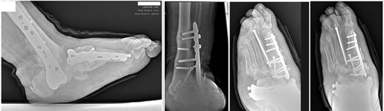
Figure 8.
One-year postoperative foot and ankle radiographs for patient 3. There is failure of screws within the lateral plate. The patient at this point had been weightbearing in diabetic shoes without bracing.
Case 4
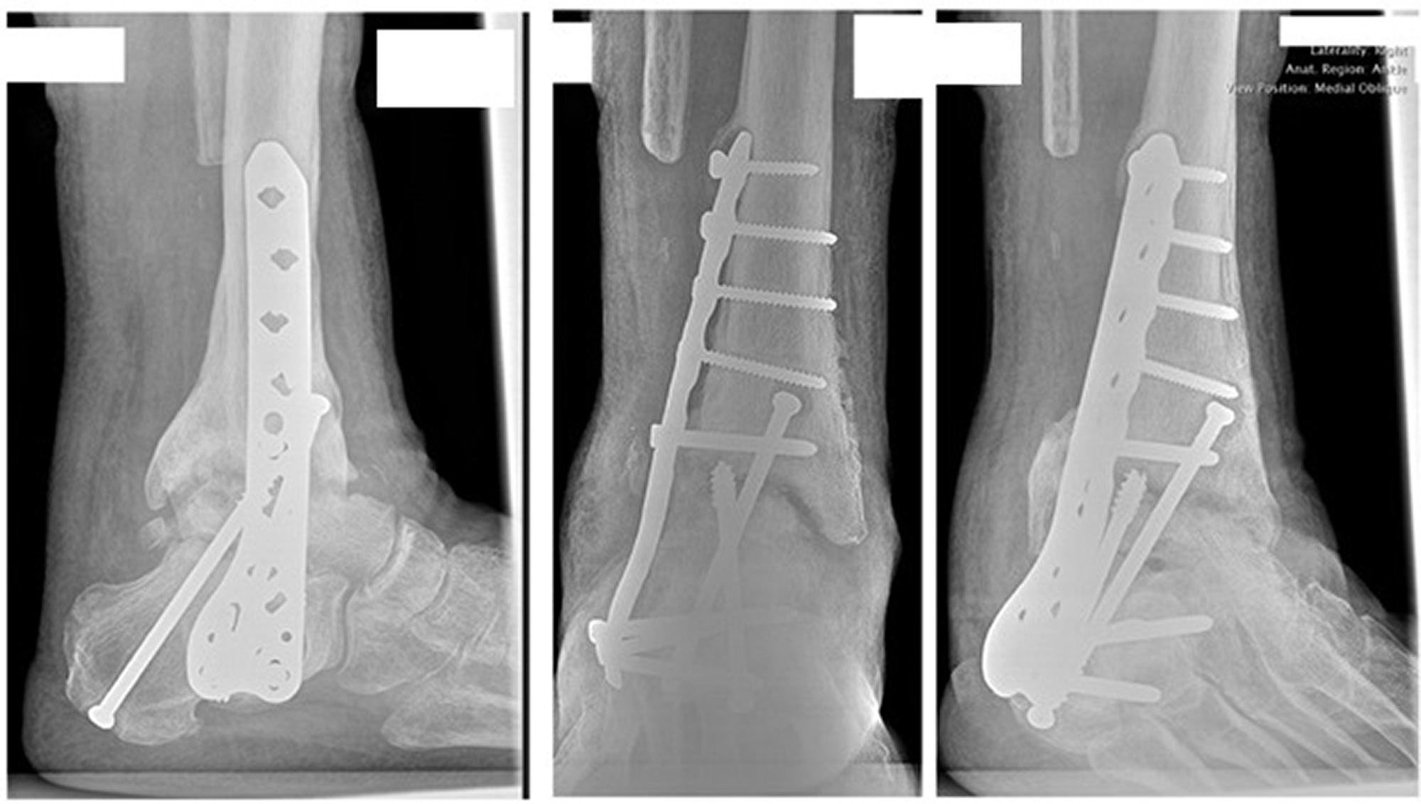
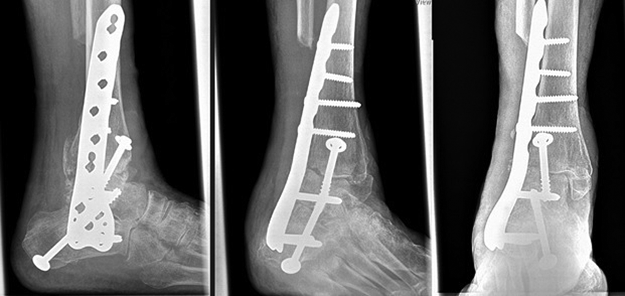
An 82-year-old woman presented who had been seen in the hospital after suffering an open right ankle fracture. A discussion was had to perform a TTCA because of the deformity and open nature of the wound. The patient had consented to the procedures with one of the authors (H.A.H.) after hearing the risks, benefits, alternatives, and postoperative course. Postoperatively, the patient had wound management by plastic surgery with allograft and vacuum-assisted closure placement. The patient was discharged to a skilled nursing facility for postoperative care. She was able to heal the wound over the course of the next 3 weeks. Short leg casts were placed every 2 weeks, for a total of 12 weeks. Radiographs were taken (Figs. 9 and 10). At 3.5 months, she was wound free and had transitioned to weightbearing with a CAM boot. She was to start physical therapy and had Lidoderm (Endo Pharmaceuticals Inc, Dublin, Ireland) patches for pain as needed. The patient has not followed up since.
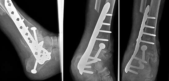
Figure 9.
Two-week postoperative ankle radiographs for patient 4. There is one screw from the tibia to the talus and one screw from the calcaneus to the talus.
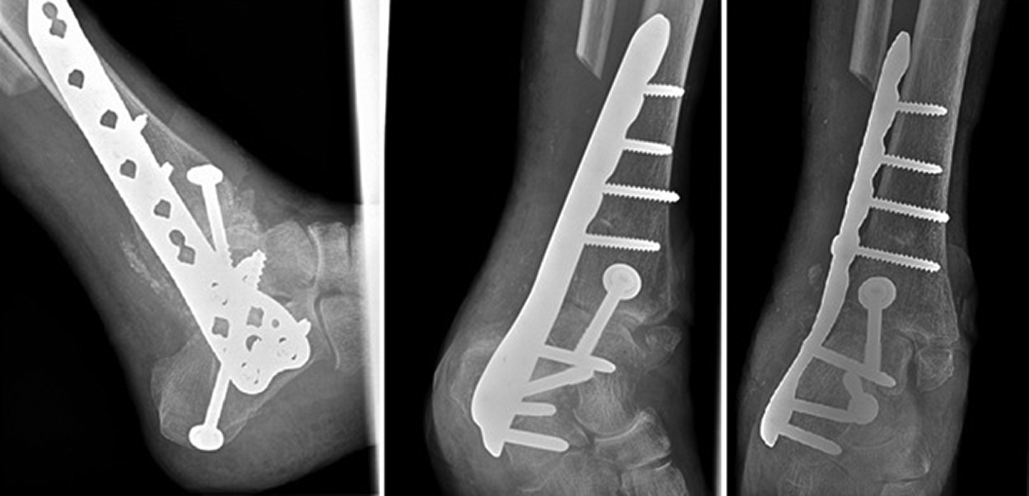
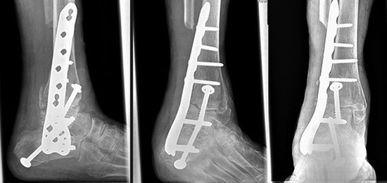
Figure 10.
Two-month postoperative ankle radiographs for patient 4.
Results
From 2018 to 2020, there were four patients (three men and one woman) who underwent a TTCA by one of the authors (H.A.H.). Their ages at the time of surgery were 53, 57, 66, and 82 years. The total follow-up time ranged from 3.5 to 16 months. Three patients are currently still following up in the office. Regarding comorbidities, three of the four had complicated DM with neuropathy, and the TTCA was performed because of Charcot neuroarthropathy of the foot or ankle and its sequelae. The patients with neuropathy had absent protective sensation as determined by with Semmes-Weinstein monofilament and absent vibratory sensation as determined by tuning fork.
Radiographs preoperatively showed that one patient had active Charcot ankle at the time of surgery (patient 1) with collapse and resorption of bone of the distal tibia and of the talar dome. Two patients (patients 2 and 3) had medial column fusions performed along with their TTCA because of foot wounds secondary to Charcot breakdown. One patient had posttraumatic arthritis without a history of DM. The three patients with DM developed postoperative complications, including one surgical-site dehiscence and two nonunions. The two patients that had active Charcot of the ankle developed nonunion; however, the hardware remained stable.
As of March 31, 2021, all four patients still have their operative limb (100% limb salvage). Two are currently weightbearing at their final follow-up, with one weightbearing with a CAM boot (patient 1) and one weightbearing without bracing (patient 4). These two patients returned to weightbearing with a CAM boot at approximately 3 months. Patient 3 was weightbearing with a CAM boot at 3 months and had transitioned to diabetic shoes. However, because of the hardware failure, he had the plate removed and an external fixator applied. The most recent patient that has undergone the TTCA again is actively healing his surgical incision with vacuum-assisted closure and is nonweightbearing, with a wheelchair.
Discussion
Going into the reconstructions, we had planned to use the more robust lateral femoral locking plate for three of the patients because of the known complications and increased complication rates associated with complicated DM. One patient was nondiabetic, but she had a significant open ankle fracture and was the oldest at 82 years at the time of surgery. Another patient had DM that was controlled on the day of surgery with an HbA1c level of 6.1%, but still had signs of peripheral neuropathy. Two of the patients (patients 2 and 3) were both type 2 diabetics on insulin (HbA1c levels of 6.7% and 7.6%, respectively) with peripheral neuropathy and had been dealing with Charcot neuroarthropathy of the midfoot with ulcerations. They were considered the highest risk going into surgery and were deemed to need the most time to heal. DeVries et al [17] found that there was a seven-fold increased odds ratio for amputation in patients with DM after TTCA with IMN. Preoperative ulceration was shown to increase the odds of failure by six [3] and three times [18]. Patients 2 and 3 both had ulcerations in the midfoot at the time of surgery; however; bone and tissue cultures were taken before the procedure to rule out any osteomyelitis before proceeding with definitive fixation. We also had made a conscious effort to hold off on performing surgery with patients who had an HbA1c level of over 8.0%. There has been plenty of research published regarding the adverse effects of DM, including fracture healing and osseous union [18,19]. Lee et al [20] showed that a postoperative blood glucose level of more than 200 mg/dL; an HbA1c value of more than 7.0%; or those with diabetic ketoacidosis, hyperosmolar hyperglycemic syndrome, or diabetic coma had a 10-fold increased odds of B/K amputation. Noninfectious complications were found to be higher in patients with poor short-term glucose control (a preoperative glucose level ≥200 mg/dL) [19]. Of the three diabetic patients, only one (patient 3) had an HbA1c level greater than 7.0% at the time of surgery (7.6%).
For all four of the patients, we used at least one screw across the AJ and STJ, which allowed us to maintain our reduction and position and to provide compression for fusion. However, even with the more robust fixation method, there was still a 100% (three of three) complication rate for our patients with DM, with two developing nonunions (patients 1 and 3) and the other developing wound dehiscence along with infection (patient 2). The union rate of 50% is low for this procedure, regardless of fixation type compared to the combined union rates being greater than 65% [5,7]. In addition, the follow-up time is still early for patient 2, as he is only 3.5 months out from the index procedure and patient 4 never followed up after 3.5 months. There is a possibility that patient 4 may have gone to nonunion and that patient 2 will go to nonunion as well. Expecting a 100% osseous union rate of the AJ and STJ in this patient population may be unrealistic. Although we have two patients with nonunions, our limb salvage rate is still 100%, with 75% able to bear weight on their affected extremity with or without assistive devices at the time of final follow-up. Obtaining a stable metal construct that allows a patient to have a functional limb with the ability to bear weight may be a more attainable goal. A metal fusion with the biomechanically advantageous locking plate may provide more benefit, as the chance of the hardware failure is decreased, especially in those that are geriatric patients with osteopenia (patient 4) or poor bone quality and bone stock from Charcot (patients 2 and 3). It may afford some stability even without osseous fusion. Super constructs for Charcot have been described by Sammarco [21] with use of more robust hardware implementations including locking plates and IMN fixation. Dujela et al [5] alluded to a functional nonunion of the subtalar joint with patients ambulating pain-free with shoe and a brace. Especially those with complete peripheral neuropathy, history of Charcot, and ulceration such as patients 2 and 3, a functional nonunion may still be an acceptable outcome for them.
In contrast, the locking plate fixation may be too stout, which may prevent secondary bone healing from taking place because of stress shielding. The femoral plating system also may be more unwieldy and may be less able to contour to the lateral aspect of the tibia, talus, and calcaneus because of the thickness and nonanatomical nature of the plate. Griffin and Coughlin [7] found that first-time fusion rates were low at 65%; however, with dynamization, fusion rates improved to 83%. Intramedullary nailing allows for the ability to dynamize to allow for secondary bone healing. The stress shielding from the locking plate may have prevented patients (ie, patients 1 and 3) from achieving union if bone had resorbed at the fusion sites removing any significant apposition, especially in the case with additional bone loss from Charcot. Patient 1 had developed significant cystic changes of the ankle joint and collapse of the talus, which is shown by the posterior-to-anterior STJ screw head being more prominent in the joint and into the tibial plafond (Fig. 1 compared to Fig. 3). Patient 3 had a significant bayonet deformity with patchy osteopenia and resorption of the talus already present preoperatively (Fig. 5). The future use of osteobiological agents may be of benefit for these patients.
There has not been a consensus on the most optimal fixation, again with each having its indications and proposed benefits. However, the goals are the same: limb salvage and a functional limb with osseous union. This case series, although limited in follow-up and size, provides some insight into an additional option for surgeons that are not comfortable using an IMN or a posterior approach. Intramedullary nailing does have a learning curve and may be harder to implement initially in those with less experience. The lateral approach is a relatively common approach for a variety of procedures, and with one incision, access is obtained for both the AJ and the STJ. Direct visualization of both joints is helpful and allows for adequate preparation of the joints and for positioning. It avoids plantar placement of a larger incision (a requirement for a retrograde IMN), which may be in the vicinity of a plantar wound in patients with midfoot Charcot. In addition, the lateral approach avoids the plantar neurovascular bundle compared to the possibility of injury with reaming and insertion of the nail. Nailing also has the risk of seeding infection within the proximal tibial canal. As described previously, research has noted an increase in the biomechanical stability of the construct compared to intramedullary nailing in patients with poor bone stock.
The cases presented here are not without their own complications. Besides changes in construct to allow for dynamization, optimization of patients preoperatively may be used to reduce overall complication rates postoperatively and improve long-term outcomes. Focusing on modifiable risk factors including history of tobacco abuse and nutritional status, and improving the short- and long-term glycemic control of patients before surgery again would be beneficial. Looking at the outcomes of those with DM versus those without and those with Charcot of the ankle may be of interest regarding this plate, as the patient (patient 4) without diabetes or Charcot seemed to heal without issue. In addition, because this is a relatively recent technical adaptation by the surgeon (H.A.H.), it may take more cases before a comfort level with the procedure and the hardware is attained. We are unable to make any definitive conclusions on whether an IMN or posterior approach would have provided better outcomes in these four patients comparatively. A larger sample size and longer follow-up is needed to further make any judgments on the effectiveness of this lateral plate for TTCAs.
Conclusions
Lateral locking plate fixation with a distal femoral locking plate system may be an alternative fixation option for tibiotalocalcaneal fusions in complex patients. However, a larger sample size with longer term follow-up is needed to make any significant recommendations on the use of this plating system.
None reported.
None reported.
References
- Burns PR, Dunse A: Tibiotalocalcaneal arthrodesis for foot and ankle deformities. Clin Podiatr Med Surg 34: 357, 2017.
- Asomugha EU, Hartog D, Bryan D, et al: Tibiotalocalcaneal fusion for severe deformity and bone loss. J Am Acad Orthop Surg 24: 125, 2016.
- Gross JB, Belleville R, Nespola A, et al: Influencing factors of functional result and bone union in tibiotalocalcaneal arthrodesis with intramedullary locking nail: a retrospective series of 30 cases. Eur J Orthop Surg Traumatol 24: 627, 2014.
- Rammelt S, Pyrc J, Agren PH, et al: Tibiotalocalcaneal fusion using the hindfoot arthrodesis nail: a multicenter study. Foot Ankle Int 34: 1245, 2013.
- Dujela M, Hyer CF, Berlet GC: Rate of subtalar joint arthrodesis after retrograde tibiotalocalcaneal arthrodesis with intramedullary nail fixation: evaluation of the RAIN database. Foot Ankle Spec 11: 410, 2018.
- Lee BH, Fang C, Kunnasegaran R, et al: Tibiotalocalcaneal arthrodesis with the hindfoot arthrodesis nail: a prospective consecutive series from a single institution. J Foot Ankle Surg 57: 23, 2018.
- Griffin MJ, Coughlin MJ: Evaluation of midterm results of the Panta Nail: an active compression tibiotalocalcaneal arthrodesis device. J Foot Ankle Surg 57: 74, 2018.
- Peterson KS, Chapman WD, Hyer CF, et al: Short-term radiographic results and technique of tibiotalocalcaneal arthrodesis with a posterior anatomic locking plate. J Foot Ankle Surg 55: 906, 2016.
- Gorman TM, Beals TC, Nickisch F, et al: Hindfoot arthrodesis with the blade plate: increased risk of complications and nonunion in a complex patient population. Clin Orthop Relat Res 474: 2280, 2016.
- Devrim O, Avni IB, Abdi K, et al: Tibiotalocalcaneal arthrodesis by using proximal humeral locking plate. Acta Orthop Traumatol Turc 50: 389, 2016.
- Fan J, Zhang X, Luo Y, et al: Tibiotalocalcaneal (TTC) arthrodesis with reverse PHILOS plate and medial cannulated screws with lateral approach. BMC Musculoskelet Disord 18: 317, 2017.
- Gutteck N, Schilde S, Reichel M, et al: Posterolateral plate fixation with Pantalarlock is more stable than nail fixation in tibiotalocalcaneal arthrodesis in a biomechanical cadaver study. Foot Ankle Surg 26: 328, 2020.
- DiDomenico LA, Wargo-Dorsey M: Tibiotalocalcaneal arthrodesis using a femoral locking plate. J Foot Ankle Surg 51: 128, 2011.
- Chiodo CP, Acevedo JI, Sammarco VJ, et al: Intramedullary rod fixation compared with blade-plate-and-screw fixation for tibiotalocalcaneal arthrodesis: a biomechanical investigation. J Bone Joint Surg Am 85: 2425, 2003.
- Doornink J, Fitzpatrick DC, Boldhaus S, et al: Effects of hybrid plating with locked and nonlocked screws on the strength of locked plating constructs in the osteoporotic diaphysis. J Trauma 69: 411, 2010.
- Zhang C, Shi Z, Mei G: Locking plate versus retrograde intramedullary nail fixation for tibiotalocalcaneal arthrodesis: a retrospective analysis. Indian J Orthop 49: 227, 2015.
- DeVries JG, Berlet GC, Hyer CF: Predictive risk assessment for major amputation after tibiotalocalcaneal arthrodesis. Foot Ankle Int 34: 846, 2013.
- Marin C, Luyten FP, Van der Schueren B, et al: The impact of type 2 diabetes on bone fracture healing. Front Endocrinol (Lausanne) 9: 6, 2018.
- Myers TG, Lowery NJ, Frykberg RG, et al: Ankle and hindfoot fusions: comparison of outcomes in patients with and without diabetes. Foot Ankle Int 33: 20, 2012.
- Lee M, Choi WJ, Han SH, et al: Uncontrolled diabetes as a potential risk factor in tibiotalocalcaneal fusion using a retrograde intramedullary nail. Foot Ankle Surg 24: 542, 2018.
- Sammarco VJ: Superconstructs in the treatment of Charcot foot deformity: plantar plating, locked plating, and axial screw fixation. Foot Ankle Clin 2009 14: 393, 2009.
© 2023 American Podiatric Medical Association