


Abstract
Background: Kaposi sarcoma (KS) has multiple clinical variants, and most frequently presents on the lower extremities. Anti–human immunodeficiency virus (HIV) therapy has significantly reduced the incidence of KS. However, KS is still prevalent in both HIV-infected and HIV-uninfected patients. This case series analysis aims to reveal the clinical presentations, differential diagnosis, and treatment options of KS on the foot and ankle. Methods: Eleven cases of KS involving the foot and ankle were retrieved from our patient database, and their clinicopathologic features were analyzed. Results: All patients were men, aged 29 to 85 years. Two types of KS were found: classic and acquired immunodeficiency syndrome–associated epidemic. The average ages of classic and epidemic KS were 65.7 and 41.8 years, respectively. Clinically, three patients manifested multiple erythematous or deep violaceous, or blue-violaceous macules on either the dorsal or plantar surfaces of both feet. Eight patients showed exophytic, pyogenic granuloma-like nodules on the plantar surface, heels, and toes. Histologically, all KSs had uniform intervening fascicles of elongated spindle cells with slit-like vascular spaces filled with red blood cells and immunoreactivity with human herpesvirus-8. The patients were treated according to HIV infection status. Human immunodeficiency virus–infected patients were treated with anti-HIV therapy after primary surgical excision or biopsy. Human immunodeficiency virus–negative patients were treated with either surgical excision, Mohs surgery, or a combination of surgical excision and local radiotherapy according to individual patient clinical presentation. Conclusions: Kaposi sarcoma is still prevalent in both HIV-infected and HIV-uninfected patients with a variety of clinical presentations. Biopsy, with histologic evaluation, in combination with immunohistochemistry is essential for the differential diagnosis. The patient should be treated according their clinical manifestation, staging, comorbidity, and immune function.
Kaposi sarcoma (KS) is a neoplasm derived from abnormal proliferation of angioendothelial cells. It is caused by KS-associated herpes virus (or human herpes virus 8 [HHV8]) [1]. Since Hungarian dermatologist Moritz Kaposi established KS as an entity featured as the idiopathic multiple pigmented sarcomas of the skin in 1872 [2], four subtypes were subsequently identified: classic, African endemic, iatrogenic (immunosuppression-related), and epidemic (acquired immunodeficiency syndrome [AIDS] related) [3]. Classic KS (CKS) occurs most frequently in senior populations of Mediterranean, Eastern European, and Middle Eastern heritage [4]. The incidence of CKS is significantly higher in men than in women. African endemic KS occurs in human immunodeficiency virus (HIV)-negative patients, in sub-Saharan Africa, where KS-associated herpesvirus infections are prevalent. This type of KS frequently occurs in younger populations, including children and women. African endemic KS has dramatically increased in Africa because of the HIV/AIDS pandemic [5], and has become the most common malignancy, in men and women, in Africa [6]. Iatrogenic KS is related to the use of immunosuppressive therapy during organ transplantation, or chemotherapy for cancer and other autoimmune diseases, which decreases the immunosurveillance, and leads to KS-associated herpes virus activation. Epidemic KS most often presents in HIV-infected men who had sex with men. Increased incidence of KS is correlated with increased incidence of multiple sexual partners and AIDS transmission among male homosexuals [7], whereas highly active antiretroviral therapy (HAART) significantly reduces the incidence of KS [8].
Kaposi sarcoma most frequently occurs on the feet, especially CKS (>60%) [4,7]. Clinically, it presents as one or more slow growing cutaneous red, purple, or brown macules, papules, plaques; and nodular, florid, infiltrative, or lymphadenopathic growths on the skin of the feet [9]. Extracutaneous organs, including oral mucosa, gastrointestinal tract, liver, lung, and lymph nodes can be involved [3]. Histologically, KS, in early stages, including presentation with macules, papules, and plaques, is characterized by endothelial cell–lined, irregularly shaped vascular vessels surrounding existing vasculature with infiltration of lymphocytes. Kaposi sarcoma, in late stages, including nodular and florid growth, is characterized by intervening fascicles of elongated spindle cells with clear nucleoli and abundant cytoplasm and slit-like vascular spaces filled with red blood cells. Immunophenotypically, the spindle cells show strong immunoexpression of CD31/CD34 and HHV8.
Because KS has a variety of clinical manifestations with different pathogeneses, patient treatment should be based on the individual’s clinical presentation, staging, tumor growth rate, HIV status, use of immunosuppression, and immune system function. Classic KS, with a single lesion on the lower extremity in patients without HIV infection, is treated with surgical excision and observation. Local radiation therapy is applied to larger or multiple lesions [4,7]. Epidemic KS, in HIV-infected patients, should be treated with HAART. Chemotherapy should be added to treat severe or progressive forms of KS in either HIV-infected or HIV-uninfected patients [3,10–12]. Immunosuppressive therapy–related iatrogenic KS needs to be treated with a decrease or discontinuation of immunosuppressive medications [13].
One of this study’s principal authors (S.R.) encountered an early case of “epidemic KS” in 1982, before HIV was identified as a unique and fatal disease [14]. The patient would die as result of AIDS within the year. Here, we review 11 cases of KS, with known HIV status, with the goal of eliciting the KS’s clinical presentation, differential diagnosis, and treatment options when these lesions are found on the foot and ankle.
Methods
Eight patients diagnosed with KS on the foot and ankle were retrieved from the Foot and Ankle Specialists of the Mid-Atlantic (FASMA) database, over a 6-year period, from September 1, 2015, to August 30, 2021. Three patients were retrieved from one clinician’s clinical practice before 2015 (S.R.). All patients’ hematoxylin and eosin and immunostaining slides were reevaluated with the confirmation of the original diagnosis (X.H.). The clinicopathologic data were obtained from the FASMA database and analyzed by one observer (X.H.), and were then confirmed by the corresponding podiatrists who treated and followed the patients. The institutional review board of FASMA composed of members of the executive board approved the case series analysis. Consents for collection of all patient specimens and clinical data were granted by patients with written documentation through our Health Insurance Portability and Accountability Act and office policy forms. The study was conducted in compliance with all ethical practices.
Results
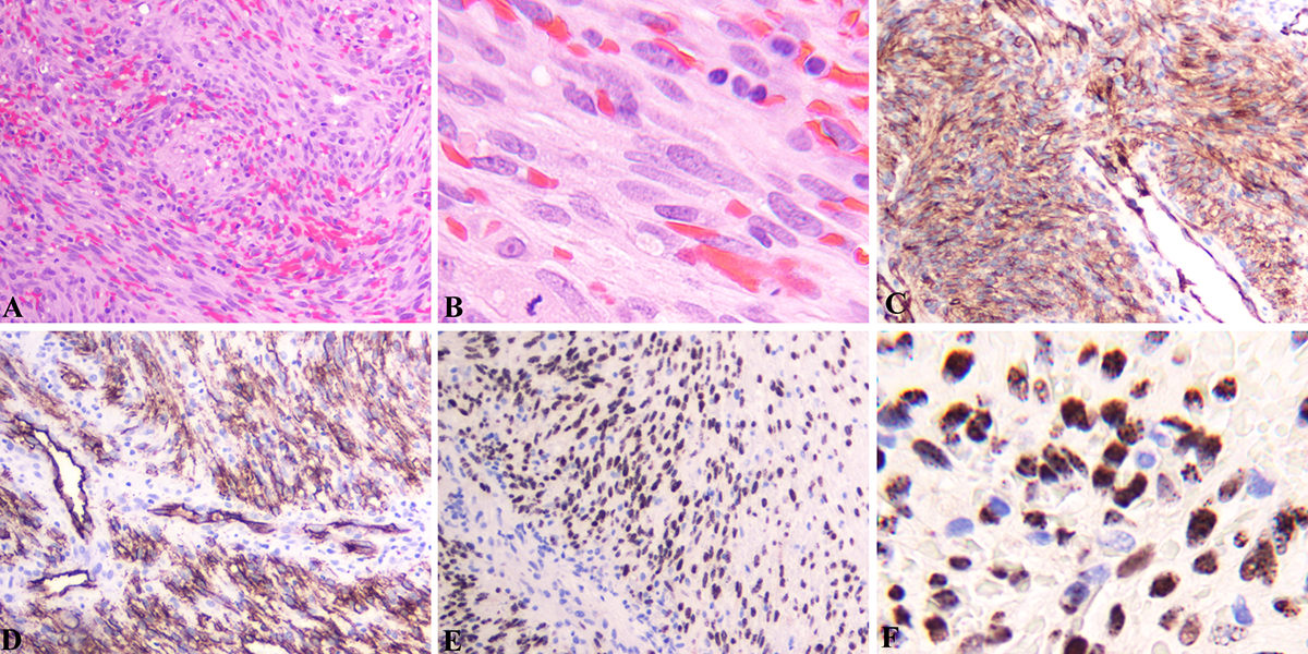
As shown in Table 1, all 11 patients were men, aged 29 to 85 years, with a mean age of 57 years and a median age of 58 years. Two types of KS were identified: classic (seven cases, HIV-negative) and AIDS-associated epidemic (four cases, HIV-positive). The ages of classic and epidemic KS ranged from 48 to 85 years, and from 29 to 51 years, respectively, with average ages being 65.7 and 41.8 years, respectively. Clinically, three patients presented with multiple dark or deep violaceous, or blue-violaceous macules, ranging in size from several millimeters to several centimeters, even confluent together, to form map-like plaques on either the dorsal or plantar surface of the foot/feet (Fig. 1). Eight patients exhibited exophytic, erythematous, pyogenic granuloma-like nodules. The nodular lesion ranged in size from 0.5 to 1.5 cm in diameter, with the mean and median sizes being 1.1 cm and 1.1 cm in diameter, respectively. The nodular lesion can occur on any part of the foot, including on the surface of the webspace, between the second and third toes (Fig. 2A), on the plantar surface (Fig. 2B), on the heels (Fig. 2C), on the second dorsal toe, and on the plantar fifth toe (Fig. 2D). The duration from when the patients noticed the macular or nodular growth to the diagnosis ranged from 2 weeks to 5 years, with a mean duration of 13.5 months and a median duration of 13.3 months. One of the HIV-negative patients had a history of melanoma. All patients were diagnosed with biopsy. Histologically, hematoxylin and eosin–stained slides showed proliferation of neoplastic endothelial cells in the dermis, forming intervening fascicles of elongated spindle cells with infiltrated lymphocytes and plasma cells. The vascular spaces had irregular and jagged outlines, forming slit-like spaces lined by thin spindle-shaped endothelial cells that were filled with red blood cells and deposits of hemosiderin (Fig. 3 A and B). Immunoperoxidase stain showed strong cytoplasmic staining of CD31 (Fig. 3C) and CD34 (Fig. 3D), and strong granular nuclear staining of HHV-8 in the endothelial and endothelial-derived spindle cells (Fig. 3 E and F).

Table 1.
Clinicopathologic Characteristics of the Patients with Kaposi Sarcoma on the Foot and Ankle
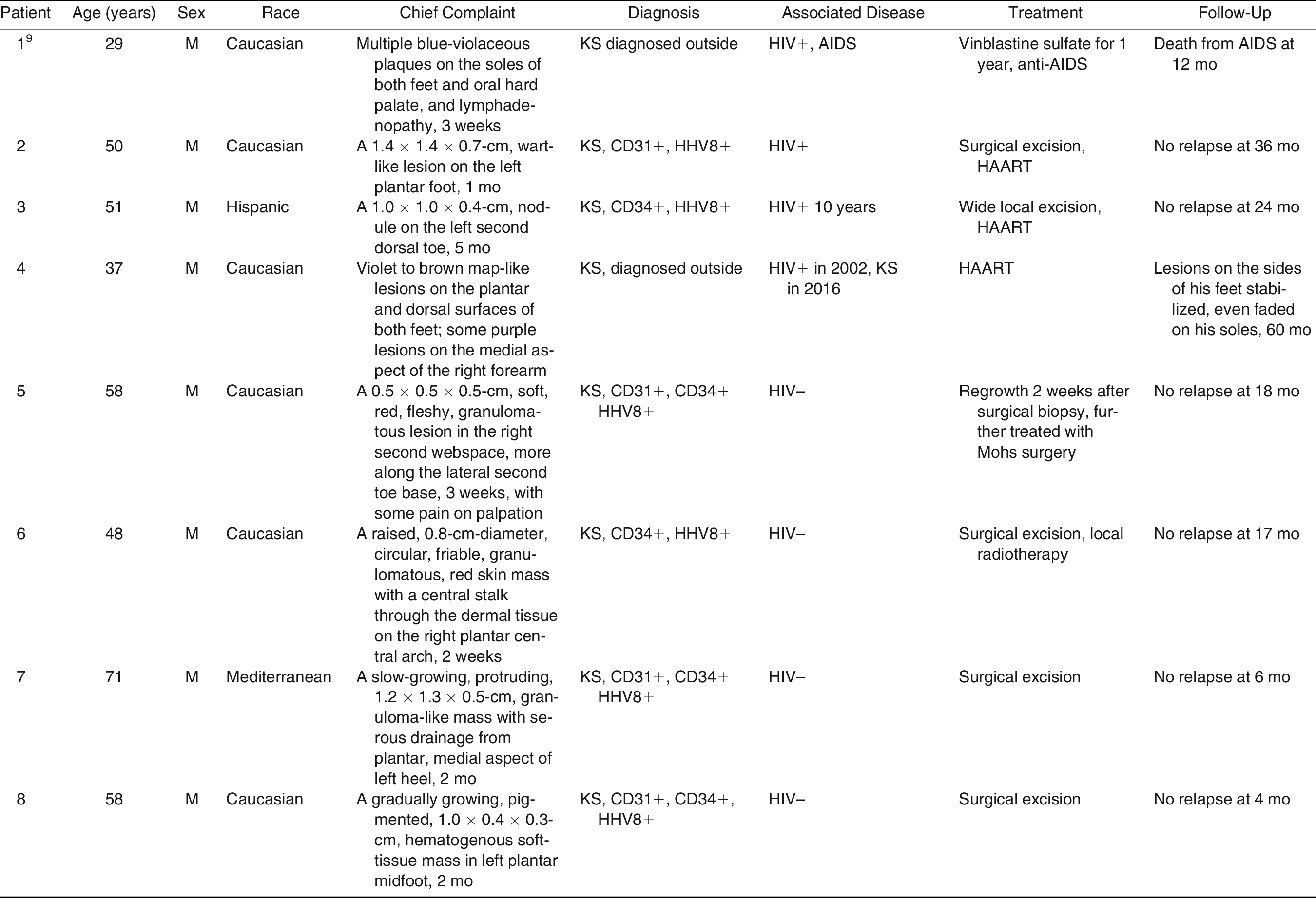
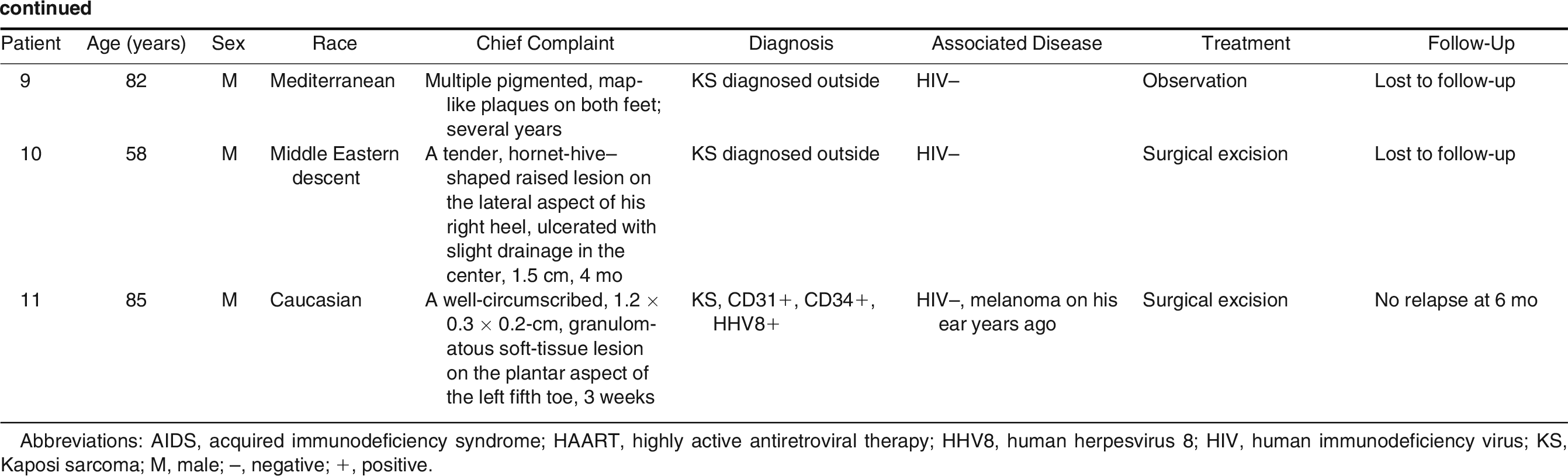
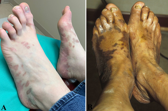
Figure 1.
Clinical presentations of Kaposi sarcoma on the foot. A, Multiple purple, different sized macules or papules on the dorsal and plantar surfaces of both feet from a 37-year-old, human immunodeficiency virus–infected man with cutaneous, disseminated Kaposi sarcoma (patient 1). B, Multiple, different sized, brown-violaceous plaques on the dorsal surfaces of both feet of an 82-year-old, human immunodeficiency virus–negative male patient of Mediterranean descent with classic Kaposi sarcoma (patient 9).
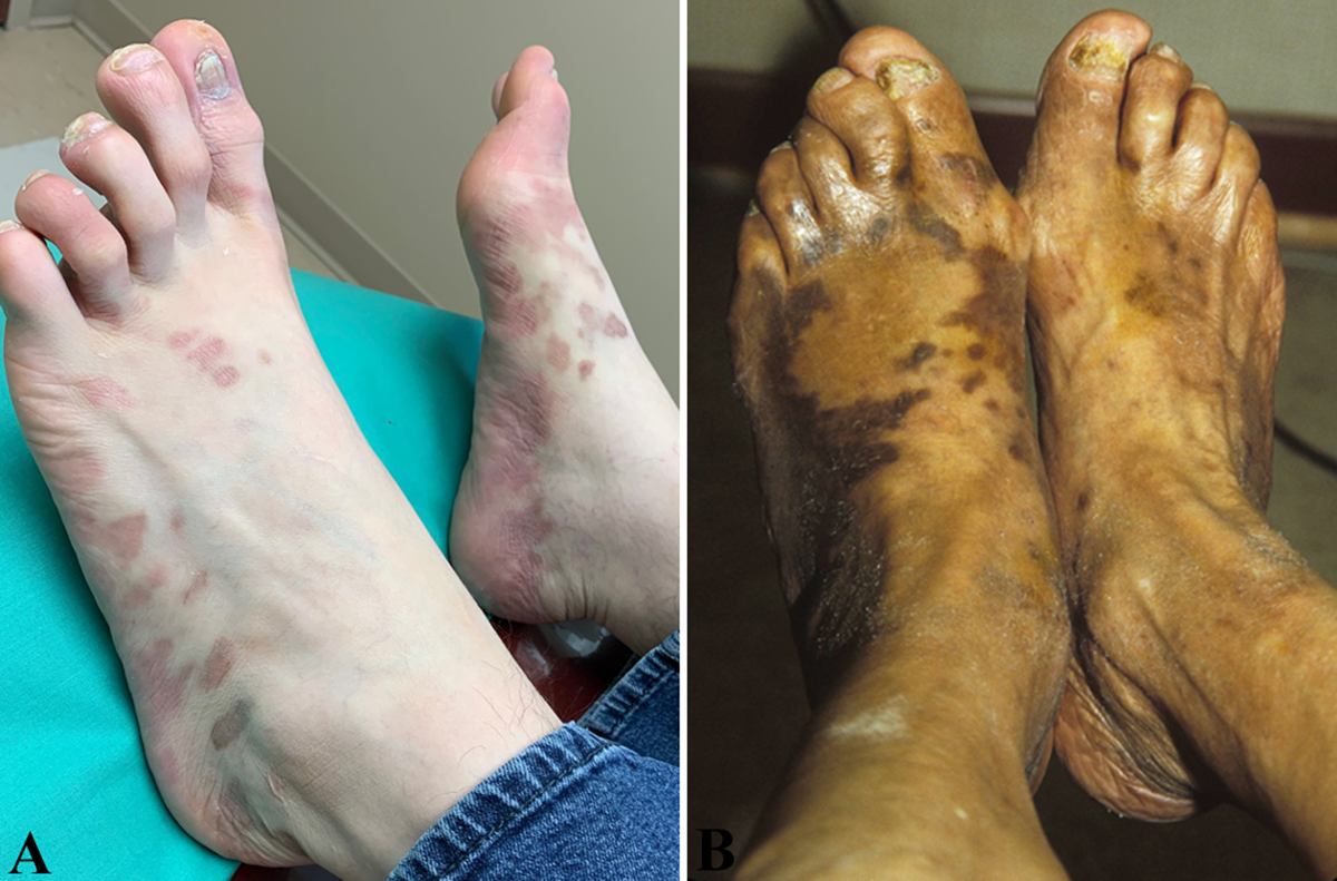
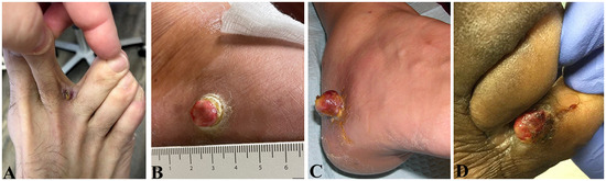
Figure 2.
Clinical presentations of Kaposi sarcoma on the foot. A, A regrowth of a nodular lesion in the webspace between the second and third right toes, 2 weeks after primary surgical excision, from a 58-year-old, human immunodeficiency virus (HIV)–negative man (patient 5). B, A raised, circular, friable, granuloma-like mass, with a central stalk through the dermal tissue, on the right plantar central arch, from a 48-year-old, HIV-negative man (patient 6). C, A raised, ulcerated lesion with slight drainage on the lateral aspect of the right heel from a 58-year-old, HIV-negative man (patient 10). D, A well-circumscribed, granulomatous mass on the plantar aspect of the left fifth toe of an 85-year-old, HIV-negative man (patient 11).
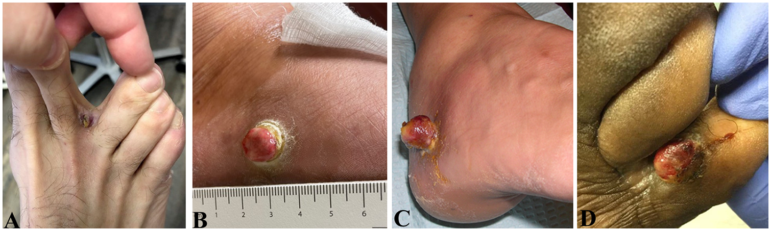
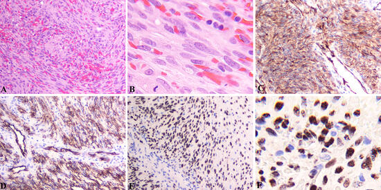
Figure 3.
Histopathology of the Kaposi sarcoma on the foot. A (H&E, ×100) and B (H&E, ×400). Intervening fascicles of elongated spindle cells, with slit-like spaces filled with extravasated red blood cells. A mitotic figure in the lower left corner and several infiltrated lymphocytes in the right upper corner in B. C, Strong cytoplasmic staining of CD31 on the endothelial cells and spindle cells (diaminobenzidine [DAB], ×100). D, Strong cytoplasmic staining of CD34 on the endothelial cells and spindle cells (DAB, ×100). E (DAB, ×100) and F (DAB, ×400), Strong granular nuclear staining of human herpes virus-8 in spindle cells.
The patients were treated according to the patient’s HIV status. All HIV-infected patients received anti-HIV therapy with HAART regimens after surgical excision. An 82-year-old HIV-negative patient refused further treatment and did not show up for follow-up visits. Four patients without HIV infection were treated with simple surgical excision and had no relapse at a follow-up ranging from 6 to 18 months. One HIV-negative patient’s lesion in the webspace, between his second and third toes, regrew rapidly 2 weeks after primary excisional surgical biopsy. His regrowth was then treated with Mohs surgery, and no recurrence was noted at a follow-up visit 18 months after the Mohs excision. Another HIV-negative patient was treated with a combination of surgical excision and local radiotherapy. This patient had no relapse at a follow-up visit 17 months later.
Discussion
In our review, we found two types of KS frequently encountered in podiatric medical practice: classic and epidemic. All patients identified were men. The average ages of classic and epidemic types are 65.7 and 41.8 years, respectively, values that are in line with previously reported age distribution of both types of KS [4,7]. Three of seven HIV-negative classic KS patients (42.9%) were of Mediterranean or Middle Eastern European descent.
Because KS has significant variation in its clinical presentation, and resembles other malignant and nonmalignant lesions (including angiosarcoma, spindle cell hemangioma, pyogenic granuloma [15,16], dermatofibroma, diabetic ulcer [17], melanoma, mycosis fungoides, and benign pigmented purpura), it is imperative to perform biopsies of the lesions, for histologic evaluation, with immunohistochemistry of HHV8, CD31, and CD34, and other diagnostic markers for a definitive diagnosis.
Treatment options depend on the HIV infection status. HIV-positive patients should continuously receive anti-HIV HAART treatment. Anti-HIV therapy significantly decreases the incidence of KS [8]. For severe or progressive KS occurring in HIV-infected patients with anti-HIV therapy, chemotherapy should be added [3,10]. Human immunodeficiency virus–negative patients can be treated with either simple surgical excision, local wide excision, Mohs surgical excision [18], or surgical excision plus local radiotherapy.
Conclusions
Kaposi sarcoma, an angioendothelial cell neoplasm, has a variety of clinical presentations. It needs to be differentiated from malignant and nonmalignant lesions. Biopsy specimens, with histologic evaluation and immunohistochemistry of HHV8, CD31, and CD34, are needed to make an accurate diagnosis. The treatment options should be based on the individual’s clinical presentation, staging, HIV infection status, and immune status.
None reported.
None reported.
References
- Chang Y, Cesarman E, Pessin MS, et al: Identification of herpesvirus-like DNA sequences in aids-associated Kaposi’s sarcoma. Science 266: 1865, 1994.
- Kaposi M: Idiopathisches multiples pigmentsarkom der haut. Arch Dermatol Syph 4: 265, 1872.
- Etemad SA, Dewan AK: Kaposi sarcoma updates. Dermatol Clin 37: 505, 2019.
- Steinfeld AD, Cooper JS: Epidemic and classic Kaposi’s sarcoma of the feet. A comparative study. JAPMA 80: 469, 1990.
- Mosam A, Aboobaker J, Shaik F: Kaposi’s sarcoma in sub-Saharan africa: a current perspective. Curr Opin Infect Dis 23: 119, 2010.
- Schneider JW, Dittmer DP: Diagnosis and treatment of Kaposi sarcoma. Am J Clin Dermatol 18: 529, 2017.
- Berlin SJ, Clancy JT, Giordano ML: Kaposi’s sarcoma of the foot. A review and report of 156 cases. JAPMA 79: 311, 1989.
- Bohlius J, Valeri F, Maskew M, et al: Kaposi’s sarcoma in HIV-infected patients in South Africa: multicohort study in the antiretroviral therapy era. Int J Cancer 135: 2644, 2014.
- Taylor JF, Templeton AC, Vogel CL, et al: Kaposi’s sarcoma in Uganda: a clinico-pathological study. Int J Cancer 8: 122, 1971.
- Gbabe OF, Okwundu CI, Dedicoat M, et al: Treatment of severe or progressive Kaposi’s sarcoma in HIV-infected adults. Cochrane Database Syst Rev 9: CD003256, 2014.
- Tourlaki A, Germiniasi F, Rossi LC, et al: Paclitaxel as first- or second-line treatment for HIV-negative Kaposi’s sarcoma: a retrospective study of 58 patients. J Dermatolog Treat 31: 183, 2020.
- Brambilla L, Romanelli A, Bellinvia M, et al: Weekly paclitaxel for advanced aggressive classic Kaposi sarcoma: experience in 17 cases. Br J Dermatol 158: 1339, 2008.
- Moosa MR: Kaposi’s sarcoma in kidney transplant recipients: a 23-year experience. QJM 98: 205, 2005.
- Rubenstein SA, Jenkin WM, Conant MA, et al: Disseminated Kaposi’s sarcoma in male homosexuals. J Am Podiatry Assoc 73: 413, 1983.
- Korekawa A, Kaneko T, Nakano H, et al: Pyogenic granuloma-like Kaposi’s sarcoma on the first toe. J Dermatol 45: e177, 2018.
- Lee MK, Ku SH, Cho EB, et al: Unusual case of pyogenic granuloma-like Kaposi’s sarcoma on the sole. J Dermatol 42: 425, 2015.
- Torrence GM, Wrobel JS: A case of mistaken identity: classic Kaposi sarcoma misdiagnosed as a diabetic foot ulcer in an atypical patient. Clin Diabetes Endocrinol 5: 8, 2019.
- Soleymani T, Bennett RG: Mohs micrographic surgery for exophytic Kaposi sarcoma. Dermatol Surg 46: 1232, 2020.
© 2023 American Podiatric Medical Association